














On-siteDPCFIFPL


Sports physiotherapy
Return to sport, performance and prevention: private practice and patient self-management
Adrien Blachère · Quentin Bouillard
Go further
A limping child or an adolescent with knee pain: SCFE, Perthes, Sinding-Larsen, osteochondritis. An approach by age, with sources verified one by one.

In the child with a limp, age guides almost the whole diagnosis: the emergency must be ruled out first, before considering growth conditions.
17weeks of mean delay between the first symptoms and the diagnosis
What follows covers each of these points in detail, with sources. It is there if you need it.
This topic is taught in a course: Return to sport, performance and prevention: private practice and patient self-management, on-site with Adrien Blachère.
1 / 12
11 article chapters · 50 min in total
Age markers◔ 4 min
Age is not one element among others: it is the only one that rules out entire diagnoses.
Referred pain◔ 4 min
The classic figure for referred pain is copied from review to review without its method being stated.
Ruling out the emergency◔ 6 min
Two diagnoses dominate: septic arthritis of the hip and acute leukaemia.
Slipped epiphysis◔ 8 min
It is a mechanical slip of the epiphysis that progresses as long as the hip is loaded.
Perthes◔ 5 min
The only defensible management on which everyone agrees: preserve range of motion.
Apicitis◔ 4 min
It is the least studied entity in the article: a founding series of eight patients, no trial.
Osteochondritis◔ 5 min
Two elements matter: effusion, and persistence of symptoms despite well-conducted treatment.
Rehabilitation◔ 4 min
None of these four conditions has a randomised controlled trial evaluating physiotherapy.
Case studies◔ 4 min
None of the six cases ends with immediate recognition of the diagnosis.
In practice◔ 3 min
If one of these signs is present, the child needs a medical opinion the same day, not rehabilitation.
Questions◔ 3 min
In a prospective study, 52 of the 122 operated hips, that is 42.6%, had no hip pain at all.
Train on this
Sports physiotherapy
Adrien Blachère · Quentin Bouillard
Next comes practice: the course that teaches this topic, with Adrien Blachère.
Our courses on this topic may be covered by DPC or FIFPL, the French continuing-education funding schemes, depending on your profession and situation.
A limping child, an adolescent with knee pain: behind these two commonplace presentations hide four conditions of the growth plate, two of which are paid for in a lost hip when they are missed. This article is built on the approach, not on a list of diseases.
A synthesis article. Every figure cited carries its source where it is written, with its sample size and its method. Where the literature on an entity is thin, that is said rather than papered over: two of these four conditions rest, in rehabilitation, on no controlled study at all.
Three figures that govern vigilance
Primary data: prospective cohort of 122 hips, series of 55 hips, retrospective cohort of 481 patients
Sources: Uvodich M et al. J Pediatr 2019;206:184-189.e1 (PMID 30454962); Loder RT et al. J Bone Joint Surg Am 1993;75(8):1134-40 (PMID 8354671); Schur MD et al. J Pediatr 2016;177:250-254 (PMID 27470686).
In this article
What the available literature allows us to state, in twelve points. Each is taken up and sourced in the chapters that follow.
Who this article is for, and what it does not cover
For physiotherapists who see children and adolescents, in the clinic as at the pitchside. It covers four growth plate conditions of the lower limb in a single text, because none of them is recognised in isolation: it is their comparison, at a given age and in a given location, that makes the diagnosis.
Osgood-Schlatter disease, the commonest apophysitis of adolescence, has its own page on this site and is not covered again here: this article covers the conditions taken for Osgood-Schlatter, that one covers Osgood-Schlatter itself and its load management. See Osgood-Schlatter disease. Benign growing pains and calcaneal apophysitis are touched on here for what they bring to the differential diagnosis; they are covered in their own right in heel growth pain in children.
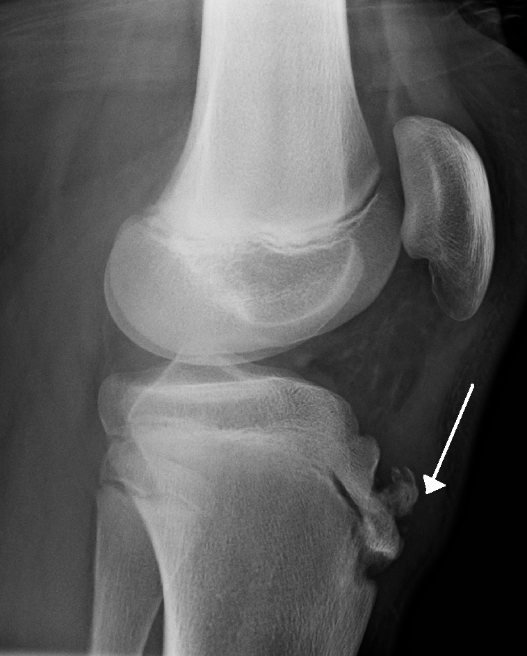
X-rayLateral knee X-ray: fragmentation of the anterior tibial tubercle with overlying soft tissue swelling, characteristic of Osgood-Schlatter disease.
Source : Houghton, Wikimedia Commons · CC BY
Because each of these conditions strikes a structure that is only vulnerable at a precise moment of skeletal maturation. Age is not one guiding element among others: it is the only one that, on its own, rules out entire diagnoses.
The growth plate is not a homogeneous structure that would weaken uniformly throughout childhood. The blood supply of the femoral head, the mechanical strength of the proximal femoral physis, the ossification of the secondary centres of the knee and the maturation of subchondral bone each follow their own calendar. A condition can therefore only occur in the window when its target structure is both present and vulnerable.
That produces a distribution by age whose clinical use is direct. Legg-Calvé-Perthes disease is a necrosis of the femoral head linked to a failure of its blood supply, in the period when that supply is terminal and precarious: in a population of nearly 800,000 children in an integrated health system in southern California, the highest incidence was 3.05 per 100,000 in 2- to 5-year-olds, and the lowest 1.06 in 9- to 12-year-olds, that is an odds ratio of 3.13 in favour of the younger12. Slipped capital femoral epiphysis is conversely a mechanical failure of the growth plate, occurring when it widens and becomes more horizontal under the effect of the hormones of puberty: the incidence reported in the United States from 1997 and 2000 hospital admissions was 10.80 per 100,000 children aged 9 to 163.
The age window of each entity
Each bar covers the band in which the measured incidence is highest, not the extreme ages reported
Sources: Kessler JI, Cannamela PC. Clin Orthop Relat Res 2018;476(12):2344-2350 (PMID 30211706) for Perthes; Lehmann CL et al. J Pediatr Orthop 2006;26(3):286-90 (PMID 16670536) for slipped epiphysis; Medlar RC, Lyne ED. J Bone Joint Surg Am 1978;60(8):1113-6 (PMID 721864) and Valentino M et al. J Ultrasound 2012;15(2):127-9 (PMID 23396672) for Sinding-Larsen; Kessler JI et al. Am J Sports Med 2014;42(2):320-6 (PMID 24272456) and Nissen CW et al. Am J Sports Med 2022;50(1):118-127 (PMID 34818065) for osteochondritis.
These incidences have a practical corollary that is often forgotten when reading review articles: these diseases are rare. A physiotherapist with an ordinary paediatric caseload will see a great deal of anterior knee pain, a few apophysitides, and will meet a slipped epiphysis or a Perthes a few times in their career. That is precisely what makes the reasoning necessary: rarity forbids relying on pattern recognition, and demands an approach triggered by criteria, not by an impression.
Sex is added to age as a second filter, and it is almost as discriminating. The odds ratio for male sex is 12.44 for Legg-Calvé-Perthes disease in the Californian cohort12, and the incidence of osteochondritis dissecans of the knee is 15.4 per 100,000 in boys against 3.3 in girls26. For slipped epiphysis, the gap exists but stays more modest: 13.35 against 8.07 per 100,0003. These gaps never allow a diagnosis to be ruled out in a girl, but they shift the starting probability, which matters when the decision is about whether to image.
Key points
The patient's age eliminates diagnoses before any examination. Before 6 years, osteochondritis dissecans of the knee has never been observed in the largest series with a known denominator, and slipped epiphysis is exceptional and suspicious of endocrinopathy. After 12 years, Legg-Calvé-Perthes disease no longer starts. Between 11 and 14 years the windows overlap and age no longer decides on its own: that is where the site and the mode of onset take over.
This is the classic trap of hip conditions in children, and the figure that describes it is copied from review to review without its method being stated. Two primary studies exist, they do not measure the same thing, and the gap between their results is entirely explained by their definitions.
The hip joint and the knee joint share their sensory innervation. The obturator nerve, the femoral nerve and the nerve to quadratus femoris give articular branches to the hip; the cutaneous and articular territories of the femoral and obturator nerves run down to the medial side of the knee. A nociceptive afferent arising in the hip capsule therefore enters the cord through roots that also receive the knee, and the brain regularly attributes the pain to the more distal segment. That is not a curiosity: it is the usual mode of presentation of a substantial share of hip conditions in children.
The first is retrospective. Matava and colleagues reviewed 106 patients with a slipped capital femoral epiphysis and counted those whose main complaint at the first consultation was knee or distal thigh pain: there were 16, that is 15 %. In the subgroup of 65 patients who had received no treatment before arriving, the proportion rose to 15 out of 65, that is 23 %. The most important result of that work is not the percentage but what goes with it: those patients received significantly more wrong diagnoses, underwent significantly more unnecessary radiographs, and arrived with radiologically more severe slips2.
The second is prospective, and that is what makes it valuable. Uvodich and colleagues had 107 children operated on for a slipped epiphysis complete a standardised body chart, giving 122 analysable hips. They did not ask for the main complaint but for the complete mapping of the painful areas. Result: 70 hips out of 122 had hip pain, so 52 hips, that is 42.6 %, had none. The knee was painful in 32 cases (26.2 %), the thigh or leg in 43 (35.2 %), the groin in 17 (13.9 %), the posterolateral region in 13 (10.7 %). A combination of several areas was present in 39.3 % of cases, and 49 % of patients had consulted more than once before the diagnosis1.
Where does a slipped epiphysis hurt? Two studies, two different questions
The percentages do not add up in the prospective study: several areas coexist in 39.3 % of hips
Sources: Matava MJ, Patton CM, Luhmann S, Gordon JE, Schoenecker PL. J Pediatr Orthop 1999;19(4):455-60 (PMID 10412993); Uvodich M, Schwend R, Stevanovic O, Wurster W, Leamon J, Hermanson A. J Pediatr 2019;206:184-189.e1 (PMID 30454962).
Why these two figures do not contradict each other
You often read that “15 % of slipped epiphyses present with knee pain”, sometimes “25 %”, sometimes “nearly half”. All three formulations circulate, and they come from the same two studies badly copied. Matava's 15 % answers the question “what is the main complaint?”; Uvodich's 26.2 % answers “is the knee painful?”, which includes patients who have pain in the hip and in the knee; the 42.6 % answers “is the hip free of pain?”. Three questions, three answers, no contradiction. What matters in practice is none of those three numbers taken alone, but their common conclusion: an examination of the painful adolescent that stops at the knee misses a substantial share of slipped epiphyses.
That mechanism does not concern only slipped epiphysis. Legg-Calvé-Perthes disease also commonly presents with isolated knee or thigh pain, which, as a literature review written for paediatricians puts it, “obliges the clinician to keep the hip in mind during the examination”20. The practical consequence is the same in both cases, and it comes down to one rule of examination.
Loss of hip internal rotation is common to both hip conditions in this group. In Legg-Calvé-Perthes disease, deficits in abduction and internal rotation are the most consistent examination findings, the Trendelenburg limp appearing only late20. In slipped epiphysis, the posterior slip of the epiphysis produces obligatory external rotation: the hip spontaneously moves into external rotation when the knee is passively flexed towards the abdomen, which is the most suggestive sign in the examination.
That examination is done supine, with the hip flexed to 90 degrees, and always compared with the opposite side. Asymmetry of internal rotation in a child with knee pain whose knee examination is normal is enough to request hip imaging. It is not enough to dispense with imaging when it is absent: internal rotation can be preserved in slips of small magnitude, and it is precisely those that need catching.
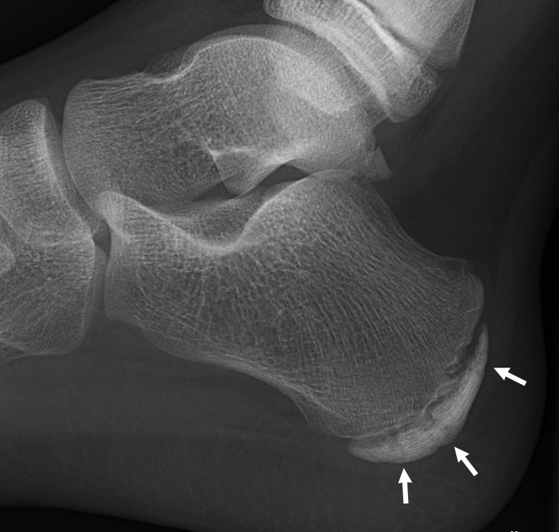
X-rayLateral foot X-ray in an 11-year-old boy: sclerosis and fragmentation of the calcaneal apophysis, an appearance suggestive of Sever's disease.
Source : Häggström, Wikimedia Commons · CC0
Before reasoning about growth conditions, you have to have ruled out what will not wait. Two diagnoses dominate that step: septic arthritis of the hip, and acute leukaemia revealed by musculoskeletal pain. Neither belongs to physiotherapy, and both present like the conditions in this chapter.
A limping child is a presentation whose first task is triage by severity, before any aetiological reasoning38. The most used prediction rule for separating septic arthritis of the hip from transient synovitis, the benign self-limiting form, rests on four variables: a history of fever, inability to bear weight, an erythrocyte sedimentation rate of at least 40 mm in the first hour, and a white cell count above 12,000 per cubic millimetre. The probability of septic arthritis goes from less than 0.2 % when no criterion is present to 3.0 % for one, 40.0 % for two, 93.1 % for three and 99.6 % for all four33.
That rule was validated prospectively in a different population, where the area under the curve fell from 0.96 to 0.86: it remains highly accurate but loses precision outside its original population, which is the expected behaviour of any prediction rule34. A later prospective study of 53 children in whom hip aspiration had been judged necessary identified fever above 38.5 degrees as the best single predictor, C-reactive protein being the only independent factor on multivariable analysis35.
Red flags in a limping child
None of these features belongs to first-line physiotherapy management. Their presence calls for a medical opinion the same day.
Decision tree for a limping child or adolescent
A physiotherapy decision path. The red outcomes fall outside first-line rehabilitation.
Built from: Kocher MS, Zurakowski D, Kasser JR. J Bone Joint Surg Am 1999;81(12):1662-70 (PMID 10608376) for septic triage; Uvodich 2019 (PMID 30454962) for the topographical proportions; Loder 1993 (PMID 8354671) for the definition of instability; Pinkowsky GJ, Hennrikus WL. J Pediatr 2013;162(4):804-7 (PMID 23149177) for the radiographic view to request.
The table below crosses the two variables that do the sorting, and adds for each entity the action that must not be missed. It includes the common benign entities, without which differential reasoning makes no sense, with links to their own page where one exists.
| Age | Site | Entity | Warning sign | What to do |
|---|---|---|---|---|
| 2 to 8 years | Hip, thigh or knee | Legg-Calvé-Perthes disease | A limp that is often painless at first, loss of abduction and internal rotation | Pelvic radiograph, AP and frog-leg lateral |
| 3 to 8 years | Hip | Transient synovitis | Abrupt onset, often after a viral episode, weight-bearing preserved | Apply the four-criterion rule before concluding it is benign |
| Any age | Hip or any joint | Septic arthritis | Fever above 38.5 degrees, refusal to bear weight, constant pain | Medical opinion the same day |
| Any age | Diffuse, often bilateral | Malignant blood disorder | Night pain that wakes the child, deterioration in general condition, subtle blood count abnormalities | Medical opinion, full blood count |
| 9 to 16 years | Hip, groin, thigh or knee | Slipped capital femoral epiphysis | Obligatory external rotation on hip flexion, overweight common | Stop weight-bearing, imaging, surgical opinion |
| 10 to 14 years | Inferior pole of the patella | Sinding-Larsen-Johansson disease | Exquisite tenderness on palpation of the patellar apex, jumping sports | Load adaptation, clinical diagnosis, ultrasound if in doubt |
| 11 to 15 years | Tibial tuberosity | Osgood-Schlatter disease | Pain and swelling of the tibial tubercle, two to three centimetres below the patella | See the dedicated article: Osgood-Schlatter disease |
| 11 to 19 years | Knee, often poorly localised | Osteochondritis dissecans | Persistent exertional pain, effusion, locking or giving way if the lesion is unstable | Imaging for any effusion or any failure at six weeks |
| 8 to 14 years | Heel, calcaneal insertion | Calcaneal apophysitis | Pain on lateral compression of the calcaneus, weight-bearing sports | Load adaptation. Mean return to play of 60.7 days in a football academy40. See the dedicated article: Sever's disease |
| 3 to 12 years | Bilateral, lower limbs | So-called growing pains | Evening or night pain, bilateral, strictly normal examination, no limp | A diagnosis of exclusion. Any limp or any unilateral pain rules it out |
One line of that table deserves reading twice. So-called growing pains are a diagnosis of exclusion whose usual criteria are themselves debated: an Australian twin study showed that those criteria take in a share of painful restless legs syndrome, and that the prevalence falls by a third as soon as the absence of motor restlessness is required41. What matters to the physiotherapist is simpler: these pains are bilateral, come in the evening, and never cause a limp. A limp, unilateral pain or mechanical daytime pain do not fit that frame, whatever label those around the child have applied.
Key points
First-line triage is not about the diagnosis but about urgency. Fever, refusal to bear weight, night pain and deterioration in general condition fall outside physiotherapy. Once those are ruled out, age points to the hip before 8 years, to slipped epiphysis between 9 and 16, to the patellar apex and osteochondritis in adolescence. Knee pain in an adolescent never allows you not to examine their hip.
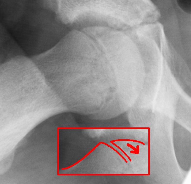
X-rayAnteroposterior hip X-ray with Klein's line drawn: slippage of the upper femoral epiphysis relative to the metaphysis, a sign of slipped capital femoral epiphysis.
Source : Lengerke, Wikimedia Commons · CC0
Because it is not an osteochondrosis. It is a mechanical slip of the femoral epiphysis on its growth plate, which progresses as long as the hip is loaded, and whose treatment is surgical. An adolescent rehabilitated for knee pain while their epiphysis slips loses hip at every session.
The other three entities in this article are disorders of ossification or of blood supply: they follow their own natural history, and physiotherapy accompanies a process. Slipped capital femoral epiphysis belongs to another category. The proximal femoral growth plate, widened and straightened by the hormonal changes of puberty, stops resisting the shear forces imposed by weight-bearing. The epiphysis, held by the ligamentum teres and the periosteum, slips backwards and downwards while the femoral neck moves forwards and into external rotation.
The practical consequence is unmatched in the rest of this article: the factor that makes the lesion worse is exactly the one rehabilitation uses as its tool. Loading, walking, weight-bearing exercises: all of these, on a growth plate that is giving way, increase the displacement. That is why unloading precedes imaging, and not the other way round.
The incidence reported in the United States from the 1997 and 2000 paediatric admission databases is 10.80 per 100,000 children aged 9 to 16, with 13.35 in boys and 8.07 in girls. That same analysis finds a relative incidence 3.94 times higher in Black children and 2.53 times higher in Hispanic children than in white children, as well as geographical and seasonal variations3.
Overweight is the most consistently associated risk factor, to the point of being built into aetiological triage rules. It is not a diagnostic criterion and its absence rules nothing out: it is an element that shifts the probability, not a necessary condition.
Loder and colleagues proposed in 1993 abandoning the traditional classification into acute, chronic and acute-on-chronic forms, based on symptom duration, in favour of a purely functional distinction. A slip is called unstable when the pain is such that weight-bearing is impossible, even with crutches; it is called stable when the patient can bear weight, with or without aids.
Across 55 hips with symptoms of less than three weeks' duration, 30 were unstable and 25 stable. The result was satisfactory for 14 of the 30 unstable hips (47 %) against 24 of the 25 stable hips (96 %). Above all, avascular necrosis occurred in 14 of the 30 unstable hips (47 %) and in none of the 25 stable hips4. That series does not allow a link to be established between early reduction and the risk of necrosis, which the authors state explicitly.
What the stability of the slip changes
A series of 55 hips with symptoms of less than three weeks' duration
Source: Loder RT, Richards BS, Shapiro PS, Reznick LR, Aronson DD. Acute slipped capital femoral epiphysis: the importance of physeal stability. J Bone Joint Surg Am 1993;75(8):1134-40. PMID 8354671
That definition is functional and not radiological, which makes it directly usable by a physiotherapist: it is made by observing whether the adolescent can put the foot down, with no investigation at all. A qualification has been added since, one team having shown that clinical stability does not perfectly predict the stability found intraoperatively10. That does not invalidate the rule, it is a reminder that a clinically stable slip is not a harmless slip.
The delay between the first symptoms and the diagnosis has been measured several times, with converging and poor results. Across 196 patients, the median delay was 8.0 weeks, it increased with the severity of the slip, and knee or distal thigh pain remained an independent multivariable predictor of a longer delay, alongside the type of health cover5.
Twelve years later, a study of 481 patients admitted to three paediatric hospitals between 2003 and 2012 showed no improvement at all : the mean delay was 17 weeks, with extremes of 0 to 169 weeks, and no significant difference between the two-year periods of the follow-up. The delay between the first professional consulted and the diagnosis was on average nil when that professional was an orthopaedic surgeon, 4 weeks for a primary care doctor and 6 weeks in the emergency department. Finally, 52 patients out of 481, that is 10.8 %, had a slip on the opposite side after treatment of the first6.
Klein's line is the classic landmark on the AP radiograph: a line drawn along the upper border of the femoral neck should cut across part of the epiphysis. In slips of small magnitude, it stays normal. One study compared the three methods across 23 hips with a slipped epiphysis: the classic Klein line allowed the diagnosis in only 9 cases (39 %), its modified version in 20 cases (87 %), and the frog-leg lateral view in all 23 cases (100 %)7. The modified version of that line, proposed to make up for the low sensitivity of the original, has been the subject of a reproducibility study that quantifies its limits: the minimum difference required between two measurements for them to be considered distinct with 95 % confidence reaches 8.80 degrees between observers for the head-neck angle45.
What each view allows you to see
23 hips with a confirmed slipped epiphysis, radiographic re-reading
Source: Pinkowsky GJ, Hennrikus WL. Klein line on the anteroposterior radiograph is not a sensitive diagnostic radiologic test for slipped capital femoral epiphysis. J Pediatr 2013;162(4):804-7. PMID 23149177. The inter-observer reliability of the angle and displacement measurements was quantified by Green DW et al. J Pediatr Orthop 2009;29(5):449-53 (PMID 19568015).
Two practical points follow. The first is that the request must explicitly mention both views, failing which an isolated AP film is common. The second is that the examination covers the whole pelvis and not the painful hip alone : bilaterality of slipped epiphysis is reported within a wide range of 12 to 80 % depending on the series9, and the opposite side is frequently affected without yet being symptomatic.
A slipped epiphysis occurring outside its usual window, or in a slim adolescent, should prompt a search for an underlying cause: hypothyroidism, growth hormone deficiency, hypogonadism, chronic kidney disease. Loder and Greenfield proposed a simple rule from 433 children, of whom 285 had idiopathic and 148 atypical slipped epiphyses, giving 612 hips. At equal weight, a child under 10 or over 16 was 4.2 times more likely to have an atypical form; at equal age, a child below the 50th percentile of weight was 8.4 times more likely.
The so-called age-weight test follows: it is negative when the child is under 16 and of a weight at least equal to the 50th percentile, positive beyond those bounds. The probability that a slipped epiphysis was idiopathic was 93 % when the test was negative, and the probability that it was atypical 52 % when it was positive8. A positive test therefore does not make the diagnosis of endocrinopathy, but it justifies investigation.
What the physiotherapist must not do
Key points
Slipped capital femoral epiphysis is a surgical condition whose worsening depends on load. It presents between 9 and 16 years, often in an overweight adolescent, and frequently with pain that is not in the hip. The distinction between stable and unstable slip is made in the clinic, on whether weight can be borne, and separates two prognoses that are opposites: 0 % against 47 % avascular necrosis. The useful imaging is a pelvic radiograph, AP and frog-leg lateral.
Far less than it is credited with, and one precise, defensible thing: preserve range of motion. It is the only point on which the literature and practitioners converge, and it is also the one that formal guidelines cannot support with a controlled trial, because none exists.
Legg-Calvé-Perthes disease is an idiopathic avascular necrosis of the femoral head in childhood. The head loses its blood supply, fragments, then rebuilds over a period measured in years, keeping the shape it had during its fragile phase. The whole prognosis rests on that final shape: a round, congruent head in a matching acetabulum ages well, a deformed head leads to impingement then osteoarthritis.
The most solid study on this disease is a British national prospective cohort, in which 143 of the 144 hospitals managing children's hips took part over 18 months. It recorded 371 children and 396 hips, that is an annual incidence of 2.48 per 100,000 children aged 0 to 14, 95 % confidence interval 2.20 to 2.7611. An American study of nearly 800,000 children aged 2 to 12 gives a similar order of magnitude, 2.84 per 100,000, with a marked male predominance, odds ratio 12.44, and a peak between 2 and 5 years12. The incidence of this disease also varies strongly with social deprivation and with region, a gradient documented in the United Kingdom on geographical and temporal trends19, which is a reminder that none of these incidences transfers directly from one country to another.
In that British cohort, the independent predictors of a worse radiological outcome at two years were female sex, odds ratio 2.27 (1.19 to 4.35), age over 6 years, odds ratio 2.62 (1.30 to 5.28), and radiological collapse of more than 50 % at inclusion, odds ratio 2.19 (0.99 to 4.83). And above all: 117 hips out of 396, that is 36.4 %, were operated on, without surgery changing the radiological outcome, odds ratio 1.03 (0.55 to 1.96)11. The authors conclude that there is enough collective uncertainty to justify a randomised trial, which is an elegant way of saying that the reference treatment is not established.
That result must be read alongside Herring's prospective multicentre study, which remains the reference on the surgical indication. It followed 438 patients and 451 hips, all aged 6.0 to 12.0 years at disease onset, across five modalities: abstention, bracing, range-of-motion exercises, femoral osteotomy and innominate osteotomy. All hips were classified using the modified lateral pillar classification and the Stulberg classification. The first part of that work is devoted to their reproducibility and introduces the intermediate group known as the B/C border: agreement between six observers reached 81 then 85 % per radiograph, for a mean weighted kappa of 0.71 then 0.7914. Its conclusions are precise and nuanced13 :
The French-language reference review draws the same line: age at onset and the lateral pillar classification are the two main prognostic factors, non-operative treatment is not effective, and surgery is useful only for pillars B and B/C starting after 8 years, abstention being recommended in the other situations16.
What “non-operative treatment is not effective” means, and does not mean
That sentence, as it appears in the orthopaedic literature, concerns the ability of a brace or an exercise programme to change the final shape of the femoral head. It does not say that physiotherapy is useless in this disease: it says that it does not contain the hip. Confusing the two leads either to promising what cannot be delivered, or to giving up what can genuinely be brought, namely preserved range, controlled pain and a child who keeps a physical life.
A prospective cohort re-examined 56 patients, that is 58 hips, at a mean of 20.4 years after inclusion, all treated without surgery by range-of-motion exercises or bracing. The mean non-arthritic hip score was 79 out of 100, and 44 % of hips showed moderate to severe osteoarthritis on radiographs. Three patients had needed arthroplasty15. The French-language review recalls for its part that osteoarthritis rarely develops before 50 years16. The two findings complement rather than contradict each other: the trouble appears early on the radiograph, disabling osteoarthritis much later.
Two recent surveys document the real state of practice, and they must be read for what they are: opinion surveys, not evidence of effectiveness. The first surveyed 160 paediatric orthopaedic surgeons from 43 countries and seven continents. It finds general agreement that restricted range and pain are what prompt a physiotherapy prescription, broad agreement on the importance of stretching in the early stages, and considerable disagreement on strengthening exercises. As for activity, there is broad agreement to allow swimming and cycling, and to discourage impact activities, school sport included, at all stages17.
The second, Swedish, surveyed 25 surgeons and 19 physiotherapists on three clinical scenarios corresponding to the initial, fragmentation and reconstruction stages. Stretching is recommended at all stages; strengthening recommendations vary at the first two stages; none of the participants recommended complete unloading of the affected hip, at any stage. Most limited trampolining, running, ball sports and gymnastics at the first two stages, and allowed swimming, short walks, cycling and horse riding without restriction18.
What there is consensus on, and what to rely on
Key points
In Legg-Calvé-Perthes disease, the physiotherapist's role is to maintain range, abduction and internal rotation in priority, to control pain and to maintain non-impact activity. It is not to change the shape of the femoral head: neither bracing nor exercises achieve that, and surgery itself has shown no overall benefit in a national prospective cohort. Two factors dominate the prognosis and cannot be treated: age at onset and the height of the lateral pillar.
Not much, and that must be said clearly. It is the entity in this article with the thinnest evidence base: a founding series of eight patients, a few case reports, no trial. Which does not prevent it from being recognised reliably, nor from knowing what becomes of it.
Sinding-Larsen-Johansson disease is distress of the proximal insertion of the patellar tendon on the inferior pole of the patella, in an adolescent in whom that pole is not yet ossified. It arises from the same mechanism as Osgood-Schlatter disease, at the other end of the same tendon: repeated traction on an ossification centre still maturing. It is the general rule for overuse injuries in the young athlete, which occur at the growth centres, epiphyses and apophyses, because those are the weakest zones of the immature skeleton25.
The original description is a prospective study of children attending with knee pain, in which eight patients, that is ten knees, showed the clinical and radiographic signs of the disease. The authors attributed the picture to a traction tendinitis with de novo calcification in the partly avulsed proximal insertion of the patellar tendon, and followed these patients to resolution: the course proved self-limiting and benign, like that of Osgood-Schlatter disease21.
Eight patients, and what that implies
A series of eight patients does not allow a frequency, a mean healing time or the effect of a treatment to be estimated. It establishes the existence of the entity and its benign nature, which is not nothing. No systematic review, no controlled trial and no clinical practice guideline deals specifically with this disease. The quantified claims that circulate about it, on its prevalence or on precise healing times, refer to no identifiable primary source: they are set aside in this article rather than repeated.
The picture combines anterior knee pain of gradual onset, made worse by jumping, running and climbing stairs, with exquisite tenderness on palpation of the inferior pole of the patella, sometimes with local swelling. The pain increases when the patellar tendon is tensioned in flexion. A case report documented by imaging describes exactly that picture in a 13-year-old footballer, with ultrasound confirmation of the characteristic lesions, and complete recovery after five months away from sport, the follow-up ultrasound showing no remaining abnormality22.
Ultrasound is not essential to the diagnosis, but it is the most suitable investigation when a doubt exists: it shows the thickening of the tendon, the ossification fragments and any associated bursitis, with no radiation.
The question comes up constantly in practice, because both entities occur in the same profile of sporting adolescent and give anterior knee pain. The distinction is made on palpation, in a few seconds, and it has a limited practical consequence for treatment but a real one for what is said to the patient and for the duration announced.
| Criterion | Sinding-Larsen-Johansson | Osgood-Schlatter |
|---|---|---|
| Tender point | Inferior pole of the patella, the patellar apex | Tibial tuberosity, two to three centimetres lower |
| Structure involved | Proximal insertion of the patellar tendon, on an unossified patellar pole | Distal insertion of the patellar tendon, on the tibial apophysis |
| Usual age | 10 to 14 years | 11 to 15 years, a little later in boys |
| Visible swelling | Slight, sometimes absent | Often obvious, a lump that may persist |
| Frequency | Markedly less common | The commonest apophysitis of adolescence |
| Course | Benign and self-limiting | Benign, with a bony prominence that is sometimes permanent |
| Dedicated page | This chapter | Dedicated article, load management and return to sport |
The two can coexist in the same adolescent, which poses no management problem since the principle is identical: adapt the load on the tendon rather than impose strict rest.
It exists, and it is documented. A 12-year-old treated conservatively for Sinding-Larsen-Johansson disease sustained, during a high jump, a sleeve avulsion of the inferior pole of the patella, a rare injury of the immature skeleton which the authors recall is easily missed on plain radiographs, with well-documented poor results when management is delayed. The diagnosis required magnetic resonance imaging, and the lesion was fixed surgically23.
That case does not call the benign nature of the apicitis into question, it defines the alarm signal: an abrupt change in the picture, pain that becomes acute and intense after an identified movement, loss of quadriceps function, inability to extend actively, fall outside the frame of a growth condition and call for an opinion. Finally, a few refractory forms have been the subject of published surgical treatment, as in a professional handball player treated arthroscopically24 : these are exceptions, cited here as a reminder that pain lasting beyond several months despite well-conducted load adaptation deserves reassessment rather than prolongation.
What is no longer an apicitis
Key points
Sinding-Larsen-Johansson disease is the patellar counterpart of Osgood-Schlatter disease, with a tender point at the inferior pole of the patella and not on the tibial tuberosity. Its diagnosis is clinical, its course benign and self-limiting, its duration measured in months. Its evidence base is thin, which forbids announcing precise timelines. An effusion or abrupt pain with loss of function take you out of the diagnosis.
By relying neither on the location of the pain, which is vague, nor on the clinical examination, which has no reliable test, but on two simple elements: the presence of an effusion, and the persistence of symptoms despite well-conducted management.
Osteochondritis dissecans is a disorder of subchondral bone and of the articular cartilage covering it. A bone fragment gradually demarcates, may partly detach, then become loose in the joint. The outcome depends on a factor the physiotherapist does not control but which governs everything: the degree of skeletal maturity. In the child whose growth plates are open, the so-called juvenile form has a potential for spontaneous healing that the adult form no longer has.
Two studies with known denominators frame the epidemiology. The first analysed a cohort of more than a million children in an integrated health system: 192 patients with 206 knee lesions, that is an incidence of 9.5 per 100,000 in 6- to 19-year-olds, with 15.4 in boys against 3.3 in girls, and 11.2 in 12- to 19-year-olds against 6.8 in 6- to 11-year-olds. The medial femoral condyle carried 63.6 % of lesions. No lesion was found in children aged 2 to 526. The second, in the general population of Olmsted County over nearly forty years, gives an incidence of 6.09 per 100,000 person-years, with a very clear peak between 11 and 15 years: 39.06 per 100,000 in boys and 16.15 in girls in that band28.
The anatomical distribution is documented precisely by the North American prospective multicentre cohort dedicated to this condition, which included 1,004 knees in 903 patients, 68.9 % of them boys, of median age 13.1 years. The lesions sat in the medial femoral condyle in 66.2 % of cases, the lateral femoral condyle in 18.1 %, the trochlea in 9.5 %, the patella in 6.0 % and the tibial plateau in 0.2 %. A majority of these patients, 68.1 %, played several sports27.
Where osteochondritis dissecans lesions of the knee sit
Prospective multicentre cohort, 1,004 knees in 903 patients, median age 13.1 years
Source: Nissen CW, Albright JC, Anderson CN, et al. Descriptive Epidemiology From the Research in Osteochondritis Dissecans of the Knee (ROCK) Prospective Cohort. Am J Sports Med 2022;50(1):118-127. PMID 34818065
Wilson's sign is the test most often cited for this condition. Described in 1967, it consists of reproducing the pain by taking the tibia into internal rotation during knee extension, between 90 and 30 degrees of flexion, then making it disappear in external rotation. Its validity was re-examined in 32 patients with osteochondritis of the medial femoral condyle, 17 aged 9 to 12 and 15 aged 13 to 17: 24 of those 32 patients, that is 75 %, had a negative Wilson sign while the lesion was visible on radiographs. The authors conclude that the sign has minimal diagnostic value, and usefulness as a follow-up marker when it is initially positive30.
There is no other validated clinical test for this condition. The diagnosis therefore rests on suspicion, triggered by the setting and by two elements: a joint effusion, and the absence of improvement in anterior knee pain that has been correctly managed.
The most complete systematic review retained 27 studies covering 908 knees. Its first result is methodological and deserves quoting as it stands: no high-level study was found, the whole consisting of 24 case series and 3 case reports. The overall healing rate is 61.4 %, with considerable variability from 10.4 to 95.8 % depending on the series. The poor prognostic factors identified are a large lesion, an advanced stage, older age and skeletal maturity reached, the presence of a discoid meniscus, and a clinical presentation combining swelling or locking29.
The most useful point for physiotherapy practice is what that review does not support. The authors conclude that restricting intense sporting activity is the most favourable approach, possibly combined with physiotherapy, but that there is no evidence that instrumental physical therapy, immobilisation or restricted weight-bearing brings any benefit29.
A systematic review devoted to return to sport after a stable lesion in adolescents, covering 13 studies, 710 patients and 783 knees aged 8 to 18, completes that picture. All patients had tried at least six weeks of non-operative treatment. The success rate of non-operative treatment ranged from 40.3 to 87.5 % depending on the series, and among those treated successfully without surgery, return to sport ranged from 84.7 to 100 %31.
| Feature | Points to patellofemoral pain | Should raise osteochondritis |
|---|---|---|
| Effusion | Absent | Present, even slight |
| Locking or giving way | Absent | Present, suggests an unstable lesion |
| Response at six weeks | Gradual improvement | Plateau or worsening despite load adaptation |
| Location of the pain | Peripatellar, often indicated with the palm | Vague, sometimes indicated on the medial side of the knee |
| Pain on prolonged loading | Stairs, prolonged sitting | Running, impacts, sustained activity |
| What to do | Rehabilitation, load progression | Imaging before continuing rehabilitation |
The talar location exists and follows similar logic, with a literature that is rarer still in the growing patient. The principle stays the same: in an adolescent, an ankle that stays persistently painful after what has been labelled a sprain, especially if it swells intermittently or locks, should raise an osteochondral lesion of the talar dome and lead to imaging. The practical point of vigilance is identical to that of the knee: it is persistence beyond the expected duration that triggers imaging, not the intensity of the pain.
Key points
Osteochondritis dissecans mainly affects boys aged 11 to 19, with two lesions in three in the medial femoral condyle. No clinical test rules it out: Wilson's sign is negative in three quarters of proven cases. First-line treatment is restriction of intense activity, with a healing rate of 61.4 % that varies greatly between series and no evidence in favour of immobilisation, unloading or instrumental physical therapy. An effusion, locking or a plateau at six weeks call for imaging.
The honest answer comes down to one sentence: none of these four entities has a randomised controlled trial evaluating physiotherapy. This chapter therefore sets out what the literature genuinely supports, graded by the solidity of what backs it, and not by habit.
The level of evidence in paediatric rehabilitation for these conditions is structurally low, for a reason that is not methodological laziness: these diseases are rare, their course is measured in years, and their relevant outcome is a joint shape at skeletal maturity. Mounting a randomised trial on an incidence of 2.5 to 10 per 100,000 with ten years of follow-up is an exercise that has succeeded only once, for the surgical question alone, and whose repetition the authors of the British cohort are now calling for11.
What unloading means, depending on the disease
The word covers two opposite realities within this same article, which is a real source of confusion. In an unfixed slipped epiphysis, unloading is imperative and immediate: it protects a growth plate that is giving way. In Legg-Calvé-Perthes disease, complete unloading is recommended by none of the 44 Swedish practitioners surveyed, at any stage18, and weight-bearing as tolerated is the dominant position in several parts of the world17. In osteochondritis dissecans, restricted weight-bearing rests on no evidence of benefit29. An unloading instruction copied from one condition to another is therefore either vital, or useless, or counterproductive depending on the diagnosis.
A quick reading of this chapter might give the impression that rehabilitation has no place here. It is the opposite, provided the objective is shifted. These children and adolescents go through long, often painful diseases that keep them away from sport at an age when physical activity structures social life. Three missions are defensible and documented, at least by consensus.
Six published cases, all verifiable by their identifier. They are chosen because each illustrates a different turning point, and because none ends with immediate recognition of the diagnosis.
A 7-year-old boy attends for recurrent left knee pain. The physical examination of the knee, the blood tests and the knee radiographs are all normal. It is the radiographs then requested on the hypothesis of hip involvement that show a focal cortical defect of the left femoral head and a larger effusion in the left hip than on the opposite side. The authors, paediatricians, publish this case precisely to recall that knee pain with no local cause must lead to examination of the hip42.
The same mechanism is the subject of a case published in a sports physiotherapy journal, in an 8-year-old athlete whose parapatellar pain was of hip origin. The author recalls that parapatellar pain in the active adolescent is usually attributed to patellofemoral pain syndrome, to Osgood-Schlatter disease, to Sinding-Larsen-Johansson disease or to a tendinopathy, and that every source of pain, primary as well as referred, must be assessed44.
What these two cases change in practice
They do not say that every child's knee pain should be radiographed. They say that a strictly normal knee examination in a child with knee pain is positive information, not an absence of information: it shifts the search to the hip.
A boy treated at 7 for Legg-Calvé-Perthes disease of the left hip by femoral osteotomy sees his femoral head heal spherical and congruent. Three years later, he develops tibia vara on the right, treated by growth modulation. At 15, he presents with a painful limp on the left and the typical signs of a slipped capital femoral epiphysis, in the very hip that had healed. The authors' conclusion deserves quoting for what it implies: a slipped epiphysis must always be considered in an adolescent with hip pain or knee pain or a gait disturbance, even if they have had other conditions in the past such as Perthes disease43.
That case is rare and the authors present it as such. Its value to the physiotherapist lies elsewhere: a known orthopaedic history is a powerful anchor. When an adolescent followed for the sequelae of Perthes starts limping again, the explanation that comes to mind is the sequelae. It can be wrong.
A 12-year-old athlete treated conservatively for Sinding-Larsen-Johansson disease is referred urgently for severe pain and loss of function after a high jump. Plain radiographs and magnetic resonance imaging confirm a sleeve avulsion of the inferior pole of the patella, fixed surgically with two transosseous bands. The authors stress that these injuries are easily missed on plain films, with well-documented poor results when there is a delay23.
Conversely, the usual course of this disease is illustrated by a 13-year-old footballer whose anterior knee pain and swelling of the inferior pole of the patella were confirmed on ultrasound, and in whom stopping sport allowed complete recovery in five months, with a normal follow-up ultrasound22. The two cases frame the clinical reality: the rule is slow recovery, the exception is the acute accident, and it is the abrupt change in the picture that separates them.
A 17-year-old elite footballer is spotted running with an antalgic gait, and then reveals knee pain that had been worsening for two months. The picture is one of patellofemoral pain with a small effusion and quadriceps wasting. After two weeks without running and classic patellofemoral treatment, there is no improvement. Magnetic resonance imaging and computed tomography reveal an osteochondritis dissecans of the trochlea, a location accounting for less than 1 % of cases. The player is operated on, follows a four-stage rehabilitation defined by objective criteria, and returns to competition 24 weeks after the operation32.
That case is the most directly transferable of all. The physiotherapist is the one who saw the antalgic gait, the one who raised the patellofemoral hypothesis, and the one who noted the absence of response. It is that last observation that produced the diagnosis. The authors conclude by recommending that osteochondritis dissecans be considered in the assessment of any persistent knee pain in the young athlete.
Key points
The six published cases share a structure: a plausible diagnosis made first, a course that does not follow, and a reassessment that puts things right. None rests on a pathognomonic sign spotted from the outset. What protects in practice is therefore not knowledge of a rare sign, but a rule of conduct: after six weeks with no improvement, you do not intensify the treatment, you reopen the diagnosis.
An operational synthesis, organised in the real order of a consultation: what you rule out, what you examine, what you request, what you say.
When hip involvement is suspected, the request must specify a pelvic radiograph, AP and frog-leg lateral view. The word “pelvic” matters, because bilaterality is common and often asymptomatic9 ; mentioning the lateral view matters more still, since an AP view alone misses more than half of slipped epiphyses7. In the child under 5 who limps, the imaging appropriateness criteria published by the American College of Radiology are a useful reference for discussing the choice of investigations with the doctor39.
For an adolescent's knee that is not improving, the most useful wording is not a presumed diagnosis but a fact: persistent exertional pain after six weeks of management, with or without effusion, in a growing patient. That is what triggers imaging in the published series31,32.
The rule that sums up the article
In children and adolescents, two reflexes cover most of the risk. The first: any knee pain means examining the hip, because 42.6 % of slipped epiphyses have no hip pain at all1. The second: six weeks with no improvement reopen the diagnosis, they do not justify intensifying the treatment.
Yes, and it is common. In a prospective study of 122 hips, 52 of them, that is 42.6 %, had no hip pain at all. The knee was painful in 26.2 % of cases, the thigh or leg in 35.2 %1. That is why examination of the hip is routine when an adolescent has knee pain.
The incidence falls markedly with age: it is 3.05 per 100,000 in 2- to 5-year-olds and 1.06 in 9- to 12-year-olds in the largest series with a known denominator12. Beyond 12 years, the disease no longer starts, and it is slipped epiphysis that becomes the diagnosis to rule out. Note however: a child may attend late for a Perthes that started earlier.
Complete unloading is recommended by none of the 44 Swedish practitioners surveyed in a dedicated study, at any stage of the disease18. Weight-bearing as tolerated is the dominant position in the British Isles and in Scandinavia, with considerable variability elsewhere17. What there is consensus on is maintaining range and avoiding impact activities during the active phases.
By palpation. The first is tender at the inferior pole of the patella, the second at the tibial tuberosity, two to three centimetres lower. Both arise from the same traction mechanism on an ossification centre and from the same logic of load adaptation. Osgood-Schlatter disease is covered in detail in its own article.
Yes, in a substantial but variable proportion. The reference systematic review reports 61.4 % healing, with a range of 10.4 to 95.8 % depending on the series, across 908 knees29. The unfavourable factors identified are a large lesion, an advanced stage, older age with skeletal maturity reached, a discoid meniscus, and a presentation with swelling or locking.
No. Across 32 patients with an osteochondritis of the medial femoral condyle visible on radiographs, 24, that is 75 %, had a negative Wilson sign. The authors conclude to minimal diagnostic value, the test retaining usefulness for follow-up when it is initially positive30.
No. The classic Klein line on the AP film allowed the diagnosis in only 9 cases out of 23, against 20 out of 23 with its modified version and 23 out of 23 on the frog-leg lateral view7. An imaging request must specify both views.
The frame exists, but its criteria are debated. A twin study showed that the usual criteria take in a share of painful restless legs syndrome, the prevalence falling by a third when motor restlessness is excluded41. For practice, what matters is simpler: these pains are bilateral, in the evening or at night, with a normal examination, and they do not cause a limp. Any limp falls outside that frame. On the overlap itself (recognising an urge to move relieved by movement, and telling it from a cramp or a neuropathy), see our article on restless legs syndrome.
In a German football academy that recorded 22 cases over ten years and 4,326 injuries, the mean time to return to play was 60.7 days with a standard deviation of 64.9 days, in athletes of mean age 11.8 years, and 13.6 % of cases were recurrences40. The order of magnitude, two months with considerable spread, is useful to announce from the outset. The diagnosis, the red flags and the load management of this apophysitis are covered in the article devoted to it.
Reopen the diagnosis rather than intensify the treatment. It is the common structure of the published cases: in that of a 17-year-old elite footballer, the absence of improvement after two weeks of unloading and classic patellofemoral treatment led to the imaging that revealed an osteochondritis of the trochlea32. The threshold commonly used in the non-operative treatment series is six weeks31.
Forty-five references, all verified through the NCBI E-utilities API at the time of writing: existence of the identifier, full author list, journal, volume, pagination, and reading of the abstract to check that the source does establish what is attributed to it. The identifiers are clickable.
Behind this article
An author who explains, a reviewer who checks.
How we write and check our content

Physiotherapist · co-founder of Physio Learning
Marked for life by his first four-hour lecture without a single image, he took a master’s in instructional design so that it would never happen to anyone again. He hunts down publication bias and unreadable slides with the same intransigence.

Head of scientific content
Physiotherapist specialising in neuro-musculoskeletal practice and holder of a master’s in public health. He checks the methodological rigour of every article: primary sources, levels of evidence, no exceptions.
