
















On-siteDPCFIFPL


Sports physiotherapy
Return to sport, performance and prevention: private practice and patient self-management
Adrien Blachère · Quentin Bouillard
Go further
Sever's disease is not a growing pain: what separates them, why the X-ray settles nothing, and the trial of 101 children where three treatments tied.
« Growing pain » misleads: two different realities under one label, and Sever's disease, the only one to truly bear the name, has almost nothing to do with growth.
What follows covers each of these points in detail, with sources. It is there if you need it.
This topic is taught in a course: Return to sport, performance and prevention: private practice and patient self-management, on-site with Adrien Blachère.
1 / 14
13 article chapters · 65 min in total
Naming◔ 6 min
« Growing pain » covers two opposite realities under one reassuring label.
Rethinking Sever's◔ 5 min
The very name of the lesion may point to the wrong anatomical structure.
Quantifying◔ 5 min
Incidence varies by a factor of forty depending on the population studied.
Diagnosing◔ 5 min
The diagnosis of Sever's disease is clinical, not radiological.
Ruling out serious disease◔ 8 min
Triage is based on how the pain behaves, never on its intensity.
Checking◔ 4 min
Weight, training volume, calf stiffness: none really holds up to scrutiny.
Evaluating◔ 8 min
Ten trials, 654 children: evidence certainty stays low to very low for almost everything.
Dosing load◔ 5 min
Removing all load only defers the problem until return to activity.
Informing◔ 3 min
Parents and children don't perceive things the same way, but the gap narrows over time.
Orienting◔ 4 min
« Growing pain of the knee » covers five situations, only one benign.
Case studies◔ 4 min
Case reports that are too clean are exactly what should raise doubt about their reliability.
Consulting◔ 3 min
The consultation follows five precise steps, with mistakes that cost time.
In practice◔ 5 min
Sever's disease is a mechanical pain, not a true growing pain.
Train on this
Sports physiotherapy
Adrien Blachère · Quentin Bouillard
Next comes practice: the course that teaches this topic, with Adrien Blachère.
Our courses on this topic may be covered by DPC or FIFPL, the French continuing-education funding schemes, depending on your profession and situation.
A ten-year-old limps after training, complains about his heel, and those around him settle it: "it's growth". The word is reassuring, and that is precisely the problem. True growing pains and Sever's disease are not the same thing, are not treated the same way, and are not missed in the same way.
Review article. Every figure carries its source where it is written, with its sample size and method. The evidence base on this condition is thin: ten randomised trials in all, most of low to very low certainty according to the most recent Cochrane review. When a statement has no support, it is flagged as such rather than papered over.
Three figures that sum the problem up
A scoping review of 145 studies, a case-control study of 45 children, a cohort of 612 players followed for ten years
Sources: O'Keeffe M et al. Pediatrics 2022;150(2) (PMID 35864176); Perhamre S et al. J Am Podiatr Med Assoc 2013;103(5):361-8 (PMID 24072363); Belikan P et al. J Orthop Surg Res 2022;17(1):83 (PMID 35139872).
What the literature says, without the usual rounding. Every point is taken up and sourced in detail in the body of the article.
For physiotherapists who see children and adolescent athletes, in the clinic as well as at the side of the pitch, and who are presented with a complaint the family has already labelled.
It deals with the heel, that is, Sever's disease, and with the term itself, that is, the confusion between benign growing pains and overload apophysitis. It does not go over adolescent knee conditions again, which have their own pages on this site: Osgood-Schlatter disease has its own, and slipped capital femoral epiphysis, Legg-Calve-Perthes disease, Sinding-Larsen-Johansson disease and osteochondritis dissecans are covered together in the article on lower limb growth conditions. The chapter devoted to the knee here does only one thing, but one that none of those pages does: it starts from the complaint as it arrives ("his knee hurts, it's growth") and says which of the conditions we are heading towards.
Because it labels two opposite clinical pictures with the same reassuring tag, and because the only one of the two that really bears the name has almost nothing to do with growth.
The term circulates in three mouths at once. Parents use it to say "it will pass". Coaches use it to say "he can carry on". And some clinicians use it to say "I found nothing worrying". These three uses do not cover the same object, and it is that imprecision that costs weeks of delay: a child who is told he has growing pain does not come back, because the word itself has removed the reason to come back.
In 2022, an international team published in Pediatrics a scoping review that does not try to treat growing pains, nor to find their cause: it simply sets out how the literature defines them. Eight databases and six diagnostic classification systems were searched from inception to January 2021; 145 studies and two systems, ICD-10 and SNOMED, were retained. Definition criteria were extracted in duplicate and grouped into eight categories: location, age at onset, pain profile, trajectory, pain types and risk factors, relation to activity, severity and impact, physical examination and further investigations1.
The result is a finding of failure, and the authors put it this way: consensus between sources is extremely poor. The only more or less stable element is the location in the lower limbs, present in 50% of sources. Then come evening or night pain (48%), an episodic or recurrent course (42%), a normal physical examination (35%) and bilateral involvement (31%). No other criterion reaches 30%. More than 80% of sources give no age at onset. And 93% make no reference to growth, even though that is the word that gives the entity its name1.
What 145 sources agree on about growing pain
Proportion of sources mentioning each criterion. No criterion is present in more than half the definitions
Source: O'Keeffe M, Kamper SJ, Montgomery L, Williams A, Martiniuk A, Lucas B, Dario AB, Rathleff MS, Hestbaek L, Williams CM. Defining Growing Pains: A Scoping Review. Pediatrics 2022;150(2) (PMID 35864176). The 7% referring to growth are the complement of the 93% reported by the authors.
A diagnosis whose published definitions fail, in 93% of cases, to mention the very thing that gives it its name is not a diagnosis: it is a way of saying that nothing was found.
The question has been asked directly, by two teams, on two continents, with two different methods.
In Denmark, a study nested in a cohort of preschool children followed 777 children aged 3 to 6 at inclusion, between 2016 and 2019. Parents received a message every two weeks asking whether the child had been in pain; when the answer was yes, a telephone interview detailed the characteristics of the pain. The prevalence of growing pains comes out between 24 and 43% depending on the definition used, which in itself restates the imprecision of the framework. The pain occurred most often one to three times a week, was mainly in the legs, could be unilateral as well as bilateral, and most often had no consequences. On the point that concerns us, the authors are explicit: they find no relation with periods of rapid growth and suggest that the term is a misnomer2.
In Taiwan, a retrospective study carried out at the national university children's hospital reviewed 268 patients under 12 diagnosed between 2006 and 2019, with a mean age of 4.7 years, and compared their weight and height z-scores at diagnosis and then up to two years later. Here again, no significant association between growing pains and rapid growth in the two years following diagnosis3.
A clinical review published in Sports Health reaches the same conclusion by another route, observing that the peak incidence of these pains does not correspond to a period of rapid growth15. This is therefore not a semantic detail: the mechanism the name suggests has never been demonstrated, and it has been looked for.
Two pictures that have nothing in common but the patient's age are filed under the same label.
On one side, so-called growing pains in the strict sense: most often bilateral, in the evening or at night, diffuse, with no precise painful point, with a strictly normal examination, no limp, and no relation to the day's exertion. The Sports Health review insists on this last point: the pain is not provoked by activity and does not cause a limp15. It is a diagnosis of exclusion, whose prevalence in 4- to 6-year-olds has been estimated at 36.9% with a 95% confidence interval of 32.7 to 41.1 from 1,445 valid parental responses in South Australia16.
On the other, overload apophysitis: mechanical pain, triggered and paced by exertion, precisely localised at a tendon insertion at a secondary ossification centre, often unilateral, with a reproducible exquisite tenderness on palpation. At the heel, this is Sever's disease. At the tibial tubercle, Osgood-Schlatter disease. At the patellar apex, Sinding-Larsen-Johansson disease.
Two pictures, a single label
What is indiscriminately called growing pain, and what separates the two on questioning
Left-hand column built from Lehman PJ, Carl RL. Sports Health 2017;9(2):132-138 (PMID 28177851) and O'Keeffe M et al. Pediatrics 2022;150(2) (PMID 35864176). Right-hand column: the proportion of unilateral involvement is that of Belikan P et al. J Orthop Surg Res 2022;17(1):83 (PMID 35139872), 20 unilateral cases out of 22 in young footballers; in an unselected population bilateral involvement is more common, 43.2% against 31.8% in the review by Fares MY et al. J Am Podiatr Med Assoc 2023;113(4) (PMID 37713414).
The difference in management is immediate. Faced with the first picture, we reassure, we do not investigate, we do not treat, and we have nothing to change about sport. Faced with the second, we have a mechanical lesion at an insertion, a causal factor which is load, a therapeutic lever which is the modulation of that load, and a prognosis counted in weeks. Saying "it's growth" to a child with calcaneal apophysitis is sending him away without changing anything about what produces his pain.
Key points
The term growing pain should apply to one precise picture only: bilateral lower limb pain, in the evening or at night, diffuse, without limp, with a normal examination and no relation to exertion. Any mechanical, unilateral pain localised at an insertion or causing a limp falls outside it. And even in its correct use the name is misleading: two independent cohorts looked for the link with rapid growth without finding it2,3.
The question looks academic. It is not: the name given to the lesion drives the logic of treatment, and the one that has been used since 1912 may point to the wrong structure.
The classic description fits into one sentence of pathophysiology. The posterior calcaneal apophysis is a secondary ossification centre, separated from the body of the calcaneus by a growth plate until it fuses with it; it receives the pull of the calcaneal tendon behind and that of the plantar fascia below. Repeated impacts and traction are said to produce inflammation there: an apophysitis.
The timing of this ossification is usually given in precise years in textbooks, without the primary source being cited, and we do not reproduce it. What is measured, and enough for practice, is simpler: ultrasound of 336 young baseball players classified this apophysis into five maturation stages, and heel pain existed at every stage except the last, that of completed fusion17. The treatment trials, for their part, recruit between 8 and 15 years11, with mean ages of 10.3 to 13.3 years10.
Ogden and colleagues performed MRI on children with a presumptive diagnosis of Sever's disease whose pain persisted despite conservative treatment. What they saw was not where the name predicted. The signal abnormality lay in the trabecular bone of the metaphyseal region, adjacent to the apophysis, with limited involvement of the secondary ossification centre itself. After immobilisation, the metaphyseal signal regressed or disappeared, in parallel with clinical improvement, whereas the apophyseal signal did not change5.
The authors draw a head-on conclusion: the disorder commonly called Sever's apophysitis would be a metaphyseal trabecular stress fracture, comparable to the calcaneal stress fracture of the toddler, and should not be called an apophysitis. They see it as an overuse lesion producing microdamage in an "equivalent" metaphyseal trabecular bone that has not yet adapted to the biomechanical demands of the growing, active child5.
What this study does not say
It is an MRI series with no matched control group and no randomised sample, in children selected by the failure of conservative treatment, hence the most resistant forms. It does not demonstrate that all cases of Sever's disease arise from this mechanism, and it says nothing about the lesion in the child who recovers quickly. It does establish, however, that the site of the abnormality, in those children, is not the one the name apophysitis designates.
If the lesion is an apophyseal inflammation, the logic of treatment leans towards anti-inflammatories and rest. If it is a bone stress lesion from insufficient adaptation, the logic flips: what matters is no longer calming an inflammation but bringing load below the threshold the bone tolerates, then raising it again slowly enough for it to adapt. That is exactly the reasoning applied to adult stress fractures, and it leads to instructions different from "rest until it stops hurting".
This hypothesis finds indirect support in the fact that the pain is paced by exertion, that it reappears on resumption if that is too quick, and that 13.6% of cases in an elite football cohort are recurrences12. It also explains why doing nothing works about as well as everything else: what heals, in the three arms of the pragmatic Dutch trial, is probably the reduction in load that each of them imposes by a different route11.
We do not treat an inflammation we have not seen. We treat a load the bone no longer tolerates, and the lever is not rest: it is dosage.
A Japanese cross-sectional study ultrasounded the calcaneal apophysis of 336 young baseball players and classified its maturation into five stages, from stage 1, apophysis not visible, to stage 5, complete fusion. Heel pain was defined as pain felt on the squeeze test. Forty-nine players had pain, a prevalence of 14.6%, of whom 18 unilaterally (5.4%) and 31 bilaterally (9.2%)17.
The main result is negative: there was no difference in maturation stage between players with pain and the others. The stage the apophysis has reached therefore does not predict pain. But a secondary result is very useful in practice: no player had pain at stage 5, that of completed fusion17. In other words, maturation does not say who will have pain, but it does say when it can no longer happen. That is what underpins what we tell the family: this pain has an expiry date, and it is the closure of the calcaneal growth plate.
The heel's window of vulnerability
Ages reported in the largest series. The limits are those of the populations studied, not physiological limits
Sources: the 8-15 year limits from the inclusion criteria of Wiegerinck JI et al. J Pediatr Orthop 2016;36(2):152-7 (PMID 25985369); means of 10.3 to 13.3 years across the 10 trials included by Williams CM et al. Cochrane Database Syst Rev 2026;7(7):CD015156 (PMID 42454655); 11.8 ± 2.1 years in Belikan P et al. J Orthop Surg Res 2022;17(1):83 (PMID 35139872); absence of pain at the stage of complete fusion in Honma Y et al. Orthop J Sports Med 2024;12(10) (PMID 39430116).
Key points
The name apophysitis describes an inflammation that MRI did not find where it was expected5. Better retained as: a stress lesion of an immature bone, whose therapeutic lever is load dosage. The window of vulnerability closes when the apophysis fuses, and none of the 336 young athletes examined at the stage of complete fusion had pain17.
The figures vary by a factor of forty depending on the population surveyed. This is not a contradiction: it is a reminder that the question "how many cases?" only makes sense alongside "among whom?".
Two studies have measured incidence outside the world of sport, and they agree.
In the Netherlands, the records of patients aged 6 to 17 from 34 general practices were analysed over three years, 2008 to 2010, using computerised registration networks. Out of 16,383 records examined, 61 children carried a diagnosis of calcaneal apophysitis, an incidence of 3.7 per 1,000 registered patients. The authors point out that the true incidence is probably higher, their inclusion criteria having been strict4.
In Istanbul, a retrospective audit of the records of all paediatric patients aged 6 to 17 seen between January 2014 and December 2017 finds 74 cases out of 20,967 children, a four-year incidence of 0.35 %. Boys were more often affected, bilateral forms were more common than unilateral ones, and consultations increased in spring, which the authors link to the resumption of physical activity18.
The figures change scale as soon as a sporting population is surveyed, and they change again according to how a case is defined.
In a German football academy, all injuries were recorded over ten years, from 2009 to 2018: 4,326 cases in 612 players, with a weekly on-site clinic. Twenty-two cases of calcaneal apophysitis were identified, an incidence of 0.36 cases per 100 athletes per year. The mean age at diagnosis was 11.8 ± 2.1 years, with unilateral involvement in 20 cases and bilateral in 212. This low figure is explained by the definition used (a case diagnosed in clinic, with time off) and not by any rarity of heel pain in this population.
That is what the Japanese study already cited shows, which did not wait for players to consult: by actively screening 336 young baseball players with a squeeze test, it finds 14.6% of children in pain17. Between 0.36% per year and 14.6% at a given moment there is no contradiction; there is the difference between the children who consult and those who have pain.
An Italian study assessed 430 young athletes aged 6 to 14 (48.1% basketball players, 29.5% footballers, 22.3% volleyball players), with a physical examination, the foot posture index and questionnaires. Prevalence was higher in the youngest, with no difference by sex, foot posture index, body mass index, surface type or sport9.
The same condition, four different figures
What each study measures determines its result: consulting, being diagnosed, or simply having pain when asked
Sources: Honma Y et al. Orthop J Sports Med 2024;12(10) (PMID 39430116); Wiegerinck JI et al. Eur J Pediatr 2014;173(5):677-9 (PMID 24297670); Ceylan HH, Caypinar B. BMC Musculoskelet Disord 2018;19(1):267 (PMID 30053810); Belikan P et al. J Orthop Surg Res 2022;17(1):83 (PMID 35139872). The bars are not on the same scale as the first block: the denominators and the periods differ.
A systematic review with thematic analysis reviewed 28 studies and 1,362 cases published up to June 2021. Boys accounted for 973 cases, or 71.4%. The presentation was bilateral in 589 cases (43.2%) and unilateral in 433 (31.8%), laterality not being specified for the rest. Radiographic imaging had been used for diagnosis in 358 cases, or 26.3%. All reported treatments were conservative, and 733 cases (53.7%) reported improvement, against 32 cases (2.3%) without improvement, outcome not being reported for 44% of cases19.
This is the question families ask first, and the honest answer comes in two parts: the order of magnitude is two months, and the spread is such that this figure predicts nothing for a given child.
The only series to have measured it in a structured sporting setting reports a mean return to play of 60.7 days with a standard deviation of 64.9 days. Athletes with a recurrence had longer recovery times and return to play than those diagnosed for the first time. Neither age nor body mass index at diagnosis had any effect on the time to return12.
A standard deviation almost equal to the mean means that the distribution is very skewed: many children resume within a few weeks, a few take six months. That is the information to convey, and not the mean alone, on pain of creating a programmed disappointment in the eighth week.
Return to play: the mean hides the essentials
Mean 60.7 days, standard deviation 64.9 days, in 22 young footballers followed for ten years
Source: Belikan P, Färber LC, Abel F, Nowak TE, Drees P, Mattyasovszky SG. J Orthop Surg Res 2022;17(1):83 (PMID 35139872). The interval shown is the mean plus or minus one standard deviation as reported; the individual distribution is not published and is therefore not depicted.
Key points
Sever's disease mainly concerns boys, between 8 and 15 years, with nearly one case in two bilateral in an unselected population. It is common when looked for (14.6% of young baseball players screened) and rare when one waits for it to prompt a consultation. The time to return to sport is around two months with considerable spread, and announcing this from the outset avoids the disappointment that makes families drop out of follow-up.
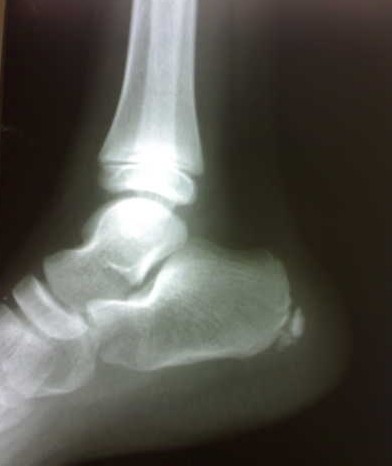
X-rayLateral foot X-ray in a child: fragmentation and sclerosis of the calcaneal apophysis, signs characteristic of Sever's disease.
Source : Sitati et al., Cases journal, 2009, figure 1 · CC BY
The diagnosis rests on three manoeuvres that take a minute. The X-ray, for its part, raises a question that two good studies answer in opposite ways, and one has to know which applies to the child in front of you.
A Swedish team assessed 30 consecutive children with a Sever's injury, all in considerable pain and very active in sport, and 15 matched control children without pain. Three clinical tests were compared, and each was set against the true status6 :
The authors propose basing the diagnosis on the history and the results of two specific clinical tests, and conclude that the diagnosis of Sever's injury is clinical and not radiological6.
Diagnostic values of the three heel tests
30 symptomatic children against 15 matched pain-free controls. Small sample: the confidence intervals are wide and were not published
Source: Perhamre S, Lazowska D, Papageorgiou S, Lundin F, Klässbo M, Norlin R. Sever's injury: a clinical diagnosis. J Am Podiatr Med Assoc 2013;103(5):361-8 (PMID 24072363). Case-control study of 45 subjects in all; these values come from a single piece of work and have not been replicated.
This is the most useful result of that same study, and it should close an old misunderstanding. Increased density and fragmentation of the calcaneal apophysis have long been presented as the radiological signs of the disease, supposedly reflecting the inflammation linked to the pull of the calcaneal tendon. The authors therefore X-rayed the controls too.
The result: all the children, cases as well as controls, had increased density of the apophysis. And half the controls with no pain at all showed fragmentation, against nearly 90% of the children with pain6. In other words, what was shown to parents as proof of the disease is, in the young athlete, a picture of normal growth.
The radiological signs do not separate the affected from the healthy
30 children with pain against 15 matched controls, all physically very active
Source: Perhamre S et al. J Am Podiatr Med Assoc 2013;103(5):361-8 (PMID 24072363). The values "nearly 90%" and "about 50%" are those reported by the authors, who do not publish exact percentages for fragmentation.
The preceding reasoning logically concludes that a child's heel pain should not be X-rayed. Another study, just as honest, reaches the opposite conclusion, and both must be read.
A team in Memphis reviewed all the records of children aged 4 to 17 who presented with heel pain, excluding acute trauma and calcaneal tendinopathies. Ninety-eight patients and 134 feet were retained, with a mean age of 10.8 years, all carrying a clinical diagnosis of calcaneal apophysitis. The radiographs were then re-read by three orthopaedic surgeons blinded to the clinical diagnosis7.
Five patients had an abnormality, all visible on the lateral view: three solitary bone cysts of the calcaneus, one non-ossifying fibroma of the distal tibia and two calcaneal stress fractures, one patient having both a cyst and a fracture in the same foot. The rate of abnormalities was therefore 5.1% of patients. These findings changed management, towards closer radiographic monitoring or immobilisation7.
The point that should hold the physiotherapist's attention is not the figure, it is the sentence that goes with it: no common feature of the history or the examination made it possible to identify the patients likely to have an abnormal X-ray7. One cannot therefore take comfort in the idea that atypical cases will be spotted on examination: in this series, they were not.
How to reconcile the two
The two studies do not answer the same question. Perhamre asks: "does the X-ray make the diagnosis of Sever's disease?", and the answer is no, its signs are present in healthy children. Rachel asks: "does the X-ray change anything in children referred for heel pain?", and the answer is yes, in 5% of cases, and unpredictably. The two answers are compatible, and they converge on one course of action: do not request imaging to confirm Sever's disease, but do not hesitate to request it to rule something else out, which is a different indication. The level of evidence remains low on both sides: 45 subjects for one, a retrospective level IV series for the other.
In practice, in the French setting, the physiotherapist does not prescribe this imaging: he has it requested. The wording matters, and "rule out a bone lesion in a child with heel pain, lateral view" obtains a more useful examination than "suspected Sever's disease".
Key points
Three tests are enough, and the best is the simplest: single-leg toe standing, with a measured sensitivity of 100%. The X-ray does not confirm the diagnosis, since density and fragmentation exist in 100% and 50% of pain-free children. It does keep a role in ruling things out: 5.1% of serious lesions in a series of 98 children, with no clinical warning sign at all.
The serious diagnoses in this chapter are rare. Yet they share with Sever's disease the age, the site and sometimes the mode of onset, and the only reliable filter is the behaviour of the pain, not its intensity.
One preliminary remark, because it changes how what follows should be read. A ten-year-old with heel pain has, in the vast majority of cases, calcaneal apophysitis. None of the pictures described here should lead to systematic investigation. What should change is the way the complaint is listened to: the question is not "is this serious?" but "does this pain behave like a mechanical pain?". A mechanical pain increases with load, decreases with rest, allows sleep, and comes with nothing else. As soon as one of those four points is missing, we are outside the frame.
Red flags in a child with heel pain
None of these features falls within first-line physiotherapy management. Their presence calls for a medical opinion, and for the first two, the same day.
A systematic review gathered 42 studies and 128 cases of paediatric calcaneal osteomyelitis published between 2000 and 2021. The sex ratio was two boys to one girl, the median age 8 years, the median duration of symptoms 0.6 months. Trauma was the main cause in 41 cases (54%), and limitation of activity the most frequent symptom, in 68 cases. Cultures were positive in 75.4% of samples, Staphylococcus aureus being the most frequent organism (57.1%). Surgery was performed in 51% of patients, and the rate of infectious recurrence was 6.8%13.
What to retain for the clinic: "limitation of activity" as the dominant symptom is exactly what a child with Sever's disease reports. What separates the two is fever, the constant character of the pain, and the absence of a mechanical rhythm. A child with heel pain at night, at rest, sitting down, and with a fever, is not a child to rehabilitate.
The Greek series of 12 children operated on for an osteoid osteoma of the foot between 1995 and 2010 is instructive less for what it describes than for what it measures. Mean age 12 years, from 8 to 16. The lesion was in the talus in 8 cases, in the calcaneus in 3, in the fourth metatarsal once. And above all: symptoms lasted on average two years, from 14 months to 4 years, before diagnosis. The authors note atypical presentations (pain referred to the hip, extensor tenosynovitis, atrophy of the calcaneal tendon) and conclude that the diversity of symptoms and the delay in diagnosis remain problematic20.
The case of calcaneal osteosarcoma published in 2023 tells the same story in a graver key: a 19-year-old woman whose heel pain had resisted analgesics for four months. The authors stress that these tumours are frequently taken clinically for a traumatic or inflammatory process, and call for osteosarcoma to be kept in the differential diagnosis of chronic heel pain in the adolescent, despite its rarity21.
The signal is not the intensity of the pain, it is its refusal to follow load. Heel pain that does not improve when load is reduced is no longer an apophysitis: it is an open question.
Faced with a child with heel pain
Physiotherapy decision path. The red outcomes fall outside first-line rehabilitation
Built from: Perhamre S et al. J Am Podiatr Med Assoc 2013;103(5):361-8 (PMID 24072363) for the clinical tests; Rachel JN et al. J Pediatr Orthop 2011;31(5):548-50 (PMID 21654464) for the yield of imaging and the absence of any predictive sign; Dhagey IA et al. BMC Infect Dis 2024;24(1):998 (PMID 39294568) and Clarke RT et al. Arch Dis Child 2016;101(10):894-901 (PMID 27647842) for the urgency criteria.
The table crosses the two variables that sort out a child's pain attributed to growth, and adds for each condition the step that must not be missed. It runs from the heel down to the knee, and refers to the dedicated page where one exists: the knee conditions are not covered again here.
| Age | Site | Condition | Warning sign | Course of action |
|---|---|---|---|---|
| 8 to 15 years | Posterior heel, calcaneal insertion | Sever's disease | Painful medio-lateral squeeze of the calcaneus, single-leg toe standing impossible, pain paced by exertion | Clinical diagnosis. Load adaptation, no complete stop, no confirmatory imaging |
| Any age, median 8 years | Heel, constant pain | Osteomyelitis of the calcaneus | Fever, non-mechanical pain, marked limitation of activity, symptoms present for less than a month13 | Medical opinion the same day |
| Any age | Diffuse, often several sites | Haematological malignancy | Night pain that wakes the child, pallor, fever, bruising, hepatosplenomegaly. Limb pain in 43% at diagnosis14 | Medical opinion, full blood count |
| 8 to 16 years | Heel or foot, often poorly localised | Osteoid osteoma | Night pain and relief with anti-inflammatories in only half of cases. Mean diagnostic delay of two years20 | Imaging and specialist opinion for any heel pain that persists without a mechanical rhythm |
| Adolescent | Heel, with swelling | Malignant bone tumour | Chronic pain resistant to analgesics, swelling, continuous worsening21 | Medical opinion without delay |
| Boys over 6 years | Entheses, asymmetrical, lower limbs | Enthesitis-related juvenile arthritis | Enthesitis and asymmetrical arthritis, morning stiffness, secondary inflammatory low back pain, family history22 | Paediatric rheumatology opinion. Screening for uveitis |
| Adolescent athlete | Body of the calcaneus | Stress fracture of the calcaneus | Pain on compression of the body of the calcaneus, recent increase in load. Found in 2 of the 98 children referred for heel pain7 | Relative unloading and lateral view imaging |
| 4 to 17 years | Calcaneus, incidental finding | Solitary bone cyst, non-ossifying fibroma | None. Three cysts and one fibroma among 98 children, with no clinical warning sign at all7 | This is the argument for a lateral view in heel pain that does not respond |
| 3 to 12 years | Diffuse, bilateral, lower limbs | So-called growing pains | Evening or night pain, strictly normal examination, no limp, no relation to exertion15 | Diagnosis of exclusion. Any limp or any mechanical pain rules it out |
| Adults mainly, rare in children | Plantar heel | Plantar fasciitis | Pain on the first steps in the morning, at the plantar insertion of the calcaneus and not its posterior border | See the dedicated article: plantar fasciitis |
| 11 to 15 years | Tibial tubercle | Osgood-Schlatter disease | Pain and swelling of the tibial tubercle, two to three centimetres below the patella | See the dedicated article: Osgood-Schlatter disease |
| 10 to 14 years | Inferior pole of the patella | Sinding-Larsen-Johansson disease | Exquisite tenderness on palpation of the patellar apex, jumping sports | See lower limb growth conditions |
| 9 to 16 years | Knee, groin, thigh or hip | Slipped capital femoral epiphysis | Obligatory external rotation on hip flexion, limp, frequent overweight | Stop weight-bearing and seek a surgical opinion. See the dedicated article |
| 11 to 19 years | Knee, often poorly localised | Osteochondritis dissecans | Persistent exertional pain, effusion, locking or giving way if the lesion is unstable | Imaging for any effusion. See the dedicated article |
| Adolescents, girls mainly | Anterior knee, around the patella | Patellofemoral pain syndrome | Pain on stairs, on squatting, on prolonged sitting. Point prevalence of 22.7% in adolescent female athletes23 | See the dedicated article: patellofemoral pain syndrome |
One line deserves to be read twice, that of the cysts and the fibroma. These are lesions that gave no distinctive sign, in children whose clinical diagnosis was calcaneal apophysitis, and that nevertheless changed management7. They do not justify irradiating every child's heel, but they explain why heel pain that does not follow load should be imaged rather than rehabilitated harder.
Key points
The sorting is not about the diagnosis but about the behaviour of the pain. A mechanical pain increases with load, eases with rest and allows sleep. Fever, constant pain, waking at night, deterioration in general condition and swelling fall outside the scope of physiotherapy. And the absence of a response after six weeks of properly dosed load is in itself a reason to reopen the diagnosis, not to intensify treatment.
The usual story fits in one sentence: a heavy child, who trains too much, with stiff calves. Three studies have tested those three elements. None comes out intact.
A systematic review published in BMJ Open searched for the risk factors, associated factors and consequences of calcaneal apophysitis in six databases from inception to April 2021. Of 736 studies identified, 11 observational studies met the criteria, totalling 1,265 participants with a mean age of 10.72 years. Four studied extrinsic factors, ten intrinsic factors, three both24.
The factors listed are limited ankle dorsiflexion, foot alignment, midfoot stiffness and mobility, plantar pressures and ground reaction forces, body mass index, age, sex, the presence of other osteochondroses and sports participation. The conclusion is cautious and worth quoting in its reservation: limited dorsiflexion is the most frequently studied intrinsic factor, followed by peak plantar pressures and foot malalignment, but disagreements exist between the investigators of the included studies, and there is no unanimity on what counts as a risk factor, an associated factor or a consequence24.
This last distinction is decisive and rarely made. A child who has had heel pain for three months walks differently, loads the heel less, and loses dorsiflexion. Measuring his dorsiflexion at the moment he consults does not say whether the stiffness caused the pain or the reverse.
An Australian prospective study compared children with Sever's disease with a non-symptomatic population of the same age, testing the causal models most cited in the literature: biomechanical foot malalignment measured by the Root method and by the foot posture index, ankle dorsiflexion measured with modified equipment, body mass index, and the volume and type of sporting activity8.
The result is clear-cut and runs against intuition: this study finds no evidence that weight and activity level are risk factors. The odds ratio for reduced ankle dorsiflexion, statistically significant on the left side, was 0.93, which the authors themselves describe as clinically negligible. Only the forefoot to rearfoot ratio emerged with a clinically stronger and bilateral odds ratio, which leads the authors to point to biomechanical malalignment as a lead to explore, and no more8.
The Italian study of 430 young athletes goes the same way and adds a frankly counter-intuitive result. Body mass index, sex, surface type, sport and the foot posture index should not be considered risk factors. On the other hand, a significantly higher risk was found in the youngest subjects, in those who had fewer training sessions per week, and in those whose sessions were shorter9.
How to read this result without over-interpreting it
That less trained children have more heel pain does not mean that training more protects them. The most economical explanation is one of exposure and adaptation: the most trained children are also the most selected and the best adapted to load, and those who train little undergo relatively more violent load spikes on a less accustomed bone. This is a cross-sectional study: it describes an association, not a causal link. It is nevertheless enough to disqualify "he does too much" as an automatic explanation.
The cross-sectional study nested in the Australian trial measured height, weight, waist circumference, body mass index, foot posture and ankle range in 124 children aged 8 to 14 with a clinical diagnosis of calcaneal apophysitis, 72 of them boys. Compared with normative values, these children had a higher body mass index, a higher weight and a greater height, all with p below 0.001, as well as differences in foot posture and ankle range25.
There is no contradiction with the previous study, but a difference of question: comparing affected children with population norms is not the same as comparing cases with controls matched from the same sporting environment. The first method also captures what distinguishes a young athlete from a child in general.
The most directly useful result of this work lies elsewhere, in the multivariate analysis: the older participants (p = 0.046) and those whose pain had lasted longer (p = 0.043) reported more intense pain. The authors conclude that early management may reduce the intensity and duration of pain25. In other words, the only clearly identified modifiable factor in this literature is the delay before management, which leads straight back to the problem of the term "growing pain", which defers the consultation.
A Japanese prospective cohort study provides the only element capable of guiding an exercise. Thirty-seven young gymnasts, that is 74 limbs, were assessed on flexibility, foot alignment, trunk function and functional balance tests, then followed for six months. The gymnasts who developed Sever's disease had a lower arch height ratio and lower values on the star excursion balance test in the anterior direction. The authors suggest that support of the medial longitudinal arch and dynamic postural balance work could prevent the condition26 : a prospective suggestion, in 37 subjects from a single sport, which has never been tested in a trial.
Key points
No risk factor is firmly established. Limited dorsiflexion is the most studied, with disagreements between studies and without our knowing whether it precedes or follows the pain. Weight and training volume do not emerge, and one study even finds more cases in the least trained children. The only clearly identified modifiable factor is the delay before management: the longer the pain has lasted, the more intense it is.
The short answer is disappointing and must be given as it stands: ten randomised trials in all, 654 children, and a certainty of evidence rated low to very low for almost everything. The long answer is more useful, because what these trials show in outline is worth more than what they show directly.
The Cochrane review devoted to non-surgical treatment of lower limb apophyseal injuries searched six databases with no language restriction up to 4 January 2025. It includes 10 randomised trials, seven of them on calcaneal apophysitis and three on traction apophysitis of the tibial tubercle, totalling 654 children with a mean age of 10.3 to 13.3 years, 73% of them boys. Risk of bias was assessed with the RoB 2 tool, and the certainty of the evidence with the GRADE method10.
The comparisons that produced a usable result are few10 :
The authors' conclusion is explicit: the evidence is limited, rated for the most part as low to very low certainty, the outcomes were heterogeneous, and the trials did not specifically target children with persistent symptoms and functional limitation. No trial measured quality of life, whereas cohort studies have shown that apophyseal injuries can affect it in the long term, and none examined the economic impact10.
The evidence map: what our recommendations rest on
Sample sizes of the comparisons that produced a result, and the GRADE certainty assigned by the 2026 Cochrane review
Source: Williams CM, Krommes K, Paterson KL, Haines T, Caserta A, Thorborg K. Non-surgical treatment for lower limb apophyseal injuries. Cochrane Database Syst Rev 2026;7(7):CD015156 (PMID 42454655). The widths are proportional to the sample sizes of the comparisons, not to the size of the effects.
It is a pragmatic Dutch single-blind trial that compared three commonly used approaches in children aged 8 to 15 with symptoms for at least four weeks and a revised faces pain scale of at least 3 points: a protocol of wait and see, or a heel lift, or an eccentric exercise programme supervised by a physiotherapist. Treatment lasted ten weeks, the primary outcome was the faces pain scale at three months, with intention-to-treat analysis. One hundred and one children were included, three lost to follow-up11.
At six weeks, the children with a heel lift were more satisfied than those in the other two groups, and improved significantly on the Oxford Ankle and Foot Questionnaire, child version, compared with wait and see; the physiotherapy group improved significantly on the parent version. But at the end of follow-up, all three approaches had produced a clinically relevant and statistically significant reduction in pain, with no clinically relevant difference between them. The authors conclude that clinicians should deliberate with patients and parents about the preferred treatment11.
This result is richer than it looks. It does not say that nothing works: it says that everything works, including doing nothing in particular, which shifts the question away from the choice of technique towards support, load dosage and the explanation given to the family.
The Australian 2 × 2 factorial trial recruited 124 children aged 8 to 14 with a clinical diagnosis of calcaneal apophysitis. Two factors were crossed: the type of in-shoe insert, heel lift or prefabricated orthosis, and whether or not the shoe was replaced. The primary outcome was functional disability, the secondary outcomes pain and dorsiflexion range, with follow-up at 1, 2, 6 and 12 months27.
At 1 and 2 months there was a main effect of the heel lift, but only in the physical domain of the Oxford questionnaire (p = 0.04). At 6 and 12 months, no main effect and no interaction remained, for any outcome. The authors conclude that beyond two months the choice of treatment may be a matter of clinical judgement, cost minimisation or patient preference27.
| Approach | Best study | What it shows | Certainty |
|---|---|---|---|
| Load adaptation and education | No dedicated randomised trial | It is the logical foundation of management, but it has never been isolated in a trial. The "usual care" of the trials includes exercise, stretching, anti-inflammatories and massage, and remains poorly described10 | not assessed |
| Supported wait and see | Pragmatic trial, n = 10111 | Clinically relevant reduction in pain at three months, with no difference from the heel lift or from physiotherapy | low |
| Heel lift | Pragmatic trial n = 10111 and factorial trial n = 12427 | Greater satisfaction at six weeks and a slight functional advantage at two months. No advantage left at 6 and 12 months | low |
| Foot orthoses | Cochrane comparison, n = 12310 | Probably little or no difference from a simple heel lift, for pain as for function | moderate |
| Supervised eccentric exercise | Arm of the pragmatic trial, n = 10111 | Improves the parent questionnaire significantly compared with wait and see at six weeks; no advantage at three months | low |
| Kinesio tape | Randomised trial, n = 2228 | Better AOFAS functional score at 1 and 3 months than sham taping, but no effect on pain. With no serious adverse effect | very low |
| Braces for barefoot sports | Randomised trial, n = 43 included, 32 analysed29 | Both braces tested improve pain and function over three months, with no difference between them | very low |
| Custom-made orthoses | Randomised trial, n = 20830 | Pressure pain threshold +53.4% and VAS −68.6% against off-the-shelf heel lifts. A result not taken up by the Cochrane synthesis, which concludes there is no difference | contested |
| Cast immobilisation | No randomised trial | Described in series of resistant cases, notably the one that made it possible to follow the regression of the MRI signal5. To be considered only in refractory forms, and on medical advice | not assessed |
A divergence not to be hidden
A CONSORT-compliant Spanish trial randomised 208 children aged 9 to 12 between custom-made polypropylene orthoses and off-the-shelf heel lifts, with twelve weeks of follow-up. It reports a 53.4% increase in the pressure pain threshold (95% CI 47.1 to 59.7) and a 68.6% fall in the VAS (95% CI −74.5 to −62.7) in favour of custom orthoses, with p below 0.00130. It is the largest sample in this whole field, and the reported effect is considerable. The Cochrane synthesis, which searched the databases up to January 2025, nevertheless concludes there is no difference between orthoses and heel lifts on the basis of a comparison of 123 participants10. We take the Cochrane review as our anchor, because it applies RoB 2 and GRADE explicitly; we flag the discrepancy rather than keeping quiet about it. A clinician who chooses a custom orthosis is not making a mistake: he is making a costly bet on a result the reference synthesis did not retain.
Key points
No treatment has proved itself superior. The only moderate-certainty result in this whole field is an absence of difference between two types of insole. A trial of 101 children shows that supported wait and see, the heel lift and supervised exercise give the same result at three months. The practical consequence is not nihilism: it is that the physiotherapist's added value lies in load dosage and support of the family, not in the choice of device.
This is the heart of the work, and it is also the part least supported by trials. We therefore have to distinguish what is demonstrated, what is deduced from a mechanism, and what rests on expert agreement, and say so to the family in those terms.
Three arguments converge, none decisive on its own.
The first is mechanistic. If the lesion is a stress lesion of an immature bone that has not yet adapted to the biomechanical demands of an active child5, then removing load entirely does not advance adaptation: it defers the problem until the resumption, where the stress met again will be relatively greater on a bone that has lost what it had gained.
The second is empirical and comes from the pragmatic Dutch trial. The "wait" arm was not a "stop sport" arm: it was a wait-and-see protocol. And it produced a clinically relevant reduction in pain, like the other two11. Nothing in this literature shows that immobilising does better.
The third concerns consequences. The American Academy of Pediatrics clinical report on overuse injuries, overtraining and burnout in young athletes recalls that taking part in sport brings the child considerable physical and psychological benefits, and that burnout, defined as a state of physical or mental exhaustion with a reduced sense of accomplishment leading to devaluation of the sport, is one of the leading causes of dropout in youth sport, and a direct threat to the aim of lifelong physical activity31. A child kept away from his club for three months for a condition whose prognosis is spontaneously favourable pays a price that appears in no trial.
What is not demonstrated, and should stop being presented as such
The numerical rules that circulate about load in the young athlete (a weekly number of hours capped by age, one rest day a week, several months a year without competition) come from expert agreement, notably the position statement of the American Medical Society for Sports Medicine on overuse injuries and burnout in youth sport32 and the clinical reports of the American Academy of Pediatrics31,33. They have never been tested in a trial in children with calcaneal apophysitis, and the few specific data even run counter to the simple idea that "too much training" explains the condition8,9. Using them as points of reference for discussion is reasonable; presenting them as validated thresholds is not.
The practical principle rests on a distinction families grasp immediately: between pain one works through and pain that settles in.
In concrete terms, the child can continue an activity that wakes a tolerable discomfort during exertion, that subsides within the following two hours, and that has not increased by the next morning. He must cut back as soon as one of those three points is crossed: pain that forces him to change his running or to limp, pain that persists in the evening, pain on the first steps the next day. This framework has not been validated by a trial in calcaneal apophysitis, none has tested it, but it follows directly from the load reasoning and it gives the child a criterion he can apply on his own, which "stop if it hurts" does not.
A child told to stop as soon as it hurts learns that he can no longer do anything. A child given three criteria learns to dose, and that is what we leave him with for the future.
Load modulation is not binary, and the most frequent error is to jump straight to cutting volume when three less costly levers exist upstream.
A quick reading of the literature could lead to discouragement: if supported wait and see does as well as a supervised programme, what is rehabilitation for? The answer is in the very structure of the trial that raises the question. All three arms included a consultation, an explanation, a framework and ten weeks of follow-up. What was compared was three techniques, not the presence against the absence of care.
And one of the few modifiable factors identified in this field points in exactly that direction: in 124 children, pain was more intense in those in whom it had lasted longer, which led the authors to recommend early management25. The physiotherapist who recognises Sever's disease in a single consultation, rules out the red flags, explains the mechanism and puts the child back on a dosed load is doing precisely what that finding suggests.
What physiotherapy really contributes here
This is the real intervention, and it is also the only one for which a measure exists: parents and children do not perceive this condition in the same way, and the gap narrows over time.
A longitudinal study nested in the Australian trial had the Oxford Ankle Foot Questionnaire, child version, completed by the child and by the parent, at inclusion and then at 1, 2, 6 and 12 months. One hundred and thirty-three children were recruited, 124 took part, 101 completed the questionnaire at all five time points34.
Agreement between child and parent ranged from poor (0.06) to good (0.77) for the physical domain, and from poor (0.09) to good (0.66) for the footwear domain. The school and emotional domains held up better, from moderate (0.46) to good (0.77). Above all: parents initially reported a greater impact than their child did, with the two viewpoints converging over the follow-up. The authors conclude that it is essential to understand the impact from the child's point of view as well as the parent's34.
The consequence in consultation is direct. Questioning only the parent overestimates the impact at the start, and leads to heavier restrictions than the child experiences. Questioning only the child, who wants to play, underestimates it. Both are needed, separately, and it helps to name the gap when it appears: "you see him suffering more than he feels it, that is usual, and the two will come closer together."
Five sentences to say, and why
This is the most often forgotten party, and the one who in practice decides the real load. Three messages are enough, and they must be phrased in terms of the session, not of the diagnosis.
The systematic review of 1,362 cases concludes, moreover, after noting the absence of any robust treatment data, that educating parents and coaches about the symptoms, aetiology and treatment of this condition is essential in order to diagnose it earlier and obtain better outcomes19. When the literature of a field cannot separate the techniques, it ends up pointing to what is left.
Key points
Parents initially overestimate the impact compared with what the child reports, and the two perceptions converge over the follow-up34 : both must be asked. The most useful message is the one that replaces "growing pain" with "load pain", because it makes action possible. The coach must receive instructions in terms of session content, not of diagnosis.
"His knee hurts, it's growth" is the second most frequent phrasing after the heel one. This chapter does not go over any of the knee conditions again: it starts from that sentence and says which condition we are heading towards, with the link to the page that covers it.
First, why this chapter is short. Each of the conditions mentioned here has its own page on this site, written in full. Going over them again would create two texts competing for the same query, and the reader would lose the depth. What was missing was not one more description: it was the triage, that is, the translation of the complaint as it arrives into a direction.
The phrase is used for at least five distinct situations, only one of which is benign by definition.
These four questions take less than two minutes and are enough to give a direction.
A remark on co-occurrence
A child can have several apophysitis sites, successively or simultaneously. The systematic review of factors associated with calcaneal apophysitis does indeed list the presence of other osteochondroses among the factors recorded24. Finding Sever's disease in a child who also has knee pain therefore does not exempt one from examining the knee in its own right, and the reverse is true.
Key points
Faced with an adolescent knee labelled "growth", four questions give the direction: is the pain bilateral, nocturnal and unrelated to exertion; can it be pointed to with one finger on an insertion; is hip internal rotation symmetrical; is there effusion, locking or giving way. Only the first combination, complete, allows a benign conclusion. Patellofemoral pain syndrome is statistically the most likely diagnosis, with a 28.9% annual prevalence in adolescents23.
Four genuinely published cases or series, with their identifier. None is reconstructed or composite: cases that are too clean are exactly what makes a review article untrustworthy.
Two 10-year-old Indian monozygotic twin sisters, from a disadvantaged rural background, developed bilateral Sever's disease at the same time. Both reported bilateral heel pain of gradual onset for seven months, worsened by walking and physical activity. On examination, average build, weights of 26 and 27 kilograms, bilateral hallux varus in both, and a positive calcaneal squeeze test on both sides, which was enough to make the diagnosis. Anteroposterior and lateral radiographs of both feet confirmed the apophysitis. Faced with an identical clinical and radiographic presentation in two twins, the authors raise the possibility of a genetic predisposition or shared biomechanical risk factors35.
What this case teaches: seven months of symptoms before diagnosis, in two children whose examination was characteristic in every respect and for whom a thirty-second test was enough. It is the exact illustration of the problem the word "growth" creates: these are not difficult cases that drag on, they are easy cases that no one brings in. And the finding that goes with it is James's: the longer the pain lasts, the more intense it is25.
A 10-year-old girl was managed for persistent left heel pain. Clinical examination and diagnostic ultrasound confirmed Sever's disease, the ultrasound also making it possible to document associated involvement of the calcaneal tendon. Treatment combined medication, immobilisation, rehabilitation modalities and exercises, with improvement after ten weeks36.
What this case teaches: the order of magnitude of the timescale, consistent with the ten weeks of treatment in the pragmatic Dutch trial11, and the value of ultrasound when the question is not to confirm Sever's disease but to find out whether something else is added to it. It is also a single case, with no comparison group: it demonstrates the effectiveness of none of the modalities used, and the authors do not claim otherwise.
Twelve children (7 girls, 5 boys) were operated on for an osteoid osteoma of the foot in a Greek department between February 1995 and February 2010, with a mean follow-up of 5 years. Mean age 12 years, from 8 to 16. The lesion was in the talus in 8 cases, in the calcaneus in 3 cases, in the fourth metatarsal once. Symptoms lasted on average two years, from 14 months to 4 years. A crucial point: night pain and relief with anti-inflammatories, held to be characteristic of this tumour, were present in only half the cases. Atypical presentations were observed: pain referred to the hip, tenosynovitis of the foot extensors, atrophy of the calcaneal tendon. En bloc excision achieved complete cure with no recurrence20.
What this series teaches: the trap is not failing to know about osteoid osteoma, it is believing that it announces itself with its classic triad. In half the cases it does not. The only reliable signal is that of the red flags chapter: foot pain that lasts and does not follow load. Two years, in a child of 12, is two sporting seasons.
A 19-year-old woman consulted for heel pain resistant to analgesics for four months. The diagnosis retained was calcaneal osteosarcoma, an extremely rare entity since it accounts for less than 1% of all osteosarcomas. The authors stress that these tumours typically present with swelling and chronic heel pain, and that they are frequently taken clinically for a traumatic or inflammatory process21.
What this case teaches: it lies outside the age window of Sever's disease, and that is precisely why it is useful. At 19, the calcaneal apophysis is fused, and chronic heel pain can no longer be an apophysitis. The child's age is as much a criterion for exclusion as for inclusion.
None is a case of spectacular diagnostic error. Three out of four share the same feature, and it is the one worth retaining: a long duration of symptoms before anyone asked the question. Seven months, two years, four months. In the first case it was an ordinary apophysitis that nobody had brought in to be looked at; in the other two, a serious lesion nobody had thought of.
The common thread is therefore not the difficulty of the diagnosis. It is the delay, and the delay has the same cause in both situations: a child's pain that is not frightening does not prompt a consultation, and "it's growth" is the sentence that prevents it.
Key points
The published cases on this subject do not tell of difficult diagnoses: they tell of late ones. Seven months for a characteristic apophysitis recognisable in thirty seconds, two years for an osteoid osteoma half of whose carriers do not have the classic triad, four months for an osteosarcoma in a patient who was no longer of an age to have an apophysitis.
What is done at first contact, in what order, and the mistakes that cost time or credibility.
Step one, four questions in the history. Since when? How does the pain behave in relation to exertion: does it increase during, after, the next morning? Does it wake him at night? Has there been fever, weight loss, unusual tiredness? These four questions separate a mechanical pain from one that is not, and that is the only separation that matters at this stage.
Step two, three manoeuvres. Single-leg toe standing, on the painful side then the other. Medio-lateral squeeze of the calcaneus between thumb and index finger. Palpation of the insertion. The measured values of these three tests are in the diagnosis chapter; the first is the most sensitive and is also the one the child understands best6. Add a comparative ankle dorsiflexion measurement and a quick examination of the hip and knee, because one apophysitis does not exclude another24.
Step three, decide whether we are leaving the frame. The red flags are in their box. In their absence, we do not request imaging to confirm, and we say so explicitly to the family, who often expect it: the X-ray does not make this diagnosis, and its signs are present in all pain-free young athletes6.
Step four, set out the load plan. With the child, not only with the parent. Name the three criteria he will apply on his own: no limp during the session, pain subsided within two hours, no pain on the first steps the next day. Choose the levers in the order of the corresponding chapter, starting with the content of the session and not its volume. Offer a heel lift if it helps: it has a measured early advantage and no reported adverse effect10,11.
Step five, give the prognosis with its spread, and set the checkpoint. About two months, with wide variability12. And an explicit rule: if after six weeks of properly dosed load nothing has changed, we do not treat harder, we reopen the diagnosis.
It must be said to the family in terms they understand, because it changes the relationship. The wording that works: "we know this condition well, we know it gets better, and we do not know well which of the things we offer speeds it up; that is why I am suggesting we choose together what suits you best." That is exactly the conclusion of the authors of the pragmatic Dutch trial, who recommend deliberating with patients and parents about the preferred treatment11, and it is also what a systematic review of eight randomised trials says, concluding that conservative treatment is effective in general without being able to single out a best one37.
No, if both terms are used strictly. So-called growing pains are bilateral, occur in the evening or at night, are diffuse, cause no limp and bear no relation to exertion; Sever's disease is a mechanical pain, localised at the calcaneal insertion and paced by activity. And the term itself is misleading for both: two independent cohorts looked for a link with rapid growth and did not find one2,3, while 93% of published definitions make no reference to growth at all1.
No to confirm it, possibly yes to rule other things out. Among 30 children with pain and 15 pain-free controls, increased density of the apophysis was present in 100% of both groups and fragmentation in half the controls6 : these signs therefore cannot establish a diagnosis. On the other hand, among 98 children referred for heel pain, 5.1% had a serious lesion that nothing in the history or the examination had signalled7. The sensible approach is not to image a typical presentation that responds to load adaptation, and to image the one that does not.
The order of magnitude is two months. In a German football academy, the mean return to play was 60.7 days with a standard deviation of 64.9 days, in athletes with a mean age of 11.8 years; 13.6% of cases were recurrences, and those took longer12. This spread means that some children resume within three weeks and others within four months: announcing the mean alone sets up disappointment.
Nothing in the literature supports that. The most relevant trial compared a wait-and-see protocol, a heel lift and an eccentric exercise programme in 101 children: all three produced a clinically relevant improvement, with no difference between them11. The useful approach is to modulate the content of sessions (impacts, sprints, jumps, work on the toes) rather than to remove them.
The question is open, and it is the only one in this whole field with a moderate-certainty result: according to the Cochrane review, foot orthoses and heel lifts probably do not differ, either for pain or for function10. A Spanish trial of 208 children nevertheless reports a marked advantage for custom-made orthoses30, a result the Cochrane synthesis did not retain. In practice, starting with the least expensive option is defensible, and switching if it does not relieve the pain is defensible too.
For function perhaps, for pain no. The only randomised trial available involves 22 young footballers and compares active taping with sham taping: AOFAS functional scores were better at one and three months in the active group, but the authors themselves state that its role on pain is limited28. The Cochrane review rates this comparison as very low certainty10. With no known adverse effect, it can be used as an adjunct, not as a treatment.
No data describe any structural sequela. The window of vulnerability closes when the apophysis fuses: none of the 336 young baseball players examined at the stage of complete fusion had heel pain17. On the other hand, the impact during the active phase is not negligible, and it is poorly measured: the Cochrane review notes that none of the ten trials assessed quality of life10.
No, it is common. In the systematic review of 1,362 published cases, the presentation was bilateral in 43.2% of cases against 31.8% unilateral19, and among 336 young players screened, bilateral forms were twice as numerous as unilateral ones17. Bilateral involvement is not in itself a warning sign; what is one is pain that does not follow load, whether one-sided or two-sided.
The data do not say so. A prospective study concludes that there is no evidence that weight and activity level are risk factors8, and a study of 430 young athletes retains neither body mass index, nor sex, nor sport, nor surface type, and even finds a higher risk in the least trained children9. Blaming the condition on the child's weight in front of him is therefore both hurtful and unfounded.
It is plausible and unproven. Limited ankle dorsiflexion is the most studied intrinsic factor, but the systematic review that lists them reports disagreements between studies and the impossibility of distinguishing what is a risk factor from what is a consequence24 : a child who has been in pain for three months may have lost range because of the pain. No trial has isolated triceps surae stretching as an intervention. It can be offered if it is comfortable, without expecting it to be the main effect.
When the pain stops behaving mechanically: waking at night, constant pain at rest, fever, deterioration in general condition, swelling, or the absence of any improvement after six weeks of properly reduced load. Calcaneal osteomyelitis in children has a median age of 8 years13, limb pain is present in 43% of children at the diagnosis of leukaemia14, and a series of 12 osteoid osteomas of the foot reports a mean diagnostic delay of two years20.
The reasoning is the same, the conditions are not. The chapter devoted to the knee gives the four triage questions. The most important is that of hip internal rotation: an adolescent with knee pain, whose knee is normal on examination and one of whose hips has lost its internal rotation, must have hip imaging before any rehabilitation; the detail of this approach is in the article devoted to lower limb growth conditions.
Thirty-seven references, all checked against the NCBI E-utilities API at the time of writing: existence of the identifier, complete author list, journal, year, volume, pagination, and reading of the abstract to check that the source does establish what is attributed to it. The identifiers are clickable.
Behind this article
An author who explains, a reviewer who checks.
How we write and check our content

Physiotherapist · co-founder of Physio Learning
Marked for life by his first four-hour lecture without a single image, he took a master’s in instructional design so that it would never happen to anyone again. He hunts down publication bias and unreadable slides with the same intransigence.

Head of scientific content
Physiotherapist specialising in neuro-musculoskeletal practice and holder of a master’s in public health. He checks the methodological rigour of every article: primary sources, levels of evidence, no exceptions.
