















In brief
Patellofemoral pain (PFP), also called anterior knee pain, is retro- or peripatellar pain provoked by activities that load the patellofemoral joint in flexion: squatting, descending stairs, running, prolonged sitting (the cinema sign). Diagnosis is essentially clinical, based on reproduction of the pain on squatting and on palpation of the facets, imaging not being recommended in routine practice; the prognosis is often unfavourable, with around half of patients still symptomatic at five years. First-line treatment combines education and therapeutic exercise, combined hip and quadriceps strengthening being superior to quadriceps alone. Its annual prevalence reaches around 22.7% in adults.
Clinical synthesis based on the JOSPT 2019 CPG (Willy), the Manchester 2016 and Gold Coast 2018 international consensus statements, and the 2022-2025 meta-analyses (Winters NMA, Mansfield NMA, Hsu prognosis).
Clinical synthesis
- PFP is retro- or peripatellar pain provoked by activities that load the joint (stairs, squatting, running, prolonged sitting: the “cinema sign”). Annual prevalence of around 22.7% in adults and 28.9% in adolescents.
- The 1.5- to 2-fold higher rate in women, particularly among adolescent female athletes and recreational female runners, is well documented. Incidence rises with training load.
- The main mechanism is an increase in stress on the patellofemoral joint (Stress = Force / Contact area), promoted by quadriceps weakness and poor control of the hip muscles.
- The prognosis is often unfavourable: around 50% of patients remain symptomatic at 5 years without structured management. PFP is a possible precursor of patellofemoral osteoarthritis.
- Diagnosis is essentially clinical, based on the location of the pain and its reproduction on squatting, on stair descent and on palpation of the patellar facets.
- The use of imaging is not recommended in routine practice since structural “abnormalities” (chondromalacia) are common in asymptomatic people and poorly correlated with pain.
- First-line treatment combines patient education and therapeutic exercise (Gold Coast 2018 consensus, JOSPT 2019 CPG, Winters 2022 network meta-analysis).
- The combination of hip + quadriceps strengthening is superior to strengthening the quadriceps alone (Nascimento 2018 JOSPT, meta-analysis of 14 trials).
- For runners: a 5-10% increase in cadence reduces patellofemoral stress and relieves pain (gait retraining Esculier 2018, Davis 2020).
- Passive therapies (taping, manual therapy, orthoses) are short-term adjuncts, never the mainstay of treatment.
- Psychosocial factors (kinesiophobia, catastrophising) predict poorer recovery and should be assessed systematically (Maclachlan 2017 SR BJSM).
- The primary prevention of PFP rests mainly on patellar braces (RR 0.40) and on running retraining (RR 0.21) according to the Culvenor 2020 BJSM SR/MA.
- The differential diagnoses to know: patellar tendinopathy, plica syndrome, Hoffa syndrome, infrapatellar saphenous neuralgia, early patellofemoral osteoarthritis.
- The red flags (major effusion, locking, trauma, fever, night pain) call for orthopaedic referral before any rehabilitation protocol.
- Return to sport should be guided by functional criteria (symmetrical strength ≥ 90%, VAS < 2/10 on sporting tasks, single-leg hop tests) and not by a calendar.
Contents
- What are the fundamentals to know about patellofemoral pain?
- How do you assess and diagnose patellofemoral pain with certainty?
- Which treatment strategies are the most effective?
- Runners and PFP: why does this subgroup deserve a dedicated approach?
- How do you secure lasting recovery and prevent flares?
- What do real clinical cases teach us?
- How do you apply these recommendations concretely in your practice?
What are the fundamentals to know about patellofemoral pain?
Patellofemoral pain syndrome (PFP), also called patellofemoral pain (PFP), or more broadly “anterior knee pain”, refers to pain located behind or around the patella, provoked or aggravated by activities that load the patellofemoral joint in flexion.¹ ² It is not an organ-based diagnosis but a clinical syndrome whose diagnostic coherence was restated by the 4th International Patellofemoral Pain Research Retreat (Manchester, 2016) and then by the JOSPT 2019 CPG.¹ ²
How is this condition defined, who is affected and what are the risk factors?
Diagnosis of PFP rests on three main elements according to the Manchester international consensus²:
- Retro- or peripatellar pain lasting more than 6 weeks, of insidious onset or linked to an increase in load.
- Pain reproduced by at least one activity that loads the patellofemoral joint in flexion such as squatting, descending stairs, running, jumping or prolonged sitting (the “cinema sign”).
- Exclusion of the other causes of anterior knee pain (patellar tendinopathy, plica, Hoffa, meniscal, ligamentous, osteochondral, neuropathic).¹
The SR/MA by Smith et al. published in PLoS One in 2018 remains the epidemiological reference: pooling 23 studies, it reports an annual prevalence of 22.7% in the general population and of 28.9% in adolescents, making PFP one of the most frequent musculoskeletal conditions of the lower limb. 🦵⁴
📊 Annual prevalence of PFP by subpopulation
Smith 2018 meta-analysis (PLoS One, 23 studies) and Boling 2009 (JUMP-ACL)
Sources: Smith BE et al. PLoS One. 2018;13(1):e0190892 (PMID 29324820) · Boling MC et al. Am J Sports Med. 2009;37(11):2108-16 (PMID 19797162). Female over-representation reaches a factor of 2.23 in the JUMP-ACL cohort (incidence 33% in women vs 15% in men during military training).
PFP preferentially affects adolescents and young adults (15-40 years), with a well-documented female over-representation . The SR/MA by Neal et al. (BJSM 2019) identified three groups of risk factors with a sufficient level of evidence:⁵
- Quadriceps weakness measured at baseline (the most consistent evidence, prospective predictor).
- Weakness of the hip muscles (abductors, external rotators), a risk factor in women in particular.
- Lateral patellar hyperpressure and altered motor control during weight-bearing tasks (single-leg squat, landing).
Conversely, several long-invoked factors have been downgraded: the Q angle is not a reliable predictor, static patellar malalignment on imaging is not correlated with pain, and excessive foot pronation has only a modest effect confined to a subgroup.⁵ ⁶ The pathomechanical model proposed by Powers, Witvrouw, Davis and Crossley (BJSM 2017) brings all these elements together in a simple equation: joint stress = force / contact area, modulated by proximal factors (hip, trunk), local factors (quadriceps, peripatellar tissues) and distal factors (foot, ankle).⁶
What happens inside the body and how does PFP evolve naturally?
PFP is neither an inflammatory condition nor an obligatory structural lesion. The concept of “chondromalacia”, long invoked, is now largely abandoned as an aetiology: 30 to 50% of asymptomatic individuals show patellofemoral cartilage abnormalities on MRI without any pain.¹ ⁷ The dominant pathophysiological model is that of tissue homeostasis proposed by Scott Dye (2005, Clin Orthop Relat Res): PFP arises when the load applied to the joint exceeds the envelope of function : the individual capacity of the tissues to tolerate stress without pain.⁷
The finite element study by Farrokhi, Keyak and Powers (Osteoarthritis Cartilage 2011) quantified this phenomenon: individuals with PFP show mean patellar stress 1.3 to 1.5 times higher than asymptomatic controls during squats at 15° and 45° of flexion.⁸ The structures responsible for the pain are multiple (subchondral bone, retinaculum, infrapatellar fat pad (Hoffa), synovium) and all richly innervated.⁷
Key point: the stress model
- PFP is not an inflammation and not necessarily a cartilage lesion.
- It results from an excess of mechanical stress on richly innervated tissues (bone, retinaculum, Hoffa fat pad).
- Load can exceed the envelope of function through ↑ force (rapid increase in training volume) or ↓ contact area (muscle weakness, dynamic knee valgus).
- An “abnormal” MRI says nothing about the prognosis: 30-50% of asymptomatic individuals have cartilage lesions.
The natural history is frequently unfavourable, which contradicts the old perception of a benign, self-limiting condition. The recent SR/MA by Hsu et al. published in JOSPT in 2025 synthesised the available prospective studies and shows that a substantial proportion of patients remain symptomatic well beyond the initial course of management.⁹ In adolescents, cohort data report up to 75% persistent pain at 1 year and 50-90% in the long term (1-20 years).¹⁰ In adults, the SR by Collins et al. (BMC Musculoskelet Disord 2010) had already identified as predictors of a poor long-term outcome: symptom duration before the first consultation, high baseline pain, and persistent quadriceps weakness.¹¹
A direct consequence: long-standing PFP is a possible risk factor for patellofemoral osteoarthritis later on (PFOA), although direct causality remains debated.² ¹² This reinforces the idea that early, structured management is not a comfort but a prognostic issue. 📉
Key points: chapter 1
- PFP is anterior knee pain linked to an excess of stress on the patellofemoral joint, and not an inflammation.
- Annual prevalence ≈ 22.7% in adults and 28.9% in adolescents (Smith 2018, PLoS One).
- The most consistent risk factors: weakness of the quadriceps and of the hip muscles (Neal 2019).
- The prognosis is unfavourable in roughly 40-50% of patients at 5 years (Hsu 2025), so early structured management is crucial.
References: chapter 1
- Willy RW, Hoglund LT, Barton CJ, et al. Patellofemoral Pain: Clinical Practice Guidelines Linked to the International Classification of Functioning, Disability and Health From the Academy of Orthopaedic Physical Therapy of the American Physical Therapy Association. J Orthop Sports Phys Ther. 2019;49(9):CPG1-CPG95. doi:10.2519/jospt.2019.0302
- Crossley KM, Stefanik JJ, Selfe J, et al. 2016 Patellofemoral pain consensus statement from the 4th International Patellofemoral Pain Research Retreat, Manchester. Part 1: Terminology, definitions, clinical examination, natural history, patellofemoral osteoarthritis and patient-reported outcome measures. Br J Sports Med. 2016;50(14):839-843. PMID 27343241
- Crossley KM, van Middelkoop M, Callaghan MJ, et al. 2016 Patellofemoral pain consensus statement from the 4th International Patellofemoral Pain Research Retreat, Manchester. Part 2: recommended physical interventions. Br J Sports Med. 2016;50(14):844-852. PMID 27247098
- Smith BE, Selfe J, Thacker D, et al. Incidence and prevalence of patellofemoral pain: A systematic review and meta-analysis. PLoS One. 2018;13(1):e0190892. PMID 29324820
- Neal BS, Lack SD, Lankhorst NE, Barton CJ, Morrissey D, van Middelkoop M. Risk factors for patellofemoral pain: a systematic review and meta-analysis. Br J Sports Med. 2019;53(5):270-281. doi:10.1136/bjsports-2017-098890
- Powers CM, Witvrouw E, Davis IS, Crossley KM. Evidence-based framework for a pathomechanical model of patellofemoral pain: 2017 patellofemoral pain consensus statement from the 4th International Patellofemoral Pain Research Retreat, Manchester, UK: Part 3. Br J Sports Med. 2017;51(24):1713-1723. PMID 29109118
- Dye SF. The pathophysiology of patellofemoral pain: a tissue homeostasis perspective. Clin Orthop Relat Res. 2005;436:100-110. PMID 15995427
- Farrokhi S, Keyak JH, Powers CM. Individuals with patellofemoral pain exhibit greater patellofemoral joint stress: a finite element analysis study. Osteoarthritis Cartilage. 2011;19(3):287-294. PMID 21172445
- Hsu HC, Chen LL, Chiu YC, et al. Prognosis of Patellofemoral Pain: A Systematic Review With Evidence- and Gap-Map. J Orthop Sports Phys Ther. 2025;55(9):568-590. doi:10.2519/jospt.2025.13491
- Rathleff MS, Vicenzino B, Middelkoop M, et al. Patellofemoral Pain in Adolescence and Adulthood: Same Same, but Different? Sports Med. 2015;45(11):1489-1495. PMID 26178330
- Vicenzino B, Collins N, Crossley K, Beller E, Darnell R, McPoil T. Foot orthoses and physiotherapy in the treatment of patellofemoral pain syndrome: a randomised clinical trial. BMC Musculoskelet Disord. 2008;9:27. PMID 18304317.
- Crossley KM. Is patellofemoral osteoarthritis a common sequela of patellofemoral pain? Br J Sports Med. 2014;48(6):409-410. PMID 24569145
- Lankhorst NE, Bierma-Zeinstra SM, van Middelkoop M. Risk factors for patellofemoral pain syndrome: a systematic review. J Orthop Sports Phys Ther. 2012;42(2):81-94. doi:10.2519/jospt.2012.3803
- Boling M, Padua D, Marshall S, Guskiewicz K, Pyne S, Beutler A. A prospective investigation of biomechanical risk factors for patellofemoral pain syndrome: the Joint Undertaking to Monitor and Prevent ACL Injury (JUMP-ACL) cohort. Am J Sports Med. 2009;37(11):2108-2116. PMID 19797162
How do you assess and diagnose patellofemoral pain with certainty?
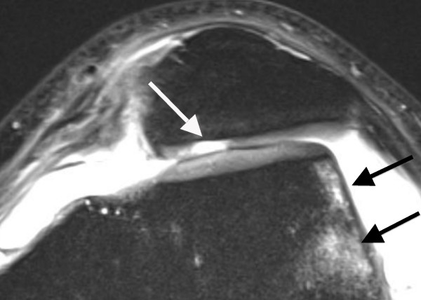
MRIAxial knee MRI: cartilage defect of the retropatellar surface and bone marrow oedema of the femoral condyle, arrows on the lesions.
Source : von Engelhardt et al., BMC musculoskeletal disorders, 2010, figure 1 · CC BY
Diagnosis of PFP is essentially clinical , one of the most robust messages of the JOSPT 2019 CPG and of the Manchester 2016 consensus.¹ ² Imaging is not recommended routinely and should be used only where there is a red flag or a suspected structural differential diagnosis.¹
Which questions should you ask to understand the patient and their history?
The history aims to reconstruct the load story and to isolate the diagnostic features:
- Location : the pain must be retro- or peripatellar. Ask the patient to point to the painful area with one finger: a patient with PFP often makes the “C” sign around the kneecap. Pain that is very localised over the inferior pole of the patella points instead to patellar tendinopathy.¹ ¹³
- Provocative activities : at least one activity loading the patellofemoral joint in flexion (descending stairs, squatting, running, landing from a jump). The prolonged sitting (“cinema sign”) is a historical but non-specific sign: Smith 2018 finds it in around 50% of PFP cases.²
- Mode of onset : insidious, most often after a rapid increase in load (going from 0 to 30 km of running per week, taking up a sport, changing shoes, changing surface).¹ ¹⁴
- Pain behaviour : worsened by loading in flexion, relieved by rest. Night pain, mechanical pain at rest, or pain unrelated to activity should raise the suspicion of a red flag.¹⁵
- Psychosocial factors : kinesiophobia, catastrophising, fear of recurrence, low self-efficacy. The SR by Maclachlan et al. (BJSM 2017) confirms their correlation with severity and persistence.¹⁶
Which clinical tests should you carry out and which other conditions must be ruled out?
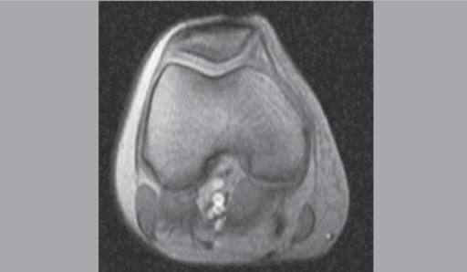
MRIAxial knee MRI: position of the patella within the femoral trochlea, image from a study on patellofemoral pain syndrome.
Source : Felicio et al., Acta ortopedica brasileira, 2014, figure 2 · CC BY-NC
No single test is sufficient: the strategy is to combine several tests to raise the cumulative diagnostic value.¹
Tests with solid diagnostic value:
- Bilateral squat has high sensitivity (≈ 80%) for reproducing PFP pain.¹
- Palpation of the patellar facets (with the patella tilted medially and laterally) reproduces the familiar pain in the majority of PFP cases.¹
- Single-leg squat / step-down test reproduces the pain under single-leg loading and allows a qualitative analysis of motor control (dynamic knee valgus, trunk lean, pelvic drop).¹
⚠️ Historical tests to abandon as a first choice, because their diagnostic value is low:
- Clarke's test (patellar grind sign, “Zohlen”): poor reproducibility, many false positives in asymptomatic people.¹⁷
- Isolated active patellar compression.
- Static measurement of the Q angle (poorly correlated with risk, poor intra-observer reliability).⁵
| Clinical test | Target | Sensitivity / usefulness | Level of evidence |
|---|---|---|---|
| Bilateral squat | Reproduction of patellofemoral pain | ≈ 80% sensitivity | A |
| Palpation of patellar facets | Local confirmation | Good reproducibility | A |
| Single-leg squat / step-down | Single-leg loading, motor control | Observable dynamic knee valgus | B |
| Clarke's test (grind) | Historically provocation | Many false positives | D: not recommended |
| Static Q angle | Historically alignment | Poorly correlated with risk/symptoms | D: not discriminating |
| Routine imaging | Structural diagnosis | Abnormalities common in asymptomatic people | D: not recommended without a red flag |
The examination is not limited to provocation tests. It must explicitly assess the modifiable deficits that will become the therapeutic targets :
- Quadriceps strength measured with a hand-held dynamometer (HHD) or a functional test under progressive load. A deficit ≥ 10% versus the healthy side is clinically relevant.¹ ⁵
- Strength of the hip abductors and external rotators is a key factor in women (Nascimento 2018).¹⁸
- Motor control under load judged on the quality of the single-leg squat / step-down (valgus, contralateral trunk drop).
- Mobility covering quadriceps flexibility (Ely test, modified Thomas test), the hamstrings and the gastrocnemius (weight-bearing dorsiflexion).
Red flags requiring medical referral
- Acute trauma with immediate haemarthrosis or inability to bear weight.
- Major joint effusion, local warmth, fever: suspected septic or inflammatory arthritis.
- True joint locking, repeated giving way: suspected meniscal or osteochondral lesion.
- Mechanical night pain, unexplained weight loss, history of cancer: work-up for tumour.
- Progressive neurological deficit, bladder or bowel disturbance: urgent spinal work-up.
- Failure of a well-conducted conservative programme (3-6 months): orthopaedic opinion for structural assessment.
The differential diagnoses to know when faced with anterior knee pain:¹ ¹³ ¹⁹
- Patellar tendinopathy gives sharp pain at the inferior pole of the kneecap, aggravated by energy storage and release (jumping, downhill work).
- Hoffa syndrome gives pain on either side of the patellar tendon, increased by forced extension.
- Medial synovial plica syndrome gives an audible snap and mediopatellar pain, sometimes resistant to standard conservative treatment. Beware, this is a diagnosis of exclusion: the plica is a normal fold, present in more than one healthy knee in three.
- Neuralgia of the infrapatellar branch of the saphenous nerve gives band-like anteromedial pain with localised hypersensitivity, often after arthroscopy or trauma.²⁰
- Early patellofemoral osteoarthritis occurs in an older patient, with morning stiffness and crepitus.
- Meniscal or osteochondral lesion involves a history of trauma, locking and recurrent effusion.
Should patients with PFP be classified, and for what benefit?
The idea of subtyping patients with PFP in order to target treatment better has been championed since 2016 by the TIPPs group (Targeted Interventions for Patellofemoral Pain) of Selfe, Janssen and Callaghan. Their observational study of 127 patients published in BJSM in 2016 identified three main subgroups according to the profile of strength, mobility and foot type.²¹
The classification approaches most discussed today:
- Overload / training error means no major muscle deficit, but a rapid increase in load. Target: load management, education.
- Strength deficit (hip and/or quadriceps): progressive strengthening and heavy resistance.
- Motor control deficit (dynamic knee valgus, trunk drop): exercises with visual and verbal feedback, trunk-hip-knee integration.
- Hypermobility / stabilisation deficit calls for proprioceptive exercises, controlled loads and avoidance of over-stretching.
The real clinical usefulness of this subclassification is still being validated.²¹ ²² A recent SR (Mansfield 2023, JOSPT NMA) showed that combinations of exercises targeting hip + knee + internal feedback do change weight-bearing kinematic parameters, supporting the idea of rehabilitation guided by the patient's motor profile rather than a standard protocol for everyone.²³
Key points: chapter 2
- Diagnosis of PFP is clinical, based on location (retro/peripatellar) plus reproduction of the pain under load in flexion (squatting, stairs).
- The use of imaging is not recommended in routine practice except in the presence of red flags or a suspected differential diagnosis.
- Useful tests: squat, palpation of the facets, single-leg squat. Obsolete tests: Clarke (grind), static Q angle.
- Always look for the differential diagnoses (patellar tendinopathy, plica, Hoffa, saphenous neuralgia, meniscal lesion).
- Clinical subtyping (overload / strength / motor control / hypermobility) is promising for personalising treatment.
References: chapter 2
- Willy RW, Hoglund LT, Barton CJ, et al. Patellofemoral Pain: CPG. J Orthop Sports Phys Ther. 2019;49(9):CPG1-CPG95. doi:10.2519/jospt.2019.0302
- Smith BE, Selfe J, Thacker D, et al. Incidence and prevalence of patellofemoral pain: SR/MA. PLoS One. 2018;13(1):e0190892. PMID 29324820
- Crossley KM, Stefanik JJ, Selfe J, et al. 2016 Patellofemoral pain consensus statement - Manchester Part 1. Br J Sports Med. 2016;50(14):839-843. PMID 27343241
- Collins NJ, Barton CJ, van Middelkoop M, et al. 2018 Consensus statement on exercise therapy and physical interventions. Br J Sports Med. 2018;52(18):1170-1178. PMID 29925502
- Neal BS, Lack SD, Lankhorst NE, et al. Risk factors for PFP: SR/MA. Br J Sports Med. 2019;53(5):270-281. doi:10.1136/bjsports-2017-098890
- Powers CM, Witvrouw E, Davis IS, Crossley KM. Evidence-based framework for a pathomechanical model of PFP. Br J Sports Med. 2017;51(24):1713-1723. PMID 29109118
- Lankhorst NE, Bierma-Zeinstra SM, van Middelkoop M. Risk factors for PFP syndrome: SR. J Orthop Sports Phys Ther. 2012;42(2):81-94. doi:10.2519/jospt.2012.3803
- Crossley KM, van Middelkoop M, Callaghan MJ, et al. 2016 Manchester Part 2. Br J Sports Med. 2016;50(14):844-852. PMID 27247098
- Esculier JF, Bouyer LJ, Dubois B, et al. Is combining gait retraining or an exercise programme with education better than education alone in treating runners with patellofemoral pain? A randomised clinical trial. Br J Sports Med. 2018;52(10):659-666. PMID 28476901
- Boling M, Padua D, Marshall S, et al. JUMP-ACL cohort. Am J Sports Med. 2009;37(11):2108-2116. PMID 19797162
- Vicenzino B, Collins N, Crossley K, Beller E, Darnell R, McPoil T. Foot orthoses and physiotherapy in the treatment of patellofemoral pain syndrome: a randomised clinical trial. BMC Musculoskelet Disord. 2008;9:27. PMID 18304317.
- Dye SF. Pathophysiology of patellofemoral pain: tissue homeostasis perspective. Clin Orthop Relat Res. 2005;436:100-110. PMID 15995427
- Cook JL, Rio E, Purdam CR, Docking SI. Revisiting the continuum model of tendon pathology: what is its merit in clinical practice and research? Br J Sports Med. 2016;50(19):1187-1191. PMID 27127294
- Rathleff MS, Vicenzino B, Middelkoop M, et al. PFP adolescence/adulthood: same same, but different? Sports Med. 2015;45(11):1489-1495. PMID 26178330
- Finucane LM, Downie A, Mercer C, et al. International framework for red flags for potential serious spinal pathologies. J Orthop Sports Phys Ther. 2020;50(7):350-372. doi:10.2519/jospt.2020.9971
- Maclachlan LR, Collins NJ, Matthews MLG, Hodges PW, Vicenzino B. The psychological features of patellofemoral pain: a systematic review. Br J Sports Med. 2017;51(9):732-742. PMID 28320733
- Cook C, Hegedus E, Hawkins R, Scovell F, Wyland D. Diagnostic accuracy and association to disability of clinical test findings associated with patellofemoral pain syndrome. Physiother Can. 2010;62(1):17-24. PMID 21197175
- Nascimento LR, Teixeira-Salmela LF, Souza RB, Resende RA. Hip and Knee Strengthening Is More Effective Than Knee Strengthening Alone for Reducing Pain and Improving Activity in Individuals With Patellofemoral Pain: A SR/MA. J Orthop Sports Phys Ther. 2018;48(1):19-31. doi:10.2519/jospt.2018.7365
- Barton CJ, Lack S, Hemmings S, Tufail S, Morrissey D. The 'Best Practice Guide to Conservative Management of Patellofemoral Pain': incorporating level 1 evidence with expert clinical reasoning. Br J Sports Med. 2015;49(14):923-934. PMID 25716151
- Trescot AM, Brown MN, Karl HW. Infrapatellar saphenous neuralgia - diagnosis and treatment. Pain Physician. 2013;16(3):E315-E324. PMID 23703431
- Selfe J, Janssen J, Callaghan M, et al. Are there three main subgroups within the patellofemoral pain population? A detailed characterisation study of 127 patients to help develop targeted intervention (TIPPs). Br J Sports Med. 2016;50(14):873-880. PMID 26834185
- Lack S, Neal B, De Oliveira Silva D, Barton C. How to manage patellofemoral pain - Understanding the multifactorial nature and treatment options. Phys Ther Sport. 2018;32:155-166. PMID 29793124
- Mansfield CJ, Vakili F, Habib N, et al. Effectiveness of Interventions Aimed at Changing Movement Patterns in People With Patellofemoral Pain: A SR With Network Meta-analysis. J Orthop Sports Phys Ther. 2023;53(12):723-739. doi:10.2519/jospt.2023.11956
Which treatment strategies are the most effective?
Treatment of PFP is now firmly consensual on its fundamentals. The Gold Coast 2018 consensus (Collins et al.) and the JOSPT 2019 CPG (Willy et al.) converge: patient education and therapeutic exercise are the unavoidable first line, everything else being adjunctive.¹ ²
Where do you start? What is the recommended hierarchy of interventions?
The evidence-based hierarchy, ranked by level of evidence:
| Intervention | Expected effect | Time horizon | Level of evidence |
|---|---|---|---|
| Patient education (load management, demedicalisation) | ↓ pain, ↑ function, ↑ adherence | Short to long term | A: strongly recommended |
| Combined hip + knee exercise | ↓ pain, ↑ strength, ↑ function | Short to long term | A: strongly recommended |
| Gait retraining in the runner (cadence ↑ 5-10%) | ↓ patellofemoral stress by up to 20% | Immediate + lasting effects | B: recommended |
| Foot orthoses (where pronation is excessive) | ↓ pain in the short term | Short term (≤ 6 weeks) | B: selectively recommended |
| Patellar taping (McConnell) | ↓ immediate pain, makes exercise easier | Short term only | B: adjunct |
| Manual therapy (patellar and hip mobilisations) | ↓ pain in the short term | Short term, little effect in the long term | C: adjunct only |
| Dry needling, Kinesio Taping | Weak to modest effects | Short term | D: insufficient evidence |
| Shockwave, ultrasound, laser | No convincing demonstrated effect | — | D: not recommended alone |
| Surgery (lateral release, MPFL, tibial tubercle transfer) | Reserved for failure of a conservative programme of ≥ 6-12 months | — | D: last resort |
The network meta-analysis (NMA) by Winters et al. published in JOSPT in 2022, which included 65 randomised trials, confirms at 3 months that 6 interventions have a positive effect on pain and function: exercise targeting the knee alone, the hip+knee combination, foot orthoses, local manual therapy, exercise combined with perineural dextrose injection, and exercise with quadriceps biofeedback.³ None is dramatically superior to the others in the short term, hence the importance of personalising the choice according to the patient's clinical profile and preferences.
What is the place of exercise and is there a superior approach?
Exercise is the most robustly validated intervention in the literature. The Cochrane review by van der Heijden et al. (2015) had already concluded that there is a moderate effect on pain and function, supported by moderate- to low-quality evidence.⁴ Since then, recent NMAs and SRs/MAs have clarified three key points:
- Hip + knee > knee alone. The meta-analysis by Nascimento et al. (JOSPT 2018, 14 trials, 673 participants) showed that strengthening hip + quadriceps is superior to strengthening the quadriceps in isolation for reducing pain and improving activity, and this holds even without any detectable increase in strength, which points to neuromuscular and behavioural mechanisms.⁵
- No superiority of open versus closed chain. The old debates about the “safe angle” (0-45° in CKC, 45-90° in OKC) have been put into perspective: as long as the load stays tolerated and progression is gradual, both modes are effective.² ⁴
- Load management > the search for perfect movement. The critical review by Lack, Neal and De Oliveira Silva (Phys Ther Sport 2018) reframed the priority: improving the capacity of the tissues to tolerate load through progressive strengthening mattered more than normalising an “ideal” movement pattern that does not exist in the population.⁶
📈 Effect of combined hip+knee exercise versus knee alone on pain
Nascimento 2018 meta-analysis (JOSPT, 14 trials, n = 673)
Source: Nascimento LR et al. J Orthop Sports Phys Ther. 2018;48(1):19-31 (DOI 10.2519/jospt.2018.7365). SMD calculated at 3 months for pain: the advantage of the combined approach is statistically and clinically significant.
A fundamental point of the 2023 update is that the NMA by Mansfield et al. (JOSPT 2023, 37 trials, n = 1235) established that interventions combining exercise + internal feedback (visual, verbal, mirror) do change frontal-plane kinematics of the knee and hip, which validates the motor-control-centred approach in cases with marked dynamic knee valgus.⁷ Conversely, braces alone and taping alone have no effect on movement kinematics.⁷
Manual therapies and technologies: how effective are they really?
Passive therapies are short-term adjuncts, never the core of treatment.
- Manual therapy (patellar mobilisations, hip mobilisations, lumbopelvic mobilisations) gives short-term pain relief, but no additional long-term benefit when added to a structured exercise programme.² Its role is essentially to reduce initial pain so that the patient can engage in exercise.
- Patellar taping (McConnell) : immediate reduction in pain documented during activity (a probable neurophysiological mechanism). No lasting long-term benefit. Kinesio Taping has not shown superiority over placebo.⁸
- Prefabricated foot orthoses give short-term pain relief in patients with excessive foot pronation shown objectively (peak eversion ≥ 4°), and little effect in the others.² ⁹
- Dry needling rests on emerging and heterogeneous evidence. A few trials show a short-term effect, but recent SRs/MAs remain cautious (poor methodological quality of the studies).¹⁰
- Shockwave, ultrasound, laser : no high-quality evidence supports their use on their own for PFP. The JOSPT 2019 CPG does not recommend them as first line.¹
- Surgery is reserved for failure of a good conservative programme run for at least 6 to 12 months (lateral release, tibial tubercle transfer, MPFL reconstruction where instability is proven).¹ ²
Beyond the physical: how do you educate the patient and address psychological factors?
The education of the patient is an active therapeutic intervention, not a mere add-on. The SR by Pazzinatto, de Oliveira Silva, Barton et al. (JOSPT 2020) showed that structured education (one to several sessions) can improve function and pain for up to 1 year alongside exercise.¹¹ The key themes to cover:
- Demedicalising the pain means explaining that pain does not equal damage, and that an “abnormal” MRI says nothing about the prognosis.
- The load model holds that pain is a signal of overload: the aim is to widen tolerance, not to avoid load.
- Progressive management follows the rule of “no more than 2/10 pain during effort, and it must settle within 24 h”.
- An active role means the patient is the main player and the physiotherapist a coach, not a repairer.
The psychosocial factors are established mediators of symptom persistence. The SR by Maclachlan, Collins, Matthews, Hodges and Vicenzino (BJSM 2017, 25 studies included) confirmed that kinesiophobia, catastrophising, anxiety and depression are high in people with PFP and correlate with severity and function.¹² A more recent SR (Matthews 2022, Phys Ther Sport) reinforces these conclusions and calls for their systematic assessment to be built in.
Key points: chapter 3
- First line = patient education + therapeutic exercise. Everything else is adjunctive.
- The combination of hip + quadriceps strengthening is superior to the quadriceps alone (Nascimento 2018, SMD −0.79 vs −0.42).
- The Winters 2022 NMA identifies 6 effective interventions at 3 months, with no dramatically superior option: personalise to the profile.
- The use of taping and manual therapy relieves pain in the short term but adds no lasting benefit to the exercise programme.
- Systematically assess and address kinesiophobia and the patient's limiting beliefs.
References: chapter 3
- Willy RW, Hoglund LT, Barton CJ, et al. Patellofemoral Pain: CPG. J Orthop Sports Phys Ther. 2019;49(9):CPG1-CPG95. doi:10.2519/jospt.2019.0302
- Collins NJ, Barton CJ, van Middelkoop M, et al. 2018 Consensus statement on exercise therapy and physical interventions to treat PFP. Br J Sports Med. 2018;52(18):1170-1178. PMID 29925502
- Winters M, Holden S, Lura CB, et al. Comparative effectiveness of treatments for patellofemoral pain: a living systematic review with network meta-analysis. Br J Sports Med. 2020;55(7):369-377. PMID 33106251. doi:10.1136/bjsports-2020-102819.
- van der Heijden RA, Lankhorst NE, van Linschoten R, Bierma-Zeinstra SM, van Middelkoop M. Exercise for treating patellofemoral pain syndrome. Cochrane Database Syst Rev. 2015;1:CD010387. PMID 25603546
- Nascimento LR, Teixeira-Salmela LF, Souza RB, Resende RA. Hip and Knee Strengthening Is More Effective Than Knee Strengthening Alone: SR/MA. J Orthop Sports Phys Ther. 2018;48(1):19-31. doi:10.2519/jospt.2018.7365
- Lack S, Neal B, De Oliveira Silva D, Barton C. How to manage patellofemoral pain - Understanding the multifactorial nature and treatment options. Phys Ther Sport. 2018;32:155-166. PMID 29793124
- Mansfield CJ, Vakili F, Habib N, et al. Effectiveness of Interventions Aimed at Changing Movement Patterns in PFP: SR With Network Meta-analysis. J Orthop Sports Phys Ther. 2023;53(12):723-739. doi:10.2519/jospt.2023.11956
- Logan CA, Bhashyam AR, Tisosky AJ, et al. Systematic Review of the Effect of Taping Techniques on Patellofemoral Pain Syndrome. Sports Health. 2017;9(5):456-461. PMID 28617653
- Barton CJ, Munteanu SE, Menz HB, Crossley KM. The efficacy of foot orthoses in the treatment of individuals with patellofemoral pain syndrome: a systematic review. Sports Med. 2010;40(5):377-395. PMID 20433211
- Rahou-El-Bachiri Y, Navarro-Santana MJ, Gómez-Chiguano GF, et al. Effects of Trigger Point Dry Needling for the Management of Knee Pain Syndromes: SR/MA. J Clin Med. 2020;9(7):2044. PMID 32610659
- de Oliveira Silva D, Pazzinatto MF, Rathleff MS, et al. Patient Education for Patellofemoral Pain: A Systematic Review. J Orthop Sports Phys Ther. 2020;50(7):388-396. doi:10.2519/jospt.2020.9400 (PMID 32349640)
- Maclachlan LR, Collins NJ, Matthews MLG, Hodges PW, Vicenzino B. The psychological features of patellofemoral pain: a systematic review. Br J Sports Med. 2017;51(9):732-742. PMID 28320733
- Lack S, Barton C, Sohan O, Crossley K, Morrissey D. Proximal muscle rehabilitation is effective for patellofemoral pain: SR/MA. Br J Sports Med. 2015;49(21):1365-1376. PMID 26175019
- Crossley KM, van Middelkoop M, Callaghan MJ, et al. 2016 Manchester Part 2 - physical interventions. Br J Sports Med. 2016;50(14):844-852. PMID 27247098
- Barton CJ, Lack S, Hemmings S, Tufail S, Morrissey D. The 'Best Practice Guide to Conservative Management of Patellofemoral Pain'. Br J Sports Med. 2015;49(14):923-934. PMID 25716151
Runners and PFP: why does this subgroup deserve a dedicated approach?
PFP is the most frequent knee condition in runners, accounting for 16 to 25% of sports medicine consultations depending on the cohort.¹ ² This over-representation justifies a dedicated focus: repetitive mechanics (1,000 to 2,000 strides per kilometre, a load equivalent to 2.5 to 5 × body weight on the kneecap) create specific conditions of cumulative overload.
Which biomechanical alterations are specific to the runner with PFP?
The SR/MA by Neal, Barton, Gallie, O'Halloran and Morrissey (Gait Posture 2016) identified three reproducible kinematic alterations in runners with PFP compared with healthy runners:¹
- ↑ hip adduction (peak hip adduction), a prospective risk factor in women.
- ↑ hip internal rotation (peak hip internal rotation).
- ↑ contralateral pelvic drop (contralateral pelvic drop), a dynamic Trendelenburg.
These three alterations converge on a dynamic knee valgus at initial contact and at mid-stance, increasing mediolateral stresses on the patellofemoral joint.¹ ³ The distal factors (peak rearfoot eversion, rearfoot versus forefoot strike) are also associated, but less consistently.¹
🏃 Biomechanical alterations in runners with PFP (versus healthy runners)
Neal 2016 meta-analysis (Gait Posture, 23 studies, 1,153 runners)
Source: Neal BS et al. Gait Posture. 2016;45:69-82 (PMID 26979886). These alterations can be modified by targeted interventions: strengthening of the hip abductors and external rotators, kinetic gait retraining, visual and verbal feedback.
The most important clinical point: these alterations can be modified. The Neal 2016 SR shows that targeted interventions (hip strengthening, gait retraining, feedback) do change running kinematics, which translates into a reduction in pain in trials at 6-12 weeks.¹ ⁴
Gait retraining and increased cadence: how much, and how?
The most robust biomechanical intervention in the runner with PFP is the 5 to 10% increase in cadence (steps per minute).⁴ ⁵ ⁶ The mechanism is mechanical: at a given speed, raising cadence shortens step length, reduces overstriding, lowers peak ground reaction forces and lowers patellofemoral stress by 15 to 20% according to biomechanical models.⁴ ⁵
The randomised trial by Esculier et al. (BJSM 2018, n = 69 runners with PFP) remains the methodological reference. It compared three arms over 8 weeks: education alone, education + exercises, education + gait retraining. All three groups improved pain and function to an equivalent degree, with no superiority for gait retraining on its own. This suggests that education about load management is the mainstay, with gait retraining a useful addition in certain profiles (runners with a low cadence < 170 spm, marked overstriding, pronounced rearfoot strike).⁴
Three main variants of gait retraining for PFP:
- Increasing cadence (the simplest to implement): aim for +5 to +10% of preferred cadence, using an auditory metronome on a smartphone. Immediate effects on kinematics, variable persistence.⁵
- Run softer (Davis): sound feedback from an accelerometer to reduce the vertical impact peak. Cuts the incident risk of PFP by around 79% (RR 0.21) according to the Culvenor 2020 BJSM SR/MA.⁷
- Transition to a midfoot or forefoot strike is controversial. It reduces patellofemoral stress but increases stress on the Achilles tendon and calf, so it should be used cautiously and progressed gradually.⁵ ⁶
Practical protocol: gait retraining through cadence
- 1. Measure preferred cadence (smartphone: 60 seconds of comfortable running).
- 2. Set a metronome to +5% of that cadence (then +10% once adapted).
- 3. Progressive runs of 5-10 min with the metronome, keeping the same average speed (check on GPS).
- 4. Pain monitoring: ≤ 2/10 during effort, settling within 24 h.
- 5. Progressive weaning off the metronome after 3-4 weeks: the new cadence becomes automatic.
For primary prevention, the Neal/Culvenor SR/MA published in BJSM in 2021 (PMID 33115705) remains the reference: across 13 trials and 6 interventions evaluated, only two interventions significantly reduce the incident risk of PFP:⁷
- Patellar braces worn during physical activity: RR 0.40 (95% CI 0.22-0.73), low-quality evidence, a reduction of around 60%.
- Gait retraining (run softer) gives RR 0.21 (95% CI 0.07-0.60), low-quality evidence, a reduction of around 79%.
Conversely, the multicomponent exercise programmes (strengthening / neuromuscular) did not significantly reduce the incident risk of PFP in the 4 included trials (n = 3,364), a surprising result that contrasts with their curative effectiveness. Foot orthoses in primary prevention showed no effect either.⁷ This finding argues for concentrating prevention on load management and technique adjustments rather than on generic exercise protocols.
Red flags specific to the runner with “anterior knee pain”
- Focal bone pain over the patella or the condyle, aggravated by single-leg hopping ➜ suspected patellar or condylar stress fracture (rare but serious).
- Onset of a persistent effusion without trauma ➜ structural work-up.
- Failure of a well-conducted gait retraining programme (8-12 weeks) with worsening symptoms ➜ reassess the diagnosis (patellar tendinopathy, Hoffa, osteochondral lesion).
- Runners ≥ 50 years with morning stiffness > 30 min ➜ consider early patellofemoral osteoarthritis.
Key points: chapter 4 (runners)
- Runners with PFP show 3 characteristic kinematic alterations , namely ↑ hip adduction, ↑ internal rotation and ↑ pelvic drop (Neal 2016).
- The 5-10% increase in cadence reduces patellofemoral stress by 15-20%: a simple and free intervention.
- For primary prevention, “run softer” (gait retraining) reduces the risk of PFP by 79% (Culvenor 2020 BJSM, RR 0.21).
- The patellar braces worn during activity reduce the incident risk by 60% (RR 0.40).
- Generic exercise programmes have not shown a preventive effect and prevention therefore runs through load management and technique adjustments.
References: chapter 4
- Neal BS, Barton CJ, Gallie R, O'Halloran P, Morrissey D. Runners with patellofemoral pain have altered biomechanics which targeted interventions can modify: A systematic review and meta-analysis. Gait Posture. 2016;45:69-82. PMID 26979886
- Taunton JE, Ryan MB, Clement DB, McKenzie DC, Lloyd-Smith DR, Zumbo BD. A retrospective case-control analysis of 2002 running injuries. Br J Sports Med. 2002;36(2):95-101. PMID 11916889
- Powers CM, Witvrouw E, Davis IS, Crossley KM. Evidence-based framework for a pathomechanical model of PFP. Br J Sports Med. 2017;51(24):1713-1723. PMID 29109118
- Esculier JF, Bouyer LJ, Dubois B, et al. Is combining gait retraining or an exercise programme with education better than education alone in treating runners with patellofemoral pain? A randomised clinical trial. Br J Sports Med. 2018;52(10):659-666. PMID 28476901
- Davis IS, Tenforde AS, Neal BS, Roper JL, Willy RW. Gait Retraining as an Intervention for Patellofemoral Pain. Curr Rev Musculoskelet Med. 2020;13(1):103-114. PMID 32170556.
- Bramah C, Preece SJ, Gill N, Herrington L. Is There a Pathological Gait Associated With Common Soft Tissue Running Injuries? Am J Sports Med. 2018;46(12):3023-3031. PMID 30193080
- Neal BS, Bartholomew C, Barton CJ, Morrissey D, Lack SD. Six Treatments Have Positive Effects at 3 Months for People With Patellofemoral Pain: A Systematic Review With Meta-analysis. J Orthop Sports Phys Ther. 2022;52(11):750-768. PMID 36070427.
- Willy RW, Hoglund LT, Barton CJ, et al. PFP CPG. J Orthop Sports Phys Ther. 2019;49(9):CPG1-CPG95. doi:10.2519/jospt.2019.0302
- Crossley KM, van Middelkoop M, Callaghan MJ, et al. 2016 Manchester Part 2. Br J Sports Med. 2016;50(14):844-852. PMID 27247098
- Heiderscheit BC, Chumanov ES, Michalski MP, Wille CM, Ryan MB. Effects of step rate manipulation on joint mechanics during running. Med Sci Sports Exerc. 2011;43(2):296-302. PMID 20581720
How do you secure lasting recovery and prevent flares?
The success of PFP treatment is not measured at the end of the clinic protocol: it is measured by the patient's ability to manage their load over the years that follow. The prospective data (Hsu 2025) are a reminder that nearly one patient in two remains symptomatic at 5 years without structured management, and that 50-90% of adolescents still have symptoms 1 to 20 years after the initial diagnosis.¹ ²
How do you make the patient an active player in their own recovery through self-management?
The transition to self-management rests on three pillars, ranked by level of evidence:
- Structured education (an active therapeutic intervention): the SR by de Oliveira Silva, Pazzinatto and Barton (JOSPT 2020) shows that well-delivered education can improve pain and function for up to 1 year.³
- Home exercise with self-managed progression: the hip + quadriceps combination remains the reference (Nascimento 2018).⁴
- Load management (training load management): learning the “no more than 10% per week” rule and how to read the pain signal.⁵
The challenge of long-term adherence is the main one. The most effective strategies:
- Collaborative definition of SMART goals by the patient (not by the physiotherapist alone).
- A simple logbook (paper or app) with self-monitoring of pain and load.
- “Booster” sessions scheduled at 3 and 6 months.
- A programme adapted to the patient's real life rhythm (3 sessions of 20 min ≫ a 1-hour programme that never gets done).
| Self-management dimension | Practical tool | Expected effect | Evidence level |
|---|---|---|---|
| Patient education | Structured booklet + 1-2 dedicated sessions | ↓ pain, ↑ function for up to 12 months | A: de Oliveira Silva 2020 |
| Home exercises | 3-5 key exercises, 3×/week, 6-12 weeks | ↑ strength, ↓ pain | A: Cochrane 2015 |
| Load management | Rule of < 10%/week, VAS monitoring | ↓ recurrence | B: Best Practice Barton 2015 |
| Long-term adherence | SMART goals, tracking app | ↑ adherence ~30-40% | B: Sancho-Velázquez 2024 |
| Recurrence prevention in runners | Patellar brace + gait retraining | RR 0.40 / RR 0.21 | B: Culvenor 2020 |
When and how do you plan a safe return to sport and activity?
Return to sport (RTS) must be based on functional criteria and not on a fixed timetable.⁶ Unlike ACL reconstruction, there is as yet no standardised, universally validated RTS test battery for PFP: practice rests on expert consensus and clinical judgement.²
The proposed RTS criteria (a synthesis of consensus recommendations):
Practical return-to-sport criteria: PFP
- Pain ≤ 2/10 on the VAS during activities of daily living and full-load strengthening exercises.
- No effusion and no joint warmth.
- Strength symmetry ≥ 90% at the quadriceps and hip abductors (the healthy side as the reference).
- Single-leg squat to 60° of flexion without pain, with symmetrical frontal-plane control.
- Single-leg hop tests (single hop, triple hop, crossover hop) with asymmetry ≤ 10%.
- Progressive resumption respecting the rule of < 10%/week increase in volume.
- Post-exercise pain resolving within 24 h and not worsening the baseline profile.
For runners, the typical progression:
- Pain-free brisk walking for 30 min ➜ alternating jog-walk (1 min running / 2 min walking) over 20 min.
- Gradual increase in running duration (1, 2, 3, 5 min and so on) over 4-8 weeks.
- Gait retraining built in (cadence +5-10%) from the very start of the return.
- Return to the usual distance before raising the pace again.
- Progressive reintroduction of specific sessions (hills, intervals, long runs), one variable at a time.
For sports with jumping and changes of direction (basketball, volleyball, football, dance), add a phase of progressive plyometric rehabilitation and a return to sport-specific training before competition.
Key points: chapter 5
- The education of the patient is an active therapy in its own right (effects lasting up to 1 year).
- Self-management of load is the key to preventing recurrence: the < 10%/week rule, pain monitoring (VAS ≤ 2/10).
- The return to sport must be validated by functional criteria (strength ≥ 90%, single-leg hop with ≤ 10% asymmetry), not by a calendar.
- In runners: patellar brace (RR 0.40) and gait retraining (RR 0.21) in secondary prevention (Culvenor 2020).
- The maintenance of long-term adherence requires SMART goals, independent monitoring and booster sessions.
References: chapter 5
- Hsu HC, Chen LL, Chiu YC, et al. Prognosis of Patellofemoral Pain: SR With Evidence- and Gap-Map. J Orthop Sports Phys Ther. 2025;55(9):568-590. doi:10.2519/jospt.2025.13491
- Rathleff MS, Vicenzino B, Middelkoop M, et al. PFP in adolescence and adulthood: same same, but different? Sports Med. 2015;45(11):1489-1495. PMID 26178330
- de Oliveira Silva D, Pazzinatto MF, Rathleff MS, et al. Patient Education for Patellofemoral Pain: A Systematic Review. J Orthop Sports Phys Ther. 2020;50(7):388-396. doi:10.2519/jospt.2020.9400
- Nascimento LR, Teixeira-Salmela LF, Souza RB, Resende RA. Hip and Knee Strengthening Is More Effective Than Knee Strengthening Alone: SR/MA. J Orthop Sports Phys Ther. 2018;48(1):19-31. doi:10.2519/jospt.2018.7365
- Barton CJ, Lack S, Hemmings S, Tufail S, Morrissey D. The 'Best Practice Guide to Conservative Management of Patellofemoral Pain'. Br J Sports Med. 2015;49(14):923-934. PMID 25716151
- Willy RW, Hoglund LT, Barton CJ, et al. PFP CPG. J Orthop Sports Phys Ther. 2019;49(9):CPG1-CPG95. doi:10.2519/jospt.2019.0302
- Esculier JF, Bouyer LJ, Dubois B, et al. Gait retraining vs exercise vs education in runners with PFP: RCT. Br J Sports Med. 2018;52(10):659-666. PMID 28476901
- Davis IS, Tenforde AS, Neal BS, Roper JL, Willy RW. Gait Retraining as an Intervention for Patellofemoral Pain. Curr Rev Musculoskelet Med. 2020;13(1):103-114. PMID 32170556.
- Culvenor AG, van Middelkoop M, Macri EM, Crossley KM. Is patellofemoral pain preventable? A systematic review and meta-analysis of randomised controlled trials. Br J Sports Med. 2020. PMID 33115705.
- Collins NJ, Barton CJ, van Middelkoop M, et al. 2018 Consensus statement Gold Coast. Br J Sports Med. 2018;52(18):1170-1178. PMID 29925502
- Mansfield CJ, Vakili F, Habib N, et al. Effectiveness of interventions changing movement patterns - NMA. J Orthop Sports Phys Ther. 2023;53(12):723-739. doi:10.2519/jospt.2023.11956
What do real clinical cases teach us?
Analysis of a “classic” case: from assessment to resolution.
The case reported by Khan et al. in Cureus in 2024 illustrates a typical presentation and evidence-based multimodal management.¹ A 21-year-old recreational female runner presented with bilateral anterior knee pain of gradual onset after a rapid increase in running volume. Clinical examination confirmed:
- Bilateral peripatellar pain reproduced on squatting and on descending stairs.
- Documented weakness of the quadriceps and hip abductors.
- Single-leg squat with marked bilateral dynamic knee valgus.
- No red flag, and an otherwise normal joint examination.
Treatment combined the consensus pillars:¹
- Education covering the load model, demedicalisation, and advice to reduce volume temporarily without stopping altogether.
- Progressive strengthening with a bilateral hip programme (clams, side-lying abduction, monster walks) + quadriceps (squats, lunges, step-ups), 3 sessions a week over 8 weeks.
- Motor control work with single-leg squats in front of a mirror and step-downs with verbal feedback.
- Stretching and mobility targeting the hip flexors and the hamstrings.
- Progressive return to running with gait retraining (increased cadence) from week 4.
Outcome was a clinically significant improvement in pain (VAS), function (AKPS/Kujala) and running capacity, with a pain-free return by the end of the protocol.¹ This case validates the consensus sequence (education → combined hip+knee exercise → progressive reintegration) and confirms the central place of gait retraining in the runner.
An older but methodologically interesting case, Esculier 2017 in IJSPT (PMC5534157), describes a runner with PFP treated with a multimodal programme addressing kinesiophobia (education + exercises + gait retraining + simple cognitive therapy), with complete resolution in 8 weeks, illustrating the importance of including psychosocial factors from the outset.²
The diagnostic challenge: when PFP mimics another condition.
The entrapment of the infrapatellar branch of the saphenous nerve (IPBSN) is a classic differential diagnosis of PFP. A recent case published in Cureus in 2025 (PMC12558126) is exemplary:³
- Presentation was anterior knee pain “of the PFP type” resistant to several months of standard conservative rehabilitation.
- Discriminating feature on palpation, exquisite tenderness at the site where the nerve pierces sartorius, with neuropathic radiation.
- Targeted imaging with high-resolution ultrasound confirming IPBSN entrapment at the level of sartorius.
- Treatment with ultrasound-guided manual mobilisation of sartorius, saphenous neurodynamics, followed by soft tissue techniques.
- Outcome was complete resolution of the pain within a few sessions.
The clinical message is clear: any PFP that does not respond to well-conducted conservative treatment after 6-8 weeks should prompt a rethink of the diagnosis. The classic traps:
- IPBSN neuralgia (Trescot 2013, Cureus 2025): band-like pain, pinpoint tenderness, often after arthroscopy.
- Medial synovial plica syndrome (Schindler 2014): snapping, mediopatellar pain, sometimes resistant.⁴ The full picture and the real value of the tests are detailed in the dedicated article.
- Patellar tendinopathy gives very focal pain at the inferior pole of the kneecap, aggravated by jumping.
- Hoffa syndrome gives pain on either side of the patellar tendon, increased by forced extension.
- Early patellofemoral osteoarthritis occurs in an older patient, with morning stiffness and audible crepitus.
Study of a complex case (hypermobility, kinesiophobia).
Two clinical situations call for a significant adaptation of the standard strategy.
Case no. 1: generalised hypermobility / hypermobile Ehlers-Danlos syndrome. Hypermobile patients are at increased risk of PFP (with or without associated patellar instability). The data from Ferrero et al. (2025) on the risk factors for patellofemoral instability in patients with hEDS underline that classic heavy-load rehabilitation may be poorly tolerated.⁵ For the current nomenclature, the limits of the Beighton score and exercise dosage in this population, see joint hypermobility syndromes. The approach must be adapted:
- Strengthening below threshold initially, with light loads and very slow progression.
- Priority emphasis on motor control and proprioception rather than pure strength.
- Work on eccentric control within limited ranges.
- Monitoring for patellar subluxation, with referral for MPFL surgery if instability is proven and disabling.
- A reinforced biopsychosocial approach (frequent chronic pain, fatigue, comorbidities).
Case no. 2: kinesiophobia / catastrophising. The case published by Carter et al. in American Journal of Case Reports in 2021 (PMC8378777) illustrates the integration of pain neuroscience education (PNE) in a patient with multi-region chronic pain (lumbopelvic, cervical, knee).⁶ The intervention combined:
- 7 sessions of structured PNE over 4 weeks (videos, metaphors, drawings, discussion).
- Manual therapy + exercises prescribed by the qualified physiotherapist.
- Graded exposure to feared movements.
Results: a clinically significant decrease in subjective pain and in kinesiophobia (Tampa Scale for Kinesiophobia), even though the reduction in fear-avoidance and perceived disability was more modest.⁶ This case supports the place of PNE in chronic PFP with a strong psychosocial component, alongside the physical programme.
When to reconsider the diagnosis of PFP
- Failure of a well-conducted conservative programme of ≥ 6-8 weeks with documented adherence.
- Appearance of new signs: persistent audible snapping, true locking, effusion.
- Neuropathic pain (burning, radiation, focal hypersensitivity).
- Night pain, weight loss, fever: work-up for tumour or inflammation.
- Atypical presentation: older patient with no triggering factor, morning stiffness > 30 min.
Key points: chapter 6
- The verified PMC case studies validate the evidence-based multimodal approach (education + combined exercise + gait retraining).
- Any PFP resistant to standard treatment means the diagnosis must be questioned again: think systematically of IPBSN neuralgia, plica, Hoffa and patellar tendinopathy.
- Patients who are hypermobile require a less aggressive approach favouring motor control and proprioception.
- The pain neuroscience education approach is useful in patients with marked kinesiophobia: build it in from the initial assessment.
References: chapter 6
- Khan A, Shah RR, Bhatti A, et al. Comprehensive Management of Patellofemoral Pain Syndrome in a Recreational Long-Distance Runner: A Case Report. Cureus. 2024;16(7):e64722. PMC11327842
- Esculier JF, Roy JS, Bouyer LJ. The effects of a multimodal rehabilitation program on pain, kinesiophobia and function in a runner with patellofemoral pain. Int J Sports Phys Ther. 2017;12(5):807-815. PMC5534157
- Ramahi A, Khan AA. Ultrasound-Guided Manual Therapy for the Infrapatellar Branch of the Saphenous Nerve Entrapment Presenting as Anterior Knee Pain: A Case Report. Cureus. 2025;17(9):e92218. PMC12558126
- Schindler OS. The Sneaky Plica revisited: morphology, pathophysiology and treatment of synovial plicae of the knee. Knee Surg Sports Traumatol Arthrosc. 2014;22(2):247-262. PMID 23381917
- Trescot AM, Brown MN, Karl HW. Infrapatellar saphenous neuralgia - diagnosis and treatment. Pain Physician. 2013;16(3):E315-E324. PMID 23703431
- Carter J. Pain Neuroscience Education Delivered by a Student Physical Therapist for a Patient with Persistent Musculoskeletal Pain: A Case Report. Am J Case Rep. 2021;22:e932212. PMC8378777
- Schmidt S, Leite CBG, Bumberger A, Franco D, Jacobs CA, Lattermann C. Risk factors of patellofemoral instability in patients with hypermobile Ehlers-Danlos syndrome. Arch Orthop Trauma Surg. 2025;145(1):403. PMID 40788412.
- Maclachlan LR, Collins NJ, Matthews MLG, Hodges PW, Vicenzino B. The psychological features of patellofemoral pain: SR. Br J Sports Med. 2017;51(9):732-742. PMID 28320733
- Willy RW, Hoglund LT, Barton CJ, et al. PFP CPG. J Orthop Sports Phys Ther. 2019;49(9):CPG1-CPG95. doi:10.2519/jospt.2019.0302
- Nascimento LR, Teixeira-Salmela LF, Souza RB, Resende RA. Hip and knee strengthening more effective than knee strengthening alone. J Orthop Sports Phys Ther. 2018;48(1):19-31. doi:10.2519/jospt.2018.7365
- Esculier JF, Bouyer LJ, Dubois B, et al. Gait retraining vs exercise vs education - RCT. Br J Sports Med. 2018;52(10):659-666. PMID 28476901
How do you apply these recommendations concretely in your practice?
When, and to which other health professionals, should you refer?
Referral is not an admission of failure but a clinical skill. Three main situations:
1. Red flags → urgent medical referral. The international Finucane 2020 framework (JOSPT) remains the reference for detecting serious pathology.¹ For PFP and anterior knee pain, remember:
- Acute trauma with inability to bear weight or haemarthrosis → orthopaedics / emergency department.
- Persistent effusion, local warmth, fever → suspected infection or inflammation.
- True joint locking, repeated giving way → suspected meniscal / osteochondral lesion.
- Mechanical night pain, weight loss, history of cancer → work-up for tumour.
- Progressive neurological deficits → spinal work-up.
2. Yellow flags → psychological collaboration. Severe kinesiophobia (Tampa Scale ≥ 37), marked catastrophising, major anxiety or depression, low self-efficacy: the Maclachlan 2017 SR confirms their impact on persistence.² A “psychologically informed” physiotherapy approach (PIPT) is useful as a first step; the most severe cases warrant referral to a psychologist or psychiatrist alongside physical rehabilitation.
3. Failure of conservative treatment → orthopaedics / sports medicine. After 3-6 months of a well-conducted conservative programme (documented adherence, genuine progression) without significant improvement, a surgical opinion may be considered to discuss targeted options (lateral release, tibial tubercle transfer, MPFL reconstruction where there is instability). Surgery remains a last resort.¹ ³
| Clinical situation | Referral | Timeframe | Documents to provide |
|---|---|---|---|
| Obvious red flag | Doctor / emergency department | Immediate | Physiotherapy assessment, history, observed signs |
| Suspected plica, saphenous neuropathy, Hoffa | Sports physician | 1-2 weeks | Clinical tests, failed rehabilitation |
| Severe kinesiophobia, depression | Psychologist / psychiatrist | 2-4 weeks | TSK, PCS, FABQ scores |
| Major hypermobility | Rheumatologist / orthopaedics | 1-3 months | Beighton score, history |
| Conservative failure ≥ 6 months | Orthopaedics / sports medicine | 1-2 months | Programme followed, repeated measures, MRI if relevant |
How do you measure outcomes and overcome the barriers to implementation?
The systematic use of validated Patient-Reported Outcome Measures (PROMs) is fundamental for assessing progress objectively and guiding clinical decisions. For PFP, the recommended tools (JOSPT 2019 CPG, Manchester 2016 consensus):³ ⁴
- Anterior Knee Pain Scale (AKPS / Kujala) has 13 items, a score of 0-100 and an MCID of ≈ 8-10 points. The historical reference, sensitive to change.
- KOOS-PF (Knee Injury and Osteoarthritis Outcome Score - Patellofemoral subscale) is a patellofemoral-specific subscale, 11 items, sensitive to clinical change.
- Visual analogue scale (VAS) records pain during different activities (rest, stairs, running), MCID ≈ 1.5-2 cm.
- Lower Extremity Functional Scale (LEFS) has 20 items, applies to any lower limb condition, MCID ≈ 9 points.
- Tampa Scale for Kinesiophobia (TSK) or Fear-Avoidance Beliefs Questionnaire (FABQ) for systematic assessment of fear of movement.
- Global Rating of Change (GRC) a subjective measure of overall improvement on 7 or 15 points.
| PROM | Items | Score | MCID | When to use it |
|---|---|---|---|---|
| AKPS / Kujala | 13 | 0-100 (100 = perfect) | 8-10 pts | The PFP reference, at every assessment |
| KOOS-PF | 11 | 0-100 | ~16 pts | Patellofemoral-specific assessment |
| Pain VAS | 1 per activity | 0-10 | 1.5-2 | At every session, for target activities |
| LEFS | 20 | 0-80 | 9 pts | Overall lower limb function |
| TSK / FABQ | 17 / 16 | 17-68 / 0-96 | ~5.5 / variable | If kinesiophobia is suspected |
| GRC | 1 | -7 – +7 | ≥ +2 = improved | Patient assessment at the end of care |
Overcoming the barriers to implementation :
- Lack of time ➜ Build short PROMs (AKPS, VAS, GRC) into the initial assessment session and every 4-6 sessions. Use pre-filled paper questionnaires in the waiting room, or digital tools.
- Continuing education ➜ Follow the recent SRs/MAs (a PubMed alert on “patellofemoral pain”), training in biopsychosocial models, practical gait retraining sessions.
- Shared decision-making ➜ Present the options and their levels of evidence, take the patient's preferences into account, build the goals together.
- Professional network ➜ Identify your contacts in advance (sports physician, orthopaedic surgeon, psychologist, podiatrist) to make referrals easier.
- Updating protocols ➜ Revise the in-house protocol with every major new SR/MA (Mansfield 2023, Hsu 2025, and so on).
Key points: chapter 7
- Identify the red flags systematically (Finucane 2020) before any rehabilitation.
- Assess the yellow flags (kinesiophobia, catastrophising): a PIPT approach as first line, referral to psychological care if severe.
- Use validated PROMs (AKPS, KOOS-PF, VAS, LEFS) at the initial assessment and every 4-6 sessions.
- The surgical option remains a last resort after failure of a well-conducted conservative programme of ≥ 6 months.
- Evidence-based implementation rests on continuing education, shared decision-making and an identified multidisciplinary network.
References: chapter 7
- Finucane LM, Downie A, Mercer C, et al. International framework for red flags for potential serious spinal pathologies. J Orthop Sports Phys Ther. 2020;50(7):350-372. doi:10.2519/jospt.2020.9971
- Maclachlan LR, Collins NJ, Matthews MLG, Hodges PW, Vicenzino B. The psychological features of patellofemoral pain: SR. Br J Sports Med. 2017;51(9):732-742. PMID 28320733
- Willy RW, Hoglund LT, Barton CJ, et al. PFP CPG. J Orthop Sports Phys Ther. 2019;49(9):CPG1-CPG95. doi:10.2519/jospt.2019.0302
- Crossley KM, Macri EM, Cowan SM, Collins NJ, Roos EM. The patellofemoral pain and osteoarthritis subscale of the KOOS (KOOS-PF): development and validation using the COSMIN checklist. Br J Sports Med. 2018;52(17):1130-1136. PMID 28258176.
- Crossley KM, Stefanik JJ, Selfe J, et al. 2016 PFP consensus statement Manchester Part 1. Br J Sports Med. 2016;50(14):839-843. PMID 27343241
- Watson CJ, Propps M, Ratner J, Zeigler DL, Horton P, Smith SS. Reliability and responsiveness of the lower extremity functional scale and the anterior knee pain scale in patients with anterior knee pain. J Orthop Sports Phys Ther. 2005;35(3):136-146. PMID 15839307
- de Oliveira Silva D, Pazzinatto MF, Rathleff MS, et al. Patient Education for PFP: SR. J Orthop Sports Phys Ther. 2020;50(7):388-396. doi:10.2519/jospt.2020.9400
- Collins NJ, Barton CJ, van Middelkoop M, et al. 2018 Consensus statement Gold Coast. Br J Sports Med. 2018;52(18):1170-1178. PMID 29925502
- Greenhalgh T, Howick J, Maskrey N; Evidence Based Medicine Renaissance Group. Evidence based medicine: a movement in crisis? BMJ. 2014;348:g3725. PMID 24927763
- Barton CJ, Lack S, Hemmings S, Tufail S, Morrissey D. Best Practice Guide PFP. Br J Sports Med. 2015;49(14):923-934. PMID 25716151
- Hsu HC, Chen LL, Chiu YC, et al. Prognosis of PFP: SR Evidence- and Gap-Map. J Orthop Sports Phys Ther. 2025;55(9):568-590. doi:10.2519/jospt.2025.13491
📚 GRADE pyramid: levels of evidence in PFP (2026 synthesis)
Horizontal cards stacked from the highest to the lowest level of evidence. Format adapted from the Oxford Centre for Evidence-Based Medicine (CEBM).
Adapted from the GRADE Working Group and CEBM Oxford. The “strong” recommendations of the JOSPT 2019 CPG rest on HIGH-MODERATE evidence; the “weak” recommendations and the emerging approaches on LOW-VERY LOW.
And after this article?
This article is part of a collection of evidence-based clinical syntheses. A question, a comment, a correction to suggest? Contact us directly through the WhatsApp button at the bottom right of the screen.

