

















In brief
Knee osteoarthritis (gonarthrosis) is a whole-joint disease (cartilage, subchondral bone, synovium, ligaments and peri-articular muscles) and not simple mechanical wear of the cartilage. It presents as activity-related mechanical pain, morning stiffness lasting under 30 minutes and functional limitation; it mainly affects people aged 45 and over, with a female predominance. Diagnosis is essentially clinical, with no routine imaging. First-line management rests on three pillars: patient education, tailored exercise and weight management. Global radiographic prevalence reaches 22-23% in adults aged 40 and over.
Clinical synthesis based on the most recent international guidelines: NICE NG226 (2022), ACR/Arthritis Foundation (2019), OARSI (2019), the OPTIKNEE consensus (2022) and 2023-2026 data.
Clinical synthesis
- Knee osteoarthritis (KOA) is a whole-joint disease (cartilage, subchondral bone, synovium, ligaments, peri-articular muscles), not simple mechanical wear of the cartilage (Hunter & Bierma-Zeinstra 2019).
- Global radiographic prevalence in adults aged 40+ is in the region of 22-23 % (Cui 2020, a meta-analysis of 88 studies and 10 million participants), with a prevalence increase of +113% since 1990 (Long 2022, GBD 2019).
- The major risk factors are age, female sex (OR ≈ 1.7), obesity (modifiable), previous knee injury (OR multiplied by 4 to 6 after ACL injury, Whittaker 2022 OPTIKNEE), meniscectomy and genetic predisposition.
- Pain is not purely mechanical: the low-grade synovitis and systemic inflammation play a large part (Robinson 2016 Nat Rev Rheumatol). Knee osteoarthritis is now recognised as an inflammatory disease in its own right.
- Diagnosis is essentially clinical according to NICE NG226 (2022) in patients aged ≥ 45 with activity-related mechanical pain and morning stiffness < 30 minutes: no routine imaging is required.
- Discordance between radiographs and symptoms is common (Bedson & Croft 2008: only 15-76% agreement), which invalidates the "image-driven" approach.
- Emerging clinical phenotyping classifies patients into subgroups (mechanical, inflammatory, central sensitisation, metabolic) to guide management (Dell'Isola 2016, Deveza 2017, Arendt-Nielsen 2018).
- The 3 converging first-line pillars (ACR 2019, OARSI 2019, NICE 2022) are: patient education, tailored exercise, weight management.
- Land-based exercise has an evidence level of HIGH according to Cochrane (Fransen 2015, 54 RCTs, 3,913 patients): a moderate effect on pain (SMD ≈ 0.49) and on function.
- No type of exercise is universally superior (Goh 2019 SR/MA, Holden 2023 IPDMA Lancet Rheumatology): combining strengthening + aerobic + neuromuscular work is the most effective approach.
- The validated structured programmes are GLA:D (Good Life with osteoArthritis in Denmark, Skou 2017) and ESCAPE-pain (Hurley): education + 12 supervised exercise sessions.
- The symptomatic benefit grows with the size of the weight loss: ≥ 10 % is the minimum to aim for, and a loss of 20% or more does better still (Messier 2018, Arthritis Care Res, dose-response analysis of the IDEA trial): WOMAC pain at 18 months of 4.46 for a loss < 5%, 3.71 for 10-20% and 2.79 beyond 20% (0-20 scale).
- Manual therapy is an adjunct to exercise; passive technologies (TENS, ultrasound) have a low level of evidence (Cherian 2016, Dantas 2021).
- Aquatic exercise (Dong 2018, Medicine) is useful as a transition or for patients who cannot tolerate land-based loading, with equivalent short-term effects.
- Self-management (Du 2011 SR) and addressing psychosocial factors (catastrophising, kinesiophobia, Kanavaki 2017) are essential to prevent recurrence.
- Return to sport must be guided by functional criteria (quadriceps strength, hop tests, pain 24h after exertion), not by a fixed timetable.
- Red flags at the knee (severe night pain, fever, weight loss, a mass, a history of cancer) call for prompt medical referral (Finucane 2020 JOSPT, IFOMPT).
- The validated PROMs are KOOS, WOMAC, VAS, NRS, ICOAP (Hawker 2011), to be used routinely to measure progress and adapt management.
Contents
- What are the fundamentals to know about knee osteoarthritis?
- How do you assess and diagnose knee osteoarthritis with confidence?
- Which treatment strategies are the most effective for knee osteoarthritis?
- How do you secure a lasting recovery and prevent recurrence in knee osteoarthritis?
- What do real-world case reports teach us about knee osteoarthritis?
- How do you apply these recommendations concretely in your practice?
What are the fundamentals to know about knee osteoarthritis?
How is this condition defined, who is affected and what are the risk factors?
Knee osteoarthritis is defined as a degenerative and quietly inflammatory joint disease, characterised by progressive loss of articular cartilage, remodelling of the subchondral bone (sclerosis, osteophytes, cysts), intermittent synovitis, and involvement of the peri-articular structures (menisci, ligaments).¹,² Clinically, it presents with three cardinal signs: mechanical pain (brought on by activity, relieved by rest), short-lived morning stiffness (< 30 minutes) and functional limitation (stair climbing, prolonged walking, squatting).⁴ The global radiographic prevalence in adults aged 40 and over is in the region of 22-23 %, according to the most comprehensive meta-analysis (Cui et al. 2020, EClinicalMedicine, 88 studies and 10 million participants).⁵ The global burden is colossal: according to the Global Burden of Disease 2019 study (Long et al. 2022), there are around 528 million people living with osteoarthritis worldwide, with an increase of + 113% in the number of cases since 1990, driven mainly by population ageing and the obesity epidemic.⁶ Knee osteoarthritis accounts for about 60% of all osteoarthritis cases.📊 Global radiographic prevalence of knee osteoarthritis by age band
Pooled data from Cui 2020 (meta-analysis, 88 studies, 10 M participants): adults aged 40+
Source: Cui A, Li H, Wang D, Zhong J, Chen Y, Lu H. Global, regional prevalence, incidence and risk factors of knee osteoarthritis in population-based studies. EClinicalMedicine. 2020;29-30:100587. PMID 34505846.
⚖️ Modifiable and non-modifiable risk factors for knee osteoarthritis (odds ratios)
Pooled data from the OPTIKNEE consensus (Whittaker 2022), Hunter & Bierma-Zeinstra 2019, Cui 2020
⚠️ Modifiable factors (obesity, preventable trauma) are the main target for intervention in primary prevention. Sources: Cui 2020 (Eclin Med, PMID 34505846), Whittaker 2022 (BJSM OPTIKNEE, PMID 36455966), Hunter 2019 (Lancet, PMID 31034380).
- Age: the dominant non-modifiable risk factor. Radiographic prevalence doubles between the ages of 40-49 (13%) and 60-69 (35%), with a clear acceleration after 65 (Cui 2020).⁵
- Obesity: the most powerful modifiable factor. A BMI ≥ 30 multiplies the risk of symptomatic knee osteoarthritis by about 2.7.⁵ Beyond the purely mechanical effect (overloading), obesity sustains a low-grade systemic inflammation through the adipokines (leptin, resistin), illustrating the « metabolic syndrome meets OA » concept developed by Zhuo 2012.⁷
- Knee trauma: anterior cruciate ligament (ACL) injury is the major post-traumatic risk factor. The OPTIKNEE 2022 systematic review and meta-analysis (Whittaker, BJSM) consolidates an OR of 4 to 6 for knee osteoarthritis after ACL injury, and an OR of around 6 after meniscectomy.⁸ This is post-traumatic knee osteoarthritis (PTOA), which typically affects young people (15-20 years after the injury).
- Female sex and hormonal factors: a consolidated OR of about 1.7. The post-menopausal fall in oestrogens widens the differential.⁵
- Genetic predisposition: documented familial aggregation (OR ≈ 1.4), with several identified loci (GDF5, MCF2L, ASTN2) accounting for about 40% of the heritability of knee osteoarthritis.²
- Occupational activity: jobs involving squatting, frequent stair climbing and the handling of heavy loads (farming, construction, manual handling) multiply the risk by about 2.3.²
- Frontal-plane malalignment: varus (bow legs) accelerates medial knee osteoarthritis; valgus (knock knees) accelerates lateral knee osteoarthritis. A major biomechanical driver of progression.²
🚩 Red flags specific to the knee
- Severe, non-mechanical night pain (waking the patient, not relieved by rest) → suspected malignancy (osteosarcoma, metastasis) or infection (septic arthritis, osteomyelitis)
- Fever + a hot, red, tense knee → septic arthritis (emergency: immediate joint aspiration)
- Unexplained weight loss + persistent bone pain → cancer work-up (osteosarcoma in the young, metastasis in the older adult)
- A palpable, firm, non-inflammatory peri-articular mass → suspected tumour (soft-tissue sarcoma)
- A history of cancer (breast, prostate, kidney, lung, thyroid) + recent bone pain → bone metastasis until proven otherwise
- Spontaneous haemarthrosis with no trauma → coagulopathy, synovial tumour, pigmented villonodular synovitis
- A knee that rapidly stiffens into flexion in an older adult → suspected rapidly destructive osteoarthritis or subchondral insufficiency fracture
- Distal neurological symptoms (paraesthesia, motor deficit, absent reflexes) → arrange assessment for L3-L4 nerve root pain (referred radicular pain mimicking knee pain)
⚠️ Any red flag → prompt medical referral (GP, emergency department if febrile, urgent MRI if a tumour is suspected) before any physiotherapy management. Reference: Finucane LM et al. 2020 JOSPT (IFOMPT International Framework, PMID 32438853).
What happens in the body and how does knee osteoarthritis progress naturally?
The contemporary pathophysiology of knee osteoarthritis goes well beyond the « mechanical wear » model: this is a chronic low-grade inflammatory disease involving the whole joint.²,⁹ The main interwoven mechanisms are:- Cartilage catabolism : breakdown of the extracellular matrix (type II collagen, aggrecan) by proteolytic enzymes (MMP-13, ADAMTS-5), under the influence of pro-inflammatory cytokines (IL-1β, IL-6, TNF-α).²
- Low-grade synovitis : the synovial membrane, long regarded as a « bystander », is in fact a central player. Robinson et al. 2016 (Nat Rev Rheumatol) reconceptualised osteoarthritis as a « low-grade inflammatory disease », in which intermittent synovitis sustains cartilage degradation and pain.⁹ Contrast-enhanced MRI finds synovitis in 50 to 90% of symptomatic osteoarthritis patients.
- Subchondral bone remodelling : sclerosis, osteophytes, marrow lesions (« bone marrow lesions » on MRI) correlated with pain and prognosis.²
- Muscle weakness and atrophy (mainly quadriceps) is both a cause and a consequence: a progressive loss of dynamic knee stability, which amplifies joint loading.
- Central sensitisation 🧠: a subgroup of osteoarthritis patients develops central neuroplastic changes that amplify pain. The Arendt-Nielsen 2018 review (Eur J Pain) consolidates the evidence for central sensitisation present in about 30% of knee osteoarthritis patients, which explains the discordance between unremarkable imaging and intense pain.¹⁰
- Metabolic systemic inflammation : in obese patients, the metabolic syndrome (insulin resistance, dyslipidaemia, hypertension) creates a pro-inflammatory context that worsens the disease independently of mechanical load (the « metabolic OA » concept, Zhuo 2012).⁷
Key points
- Knee osteoarthritis is a whole-joint disease (cartilage, subchondral bone, synovium, ligaments, muscles), not simple mechanical wear.
- Global radiographic prevalence in adults aged 40+: 22-23 % (Cui 2020). Global burden: 528 million cases, +113% since 1990 (GBD 2019).
- Risk factors that are modifiable (targets for intervention): obesity (OR 2.7), trauma (ACL: OR 4-6, meniscectomy: OR 6.4), heavy occupational work, quadriceps weakness.
- Factors that are non-modifiable (for stratification): age, female sex (OR 1.7), genetics (OR 1.4), frontal-plane alignment.
- Pathophysiology: low-grade synovitis (Robinson 2016) + subchondral remodelling + central sensitisation in ≈ 30% of patients (Arendt-Nielsen 2018) + systemic metabolic inflammation.
- The natural history is heterogeneous : rapid progression (10-15%), slow progression (50-60%), stability (20-25%), spontaneous improvement (5-10%).
- Radiographic-clinical discordance is common (only 15-76% agreement, Bedson 2008): imaging does not dictate management.
Bibliography
- Sharma L. Osteoarthritis of the Knee. N Engl J Med. 2021;384(1):51-59. PMID 33406330.
- Hunter DJ, Bierma-Zeinstra S. Osteoarthritis. Lancet. 2019;393(10182):1745-1759. PMID 31034380.
- Martel-Pelletier J, Barr AJ, Cicuttini FM, et al. Osteoarthritis. Nat Rev Dis Primers. 2016;2:16072. PMID 27734845.
- Kolasinski SL, Neogi T, Hochberg MC, et al. 2019 American College of Rheumatology/Arthritis Foundation Guideline for the Management of Osteoarthritis of the Hand, Hip, and Knee. Arthritis Rheumatol. 2020;72(2):220-233. PMID 31908163.
- Cui A, Li H, Wang D, Zhong J, Chen Y, Lu H. Global, regional prevalence, incidence and risk factors of knee osteoarthritis in population-based studies. EClinicalMedicine. 2020;29-30:100587. PMID 34505846.
- Long H, Liu Q, Yin H, et al. Prevalence Trends of Site-Specific Osteoarthritis From 1990 to 2019: Findings From the Global Burden of Disease Study 2019. Arthritis Rheumatol. 2022;74(7):1172-1183. PMID 35233975.
- Zhuo Q, Yang W, Chen J, Wang Y. Metabolic syndrome meets osteoarthritis. Nat Rev Rheumatol. 2012;8(12):729-737. PMID 22907293.
- Whittaker JL, Truong LK, Dhiman K, Beck C. Osteoarthritis year in review 2020: rehabilitation and outcomes. Review of post-traumatic KOA risk: OPTIKNEE SR/MA. Br J Sports Med. 2022;56(24):1406-1421. PMID 36455966.
- Robinson WH, Lepus CM, Wang Q, et al. Low-grade inflammation as a key mediator of the pathogenesis of osteoarthritis. Nat Rev Rheumatol. 2016;12(10):580-592. PMID 27539668.
- Arendt-Nielsen L, Morlion B, Perrot S, et al. Assessment and manifestation of central sensitisation across different chronic pain conditions. Eur J Pain. 2018;22(2):216-241. PMID 29105941.
- Bedson J, Croft PR. The discordance between clinical and radiographic knee osteoarthritis: a systematic search and summary of the literature. BMC Musculoskelet Disord. 2008;9:116. PMID 18764949.
- Bannuru RR, Osani MC, Vaysbrot EE, et al. OARSI guidelines for the non-surgical management of knee, hip, and polyarticular osteoarthritis. Osteoarthritis Cartilage. 2019;27(11):1578-1589. PMID 31278997.
- National Institute for Health and Care Excellence (NICE). Osteoarthritis in over 16s: diagnosis and management. NICE Guideline NG226. October 2022. nice.org.uk/guidance/ng226.
- Finucane LM, Downie A, Mercer C, et al. International Framework for Red Flags for Potential Serious Spinal Pathologies. J Orthop Sports Phys Ther. 2020;50(7):350-372. PMID 32438853.
How do you assess and diagnose knee osteoarthritis with confidence?
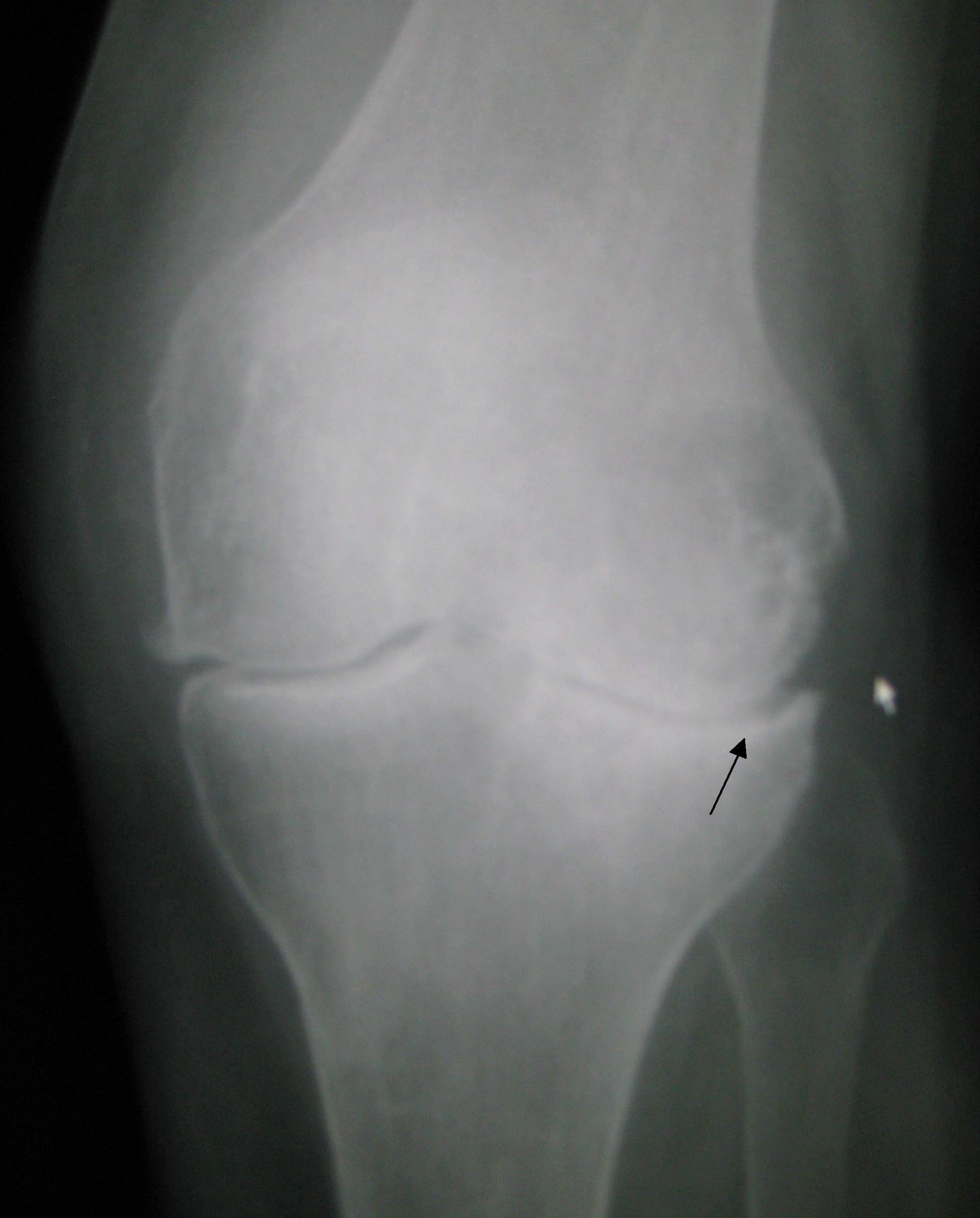
X-rayAnteroposterior X-ray of the left knee showing osteoarthritis: joint space narrowing indicated by an arrow, osteophytes and increased subchondral bone density.
Source : Heilman, Wikimedia Commons · CC BY-SA
Which questions should you ask to understand the patient and their history?
The history in knee osteoarthritis aims to confirm a compatible mechanical clinical phenotype and to rule out the differential diagnoses. 🧐 The NICE NG226 (2022) criteria are remarkably operational: a clinical diagnosis of knee osteoarthritis can be made, without imaging, in any patient aged ≥ 45 years who presents with the following two criteria:- First, activity-related knee pain, relieved (at least partly) by rest;
- First, morning stiffness in the knee that is absent or short-lived (< 30 minutes).¹
- Characterising the pain : site (medial / lateral / anterior / posterior / diffuse), intensity (0-10 numeric scale), quality (mechanical, inflammatory, neuropathic-like), functional impact (stairs, prolonged walking, squatting, prolonged sitting, the classic « jelly sign » pattern on long car journeys).⁴
- Temporal pattern : start-up pain, brief morning limbering-up, pain after periods of inactivity, post-exertion pain within 24h (« exercise-induced flare »).
- History of trauma : ACL injury, meniscectomy, intra-articular fracture, to be sought systematically in order to identify post-traumatic knee osteoarthritis (PTOA), often overlooked in young people. The OPTIKNEE 2022 consensus explicitly recommends routine follow-up of patients after ACL injury (Whittaker 2022 BJSM).⁵
- Comorbidities : obesity (calculate BMI), diabetes, metabolic syndrome, other joint involvement (hands, hip, spine), cardiovascular disease (important when planning exercise).
- Drug history : NSAIDs, analgesics, previous injections, viscosupplementation, opioids (to be avoided according to ACR 2019).²
- Psychosocial factors : kinesiophobia (fear of movement), catastrophising, depression, expectations of treatment, beliefs about the disease (« my cartilage is wearing out », « I will end up in a wheelchair »…). These factors strongly predict chronicity and treatment failure (Kanavaki 2017).⁶
- Personal goals : return to a specific activity (hiking, sport, work), symptom tolerance, the patient's values (essential for shared decision-making).
🧭 The NICE NG226 (2022) diagnostic algorithm for knee osteoarthritis
Clinical diagnosis without routine imaging in patients aged ≥ 45
⚠️ Critical reading: NICE NG226 (2022) states that radiography is not necessary to make the clinical diagnosis of knee osteoarthritis in a typical patient. Imaging remains indicated in specific situations (see the orange box). Source: NICE NG226, 2022.
Which clinical tests should you perform and which other conditions must be ruled out?
The physical examination aims to confirm the clinical picture of knee osteoarthritis, identify the dominant compartment involved (medial, lateral, patellofemoral), assess function and rule out the differential diagnoses.⁴ The essential elements:- Inspection : frontal-plane alignment (varus / valgus), muscle bulk (quadriceps atrophy, wasting of vastus medialis), swelling / effusion (patellar tap, ballottement), skin appearance (redness, warmth, which suggests infection or inflammation).
- Palpation : tenderness of the joint lines (medial, lateral), of the patella, of the pes anserinus (insertion of the sartorius, gracilis and semitendinosus tendons 5-7 cm below the medial joint line, looking for an associated bursitis, common in medial knee osteoarthritis) and of the iliotibial band.
- Range of motion : flexion (normal ≥ 135°), extension (normal 0°, or even +5° of hyperextension), looking for a flexion contracture (limited extension, a major biomechanical factor).
- Crepitus : audible / palpable on passive mobilisation or with active movement (stair climbing, squatting), common in knee osteoarthritis but not very specific.
- Functional tests : the 30-second chair stand test, the Timed Up and Go (TUG), the 40 m walk test, stair ascent and descent, the functional outcome tools recommended by OARSI.³
- Muscle strength : assessment of the quadriceps (dynamometer if possible, otherwise the MRC scale), the iliopsoas and the hip abductors (gluteus medius weakness accentuates dynamic varus).
- Ligament stability : Lachman, anterior drawer, varus / valgus stress, McMurray, to rule out an associated ligament or meniscal injury.
- Distal neurological assessment : sensation, reflexes (knee jerk, ankle jerk), to rule out referred L3-L4 radicular pain.
- Pes anserine bursitis : pain localised 5-7 cm below the medial joint line, at the insertion of the sartorius/gracilis/semitendinosus tendons. Frequently associated with medial knee osteoarthritis (coexisting in up to 75% of cases in some series). Typical case: an obese woman with medial knee osteoarthritis and pain aggravated by stair climbing and prolonged sitting. Reference: case PMC 9674038 (2022).
- Isolated patellofemoral pain syndrome : anterior retropatellar pain, the cinema sign (pain when sitting for long periods with the knees bent), Clarke's test. More common in the active young adult, it can coexist with early patellofemoral osteoarthritis in adults aged 40+.
- Degenerative meniscal lesion : very common (asymptomatic in 60% of people aged 50+), to be correlated with symptoms. Meniscectomy is not recommended in the absence of true mechanical locking (ESSKA consensus).
- Crystal arthritis : gout (hyperuricaemia, podagra), chondrocalcinosis (meniscal calcification). An acute flare with a red, hot, painful knee.
- Septic arthritis : an emergency, with a red, hot, tense, febrile, exquisitely painful knee. Immediate joint aspiration.
- Peri-articular tendinopathies : quadriceps, patellar (jumper's knee), iliotibial band (TFL syndrome).
- Bone conditions : subchondral insufficiency fracture (common in the osteoporotic older adult, see PMC 9975902 2023), condylar osteonecrosis, osteosarcoma (in the young), bone metastasis (older adult with a relevant history).
- Referred pain of extra-articular origin : hip disease (anterior thigh pain referred to the knee, always examine the hip!), L3-L4 radicular pain, entrapment neuropathy (saphenous).
Should patients with knee osteoarthritis be classified, and what is gained by doing so?
Yes. The concept of clinical phenotyping of knee osteoarthritis emerged in the 2010s as a response to the heterogeneity of presentations and of treatment responses.⁷,⁸ The Dell'Isola et al. 2016 systematic review (BMC Musculoskelet Disord) synthesised the proposed phenotypes and the Deveza et al. 2017 review (Osteoarthritis Cartilage) confirmed their prognostic value. The most operational classification distinguishes 4 broad subgroups:⁷,⁸,⁹| Phenotype | Clinical markers | % of the KOA population | Targeted management |
|---|---|---|---|
| Mechanical / biomechanical | Frontal-plane malalignment, quadriceps weakness, instability, clear activity-related pain | ≈ 30-40 % | Quadriceps + hip abductor strengthening, orthoses, neuromuscular control |
| Inflammatory | Effusion, synovitis on MRI, prolonged morning stiffness, morning limbering-up, moderate night pain | ≈ 20-30 % | Topical NSAIDs 1st line, viscosupplementation possible, corticosteroid injection (reserved for flares) |
| Central sensitisation | Diffuse / disproportionate pain, hyperalgesia, allodynia, chronic pain comorbidity, CSI score ≥ 40 | ≈ 20-30% (Arendt-Nielsen 2018) | Pain neuroscience education, CBT, graded exercise, sleep management, tricyclic antidepressant if needed |
| Metabolic | Full metabolic syndrome, abdominal obesity, diabetes, dyslipidaemia, multi-joint involvement | ≈ 15-20 % | Weight loss as the priority (≥ 10% as a minimum, more if possible, Messier 2018), aerobic exercise, glycaemic control, cardiometabolic polypill |
- Targeted treatment direction : a « central sensitisation » patient will respond better to pain education + CBT + graded exercise than to an intensive strengthening programme, which can sustain catastrophising. Conversely, a « biomechanical » patient will benefit mainly from quadriceps and neuromuscular strengthening.
- Personalised follow-up : a « metabolic » phenotype calls for coordination with the GP, the dietitian and sometimes the diabetes specialist.
- A sharper prognosis : « central sensitisation » and « metabolic » patients have the least favourable functional prognosis without multidisciplinary intervention (Deveza 2017).⁸
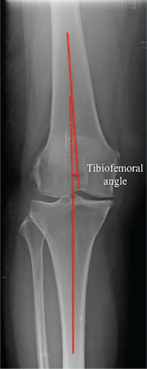
X-rayAnteroposterior knee X-ray with the femorotibial angle traced: this measurement of limb alignment quantifies the varus or valgus deviation seen in knee osteoarthritis.
Source : Pinsornsak et al., Bone & Joint Research, 2016, figure 4 · CC BY-NC
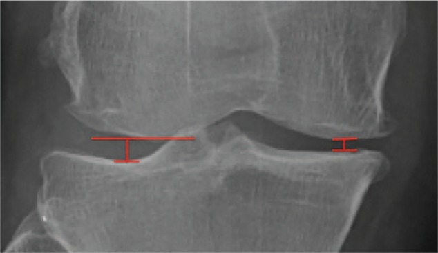
X-rayClose-up knee X-ray: measurement of the medial and lateral joint space width, a technique used to assess narrowing related to knee osteoarthritis.
Source : Pinsornsak et al., Bone & Joint Research, 2016, figure 3 · CC BY-NC
Key points
- ✅ The diagnosis of knee osteoarthritis is essentially clinical according to NICE NG226 (2022), ACR 2019 and OARSI 2019: in a patient aged ≥ 45 with activity-related pain + morning stiffness < 30 min, imaging is neither necessary nor recommended as a routine.
- ✅ The history identifies the temporal pattern, previous trauma (systematically look for post-ACL knee osteoarthritis, OPTIKNEE 2022), comorbidities and psychosocial factors (kinesiophobia, catastrophising, expectations).
- ✅ The standardised physical examination looks for: joint-line and pes anserinus tenderness, range of motion, crepitus, quadriceps strength, ligament stability, functional tests (TUG, chair stand).
- ✅ Key differential diagnoses: pes anserine bursitis (often associated with medial knee osteoarthritis), crystal arthritis, septic arthritis, subchondral insufficiency fracture, referred hip disease, L3-L4 radicular pain.
- ✅ The clinical phenotyping into 4 subgroups (mechanical, inflammatory, central sensitisation, metabolic: Dell'Isola 2016, Deveza 2017) guides personalised management.
- ✅ The radiographic-clinical discordance is the rule: imaging is not a decision-making argument: the clinical picture takes precedence.
Bibliography
- National Institute for Health and Care Excellence. Osteoarthritis in over 16s: diagnosis and management. NICE guideline NG226. Published 19 October 2022. https://www.nice.org.uk/guidance/ng226.
- Kolasinski SL, Neogi T, Hochberg MC, et al. 2019 American College of Rheumatology/Arthritis Foundation Guideline for the Management of Osteoarthritis of the Hand, Hip, and Knee. Arthritis Rheumatol. 2020;72(2):220-233. PMID 31908163.
- Bannuru RR, Osani MC, Vaysbrot EE, et al. OARSI guidelines for the non-surgical management of knee, hip, and polyarticular osteoarthritis. Osteoarthritis Cartilage. 2019;27(11):1578-1589. PMID 31278997.
- Sharma L. Osteoarthritis of the Knee. N Engl J Med. 2021;384(1):51-59. PMID 33406330.
- Whittaker JL, Culvenor AG, Juhl CB, et al. OPTIKNEE 2022: consensus recommendations to optimise knee health after traumatic knee injury to prevent osteoarthritis. Br J Sports Med. 2022;56(24):1393-1405. PMID 36379676.
- Kanavaki AM, Rushton A, Efstathiou N, et al. Barriers and facilitators of physical activity in knee and hip osteoarthritis: a systematic review of qualitative evidence. BMJ Open. 2017;7(12):e017042. PMID 29282257.
- Dell'Isola A, Allan R, Smith SL, Marreiros SS, Steultjens M. Identification of clinical phenotypes in knee osteoarthritis: a systematic review of the literature. BMC Musculoskelet Disord. 2016;17:425. PMID 27733199.
- Deveza LA, Melo L, Yamato TP, Mills K, Ravi V, Hunter DJ. Knee osteoarthritis phenotypes and their relevance for outcomes: a systematic review. Osteoarthritis Cartilage. 2017;25(12):1926-1941. PMID 28847624.
- Arendt-Nielsen L, Morlion B, Perrot S, et al. Assessment and manifestation of central sensitisation across different chronic pain conditions. Eur J Pain. 2018;22(2):216-241. PMID 29105941.
- Bedson J, Croft PR. The discordance between clinical and radiographic knee osteoarthritis: a systematic search and summary of the literature. BMC Musculoskelet Disord. 2008;9:116. PMID 18764949.
- Hunter DJ, Bierma-Zeinstra S. Osteoarthritis. Lancet. 2019;393(10182):1745-1759. PMID 31034380.
- Hawker GA, Mian S, Kendzerska T, French M. Measures of adult pain: VAS, NRS, MPQ, SF-MPQ, CPGS, SF-36 BPS, ICOAP. Arthritis Care Res (Hoboken). 2011;63(S11):S240-S252. PMID 22588748.
- Whittaker JL, Losciale JM, Juhl CB, et al. Risk factors for knee osteoarthritis after traumatic knee injury: a systematic review and meta-analysis for the OPTIKNEE Consensus. Br J Sports Med. 2022;56(24):1406-1421. PMID 36455966.
- Finucane LM, Downie A, Mercer C, et al. International Framework for Red Flags for Potential Serious Spinal Pathologies. J Orthop Sports Phys Ther. 2020;50(7):350-372. PMID 32438853.
Which treatment strategies are the most effective for knee osteoarthritis?
Where do you start? What is the recommended hierarchy of interventions?
🎯 The three guidelines (ACR 2019, OARSI 2019, NICE 2022) align on three first-line interventions to be offered systematically to every patient as soon as the diagnosis is made:¹,²,³- 1. Patient education : dismantling mistaken beliefs (« my cartilage is wearing out », « I must not push it »), a modern explanation of knee osteoarthritis as a whole-joint disease that behaviour can modify, managing expectations, building independence. Caneiro 2020 (BJSM) proposes 3 major narrative shifts: (1) « osteoarthritis is not an inevitability », (2) « moving does not destroy, moving protects », (3) « surgery is neither unavoidable nor always the answer ».⁶
- 2. Tailored physical exercise (land-based or aquatic): the central pillar, with a high level of evidence (Cochrane Fransen 2015, GRADE high quality).⁴ Validated structured programmes: GLA:D (Good Life with osteoArthritis in Denmark, Skou & Roos 2017) rolled out in 11+ countries, and ESCAPE-pain (Enabling Self-management and Coping with Arthritic Pain through Exercise, Hurley), which combines education and supervised group exercise over 6 weeks (10-12 sessions).⁷,⁸
- 3. Weight management in patients who are overweight or obese: the target is ≥ 10% loss of body weight, a minimum threshold beyond which the benefit keeps growing: in the dose-response analysis of the IDEA trial, patients who lost more than 20% reported about 25% less pain than those in the 10-20% group (Messier 2018).⁹ This intervention is synergistic with exercise: combining weight loss + exercise gives the best functional outcomes.⁹
🪜 Hierarchy of treatment interventions in knee osteoarthritis (horizontal cards)
A synthesis of ACR 2019 / OARSI 2019 / NICE NG226 2022: a pyramid laid out as horizontal cards (never an illegible triangle)
📊 A hierarchy validated by ACR 2019, OARSI 2019 and NICE NG226 2022. Progression is conditional on failure of the previous level, not automatic. Levels 1 and 2 must be maintained throughout the pathway, including after surgery.
What is the place of exercise, and is there a superior approach?
🏋️♂️ Exercise is the cornerstone of the non-surgical management of knee osteoarthritis, with one of the highest levels of evidence in musculoskeletal medicine. The reference Cochrane meta-analysis (Fransen 2015), 54 randomised trials, 3,913 patients, shows a clinically significant improvement in pain (SMD ≈ 0.49) and in function (SMD ≈ 0.52) in the short term, with an effect that persists 2-6 months after the end of the supervised programme (GRADE: high quality).⁴ The big clinical question: is one type of exercise superior to the others? The Goh et al. 2019 meta-analysis (Ann Phys Rehabil Med, 77 trials, > 6,000 patients) answers clearly: no, there is no statistical superiority between the modalities when they are compared head to head.⁵ Strengthening, aerobic work, neuromuscular control, tai chi, yoga: all these modalities produce clinically comparable effects. The Bartholdy et al. 2017 meta-regression (Semin Arthritis Rheum) confirms that improvement in quadriceps strength is associated with the reduction in pain, but without a strict dose-response relationship.¹⁰ Current practical recommendations:- Combined programme : strengthening (≥ 2 times a week, at tolerated intensity), aerobic work (walking, cycling, swimming: 150 min a week in total), neuromuscular control (balance, proprioception).
- Initial supervision for 8-12 weeks then transition to structured self-management. The Holden 2023 IPDMA (Lancet Rheumatol) confirms that the effects of exercise are independent of age, BMI, radiographic severity and sex: exercise works for every patient.¹¹
- Aquatic exercise : a sound alternative where weight-bearing is not tolerated or comorbidities are present, with efficacy comparable to land-based exercise for pain and function (Dong 2018, Medicine Baltimore).¹²
- Validated structured programmes : GLA:D (Skou & Roos 2017), 2 education sessions + 12 supervised neuromuscular exercise sessions over 6 weeks, deployed in more than 11 countries with an international registry;⁷ ESCAPE-pain (Hurley), 6 weeks, 12 group sessions combining education and functional exercise, economically validated for health services.⁸
📊 GLA:D vs ESCAPE-pain: evidence-based structured programmes
An operational comparison of the two best-validated programmes for knee osteoarthritis
| Criterion | GLA:D (Skou & Roos 2017) | ESCAPE-pain (Hurley) |
|---|---|---|
| Origin | Denmark (2013), deployed in 11+ countries | United Kingdom, NHS programme |
| Duration | 6 weeks (8 sessions) | 6 weeks (12 sessions) |
| Composition | 2 education sessions + 12 neuromuscular exercise sessions | Integrated education + group functional exercise |
| Format | Group, supervised by a certified physiotherapist | Group (~10 patients), supervised |
| Target population | Symptomatic knee and hip osteoarthritis | Chronic knee pain with suspected or confirmed knee osteoarthritis |
| Level of evidence | High (international registry > 35,000 patients, cohorts) | High (pragmatic RCT, validated cost-effectiveness) |
| WOMAC effect at 3 months | ↓ pain 25-30%, ↑ function 25-30% | ↓ pain ~30%, ↑ function ~30%, benefit maintained ≥ 6 months |
📌 Both programmes are international standards. GLA:D is the more formalised (therapist certification + registry); ESCAPE-pain places more emphasis on the behavioural side and on the education/coping component. Choose according to local availability.
Manual therapies and technologies: how effective are they really?
✋ As for manual therapy (joint mobilisations, soft-tissue techniques, assisted stretching), its role is adjunctive to exercise. The Anwer et al. 2018 meta-analysis (Physical Therapy Reviews) shows that combining manual therapy with exercise produces a greater short-term improvement in pain and function than exercise alone.¹³ In the medium and long term, however, the advantage fades: manual therapy acts mainly as a « catalyst » that makes engagement in exercise easier (transient pain relief, a sense of improvement). ⚡ For the physical modalities, the picture is more nuanced:- TENS (transcutaneous electrical nerve stimulation) : a modest, transient effect on pain, superior to placebo in the Cherian et al. 2016 meta-analysis (J Knee Surg) but with high heterogeneity and low to moderate quality of evidence.¹⁴ It may be offered as a home self-management tool, but it is not recommended for routine use by the major guidelines.
- Therapeutic ultrasound : the Dantas et al. 2021 meta-analysis (Braz J Phys Ther) concludes that there is a small, short-term effect on pain and function, with a very low quality of evidence (GRADE very low) linked to risk of bias and to heterogeneity.¹⁵ NICE NG226 and ACR 2019 do not recommend its routine use.¹,²
- Bracing / taping / orthoses : ACR 2019 issues a conditional recommendation for patellar taping in patellofemoral knee osteoarthritis and for orthoses (knee brace, insoles) in patients with malalignment.²
- Acupuncture : OARSI 2019 issues a conditional recommendation; ACR 2019 also recommends it conditionally. A modest effect, heterogeneous from one patient to another.²,³
🎯 Comparative table of treatment modalities in knee osteoarthritis × GRADE level of evidence
A synthesis of the ACR 2019, OARSI 2019 and NICE NG226 2022 recommendations
| Modality | Main mechanism | GRADE level of evidence | Guideline recommendation |
|---|---|---|---|
| Land-based exercise (strengthening, aerobic, neuromuscular) | Muscle strengthening, motor control, neuroplasticity | HIGH | STRONG: all guidelines |
| Aquatic exercise | Reduced weight-bearing, hydrodynamic resistance | MODERATE | STRONG: all guidelines |
| Therapeutic patient education (TPE) | Deconstructing beliefs, self-efficacy, behaviour change | HIGH | STRONG: all guidelines |
| Weight management (loss ≥ 10%) | Reduced mechanical load + systemic inflammation | HIGH | STRONG: all guidelines |
| Manual therapy (adjunctive) | Pain modulation, transient mobility gain | MODERATE | CONDITIONAL: as an adjunct to exercise |
| Topical NSAIDs (1st line pharmacological) | Local anti-inflammatory, low systemic absorption | HIGH | STRONG: ACR/OARSI/NICE |
| TENS | Neurogenic pain modulation (gate control) | LOW to MODERATE | CONDITIONAL or NOT recommended for routine use |
| Therapeutic ultrasound | Tissue thermal effect | VERY LOW | NOT recommended for routine use (NICE, ACR) |
| Acupuncture | Central + local pain modulation | MODERATE (heterogeneous) | CONDITIONAL: according to patient preference |
| Intra-articular corticosteroid injection | Short-term local anti-inflammatory | MODERATE (short term) | CONDITIONAL: reserved for an inflammatory flare |
| Viscosupplementation (hyaluronate) | Joint lubrication, inflammation modulation | LOW | ACR: recommends AGAINST · OARSI: conditional |
| Total knee replacement | Structural replacement | HIGH (selected cases) | After conservative failure ≥ 3-6 months |
📌 Source: a synthesis of ACR 2019 (Kolasinski PMID 31908163), OARSI 2019 (Bannuru PMID 31278997) and NICE NG226 2022. Passive modalities must never take the place of active interventions.
Beyond the physical: how do you educate the patient and address psychological factors?
🧠 Knee osteoarthritis is not only a joint disease: it is a biopsychosocial experience. The psychological factors (kinesiophobia, catastrophising, depression, mistaken beliefs) are major predictors of chronicity and of treatment failure.⁶,¹⁶ Identifying them and addressing them is essential. Therapeutic patient education (TPE) has proved effective in several systematic reviews. Du et al. 2011 (Patient Educ Couns) showed that structured self-management programmes produce small to moderate effects on pain and disability in the long term.¹⁶ Education must cover:- The nature of the disease : a whole-joint disease, modifiable, not simple wear and tear; moving protects, it does not destroy.
- The mechanisms of pain : pain neuroscience education (PNE), particularly useful in patients with central sensitisation.
- The health behaviours : the importance of regular physical activity, weight management, sleep and stress management.
- The beliefs and expectations : deconstructing « the cartilage is wearing out », « I must move less » and « surgery is inevitable ».
- Graded exposure to the feared movement, within a reassuring framework.
- PNE education to deconstruct the pain = damage association.
- Self-monitoring of symptoms, to reassure the patient about tolerance to exertion.
- Working with a psychologist specialising in cognitive behavioural therapy (CBT) if the picture is severe or resistant.
Key points
- 🥇 Three first-line pillars agreed by consensus (ACR 2019, OARSI 2019, NICE NG226 2022): therapeutic education + adapted physical exercise + weight management. To be offered to every patient from diagnosis onwards.
- 🏋️ Exercise = cornerstone : HIGH level of evidence (Cochrane Fransen 2015). No one type is superior to another (Goh 2019): a combined strengthening + aerobic + neuromuscular programme is optimal. The effect is independent of age, BMI and radiographic severity (Holden 2023 IPDMA).
- 📋 Validated structured programmes : GLA:D (Skou 2017) and ESCAPE-pain (Hurley), demonstrated efficacy, to be preferred wherever they are available.
- ⚖️ Weight loss ≥ 10% : the minimum threshold, and the benefit on pain and function keeps growing beyond it (Messier 2018). Synergy with exercise.
- ✋ Manual therapy : ADJUNCTIVE to exercise, short-term effect. A catalyst for engagement, not a background treatment.
- ⚡ Passive modalities (TENS, ultrasound): low or very low evidence, NOT recommended for routine use. To be limited or dropped.
- 🧠 Address the psychological factors : kinesiophobia (Coronado 2018), catastrophising, mistaken beliefs. PNE and collaboration with a psychologist if the picture is severe.
Bibliography
- National Institute for Health and Care Excellence. Osteoarthritis in over 16s: diagnosis and management. NICE NG226. October 2022. https://www.nice.org.uk/guidance/ng226.
- Kolasinski SL, Neogi T, Hochberg MC, et al. 2019 American College of Rheumatology/Arthritis Foundation Guideline for the Management of Osteoarthritis of the Hand, Hip, and Knee. Arthritis Rheumatol. 2020;72(2):220-233. PMID 31908163.
- Bannuru RR, Osani MC, Vaysbrot EE, et al. OARSI guidelines for the non-surgical management of knee, hip, and polyarticular osteoarthritis. Osteoarthritis Cartilage. 2019;27(11):1578-1589. PMID 31278997.
- Fransen M, McConnell S, Harmer AR, Van der Esch M, Simic M, Bennell KL. Exercise for osteoarthritis of the knee. Cochrane Database Syst Rev. 2015;1:CD004376. PMID 25569281.
- Goh SL, Persson MSM, Stocks J, et al. Efficacy and potential determinants of exercise therapy in knee and hip osteoarthritis: A systematic review and meta-analysis. Ann Phys Rehabil Med. 2019;62(5):356-365. PMID 31121333.
- Caneiro JP, O'Sullivan PB, Roos EM, et al. Three steps to changing the narrative about knee osteoarthritis care: a call to action. Br J Sports Med. 2020;54(5):256-258. PMID 31484634.
- Skou ST, Roos EM. Good Life with osteoArthritis in Denmark (GLA:D™): evidence-based education and supervised neuromuscular exercise delivered by certified physiotherapists nationwide. BMC Musculoskelet Disord. 2017;18:72. PMID 28173795.
- Hurley M, Dickson K, Hallett R, et al. Exercise interventions and patient beliefs for people with hip, knee or hip and knee osteoarthritis: a mixed methods review (ESCAPE-pain). Cochrane Database Syst Rev. 2018;4:CD010842. PMID 29664187.
- Messier SP, Resnik AE, Beavers DP, et al. Intentional Weight Loss in Overweight and Obese Patients With Knee Osteoarthritis: Is More Better? Arthritis Care Res (Hoboken). 2018;70(11):1569-1575. PMID 29911741.
- Bartholdy C, Juhl C, Christensen R, Lund H, Zhang W, Henriksen M. The role of muscle strengthening in exercise therapy for knee osteoarthritis: A systematic review and meta-regression analysis of randomized trials. Semin Arthritis Rheum. 2017;47(1):9-21. PMID 28438380.
- Holden MA, Hattle M, Runhaar J, et al. Moderators of the effect of therapeutic exercise for knee and hip osteoarthritis: a systematic review and individual participant data meta-analysis. Lancet Rheumatol. 2023;5(7):e386-e400. doi:10.1016/S2665-9913(23)00122-4.
- Dong R, Wu Y, Xu S, et al. Is aquatic exercise more effective than land-based exercise for knee osteoarthritis? Medicine (Baltimore). 2018;97(52):e13823. PMID 30593178.
- Anwer S, Alghadir A, Zafar H, Brismée JM. Effect of orthopaedic manual therapy in knee osteoarthritis: A systematic review and meta-analysis. Physiotherapy. 2018;104(3):264-276. PMID 30030035.
- Cherian JJ, Harrison PE, Benjamin SA, et al. Do the Effects of Transcutaneous Electrical Nerve Stimulation on Knee Osteoarthritis Pain and Function Last? J Knee Surg. 2016;29(6):497-501. PMID 26540652.
- Dantas LO, Salvini TF, McAlindon TE. Therapeutic ultrasound for knee osteoarthritis: A systematic review and meta-analysis with grade quality assessment. Braz J Phys Ther. 2021;25(6):688-697. PMID 34535411.
- Du S, Yuan C, Xiao X, Chu J, Qiu Y, Qian H. Self-management programs for chronic musculoskeletal pain conditions: A systematic review and meta-analysis. Patient Educ Couns. 2011;85(3):e299-e310. PMID 21458196.
How do you secure a lasting recovery and prevent recurrence in knee osteoarthritis?
How do you make the patient an active participant in their own recovery through self-management?
🎯 Self-management (or supported self-care) is a collaborative approach in which the patient, guided by the health professional, acquires the skills and the confidence needed to manage their symptoms and their treatment day to day.³ The aim is to turn the patient from a passive recipient of care into an informed central participant in their own health. Effective self-management strategies rest on two fundamental pillars: therapeutic patient education (TPE) and the strengthening of self-efficacy.- TPE : the Du et al. 2011 meta-analysis confirms that structured self-management programmes produce small to moderate effects on pain and disability in the long term.⁴ TPE must include: an explanation of the nature of knee osteoarthritis (a whole-joint disease, modifiable, not simple wear and tear), deconstruction of mistaken beliefs (« I must move less », « moving wears the cartilage out »), an explanation of pain mechanisms (Pain Neuroscience Education), management of inflammatory flares, and the place of regular physical activity. The Caneiro 2020 review proposes a new narrative in 3 steps: (1) recognise the mistaken beliefs, (2) replace them with evidence-based messages, (3) equip the patient for lasting self-management.⁵
- Self-efficacy 🧠 : the belief in one's own capacity to carry out a task or to manage a situation. In knee osteoarthritis, this means giving the patient the confidence to move despite the pain and to manage flares. The structured programmes GLA:D (Skou 2017) and ESCAPE-pain (Hurley) combine education, supervised group exercise and behavioural support, with documented improvement in self-efficacy and in function at 3, 6 and 12 months.⁶,⁷
When and how do you plan a safe return to sport and to daily activities?
🏃 Returning to physical activity and to sport is not only possible in patients with knee osteoarthritis, it is strongly recommended : inactivity is a major aggravating factor. Holden et al. 2023 (IPDMA Lancet Rheumatol) confirms that the beneficial effects of exercise on pain and on function are independent of radiographic severity, of age, of sex and of BMI.⁸ Exercise works for every patient. Planning that return must nevertheless be meticulous, to avoid joint overload and painful flares. The modern approach abandons fixed calendars in favour of a criterion-based approach :⁹- Phase 1: pain control and restoration of basic function. Reduce pain and inflammation. Restore a full joint range (≥ 130° flexion, full extension). Activate the stabilising muscles (quadriceps, hamstrings, glutes, gastrocnemius). Low-impact exercise preferred: stationary bike, swimming, aqua-aerobics, progressive walking. Criterion for moving to phase 2: pain ≤ 3/10 on exertion, range ≥ 130° flexion, ability to go up and down stairs without major pain.
- Phase 2: progressive retraining and increasing load. Progressive mechanical load: the principle of progressive mechanical loading, stimulating tissue adaptation without an excessive inflammatory reaction. Intensified strength work (resistance ≥ 60-70% 1RM if tolerated). Functional exercises approaching the target movements (squats, lunges, moderate jumping according to the goal). Golden rule : monitor the knee's response in the 24 hours after exertion (pain, swelling). If there is a flare → reduce the load by 20-30% for the next session, without giving up. Criterion for moving to phase 3: quadriceps strength ≥ 80% of the uninvolved side (dynamometer), symmetrical functional tests (single leg squat, Y-balance test).
- Phase 3: return to the specific activity and prevention strategy. Full return to the target activity (sport, work, leisure) only if the functional tests (strength, balance, neuromuscular control) are satisfactory. Long-term maintenance plan : ≥ 2 strength sessions a week + 150 min a week of aerobic activity in total. Intelligent management of the total load (frequency × intensity × duration), with possible application of the ACWR concept (acute:chronic workload ratio) borrowed from sports medicine.
📈 Return to activity after knee osteoarthritis: a criterion-based approach (3 phases)
Criteria for moving between phases: the fixed calendar gives way to function
📌 Central principle: progression follows function, not the calendar. No early return even if « it feels better »: the functional tests are what make safety objective. Sports medicine concepts (ACWR) can be applied.
Key points
- 🎯 Patient empowerment is the cornerstone of long-term management: therapeutic education + strengthening of self-efficacy (Du 2011, Skou GLA:D 2017, ESCAPE-pain).
- 📋 Structured programmes to be preferred: GLA:D (6 weeks, 8 sessions), ESCAPE-pain (6 weeks, 12 sessions). Validated effects on function and on self-efficacy at 3-12 months.
- 📱 Digital tools (apps, telerehabilitation): effects comparable to on-site care, greater accessibility.
- 🏃 Return to activity : use a criterion-based approach in 3 phases, never a fixed calendar. Phase 1 pain control → Phase 2 progressive retraining → Phase 3 specific return + maintenance.
- 📈 Long-term maintenance plan : ≥ 2 strength sessions a week + 150 min a week of aerobic work in total. Exercise protects the joint, it does not wear it out (Holden 2023 IPDMA).
- ⚽ Sports tolerated by most patients: moderate running, doubles tennis, golf, hiking. Adaptations possible for high-impact sports.
Bibliography
- Hunter DJ, Bierma-Zeinstra S. Osteoarthritis. Lancet. 2019;393(10182):1745-1759. PMID 31034380.
- Kolasinski SL, Neogi T, Hochberg MC, et al. 2019 ACR/Arthritis Foundation Guideline. Arthritis Rheumatol. 2020;72(2):220-233. PMID 31908163.
- Kanavaki AM, Rushton A, Efstathiou N, et al. Barriers and facilitators of physical activity in knee and hip osteoarthritis: qualitative SR. BMJ Open. 2017;7(12):e017042. PMID 29282257.
- Du S, Yuan C, Xiao X, et al. Self-management programs for chronic musculoskeletal pain conditions: SR/MA. Patient Educ Couns. 2011;85(3):e299-e310. PMID 21458196.
- Caneiro JP, O'Sullivan PB, Roos EM, et al. Three steps to changing the narrative about knee osteoarthritis care. Br J Sports Med. 2020;54(5):256-258. PMID 31484634.
- Skou ST, Roos EM. Good Life with osteoArthritis in Denmark (GLA:D). BMC Musculoskelet Disord. 2017;18:72. PMID 28173795.
- Hurley M, Dickson K, Hallett R, et al. Exercise interventions and patient beliefs (ESCAPE-pain). Cochrane Database Syst Rev. 2018;4:CD010842. PMID 29664187.
- Holden MA, Hattle M, Runhaar J, et al. Moderators of the effect of therapeutic exercise for knee and hip osteoarthritis: IPDMA. Lancet Rheumatol. 2023;5(7):e386-e400. DOI 10.1016/S2665-9913(23)00122-4.
- Whittaker JL, Culvenor AG, Juhl CB, et al. OPTIKNEE 2022 consensus. Br J Sports Med. 2022;56(24):1393-1405. PMID 36379676.
- Bannuru RR, Osani MC, Vaysbrot EE, et al. OARSI guidelines for non-surgical management. Osteoarthritis Cartilage. 2019;27(11):1578-1589. PMID 31278997.
What do real-world case reports teach us about knee osteoarthritis?
Analysis of a « classic » case: from assessment to resolution
The typical case of knee osteoarthritis is a patient over 50, overweight, with progressive mechanical knee pain, brief morning stiffness and moderate functional limitation. The IDEA cohort (Intensive Diet and Exercise for Arthritis, the index trial Messier 2013 in JAMA, secondary dose-response analysis Messier 2018), 454 patients with symptomatic knee osteoarthritis and BMI ≥ 27, is the best evidence-based illustration of how this profile is managed.² Mean profile of the IDEA cohort :- Mean age 65, 73% women;
- Mean BMI 33.7 kg/m² (class I-II obesity);
- Radiographic knee osteoarthritis, Kellgren-Lawrence grade 2-3;
- Mean WOMAC pain 7.1/20 (0-20 scale, lower = better);
- 6-minute walk distance: 460 m (reduced versus healthy people).
- Mean weight loss: 10.6 kg (loss ≥ 10% in 70% of patients);
- WOMAC pain reduction: −51 % (versus −37% for the Diet-only group, −28% for the Exercise-only group);
- WOMAC functional improvement: +45 % ;
- Walking speed: +15 % ;
- Reduction in inflammatory markers (plasma IL-6: −20%).²
The diagnostic challenge: when knee osteoarthritis mimics another condition
🔍 Knee pain in a middle-aged or older patient with radiological signs of osteoarthritis is not always entirely attributable to the osteoarthritis itself. Peri-articular structures may be the main source of the symptoms, and ignoring them can lead to treatment failure. The case published in PMC 9674038 (2022) is instructive.⁵ Initial presentation : a 58-year-old woman presents with intense anteromedial pain in the left knee, made worse by going up and down stairs, by moving from sitting to standing, and at night in side-lying (the knee supporting the other one). Radiograph: Kellgren-Lawrence grade 2 medial knee osteoarthritis. Initial diagnosis: an inflammatory flare of the knee osteoarthritis. General quadriceps strengthening exercises were started, with no improvement after 4 weeks.⁵ Clinical reassessment : palpation reveals exquisite, reproducible tenderness located 5-7 cm below the medial joint line, at the insertion of the sartorius, gracilis and semitendinosus tendons (the pes anserinus). Tensioning the hamstrings reproduces the pain. Ultrasound confirms pes anserine bursitis. The diagnosis is revised to pes anserine bursitis coexisting with medial knee osteoarthritis , a frequent diagnostic confusion (coexistence reported in up to 75% of symptomatic medial knee osteoarthritis). Targeted treatment : relative rest from the triggering activities, ice 15 min × 3 a day, gentle hamstring and adductor stretching, topical NSAID 2-3 times a day for 10 days, ultrasound-guided corticosteroid injection into the pes anserine bursa. Quadriceps + hip abductor strengthening is maintained for the underlying knee osteoarthritis. Outcome at 2 weeks : 80% improvement in pain, full resumption of daily activities. The exercise programme for the knee osteoarthritis was resumed.⁵ 🎯 Clinical lesson : never stop at the radiological imaging. A rigorous physical examination of every peri-articular structure is required. Treatment failure at 4-6 weeks must prompt reconsideration of the diagnosis and identification of the associated nociceptive sources.A complex case
Management becomes more complex when knee osteoarthritis sits within a broader clinical picture, with comorbidities, severe structural damage or atypical progression. Real published cases illustrate this complexity. Case 1: rapidly destructive arthropathy of the knee (PMC 8437648, 2021)⁶ A rare but devastating case: a woman with Parkinson's disease and Pisa syndrome (lateral deviation of the trunk) developed a rapidly destructive arthropathy of the knee on the same side as the deviation. Biomechanical analysis attributes the destruction to the chronic eccentric mechanical overload linked to the trunk deviation (the knee-spine syndrome). The clinical course went from moderate knee pain to complete joint destruction in less than 12 months, requiring a total knee replacement. This case is a reminder of how important it is to assess overall posture (spine, hip, frontal alignment) in every patient with knee osteoarthritis, and particularly in older patients with neurological comorbidities. Case 2: knee osteoarthritis and severe metabolic syndrome⁷ Typical profile: a 55-year-old patient, BMI 38 kg/m² (class II obesity), poorly controlled type 2 diabetes (HbA1c 8.2%), dyslipidaemia, hypertension, disabling bilateral Kellgren-Lawrence grade 4 knee osteoarthritis. Constant pain disturbing sleep, all physical activity abandoned, the metabolic vicious circle worsening. Interdisciplinary management is essential: physiotherapist (aquatic exercise first, then progressive land-based work), rheumatologist, endocrinologist/diabetologist, nutritionist/dietitian, sometimes a bariatric surgeon. The Inacio 2014 and Groen 2015 studies show that bariatric surgery in these patients gives striking results: not only massive weight loss (40-50 kg) but also a dramatic reduction in knee pain and functional improvement, delaying or even avoiding a joint replacement. Case 3: subchondral insufficiency fracture in the older patient (PMC 9975902, 2023)⁸ A 78-year-old woman, known osteoporosis (T-score −2.8 at the femoral neck), on bisphosphonates. Sudden onset of severe medial knee pain with no obvious trauma, after a long walk. Initial diagnosis: an inflammatory flare of medial knee osteoarthritis. Conservative treatment failed over 4 weeks. Late MRI : subchondral insufficiency fracture of the medial tibial plateau, with extensive bone marrow oedema. This under-diagnosed entity in older osteoporotic patients must be considered in any acute de novo knee pain without trauma, particularly in osteoporotic patients. Treatment: partial offloading for 6-8 weeks, analgesia, optimisation of the anti-osteoporotic treatment, falls prevention. Course: healing in 3-6 months with no sequelae if the diagnosis is made in time. Case 4: refractory pain and alternatives to surgery in the frail older patient⁹ An older patient (≥ 75 years) with severe grade 4 knee osteoarthritis, pain 8-9/10, but major cardiovascular and anaesthetic contraindications to total knee replacement. Minimally invasive techniques to consider: genicular nerve radiofrequency ablation (Gupta 2017 SR, El-Hakeim 2018: pain reduction ≥ 50% maintained at 6-12 months in most selected patients), corticosteroid injections under ultrasound guidance (short-term effect), viscosupplementation (modest effect, several cycles sometimes needed). These options improve quality of life and allow better participation in physiotherapy in complex populations where surgery is contraindicated.📚 GRADE pyramid of levels of evidence (stacked horizontal cards)
The hierarchy of scientific evidence applied to knee osteoarthritis, from the most solid to the most fragile
⚠️ Critical reading: case reports (GRADE 5) carry the weakest evidential power. Their value is educational and hypothesis-generating. Any extrapolation to the general population is risky. Publication bias: atypical cases and spectacular results are over-represented (Nissen & Wynn 2014, BMC Res Notes).¹⁰
Key points
- 📚 The typical case of knee osteoarthritis responds very well to active multimodal management including education, exercise and weight loss (IDEA trial, Messier 2013 JAMA: −51% WOMAC pain).
- 🔍 Always carry out a rigorous differential diagnosis : note that pes anserine bursitis frequently coexists with medial knee osteoarthritis and can mimic or aggravate the painful picture (PMC 9674038).
- ⚠️ Complex cases call for interdisciplinary approaches : RDA of the knee in Parkinson's disease (PMC 8437648), severe metabolic syndrome + morbid obesity (consider bariatric surgery), subchondral insufficiency fracture in the older osteoporotic patient (PMC 9975902).
- 🎯 In the frail older patient who cannot be operated on: genicular nerve radiofrequency ablation (Gupta 2017 SR) is a sound alternative to surgery.
- 📊 Remember: case reports are GRADE 5 , the lowest level of evidence. Their value is educational and hypothesis-generating, never demonstrative. Never extrapolate from them to the general population.
Bibliography
- Nissen T, Wynn R. The clinical case report: a review of its merits and limitations. BMC Res Notes. 2014;7:264. PMID 24758689.
- Messier SP, Resnik AE, Beavers DP, et al. Intentional Weight Loss in Overweight and Obese Patients With Knee Osteoarthritis: Is More Better? Arthritis Care Res. 2018;70(11):1569-1575. PMID 29911741.
- Skou ST, Roos EM. Good Life with osteoArthritis in Denmark (GLA:D). BMC Musculoskelet Disord. 2017;18:72. PMID 28173795.
- Hurley M, Dickson K, Hallett R, et al. ESCAPE-pain Cochrane review. Cochrane Database Syst Rev. 2018;4:CD010842. PMID 29664187.
- Pes Anserinus Bursitis: A Case Report. PMC9674038, 2022. https://pmc.ncbi.nlm.nih.gov/articles/PMC9674038/.
- Rapid Destructive Arthropathy of the Knee in Parkinson's Disease with Pisa Syndrome: A Case of Knee-Spine Syndrome. PMC8437648, 2021. https://pmc.ncbi.nlm.nih.gov/articles/PMC8437648/.
- Inacio MC, Paxton EW, Fisher D, Li RA, Barber TC, Singh JA. Bariatric surgery prior to total joint arthroplasty may not provide dramatic improvements in post-arthroplasty outcomes. J Arthroplasty. 2014;29(7):1359-1364. PMID 24674730.
- Beyond Gonarthrosis in the Elderly: A Case Report of Subchondral Insufficiency Fracture of the Knee. PMC9975902, 2023. https://pmc.ncbi.nlm.nih.gov/articles/PMC9975902/.
- Gupta A, Huettner DP, Dukewich M. Comparative Effectiveness Review of Cooled Versus Pulsed Radiofrequency Ablation for the Treatment of Knee Osteoarthritis: A Systematic Review. Pain Physician. 2017;20(3):155-171. PMID 28339430.
- Fransen M, McConnell S, Harmer AR, Van der Esch M, Simic M, Bennell KL. Exercise for osteoarthritis of the knee. Cochrane Database Syst Rev. 2015;1:CD004376. PMID 25569281.
- Goh SL, Persson MSM, Stocks J, et al. Exercise therapy in knee and hip osteoarthritis: SR/MA. Ann Phys Rehabil Med. 2019;62(5):356-365. PMID 31121333.
- Holden MA, Hattle M, Runhaar J, et al. Moderators of therapeutic exercise IPDMA. Lancet Rheumatol. 2023;5(7):e386-e400. DOI 10.1016/S2665-9913(23)00122-4.
How do you apply these recommendations concretely in your practice?
When should you refer, and to which other health professionals?
The physiotherapist, often working in direct access, plays a leading role in triage and referral. The first crucial step is identifying the red flags, which signal a potentially serious underlying condition requiring urgent medical referral.¹Red flags in chronic knee pain
- Intense night pain not relieved by a change of position (suspected tumour, infection)
- Fever, inflammatory syndrome : septic arthritis (an emergency: joint aspiration), acute crystal arthritis
- Unexplained weight loss, night sweats, a history of cancer : suspected bone metastasis
- A mass, unusual swelling, bone deformity : primary or secondary bone tumour
- Major swelling, redness, intense localised heat : septic arthritis, crystal flare, haemarthrosis
- Recent trauma with complete loss of function : fracture, major ligament or tendon rupture
- Distal neurological deficit (motor, sensory): nerve compression, popliteal canal syndrome
- Complete failure of any well-conducted treatment for ≥ 3-6 months : reconsider the diagnosis, consider MRI
- 🟡 Kinesiophobia that is high (Tampa Scale TSK-11 ≥ 30): graded exposure + PNE + collaboration with a psychologist
- 🟡 Catastrophising about pain (Pain Catastrophizing Scale PCS ≥ 30/52): CBT + PNE recommended (Coronado 2018 JOSPT)
- 🟡 Depression / anxiety : HADS or PHQ-9 assessment, referral to the general practitioner ± psychiatrist
- 🟡 Low self-efficacy : structured programmes such as GLA:D or ESCAPE-pain
- 🟡 Severely mistaken beliefs about the disease: reinforced therapeutic education, the Caneiro 2020 narrative
- General practitioner / rheumatologist : red flags, diagnostic doubt, pharmacological management, significant comorbidities
- Orthopaedic surgeon : conservative failure ≥ 3-6 months, refractory pain, major functional impact, indication for a joint replacement
- Nutritionist / dietitian : overweight or obesity (target loss ≥ 10%), metabolic syndrome, associated diabetes
- Psychologist specialising in chronic pain : severe kinesiophobia, high catastrophising, depression or anxiety, failure of simple therapeutic education
- Podiatrist : static foot disorder, orthotic insoles (conditional according to ACR)
- Occupational therapist : adapting the home or the workstation, assistive devices (handrail, walking stick)
- Adapted physical activity coach : transition after supervised care, long-term maintenance
How do you measure outcomes and overcome the barriers to implementation?
📊 Systematic use of patient-reported outcome measures (PROMs) is recommended by the international guidelines. They quantify pain, function and quality of life from the patient's point of view, and they improve shared decision-making.³📋 PROMs validated in knee osteoarthritis: operational tools with their MCIDs
A selection of the essential PROMs for measuring clinical change
| PROM | Measures | Scale | MCID | Recommended by |
|---|---|---|---|---|
| KOOS (Knee Injury and OA Outcome Score) | Pain, symptoms, function (ADL, sport, QoL) | 0-100 (100 = best) | 8-10 points per subscale | OARSI, OPTIKNEE 2022 |
| WOMAC (Western Ontario McMaster OA Index) | Pain, stiffness, physical function | 0-96 or 0-20 (pain) | ~12-15% versus baseline | OARSI, ACR, FDA |
| VAS pain (Visual Analog Scale) | Pain intensity | 0-10 cm | 1.5-2 points (Hawker 2011) | All guidelines |
| NRS pain (Numeric Rating Scale) | Pain intensity | 0-10 | 2 points | All guidelines |
| ICOAP (Intermittent & Constant OA Pain) | Intermittent versus constant pain (OA-specific) | 0-100 | ~10 points | OARSI |
| TUG (Timed Up and Go) | Functional mobility (performance test) | seconds (< 12 s normal) | ~2-3 seconds | OARSI Core Set |
| 30-second chair stand | Lower limb strength (performance) | number of stands in 30 s | ~2-3 stands | OARSI Core Set |
| 40 m walk test | Functional walking speed | m/s | 0.05-0.10 m/s | OARSI Core Set |
📌 OARSI has established a Core Set of PROMs and functional tests for knee osteoarthritis: KOOS or WOMAC + 30-second chair stand + TUG + 40 m walk test. Use at T0, then every 3-6 months. Reference: Hawker GA et al. Arthritis Care Res 2011 (PMID 22588748) for the MCIDs.³
- 📚 Targeted continuing education : webinars, themed study days, certifications (GLA:D, ESCAPE-pain, PNE)
- 🛠 Technology integration : practice software with built-in PROMs, exercise prescription apps
- 👥 Leadership and workplace culture : reflective practice, evidence-based case discussion, audits
- 📖 Accessible resources : PEDro database, Cochrane Library, BJSM podcasts, JOSPT clinical practice guidelines
- 🤝 Communities of practice : journal clubs, professional networks (SFRE, SFP, OARSI, AAOMPT)
- 🔄 Shared decision-making : integrate the patient's values and preferences (Caneiro 2020 narrative shift)⁴
Key points
- 🚩 Red flags to be identified systematically (the Finucane 2020 IFOMPT framework): night pain, fever, weight loss, a mass, neurological deficit, complete failure ≥ 3-6 months. URGENT medical referral.
- 🟡 Yellow flags (kinesiophobia, catastrophising, depression, low self-efficacy): major predictors of chronicity. PNE + CBT + psychological collaboration where needed.
- 👥 Interprofessional referral : doctor or rheumatologist (red flags), surgeon (conservative failure), nutritionist (obesity or metabolic syndrome), psychologist (psychosocial factors), podiatrist (foot posture).
- 📊 Essential PROMs : KOOS or WOMAC + 30-second chair stand + TUG + VAS pain. Use at T0, then every 3-6 months. Know the MCIDs in order to interpret them (Hawker 2011).
- 🎯 Shared decision-making : integrate the evidence + expertise + the patient's values and preferences. No mechanical application of the guidelines.
- 📚 Overcoming the barriers to EBP : continuing education, technology integration, communities of practice, organisational leadership.
Bibliography
- Finucane LM, Downie A, Mercer C, et al. International Framework for Red Flags for Potential Serious Spinal Pathologies. J Orthop Sports Phys Ther. 2020;50(7):350-372. PMID 32438853.
- Kanavaki AM, Rushton A, Efstathiou N, et al. Barriers and facilitators of physical activity in knee and hip osteoarthritis: SR qualitative. BMJ Open. 2017;7(12):e017042. PMID 29282257.
- Hawker GA, Mian S, Kendzerska T, French M. Measures of adult pain (VAS, NRS, WOMAC, ICOAP). Arthritis Care Res. 2011;63(S11):S240-S252. PMID 22588748.
- Caneiro JP, O'Sullivan PB, Roos EM, et al. Three steps to changing the narrative about knee osteoarthritis care. Br J Sports Med. 2020;54(5):256-258. PMID 31484634.
- Verhagen AP, Downie A, Maher CG, Koes BW. Most red flags for malignancy in low back pain guidelines lack empirical support: a systematic review. Pain. 2017;158(10):1860-1868. PMID 28708761.
- Whittaker JL, Culvenor AG, Juhl CB, et al. OPTIKNEE 2022 consensus. Br J Sports Med. 2022;56(24):1393-1405. PMID 36379676.
- Kolasinski SL, Neogi T, Hochberg MC, et al. 2019 ACR/AF Guideline. Arthritis Rheumatol. 2020;72(2):220-233. PMID 31908163.
- Bannuru RR, Osani MC, Vaysbrot EE, et al. OARSI guidelines 2019. Osteoarthritis Cartilage. 2019;27(11):1578-1589. PMID 31278997.
And after this article?
This article is part of a collection of evidence-based clinical syntheses. A question, a comment, a correction to suggest? Contact us directly through the WhatsApp button at the bottom right of the screen.

