















In brief
Degenerative meniscal lesions (DML) are a progressive structural failure of meniscal tissue, of multifactorial origin (age, repeated mechanical loading, alignment), distinct from the acute traumatic tears seen in young people. Often asymptomatic, they are frequently associated with tibiofemoral osteoarthritis. Diagnosis is above all clinical, MRI not being a routine investigation because incidental findings are common. First-line treatment is conservative: structured supervised exercise (8 to 12 weeks), education and load management, arthroscopic partial meniscectomy being no better than physiotherapy. In the Framingham cohort, 61 % of subjects with a meniscal tear on MRI were asymptomatic.
Clinical synthesis based on the most recent meta-analyses, randomised trials and international consensus statements: FIDELITY, ESCAPE, METEOR, DREAM, ESSKA-AOSSM-AASPT 2024.
Clinical synthesis
- The degenerative meniscal lesion (DML) is a progressive structural failure of meniscal tissue, of multifactorial origin (age, repeated mechanical loading, alignment), distinct from acute traumatic tears.
- Its prevalence rises sharply with age : present in approximately 19 % of women and 56 % of men aged 50-90 years in the Framingham cohort, most often asymptomatic.
- Englund 2008 (NEJM) remains the founding reference: 61 % of subjects with a meniscal tear on MRI had had no pain in the preceding month.
- The major risk factors: advanced age, male sex, high BMI, varus/valgus, occupational demands (squatting, load carrying), tibiofemoral osteoarthritis.
- Meniscal extrusion > 3 mm is a key sign of loss of shock-absorbing function, strongly associated with progression of knee osteoarthritis.
- The natural history is often benign and fluctuating ; many patients improve spontaneously or with well-conducted conservative management.
- Diagnosis is first and foremost clinical (history + physical examination). The accuracy of special tests (McMurray, Thessaly) is moderate; no single test is decisive.
- Of these, joint-line palpation is the most sensitive test but poorly specific. A combination of clinical features outperforms any single test.
- Knee osteoarthritis is the main differential diagnosis and coexists very frequently with DML: the symptoms overlap extensively.
- In practice,MRI is NOT a routine investigation. Its high sensitivity and the prevalence of incidental findings make it a misleading tool when used without a strong clinical hypothesis.
- Patient education aims to defuse the imaging report: the lesion is to knees what grey hair is to hair: a sign of ageing, not an inevitability.
- The first-line treatment is conservative : structured supervised exercise (8-12 weeks), education, load management. High level of evidence (GRADE moderate-high).
- Crucially, arthroscopic partial meniscectomy (APM) is NOT superior to physiotherapy or to placebo surgery (FIDELITY 2013/2020, ESCAPE 2018/2022, Kise 2016, METEOR 2013).
- APM is reserved for a narrow subgroup: persistent true mechanical locking after 3-6 months of failed, well-conducted conservative management.
- Return to activity/sport is guided by objective functional criteria (strength ≥ 90 % LSI quadriceps/hamstrings, symmetrical hop tests, psychological confidence) and not by a fixed time frame.
- Separately, meniscal root lesions (root tears) are a distinct entity with major prognostic impact (massive extrusion, accelerated osteoarthritis). Root repair may be superior to APM/non-operative management in selected patients (Lee/Krych 2025 SR).
- Knee red flags: suspected vascular (claudication, popliteal artery entrapment syndrome), neurological (L3-L4 radiculopathy) or bone pathology (spontaneous osteonecrosis SONK, fracture). Immediate medical referral.
Contents
- What are the fundamentals to know about degenerative meniscal lesions (DML)?
- How can DML be assessed and diagnosed with confidence?
- Which treatment strategies are most effective for DML?
- How to ensure lasting recovery and plan return to sport?
- How to manage DML in the context of osteoarthritis or a root lesion?
- What do real-world case studies teach us about DML?
- How can these recommendations be applied concretely in your practice?
What are the fundamentals to know about degenerative meniscal lesions (DML)?
How is this condition defined, who is affected and what are the risk factors?
The operational definition of a DML rests on three features: insidious onset (with no identifiable traumatic event), age ≥ 40 years, and a degenerative MRI appearance (horizontal cleavage, complex lesion, grade 3 intrameniscal signal on the Stoller classification, possible extrusion).² It stands in categorical contrast to the vertical longitudinal traumatic tears of the young athlete. The prevalence is massive and rises sharply with age. In the Framingham cohort (Englund 2008 NEJM, n = 991 subjects aged 50 to 90 years), the prevalence of a meniscal tear or meniscal destruction visible on MRI reached 19 % in women and 56 % in men aged 70-90 years, with a continuous gradient across age.³ The Guermazi 2012 meta-analysis (BMJ, Framingham OA Study, n = 710 adults aged 50+ with no radiographic sign of osteoarthritis) confirms that the majority of knees in middle-aged and older adults show structural lesions on MRI, irrespective of pain.⁴📊 Prevalence of meniscal lesions by age and sex (Framingham)
Percentage of subjects with meniscal tear or destruction on MRI: Englund 2008 NEJM (n = 991)
Source: Englund M, Guermazi A, Gale D, et al. N Engl J Med. 2008;359(11):1108-1115. PMID 18784100. Framingham OA Study data, 1.5 T MRI, n = 991 subjects not selected on pain.
- Advanced age : the strongest and most consistent factor. The risk roughly doubles every 10 years between 40 and 70 years of age.⁵
- Male sex : OR ≈ 1.5-2 across all forms, linked to occupational mechanical exposure.⁵
- High body mass index : overweight and obesity significantly increase the risk (OR ≈ 1.3-1.8 per 5 kg/m² increment).⁵
- Occupational activities involving squatting, prolonged kneeling and carrying heavy loads: construction workers, miners, farmers, paviours.⁵
- Concomitant tibiofemoral osteoarthritis : DML and osteoarthritis share common mechanisms and coexist in the majority of symptomatic cases after the age of 50.⁶
- Past traumatic injury (sprain, ACL rupture): significant acceleration of the degenerative process at 5-10 years.
⚖️ Consolidated risk factors for DML (pooled ORs)
An OR > 1 means excess risk relative to the reference: Snoeker 2013 JOSPT, SR/MA of 22 studies
Source: Snoeker BA, Bakker EW, Kegel ÇA, Lucas C. J Orthop Sports Phys Ther. 2013;43(6):352-67. PMID 23628788. ORs consolidated from the meta-analysis of 22 observational studies.
What happens in the body and how do DML evolve naturally?
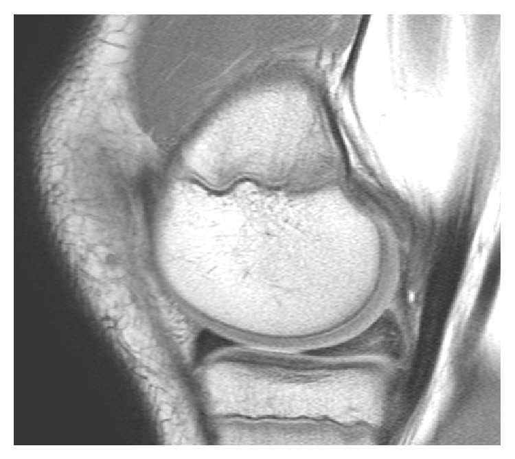
MRISagittal knee MRI: intrasubstance degeneration of the posterior horn of the medial meniscus, without a clear surface tear line.
Source : Ahmed Ali et al., Case reports in orthopedics, 2014, figure 1 · CC BY
- The Framingham cohort (Englund 2008) documented that 61 % of subjects with a meniscal tear on MRI had had no pain, stiffness or discomfort in the preceding month, and only 32 % reported meniscal symptoms after adjustment for osteoarthritis.³
- The 2-year follow-up of the ESCAPE patients (van de Graaf 2018, JAMA) shows that most degenerative tears remain radiologically stable and that symptoms evolve independently of MRI changes.⁸
- Fluctuation is the rule: frequent periods of spontaneous remission, transient flare-ups linked to specific loads, with no linear trend towards worsening in most middle-aged patients.
Key points
- DML are a progressive structural failure of meniscal tissue (loss of proteoglycans, collagen disorganisation), distinct from the traumatic tears of the young athlete.
- Framingham prevalence (Englund 2008 NEJM): 19 % in women, 56 % in men aged 50-90 years, with a continuous gradient across age. 61 % of these subjects are completely asymptomatic.
- Ranked risk factors (Snoeker 2013 JOSPT): age > 60 years (OR 2.5), associated osteoarthritis (OR 2.3), occupational squatting (OR 2.2), previous ACL injury (OR 2.0), male sex (OR 1.8), BMI > 30 (OR 1.7).
- In turn,meniscal extrusion > 3 mm is a key marker of loss of shock-absorbing function, predictive of accelerated osteoarthritic progression.
- The natural history is generally benign and fluctuating : symptoms poorly correlated with imaging, frequent spontaneous remissions, rarely a linear course.
Chapter 1 bibliography
- Beaufils P, Becker R, Kopf S, et al. The knee meniscus: management of traumatic tears and degenerative lésions. EFORT Open Rev. 2017;2(5):195-203. PMID 28698804.
- Fox AJS, Bedi A, Rodeo SA. The basic science of human knee menisci: structure, composition, and function. Sports Health. 2012;4(4):340-351. PMID 23016106.
- Englund M, Guermazi A, Gale D, et al. Incidental méniscal findings on knee MRI in middle-aged and elderly persons. N Engl J Med. 2008;359(11):1108-1115. PMID 18784100.
- Guermazi A, Niu J, Hayashi D, et al. Prévalence of abnormalities in knees detected by MRI in adults without knee osteoarthritis: population based observational study (Framingham Osteoarthritis Study). BMJ. 2012;345:e5339. PMID 22932918.
- Snoeker BA, Bakker EW, Kegel ÇA, Lucas C. Risk factors for méniscal tears: a systematic review including meta-analysis. J Orthop Sports Phys Ther. 2013;43(6):352-67. PMID 23628788.
- Englund M, Guermazi A, Lohmander LS. The meniscus in knee osteoarthritis. Rheum Dis Clin North Am. 2009;35(3):579-590. PMID 19931804.
- Roemer FW, Kwoh CK, Hannon MJ, et al. Partial meniscectomy is associated with increased risk of incident radiographic osteoarthritis and worsening cartilage damage in the following year. Eur Radiol. 2017;27(1):404-413. PMID 27121931.
- van de Graaf VA, Noorduyn JCA, Willigenburg NW, et al. Effect of Early Surgery vs Physical Therapy on Knee Function Among Patients With Nonobstructive Méniscal Tears: The ESCAPE Randomized Clinical Trial. JAMA. 2018;320(13):1328-1337. PMID 30285177.
How can degenerative meniscal lesions (DML) be assessed and diagnosed with confidence?
What questions should be asked to properly understand the patient and their history?
The history is the cornerstone of the diagnostic process in a patient with suspected DML. Attentive listening and targeted questions build a solid clinical hypothesis, often more discriminating than special tests taken in isolation.¹ 🧐- Pain description : precise location (medial or lateral joint line, typically posteromedial), type (dull / sharp / mechanical), intensity (numerical rating scale 0-10), pattern (onset after exertion, persistence at rest, at night). Night pain or pain at rest points to an inflammatory component (advanced osteoarthritis, synovitis).
- Aggravating factors : rising from a low chair, pivoting under load, going up and down stairs, squatting, carrying loads. The absence of a clear initial injury is the main distinguishing feature versus a traumatic lesion.
- Mechanical symptoms : the presence of true locking (inability to fully extend the knee without a specific manoeuvre) is highly suggestive but rare in the degenerative setting. Sensations of pseudo-locking, catching and giving way are more common and less specific.³
- Swelling (effusion) : typically intermittent, appearing 12-24 h after activity and partly resolving with rest. A major or persistent effusion should prompt reconsideration of the diagnosis (inflammatory osteoarthritic flare, infection, occult fracture).
- History and context : age ≥ 40-45 years, occupational profile (squatting, load carrying), level of sporting activity, past injuries (ACL rupture, severe sprain), history of osteoarthritis, BMI, alignment (varus/valgus).⁴
- Yellow flags : fear of movement, catastrophising, belief in a "serious lesion that needs surgery", expectations regarding imaging.
Which clinical tests should be performed and which other conditions must be ruled out?
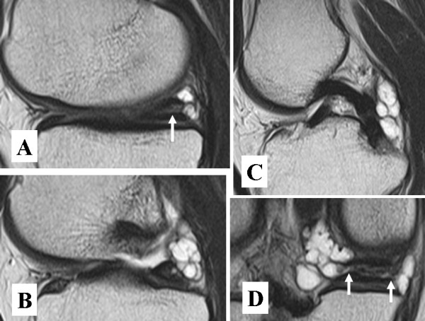
MRIKnee MRI, four images: horizontal tear of the medial meniscus (arrow, A) and a multilobulated meniscal cyst communicating with the tear (B, C, arrows in D).
Source : Ohishi et al., Sports medicine, arthroscopy, rehabilitation, therapy & technology (SMARTT), 2010, figure 1 · CC BY
- Joint-line palpation : the most sensitive test (pooled sensitivity ≈ 76-83 % per Hegedus 2007 SR). Reproducible pain on palpation of the medial or lateral joint line is found in the great majority of symptomatic patients. But its specificity is low (≈ 29-42 %) because the pain may arise from osteoarthritis, from pes anserinus tendinopathy or from the iliotibial band.
- McMurray test : sensitivity 24-71 %, specificity 60-95 %, highly variable across examiners and test versions. The "felt" McMurray (palpation + flexion-rotation) performs better than the "audible" McMurray (click alone). Pooled LR+ ≈ 1.3-3.0, insufficient to confirm on its own.
- Thessaly test (5° and 20° weight-bearing): initially described with a sensitivity > 90 % by Karachalios 2005, but later replications in real DML populations found far more modest performance (sensitivity ≈ 64 %, specificity ≈ 53 %). Its value remains debatable in routine practice.
- Apley compression test : sensitivity ≈ 60 %, specificity ≈ 70 %, overall intermediate performance.
- Combination of tests : the recommended practice is to combine a typical history + joint-line palpation + one or two dynamic tests. The combination outperforms any single test and significantly increases the post-test probability if all of them agree.
🧭 Simplified diagnostic algorithm for suspected DML
From clinical screening to the treatment decision, without routine first-line MRI
Summary algorithm adapted from the Siemieniuk 2017 recommendations (BMJ Rapid Recommendations) and the ESSKA-AOSSM-AASPT 2024 Consensus (Prill 2025 KSSTA).
- Tibiofemoral osteoarthritis : the leading differential diagnosis. More diffuse pain, morning stiffness > 30 min, restricted range of motion, crepitus. Coexists very frequently with DML: both often contribute to the symptoms simultaneously.⁵
- Meniscal root lesion (root tear) : a distinct entity with major prognostic impact. Acute posterior pain, a sensation of "popping", massive extrusion on MRI, accelerated osteoarthritis. Covered in chapter 5.
- Spontaneous osteonecrosis of the knee (SONK, Ahlback disease) : intense acute night pain over the medial femoral condyle, often without trauma, peak incidence 60+ years. MRI shows characteristic bone oedema.
- Tendinopathies : pes anserinus (pain over the medial tibial surface 5-7 cm below the joint line), iliotibial band (lateral aspect, cyclical pain), quadriceps or patellar tendon (anterior pain).
- Vascular conditions : popliteal artery entrapment syndrome in the young sporting adult (exertional claudication, calf pain).
- Neurological conditions : L3-L4 radiculopathy can refer pain to the anterior or medial aspect of the knee, without significant low back pain.
- Patellofemoral pain syndrome : anterior pain aggravated by prolonged sitting and by descending stairs, but it can radiate.
- Infrapatellar / prepatellar bursitis : localised pain, focal swelling, inflammatory signs.
Knee red flags requiring medical referral
- Recent significant trauma (fall, accident) with inability to weight-bear or to flex actively: suspected occult fracture (Ottawa Knee Rules).
- Fever, chills, weight loss that is unexplained: suspected septic arthritis, malignancy, systemic inflammatory process.
- Severe non-mechanical night pain, persistent bone pain: bone tumour, osteonecrosis, stress fracture.
- Vascular symptoms : intermittent claudication, asymmetrical pulses, distal discolouration, popliteal artery entrapment syndrome, ischaemia.
- Progressive neurological symptoms : widespread motor weakness, dermatomal sensory disturbance, bladder or bowel dysfunction, spinal pathology (cauda equina syndrome), central pathology.
- True joint locking persisting in extension: suspected bucket-handle tear, loose body, displaced root lesion.
- Major effusion of rapid onset without trauma: suspected haemarthrosis (anticoagulant, clotting disorder), crystal arthritis.
Should patients with DML be classified, and what are the benefits?
The clinical stratification of DML patients is increasingly recommended, not to apply a rigid label but to guide shared treatment decision-making.⁶ The aim is to distinguish the patients who will benefit most from a conservative approach from those for whom a surgical discussion might be relevant (a minority subgroup). Pragmatic "mechanical profile vs osteoarthritic profile" classification:- Mechanical profile (narrow subgroup):
- Marked mechanical symptoms: frequent pseudo-locking, reproducible giving way, precise painful clicks;
- Pain well localised to the joint line;
- Clear intermittent effusion after certain activities;
- Examination reliably reproducing the symptom;
- Little or no sign of advanced osteoarthritis;
- => Possible response to well-conducted PT; if this fails at 3-6 months, a surgical discussion may be considered.
- Osteoarthritic profile (the majority):
- Diffuse pain, morning stiffness, widespread bony tenderness;
- Crepitus, restricted range of motion;
- Radiographic signs of osteoarthritis (Kellgren-Lawrence ≥ 2);
- Metabolic comorbidities (obesity, diabetes);
- => DML = epiphenomenon ; management targets the overall osteoarthritis (weight, strength, education) and not the isolated meniscal lesion.
Critique and controversy: the tyranny of MRI
The major controversy in the diagnosis of DML lies in the frequent discordance between imaging findings and the clinical presentation. The advent of MRI has led to a "tyranny of the image": the presence of a meniscal tear is automatically equated with the source of the pain, triggering a cascade of interventions. Yet the epidemiological data are unequivocal: 61 % of subjects with a meniscal tear on MRI had had no pain in the preceding month (Englund 2008 NEJM).³ The Guermazi 2012 study (BMJ, Framingham) shows that most knees in adults ≥ 50 years show abnormalities on MRI irrespective of symptoms.⁴ The real diagnostic challenge is not "finding the tear" but determining whether it is clinically relevant. The modern consensus therefore argues for a clinical diagnosis first and foremost, with MRI reserved for very selected cases: failure of well-conducted PT at 3 months, suspicion of another condition (root tear, SONK, osteochondritis, ligament lesion), and preoperative planning in a patient selected for surgical discussion.Key points
- The history is king : age ≥ 40 years, insidious-onset joint-line pain with no trauma, triggered by squatting/pivoting/stairs, fluctuating.
- Clinical tests have limits : joint-line palpation is sensitive but poorly specific; McMurray and Thessaly have moderate accuracy. A combination of tests outperforms any single test.
- Always think of osteoarthritis : DML and knee osteoarthritis are frequent companions. The pain often stems from both.
- MRI is NOT a routine investigation : high prevalence of asymptomatic abnormalities (Englund 2008, Guermazi 2012). Reserve it for selected situations.
- Classifying "mechanical vs osteoarthritic" helps to direct patients towards PT (the large majority) and to reserve the surgical discussion for a narrow subgroup with persistent true locking.
Chapter 2 bibliography
- Hegedus EJ, Cook C, Hasselblad V, et al. Physical examination tests for assessing a torn meniscus in the knee: a systematic review with meta-analysis. J Orthop Sports Phys Ther. 2007;37(9):541-550. PMID 17939613.
- Smith BE, Thacker D, Crewesmith A, Hall M. Special tests for assessing meniscal tears within the knee: a systematic review and meta-analysis. Evid Based Med. 2015;20(3):88-97. PMID 25724195.
- Beaufils P, Becker R, Kopf S, et al. The knee meniscus: management of traumatic tears and degenerative lésions. EFORT Open Rev. 2017;2(5):195-203. PMID 28698804.
- Englund M, Guermazi A, Gale D, et al. Incidental méniscal findings on knee MRI in middle-aged and elderly persons. N Engl J Med. 2008;359(11):1108-1115. PMID 18784100.
- Guermazi A, Niu J, Hayashi D, et al. Prévalence of abnormalities in knees detected by MRI in adults without knee osteoarthritis: population based observational study (Framingham Osteoarthritis Study). BMJ. 2012;345:e5339. PMID 22932918.
- Englund M, Guermazi A, Lohmander LS. The meniscus in knee osteoarthritis. Rheum Dis Clin North Am. 2009;35(3):579-590. PMID 19931804.
- Siemieniuk RAC, Harris IA, Agoritsas T, et al. Arthroscopic surgery for degenerative knee arthritis and méniscal tears: a clinical practice guideline. BMJ. 2017;357:j1982. PMID 28490431.
- Thorlund JB, Juhl CB, Roos EM, Lohmander LS. Arthroscopic surgery for degenerative knee: systematic review and meta-analysis of benefits and harms. BMJ. 2015;350:h2747. PMID 26080045.
- Prill R, Ma CB, Wong SE, et al. The formal EU-US Meniscus Rehabilitation 2024 Consensus: An ESSKA-AOSSM-AASPT initiative. Part II. Knee Surg Sports Traumatol Arthrosc. 2025;33(8):3014-3024. PMID 40519541.
Which treatment strategies are most effective for degenerative meniscal lesions (DML)?
Where to start? What is the hierarchy of recommended interventions?
The hierarchy is clear and is the subject of growing international consensus. The first-line intervention is a structured, supervised conservative treatment programme lasting 3 to 6 months before any surgical discussion.⁴,⁵ Summary of the pivotal randomised trials:- FIDELITY 2013 (Sihvonen R et al. NEJM, n = 146, Finland): APM vs placebo surgery. No significant difference at 12 months in pain or function (WOMAC).⁶
- FIDELITY 5 years (Sihvonen R et al. BJSM 2020): confirmation at 5 years: no benefit of APM over placebo surgery. The first study in the world to test APM vs sham surgery over 5 years.⁷
- METEOR 2013 (Katz JN et al. NEJM, n = 351, USA): APM + PT vs PT alone in DML + osteoarthritis. WOMAC difference at 6 months = 2.4 points (not clinically significant). 30 % crossover from the PT group to surgery at 12 months.⁸
- Kise 2016 (BMJ, n = 140, Norway): 12 weeks of supervised exercise vs APM. No KOOS difference at 2 years. The exercise group had significantly greater quadriceps strength at 3 and 12 months.⁹
- ESCAPE 2018 (van de Graaf VA et al. JAMA, n = 321, Netherlands): PT 16 sessions vs early APM. IKDC difference at 24 months = 3.6 points (below the clinically relevant threshold of 8). 29 % crossover from PT to surgery.¹
- ESCAPE 5 years (Noorduyn JCA et al. JAMA Netw Open 2022): confirmation at 5 years: non-inferiority of PT vs APM maintained, with no clinically significant difference.⁵
📊 Summary of the randomised trials of APM vs PT for DML
Between-group difference in effect on function (KOOS/IKDC/WOMAC): the MCID threshold is usually 8-10 points
Visual summary: every APM minus PT difference falls below the minimal clinically important difference (MCID) threshold of 8-10 points on the KOOS/IKDC. Sources: Sihvonen 2013 NEJM PMID 24369076; Sihvonen 2020 BJSM PMID 32855201; Katz 2013 NEJM PMID 23506518; Kise 2016 BMJ PMID 27440192; van de Graaf 2018 JAMA PMID 30285177; Noorduyn 2022 JAMA Netw Open PMID 35802374.
What is the role of exercise and is there a superior approach?
Therapeutic exercise is the cornerstone of conservative treatment. Its aim is not to "heal" the meniscus (impossible) but to improve the overall functional capacity of the knee, reduce the load on sensitised structures, modulate pain and restore confidence. Recommended components (ESSKA-AOSSM-AASPT 2024 consensus Part II, Prill 2025 KSSTA):⁷- Progressive muscle strengthening : focus on the quadriceps (seated extension, leg press, partial squat), hamstrings (curl, modified deadlift), glutes (bridge, monster walk, hip thrust) and calves. Progressive intensity following the principle of graded overload.
- Neuromuscular control : proprioceptive exercises (double-leg then single-leg, on a stable and then an unstable surface), dynamic control (step-down, single-leg squat with valgus control), progressive agility work.
- Range of motion : full extension is the priority (an extension deficit perpetuates pain), then progressive flexion.
- Low-impact aerobic activity : cycling (patellofemoral load, but usually well tolerated), swimming, walking, cross-trainer. Encourage the maintenance of general physical activity.
- Education + home exercises : essential for long-term adherence.
- OMEX (Stensrud 2012) : 12 weeks, 2-3 sessions/week, the basis used in Kise 2016.
- GLA:D (Good Life with osteoArthritis in Denmark) : a structured 6-8 week programme, education + neuromuscular exercise, rolled out in dozens of countries. Skou & Roos 2017 BMC Musculoskelet Disord demonstrated robust 1-year outcomes in 9,825 patients with osteoarthritis/DML.
- ESCAPE-knee-pain programmes : a group approach combining exercise + education + self-management.
Manual therapies and technologies: how effective are they really?
Passive approaches may be used as short-term adjuncts to facilitate engagement with the active programme, but must never form the core of treatment.- Manual therapy (patellofemoral/tibiofemoral joint mobilisations, soft-tissue techniques, mobilisation with movement): a short-term effect on pain and range of motion. Limited evidence as a stand-alone treatment (Pollard 2018 review and Pollard meta-analysis).⁸
- TENS, ultrasound, shockwave therapy : no high-quality evidence specific to DML. Symptomatic relief is possible for some patients, but they do not change the course of the condition.
- Intra-articular injections : corticosteroids give a short-lived effect (weeks), hyaluronic acid a limited benefit, and the PRP data remain heterogeneous and of low quality. Discussion case by case, never routinely.
- Thermal modalities (cryotherapy, heat): symptomatically useful, with no impact on the natural history.
Beyond the physical: how to educate the patient and address psychological factors?
In this respect,therapeutic patient education (ETP) is probably the most powerful and the most under-used lever. The patient's beliefs and emotions have a considerable impact on outcomes, often greater than structural indicators.⁹ 🧠 Key messages to convey:- Defuse the imaging report : explain in simple terms that degenerative lesions are to knees what grey hair is to hair, a sign of ageing, present in the majority of people of the same age and most often asymptomatic. Quote the figures: 19 % of women and 56 % of men aged 50-90 have a DML on MRI, of whom 61 % are pain-free (Englund 2008).
- Explain pain : pain = a multifactorial protective experience, not a direct signal of damage. Modern concepts (pain neuroscience education, Louw et al.): fear of movement (kinesiophobia) maintains and amplifies pain.
- Promote self-efficacy : give the patient the tools and the confidence to manage their symptoms. Concrete, progressive functional goals (climbing a flight of stairs without holding on, walking 30 minutes without pain, resuming Nordic walking twice a week).
- Avoid nocebo language : ban "your meniscus is torn / worn out / damaged", prefer "you have age-related changes in your meniscus, which are common and generally well tolerated with a good exercise programme".
🏠 GRADE pyramid of treatment modalities for DML
Consolidated level of evidence: shown as horizontal cards for readability
GRADE summary adapted from Thorlund 2015 BMJ, Siemieniuk 2017 BMJ, Abram 2020 BJSM, Prill 2025 KSSTA. The higher and greener the card, the stronger the evidence.
| Modality | Effect on pain | Effect on function | Level of evidence | Adverse effects |
|---|---|---|---|---|
| Supervised exercise 8-12 weeks | Significant reduction | KOOS/IKDC improvement | HIGH | Minimal (acceptable pain) |
| Therapeutic patient education | Reduces catastrophising | Increases self-efficacy | HIGH | None |
| Adjunctive manual therapy | Short term only | Improved range of motion | MODERATE | Rare |
| TENS / Ultrasound | Modest / transient | Marginal | LOW | Rare |
| Corticosteroid injection | Short term (weeks) | Poorly durable | LOW | Risk of cartilage atrophy |
| APM (partial meniscectomy) | Equivalent or inferior to PT | Equivalent or inferior to PT | MODERATE AGAINST | DVT, infection, accelerated osteoarthritis |
| Placebo APM (sham) | Equivalent to real APM | Equivalent | HIGH (vs sham) | Identical |
Critique and controversy: the persistent overuse of surgery
Despite the accumulating evidence, APM remains one of the most frequently performed orthopaedic procedures in the world. Several factors perpetuate this gap between science and practice:- The tyranny of MRI : the presence of a "tear" on the report creates a strong pull towards surgery, both for the patient (anxiety) and for the surgeon (a repair mindset). Yet in most cases this abnormality is an incidental finding.¹⁰
- Patient expectations : belief in a surgical "quick fix" remains widespread. A 12-week exercise programme will seem less "tangible" than an operation.
- Heterogeneity of physiotherapy : "physiotherapy" may mean a high-quality structured active programme or an outdated passive treatment. The APM vs PT trials tested highly standardised and supervised programmes; transposing those results to unstructured PT is risky.
- Economic factors : in many systems surgery is better remunerated than lengthy rehabilitation, creating an incentive bias.
- The placebo effect of surgery : FIDELITY showed that surgical placebo produces the same results as real APM, suggesting that the perceived benefit comes from the symbolic intervention, the postoperative rest and the rehabilitation that follows, NOT from the technical act on the meniscus.⁶,⁷
Key points
- ✅ Overall,supervised exercise + education is the first-line treatment (GRADE high), with a level of evidence equal to or greater than surgery in the short, medium and long term.
- 🏋️ Exercise combines strengthening (quadriceps, hamstrings, glutes) + neuromuscular control, over 8-12 weeks. No modality is intrinsically superior; individualisation comes first.
- ❌ By contrast, arthroscopic partial meniscectomy (APM) is NOT superior to PT or to placebo surgery. Reserved for persistent true locking after 3-6 months of failure.
- 🧠 Alongside this,therapeutic patient education is crucial: defuse the MRI, explain pain, promote self-efficacy. Avoid nocebo language.
- 👍 Finally, manual therapies and passive modalities are potential short-term adjuncts, but must NOT replace the active approach.
Chapter 3 bibliography
- van de Graaf VA, Noorduyn JCA, Willigenburg NW, et al. Effect of Early Surgery vs Physical Therapy on Knee Function Among Patients With Nonobstructive Méniscal Tears: The ESCAPE Randomized Clinical Trial. JAMA. 2018;320(13):1328-1337. PMID 30285177.
- Kise NJ, Risberg MA, Stensrud S, Ranstam J, Engebretsen L, Roos EM. Exercise therapy versus arthroscopic partial meniscectomy for degenerative méniscal tear in middle aged patients: randomised controlled trial with two year follow-up. BMJ. 2016;354:i3740. PMID 27440192.
- Thorlund JB, Juhl CB, Roos EM, Lohmander LS. Arthroscopic surgery for degenerative knee: systematic review and meta-analysis of benefits and harms. BMJ. 2015;350:h2747. PMID 26080045.
- Beaufils P, Becker R, Kopf S, et al. Surgical management of degenerative meniscus lésions: the 2016 ESSKA meniscus consensus. Knee Surg Sports Traumatol Arthrosc. 2017;25(2):335-346. PMID 28210788.
- Noorduyn JCA, van de Graaf VA, Willigenburg NW, et al. Effect of Physical Therapy vs Arthroscopic Partial Meniscectomy in People With Degenerative Méniscal Tears: Five-Year Follow-up of the ESCAPE Randomized Clinical Trial. JAMA Netw Open. 2022;5(7):e2220394. PMID 35802374.
- Sihvonen R, Paavola M, Malmivaara A, et al. Arthroscopic partial meniscectomy versus sham surgery for a degenerative méniscal tear. N Engl J Med. 2013;369(26):2515-2524. PMID 24369076.
- Sihvonen R, Paavola M, Malmivaara A, et al. Arthroscopic partial meniscectomy for a degenerative meniscus tear: a 5 year follow-up of the placebo-surgery controlled FIDELITY trial. Br J Sports Med. 2020;54(22):1332-1339. PMID 32855201.
- Katz JN, Brophy RH, Chaisson CE, et al. Surgery versus physical therapy for a méniscal tear and osteoarthritis. N Engl J Med. 2013;368(18):1675-1684. PMID 23506518.
- Siemieniuk RAC, Harris IA, Agoritsas T, et al. Arthroscopic surgery for degenerative knee arthritis and méniscal tears: a clinical practice guideline. BMJ. 2017;357:j1982. PMID 28490431.
- Abram SGF, Hopewell S, Monk AP, Bayliss LE, Beard DJ, Price AJ. Arthroscopic partial meniscectomy for méniscal tears of the knee: a systematic review and meta-analysis. Br J Sports Med. 2020;54(11):652-663. PMID 30796103.
- Pujol N, Giordano AO, Wong SE, et al. The formal EU-US Meniscus Rehabilitation 2024 Consensus: An ESSKA-AOSSM-AASPT initiative. Part I. Knee Surg Sports Traumatol Arthrosc. 2025;33(8):3002-3013. PMID 40353298.
- Prill R, Ma CB, Wong SE, et al. The formal EU-US Meniscus Rehabilitation 2024 Consensus: An ESSKA-AOSSM-AASPT initiative. Part II. Knee Surg Sports Traumatol Arthrosc. 2025;33(8):3014-3024. PMID 40519541.
- Skou ST, Roos EM. Good Life with osteoArthritis in Denmark (GLA:D): evidence-based éducation and supervised neuromuscular exercise delivered by certified physiotherapists nationwide. BMC Musculoskelet Disord. 2017;18(1):72. PMID 28173795.
- Stensrud S, Roos EM, Risberg MA. A 12-week exercise therapy program in middle-aged patients with degenerative meniscus tears: a case series with 1-year follow-up. J Orthop Sports Phys Ther. 2012;42(11):919-31. PMID 22960783.
- Louw A, Zimney K, Puentedura EJ, Diener I. The efficacy of pain neuroscience éducation on musculoskeletal pain: A systematic review of the literature. Physiother Theory Pract. 2016;32(5):332-355. PMID 27351541.
How to ensure lasting recovery and plan the return to sport?
How to make the patient an active participant in their recovery through self-management?
Ultimately,patient self-management is crucial for preventing symptomatic recurrence. It turns a passive recipient of care into an active manager of their own joint health. It rests on two inseparable pillars: education + a structured, progressive exercise programme. 🧠 1. Therapeutic education: a change of paradigm- Explain that a DML is an integral part of the normal ageing of the knee, that it is very common and very often asymptomatic (Englund 2008 NEJM: 61 % of subjects with a DML on MRI had no pain).³
- Shift attention away from anatomical structure and towards function and capacity : what the knee can do matters more than what the MRI shows.
- A simple understanding of pain mechanisms (pain is protective and modifiable, not synonymous with damage), reducing kinesiophobia.⁴
- Non-nocebo communication: prefer "you have age-related changes that are well tolerated with the right programme" rather than "your meniscus is torn".
- Progressive neuromuscular strengthening : quadriceps, hamstrings, glutes, trunk, calves. Fundamental for improving shock absorption and dynamic knee control (Bartholdy 2017 review).⁷
- Load management : the concept of "acceptable pain" (≤ 3/10 on the VAS during and after exercise) as a tool for progressing without triggering a flare.⁸ It allows exercise in the presence of tolerable pain, demystifying the fear of "doing harm".
- Long-term adherence : continuing the exercises beyond the supervised phase. Patients who build in a routine of 2-3 strengthening sessions a week have the best outcomes at 1-5 years.⁹
- Self-monitoring : an exercise diary, pain VAS, walking distance, sensations on exertion. Helps to identify a flare-up quickly and to modulate the load.
- Reintegration of valued activities : walking, hiking, cycling, gardening, moderate-impact sports. Sedentary behaviour is the main enemy in the medium term.
When and how to plan a safe return to sport and activity?
Return to sport (RTS) or to meaningful activities after a DML should not be dictated by a fixed timetable, but by the achievement of objective functional criteria. A criteria-based approach minimises the risk of symptomatic recurrence and ensures that the knee tolerates the specific loads.¹⁰ 🏃 Key criteria for clearing a safe return (adapted from the ACL literature/ESSKA-AOSSM-AASPT 2024 Consensus Part II):¹¹- Basic clinical criteria:
- Pain almost or entirely absent during activities of daily living (VAS ≤ 1-2/10);
- No significant joint effusion ("zero" rule on the stroke test);
- Full and symmetrical range of motion (passive extension in particular);
- Subjective confidence in the knee.
- Strength criteria (Limb Symmetry Index, LSI):
- Isokinetic or isometric quadriceps strength ≥ 90 % of the uninvolved side ;
- Hamstring strength ≥ 90 % of the uninvolved side;
- H/Q ratio ≥ 0.6 (asymmetry avoided).
- Functional performance criteria (hop tests):
- Single leg hop for distance ≥ 90 % LSI;
- Triple hop for distance ≥ 90 % LSI;
- Crossover hop ≥ 90 % LSI;
- Y-balance test ≥ 90 % LSI.
- Psychological criteria:
- A successful sport-specific test:
- The key movement reproducible (pivot, squat, technical descent) without apprehension;
- Enough endurance to last a full training session.
📊 Functional criteria for return to activity after a DML
Visual summary: the minimum LSI targets to reach before each level of activity
Summary drawn from the ESSKA-AOSSM-AASPT 2024 consensus (Pujol 2025 KSSTA Part I PMID 40353298) and van Melick 2016 BJSM (ACL RTS recommendations transferable to post-surgical DML).
Critique and controversy: the grey areas of RTS criteria
Although functional criteria are recommended, several limitations deserve reflection:- Transposing from ACL to DML : almost all the detailed criteria come from the ACL literature. Their specific validity for DML is extrapolated but little validated by prospective studies.
- Vague definition of PT failure : "failure of well-conducted PT" remains subjective (adherence, intensity, psychosocial management). This subjectivity can lead to premature surgical referrals.
- Feasibility of the tests : hop tests and isokinetic testing require equipment and time. In routine practice many physiotherapists have no access to these tools, leading to suboptimal RTS decisions.
- Uncertain mechanism of action : the benefits of exercise are attributed to strength gains and neuromuscular control, but other mechanisms may be at work (central effects, local anti-inflammatory effects, self-efficacy).
Key points
- ✅ Above all,therapeutic patient education is fundamental in order to defuse the MRI and refocus treatment on function.
- 🏋️ An exercise programme combining neuromuscular strengthening and progressive load management is the first-line treatment (evidence ≥ APM in the long term).
- ❌ Conversely,APM should be avoided as an initial treatment for DML in the absence of true mechanical locking.
- 📊 Return to activity/sport is guided by functional performance criteria (strength ≥ 90 % LSI, symmetrical hop tests) and not by a fixed time frame.
- 🔁 Preventing recurrence depends on long-term adherence to a maintenance programme (2-3 strengthening sessions a week, indefinitely).
Chapter 4 bibliography
- van de Graaf VA, Noorduyn JCA, Willigenburg NW, et al. Effect of Early Surgery vs Physical Therapy on Knee Function Among Patients With Nonobstructive Méniscal Tears: The ESCAPE Randomized Clinical Trial. JAMA. 2018;320(13):1328-1337. PMID 30285177.
- Kise NJ, Risberg MA, Stensrud S, Ranstam J, Engebretsen L, Roos EM. Exercise therapy versus arthroscopic partial meniscectomy for degenerative méniscal tear in middle aged patients: randomised controlled trial with two year follow-up. BMJ. 2016;354:i3740. PMID 27440192.
- Englund M, Guermazi A, Gale D, et al. Incidental méniscal findings on knee MRI in middle-aged and elderly persons. N Engl J Med. 2008;359(11):1108-1115. PMID 18784100.
- Louw A, Zimney K, Puentedura EJ, Diener I. The efficacy of pain neuroscience éducation on musculoskeletal pain: A systematic review of the literature. Physiother Theory Pract. 2016;32(5):332-355. PMID 27351541.
- Sihvonen R, Paavola M, Malmivaara A, et al. Arthroscopic partial meniscectomy for a degenerative meniscus tear: a 5 year follow-up of the placebo-surgery controlled FIDELITY trial. Br J Sports Med. 2020;54(22):1332-1339. PMID 32855201.
- Noorduyn JCA, van de Graaf VA, Willigenburg NW, et al. Effect of Physical Therapy vs Arthroscopic Partial Meniscectomy in People With Degenerative Méniscal Tears: Five-Year Follow-up of the ESCAPE Randomized Clinical Trial. JAMA Netw Open. 2022;5(7):e2220394. PMID 35802374.
- Bartholdy C, Juhl C, Christensen R, Lund H, Zhang W, Henriksen M. The role of muscle strengthening in exercise therapy for knee osteoarthritis: A systematic review and meta-regression analysis of randomized trials. Semin Arthritis Rheum. 2017;47(1):9-21. PMID 28438380.
- Smith BE, Hendrick P, Smith TO, et al. Should exercises be painful in the management of chronic musculoskeletal pain? A systematic review and meta-analysis. Br J Sports Med. 2017;51(23):1679-1687. PMID 28596288.
- Skou ST, Roos EM. Good Life with osteoArthritis in Denmark (GLA:D): evidence-based éducation and supervised neuromuscular exercise delivered by certified physiotherapists nationwide. BMC Musculoskelet Disord. 2017;18(1):72. PMID 28173795.
- van Melick N, van Cingel RE, Brooijmans F, et al. Evidence-based clinical practice update: practice guidelines for anterior cruciate ligament rehabilitation based on a systematic review and multidisciplinary consensus. Br J Sports Med. 2016;50(24):1506-1515. PMID 27539507.
- Pujol N, Giordano AO, Wong SE, et al. The formal EU-US Meniscus Rehabilitation 2024 Consensus: An ESSKA-AOSSM-AASPT initiative. Part I. Knee Surg Sports Traumatol Arthrosc. 2025;33(8):3002-3013. PMID 40353298.
- Gabbett TJ. The training-injury prévention paradox: should athletes be training smarter and harder? Br J Sports Med. 2016;50(5):273-280. PMID 26758673.
How to manage DML in the context of osteoarthritis or a root lesion?
DML + osteoarthritis: what overall management?
Epidemiological observation : DML coexists with radiographic knee osteoarthritis in the majority of symptomatic middle-aged and older patients. The OAI (Osteoarthritis Initiative) and the Framingham cohort show that more than 80 % of patients with Kellgren-Lawrence (KL) ≥ 2 knee osteoarthritis have concomitant meniscal involvement on MRI, and that DML is a predictor of osteoarthritic progression.¹,² Major clinical implication : in the patient with moderate to severe osteoarthritis (KL ≥ 3, joint space narrowing, osteophytosis), treating the meniscal lesion in isolation with surgery is not only ineffective but potentially harmful, by accelerating the progression of the osteoarthritis.³ The METEOR trial (Katz 2013) was conducted specifically in this population: no added benefit of APM over PT alone at 6 months or in the long term.⁴ Recommended management in the setting of DML + osteoarthritis (consolidated data from the OARSI 2019 guideline + ESSKA-AOSSM-AASPT 2024 Part II):⁵- Weight management : a reduction of 5-10 % of initial body weight significantly reduces pain (Hall 2018 review, GLA:D programme). A 1 kg reduction = 4 kg less load per knee when walking.
- Structured therapeutic exercise : a programme combining strengthening (quadriceps first), low-impact aerobic work (cycling, swimming, pool walking) and functional exercises. An effect equivalent to or greater than NSAIDs for pain at 3-6 months.
- Osteoarthritis education : structured programmes such as GLA:D, Good Life with Arthritis, ESCAPE-knee-pain (combining exercise + education + self-management).⁶
- Activity modification : ergonomic advice (avoid prolonged squatting), assistive devices (contralateral walking stick, varus unloader brace, insoles), changes to sporting activity (Nordic walking instead of running).
- Targeted analgesia : paracetamol is of limited value but useful in acute flares, topical NSAIDs (1st line), a short course of oral NSAIDs if necessary, selective corticosteroid injections (effect 4-12 weeks).
- Red flags specific to osteoarthritis : a severe inflammatory flare (suspected crystal arthropathy, to be investigated), progressive fixed loss of extension, rapid deformity (suspected necrosis, subchondral fracture).
Meniscal root lesion (root tear): a distinct, high-risk entity
The meniscal root lesion (meniscal root tear) is a distinct clinical entity from the classic DML, with a major biomechanical and prognostic impact. The meniscal root is the posterosuperior bony attachment of the meniscus to the tibial plateau; a complete rupture is functionally equivalent to a total meniscectomy, with loss of shock-absorbing function, massive meniscal extrusion and accelerated osteoarthritis.⁷ ⚠️ Clinical features specific to root tears:- Population : mainly women aged 50-65 (medial root); varus, BMI > 30, an initial squat (a minor event is often reported: rising from a low chair, squatting, the first step in the morning).
- Symptoms : acute posteromedial pain (medial root), a perceptible "pop", a sensation of the knee "giving way". Night waking is possible.
- MRI : meniscal extrusion > 3 mm (often ≥ 5 mm), loss of the posteromedial radial signal, ipsilateral subchondral bone oedema, root tear with low T2 signal.
- Natural prognosis : accelerated osteoarthritic progression without specific treatment. KL progression of +1-2 grades over 2-5 years in several series.⁸
- A steeper medial tibial plateau slope;
- More marked varus alignment;
- Reduced intercondylar distance;
- Reduced intercondylar notch width.
- Root repair (transosseous refixation): better radiological outcomes (reduced extrusion), better clinical outcomes (KOOS, IKDC) and fewer conversions to arthroplasty in the medium term;
- APM (partial meniscectomy) : intermediate results, functionally acceptable in the short term but with accelerated osteoarthritis in the long term;
- Non-operative treatment : pain initially controlled but substantial conversion to arthroplasty at 5-10 years (up to 35-50 % depending on the series).
- An active patient < 65-70 years;
- KL ≤ 2 (little or no advanced tibiofemoral osteoarthritis);
- Acceptable alignment (varus ≤ 5°) or a feasible simultaneous correction (high tibial osteotomy);
- Recent symptoms with a root tear identified on MRI;
- A patient willing to follow a demanding postoperative rehabilitation protocol (6-8 weeks of partial weight-bearing, 4-6 months to RTS).
🧠 Decision tree: typical DML vs root tear vs DML + osteoarthritis
Distinguishing these three entities radically changes the treatment direction
Summary decision tree based on Krych/Lee 2025 (root tear), Katz 2013 NEJM (DML + osteoarthritis), Siemieniuk 2017 BMJ (isolated DML). KL = Kellgren-Lawrence (radiographic osteoarthritis grading).
Red flags specific to root tears and advanced osteoarthritis
- Acute onset of posteromedial pain in a woman aged 50-65 (often high BMI, varus) during a trivial movement (squatting, rising from a chair) => medial root tear very likely; prompt MRI so as not to miss the window for root repair.
- Meniscal extrusion > 5 mm on MRI without major pain => high risk of accelerated osteoarthritic progression; education and close follow-up.
- Loss of active extension that is fixed and progressive without mechanical locking => suspected advanced osteoarthritis with contracture, or osteochondritis with a loose body.
- Intense night pain that is non-mechanical in a patient > 60 years => suspected osteochondritis, subchondral fracture (bone insufficiency), spontaneous osteonecrosis (SONK), malignancy.
- Rapid deformity in progressive varus/valgus => joint collapse, extensive necrosis, subchondral fracture => urgent surgical referral.
Key points
- 👥 In the patient with DML + osteoarthritis KL ≥ 2-3, the meniscal lesion is an epiphenomenon ; management targets the overall osteoarthritis (weight, exercise, GLA:D-style education, targeted analgesia).
- ❌ APM in the setting of moderate to severe osteoarthritis is not recommended (Katz 2013 METEOR, Siemieniuk 2017 BMJ Rapid Recs) and potentially harmful (accelerated osteoarthritis).
- ⚠️ The meniscal root lesion (root tear) is a DISTINCT entity: a woman aged 50-65, varus, raised BMI, acute posterior pain with a "pop", extrusion > 3 mm on MRI.
- 🔍 Early diagnosis of a root tear is crucial: the window for intervention is limited if a root repair is to be discussed (KL ≤ 2, age < 65 years).
- 📈 According to the Lee/Krych 2025 SR, root repair shows superior outcomes to APM and to non-operative treatment in selected patients.
- 📊 Predisposing morphological factors (Oeding 2025): a steep medial tibial slope, varus, reduced intercondylar distance, a narrow notch.
Chapter 5 bibliography
- Englund M, Guermazi A, Lohmander LS. The meniscus in knee osteoarthritis. Rheum Dis Clin North Am. 2009;35(3):579-590. PMID 19931804.
- Guermazi A, Niu J, Hayashi D, et al. Prévalence of abnormalities in knees detected by MRI in adults without knee osteoarthritis: population based observational study (Framingham Osteoarthritis Study). BMJ. 2012;345:e5339. PMID 22932918.
- Roemer FW, Kwoh CK, Hannon MJ, et al. Partial meniscectomy is associated with increased risk of incident radiographic osteoarthritis and worsening cartilage damage in the following year. Eur Radiol. 2017;27(1):404-413. PMID 27121931.
- Katz JN, Brophy RH, Chaisson CE, et al. Surgery versus physical therapy for a méniscal tear and osteoarthritis. N Engl J Med. 2013;368(18):1675-1684. PMID 23506518.
- Bannuru RR, Osani MC, Vaysbrot EE, et al. OARSI guidelines for the non-surgical management of knee, hip, and polyarticular osteoarthritis. Osteoarthritis Cartilage. 2019;27(11):1578-1589. PMID 31278997.
- Skou ST, Roos EM. Good Life with osteoArthritis in Denmark (GLA:D): evidence-based éducation and supervised neuromuscular exercise delivered by certified physiotherapists nationwide. BMC Musculoskelet Disord. 2017;18(1):72. PMID 28173795.
- LaPrade RF, Floyd ER, Carlson GB, Moatshe G, Chahla J, Monson JK. Méniscal Root Tears: Solving the Silent Epidemic. J Arthrosc Surg Sports Med. 2021;2(1):47-57. DOI 10.25259/JASSM_55_2020.
- Krych AJ, Reardon PJ, Johnson NR, et al. Non-operative management of medial meniscus posterior horn root tears is associated with worsening arthritis and poor clinical outcome at 5-year follow-up. Knee Surg Sports Traumatol Arthrosc. 2017;25(2):383-389. PMID 27761625.
- Oeding JF, Dean MC, Hevesi M, Chahla J, Krych AJ. Steeper Slope of the Medial Tibial Plateau, Greater Varus Alignment, and Narrower Intercondylar Distance and Notch Width Increase Risk for Medial Meniscus Posterior Root Tears: A Systematic Review. Arthroscopy. 2025. PMID 39505159.
- Lee DR, Lu Y, Reinholz AK, Till SE, Lamba A, Saris DBF, Camp CL, Krych AJ. Root Repair Has Superior Radiological and Clinical Outcomes Than Partial Meniscectomy and Nonoperative Treatment in the Management of Meniscus Root Tears: A Systematic Review. Arthroscopy. 2025. PMID 38401664.
- Hall M, Castelein B, Wittoek R, Calders P, Van Ginckel A. Diet-induced weight loss alone or combined with exercise in overweight or obese people with knee osteoarthritis: A systematic review and meta-analysis. Semin Arthritis Rheum. 2019;48(5):765-777. PMID 30072112.
What do real-world case studies teach us about DML?
Analysis of a classic case: from assessment to resolution
Typical profile (synthesis of the Kise 2016 and ESCAPE van de Graaf 2018 trials):- Patient : a woman or man aged 50-60, BMI 26-30, office-based or semi-active occupation, moderate level of physical activity (regular walking, occasional cycling).
- Complaint : pain over the medial aspect of the right knee, appearing 4-6 months ago with no identifiable traumatic event, initially triggered when going down stairs, and progressively causing discomfort on prolonged walking (> 30 min) and when squatting.
- Clinical examination : pain localised on palpation of the medial joint line, equivocal McMurray (painless click), positive medial Thessaly at 5°, preserved range of motion (full extension, flexion 130°), no notable effusion, quadriceps strength 75 % of the uninvolved side.
- No red flags ; no true mechanical locking.
- A supervised programme of 12 weeks, 2 sessions of 60 min/week with the physiotherapist;
- Progressive strengthening: quadriceps, hamstrings, glutes, calves, trunk ;
- Neuromuscular control: proprioceptive exercises, double-leg then single-leg, dynamic knee control in step-down/step-up;
- Functional exercises: partial squat, progressive lunge, stair technique going up and down;
- Education: understanding of the condition, defusing anxiety, encouragement to keep active;
- Home programme: 3 sessions a week of 30 min between the supervised sessions.
- W2-W4: progressive reduction in pain (VAS 6/10 then 4/10);
- W6-W8: functional improvement (stairs easier, prolonged walking better tolerated);
- W10-W12: KOOS pain +20 points on average, function +15 points;
- 3-6 months: home exercises continued 2-3x a week, Nordic walking reintroduced, moderate hikes;
- 2-5 years: benefits maintained where adherence continues (confirmed by ESCAPE 5y, Noorduyn 2022; FIDELITY 5y, Sihvonen 2020).⁴,⁵
The diagnostic challenge: when DML mimics another condition
The high prevalence of asymptomatic DML (61 % of subjects with a tear on MRI, Englund 2008)¹ creates a major risk of causal attribution bias : real pain of another origin is attributed to a DML visible on MRI, delaying the correct diagnosis. Other classic pitfalls:- Spontaneous osteonecrosis of the knee (SONK) : a woman > 60 years, intense acute pain over the medial femoral condyle, often at night, without trauma. MRI is crucial: characteristic bone oedema. The pain can mimic an acute posteromedial DML.
- L3-L4 radiculopathy : nerve compression in the lumbar spine that can produce referred pain over the anterior or medial aspect of the knee, without significant low back pain. A targeted neurological examination (patellar reflexes, L3-L4 sensation) should be routine whenever there is no clear local sign.
- Pes anserine bursitis : pain over the medial tibial surface 4-6 cm below the joint line, with local tenderness at the insertion of the pes anserinus tendons. Common in women > 50 years with osteoarthritis, often mistaken for a medial DML.
- Iliotibial band tendinopathy at Gerdy's tubercle: lateral pain on repeated flexion, sometimes mistaken for a lateral DML.
- Acute meniscal root lesion (see chapter 5): a woman aged 50-65, audible pop, acute posterior pain => a distinct, high-risk entity; prompt MRI.
- Pigmented villonodular synovitis (PVNS): rare, but to be considered with an unexplained recurrent effusion.
A complex case study: DML + osteoarthritis + comorbidities
Profile (case-based synthesis, OAI/Framingham cohort + OARSI 2019 guidelines):⁶- Patient : a 64-year-old woman, BMI 32, treated hypertension, type 2 diabetes (HbA1c 7.2 %), history of a right knee sprain at the age of 25;
- Complaint : right knee pain for 8 months, initially intermittent, now almost constant. Waking at night when changing position, morning stiffness 25 min;
- Examination : moderate varus of the right lower limb (8°), quadriceps girth -2 cm versus the uninvolved side, diffuse pain on periarticular palpation, poorly reproducible McMurray, crepitus on flexion-extension, 10° loss of extension (fixed deficit), moderate effusion;
- Weight-bearing radiographs : medial tibiofemoral joint space narrowing ≥ 50 %, osteophytosis, KL grade 3;
- MRI : complex DML of the medial posterior horn + 4 mm extrusion + focal cartilage loss in the medial compartment + condylar osteophytes.
- The main clinical diagnosis is KL 3 medial compartment knee osteoarthritis in varus, with an associated DML that is an epiphenomenon of the global osteoarthritic process;
- APM would be not recommended and even harmful here (METEOR Katz 2013: no benefit; Roemer 2017: risk of accelerated osteoarthritis after partial meniscectomy).⁷
- Overall treatment plan:
- Education : explaining the condition as a multifactorial process, defusing the myths around MRI;
- Weight management : target a loss of 7-10 % of initial body weight over 6-12 months (dietary advice, referral to a structured programme);
- A 12-week supervised exercise programme (GLA:D-style) : quadriceps strengthening as the priority, neuromuscular control, low-impact aerobic work (cycling 3x a week);
- Activity modification : avoid prolonged squatting, limit stairs during flares, lateral-wedge insoles to unload the medial compartment;
- Analgesia : topical NSAID first line, paracetamol, a short course of oral NSAID if necessary; possibly a corticosteroid injection during an inflammatory flare;
- Follow-up : reassessment at 3 months (KOOS, VAS, quadriceps girth), discussion of total knee replacement if there is failure at 6-12 months and a major impact on independence.
Key points
- ✅ The typical DML case responds well to 12 weeks of supervised PT combining strengthening and neuromuscular control (the Kise 2016, ESCAPE model).
- ❌ Meniscectomy shows no superior benefit over PT or over placebo surgery (FIDELITY) for the great majority of DML.
- 🔍 A meniscal lesion on MRI after the age of 40 is often an incidental finding and not the cause of the pain (Englund 2008: 61 % asymptomatic).
- 🤔 The differential diagnosis is crucial so as not to attribute to the meniscus symptoms arising from another condition: vascular (PAES), neurological (L3-L4 radiculopathy), bone (SONK), tendinous (pes anserine bursitis, ITBS), articular (root tear).
- 🦵 In the setting of advanced osteoarthritis (KL ≥ 2-3), management targets the joint as a whole, not the isolated meniscal lesion.
Chapter 6 bibliography
- Englund M, Guermazi A, Gale D, et al. Incidental méniscal findings on knee MRI in middle-aged and elderly persons. N Engl J Med. 2008;359(11):1108-1115. PMID 18784100.
- Guermazi A, Niu J, Hayashi D, et al. Prévalence of abnormalities in knees detected by MRI in adults without knee osteoarthritis: population based observational study (Framingham Osteoarthritis Study). BMJ. 2012;345:e5339. PMID 22932918.
- Kise NJ, Risberg MA, Stensrud S, Ranstam J, Engebretsen L, Roos EM. Exercise therapy versus arthroscopic partial meniscectomy for degenerative méniscal tear in middle aged patients: randomised controlled trial with two year follow-up. BMJ. 2016;354:i3740. PMID 27440192.
- Noorduyn JCA, van de Graaf VA, Willigenburg NW, et al. Effect of Physical Therapy vs Arthroscopic Partial Meniscectomy in People With Degenerative Méniscal Tears: Five-Year Follow-up of the ESCAPE Randomized Clinical Trial. JAMA Netw Open. 2022;5(7):e2220394. PMID 35802374.
- Sihvonen R, Paavola M, Malmivaara A, et al. Arthroscopic partial meniscectomy for a degenerative meniscus tear: a 5 year follow-up of the placebo-surgery controlled FIDELITY trial. Br J Sports Med. 2020;54(22):1332-1339. PMID 32855201.
- Bannuru RR, Osani MC, Vaysbrot EE, et al. OARSI guidelines for the non-surgical management of knee, hip, and polyarticular osteoarthritis. Osteoarthritis Cartilage. 2019;27(11):1578-1589. PMID 31278997.
- Roemer FW, Kwoh CK, Hannon MJ, et al. Partial meniscectomy is associated with increased risk of incident radiographic osteoarthritis and worsening cartilage damage in the following year. Eur Radiol. 2017;27(1):404-413. PMID 27121931.
- Katz JN, Brophy RH, Chaisson CE, et al. Surgery versus physical therapy for a méniscal tear and osteoarthritis. N Engl J Med. 2013;368(18):1675-1684. PMID 23506518.
How can these recommendations be applied concretely in your practice?
When and to which other health professionals should you refer?
The physiotherapist is often in the front line for DML, but their practice sits within a coordinated care network. Appropriate referral is a guarantee of safety and effectiveness. 🧐 Identifying knee red flags = a non-negotiable step before starting any treatment.¹ These signs may point to serious pathology requiring immediate medical care:- Significant acute trauma (fall, accident) with inability to weight-bear or to flex actively: apply the Ottawa Knee Rules to decide on a radiograph: suspected fracture if ≥ 1 criterion is positive.²
- Suspected vascular cause : intermittent claudication, asymmetrical pedal pulses, distal cyanotic discolouration, urgent referral to vascular surgery (PAES, ischaemia).
- Suspected neurological cause : widespread motor weakness, dermatomal sensory disturbance, bladder or bowel dysfunction, suspected compressive radiculopathy, cauda equina syndrome.
- Systemic inflammatory syndrome : fever, chills, unexplained weight loss, night sweats, suspected septic arthritis, rheumatoid arthritis, malignancy.
- Severe non-mechanical night bone pain : suspected osteochondritis, insufficiency stress fracture, malignancy (primary, or metastatic in a patient with a cancer history).
- True joint locking persisting in extension (suspected bucket-handle tear, osteochondral loose body, displaced root lesion), prompt MRI + surgical opinion.
- Major effusion of rapid onset without trauma: suspected haemarthrosis (anticoagulant, clotting disorder), crystal arthritis (chondrocalcinosis, gout), infection.
- Progressive deformity of the knee (varus, valgus, recurvatum) over weeks to months: suspected joint collapse, extensive osteonecrosis, subchondral fracture.
- Failure of well-conducted conservative treatment : no clinically significant improvement after 3 to 6 months of structured supervised PT with satisfactory adherence.³
- Persistent true mechanical locking that cannot be reduced (a specific but rare sign in degenerative disease).
- Suspected meniscal root lesion in a patient with KL ≤ 2, age < 65 years (the window of opportunity for root repair).
- Disabling advanced osteoarthritis with a major impact on independence and quality of life, after conservative optimisation: discussion of total knee replacement.⁴
- Associated ligament injury that is unstable (ACL, grade III MCL).
- A psychologist or pain specialist therapist when yellow flags predominate: marked catastrophising (PCS ≥ 30), severe kinesiophobia (TSK-11 ≥ 41), clinically significant depression or anxiety.⁵
- An occupational physician / ergonomist to analyse and adapt the workstation where occupational demands (squatting, load carrying) are a maintaining factor.
- A dietitian where BMI > 28-30: a 5-10 % weight loss has a major impact on pain in DML with osteoarthritis (Hall 2018, Bannuru OARSI 2019).⁶,⁷
- An endocrinologist where diabetes is poorly controlled (impact on tissue healing and on associated sarcopenia).
- A sports physician for very active patients wishing to optimise their return to specific sporting activities.
How to measure outcomes and overcome barriers to implementation?
To confirm the effectiveness of a course of management and justify continuing it, it is essential to measure progress objectively. In parallel, the barriers that prevent best practice from being applied must be identified and overcome. 📈 Recommended measurement tools for DML: Self-administered questionnaires (Patient-Reported Outcome Measures, PROMs) are strongly recommended for assessing the impact on function and quality of life.⁸ For the knee, and more specifically for DML, the best validated are:- KOOS (Knee injury and Osteoarthritis Outcome Score) : 42 items, 5 subscales (pain, symptoms, ADL, sport/recreation, quality of life), score 0-100 (100 = best). The international reference for DML and knee osteoarthritis. MCID ≈ 8-10 points for pain and function.⁹
- IKDC subjective knee form : 18 items, score 0-100, focused on symptoms and function in meniscal and ligamentous conditions. Often used as the primary outcome in trials (ESCAPE).
- Lysholm Knee Scoring Scale : 8 items, score 0-100, a classic in orthopaedics, useful for tracking change.
- Tegner Activity Scale : a 0-10 scale of sporting/occupational activity level. Important for monitoring the return to activity.
- Pain VAS (visual analogue scale 0-10) or NRS (numerical 0-10): a quick, reproducible assessment at every session.
- WOMAC (Western Ontario McMaster Universities Osteoarthritis Index) : 24 items (pain, stiffness, function), specific to knee osteoarthritis, useful in the DML + osteoarthritis setting.
- Range of motion (extension, flexion);
- Isometric or isokinetic strength of the quadriceps and hamstrings (LSI %);
- Thigh girth 10 cm above the upper pole of the patella (a proxy for atrophy);
- Functional tests: 30-second chair stand test, stair climb test, 6-minute walk test, and hop tests for more advanced levels.
- Time pressure : the time constraint per session, particularly difficult for long programmes (12 weeks) with quality education and supervised exercise.
- Lack of knowledge / training : difficulty taking in recent literature, appraising evidence critically, and updating protocols.
- Clinician beliefs and attitudes : entrenched habits, cognitive biases (a preference for passive modalities), professional inertia in the face of data that challenge long-standing practice.
- Patient expectations : recurring requests for an MRI "to see", for surgery "to repair", for passive treatments (mesotherapy, injections without a clear indication). The social pressure of "something must be done".
- Organisational barriers : payment structures that reward passive, fragmented care, limited facilities for hop tests and isokinetic testing, poor coordination with doctor and surgeon.
- The gap between trials and real life : the trials (ESCAPE, Kise, FIDELITY) recruit motivated patients, without major comorbidities, who have access to structured PT. Transposing those results to the multimorbid patient in a deprived neighbourhood remains a challenge.
- Targeted continuing education : webinars on therapeutic exercise for DML, practical workshops on hop tests, an annual literature update;
- Peer support and mentoring : discussion groups between physiotherapists, case presentations, regular clinical supervision;
- Decision support tools : summary algorithms (ESSKA, BMJ Rapid Recs), red flag checklists, downloadable patient handouts;
- Patient education from the very first session : set out the evidence-based framework, explain why there is no routine MRI, present the favourable prognosis. Aligning expectations from the outset reduces later conflict;
- Report templates that include the PROMs: making communication with doctor and surgeon easier around objective criteria;
- Multidisciplinary network working : coordination meetings, a patient pathway defined with the general practitioner, the surgeon and the dietitian.
Critique and controversy: persistent tensions
While this framework looks structured, the reality is more complex. Several tensions deserve reflection:- The PROM paradox : their usefulness is demonstrated but their routine use remains low (time pressure, the perception of an "administrative chore"). The challenge is to turn them into a tool for dialogue that enriches shared decision-making rather than into a burden.
- The grey area of surgical referral : "failure of well-conducted PT" remains subjective. Adherence, exercise intensity, management of psychosocial factors: all influence the result. There is considerable variability in referral decisions between practitioners.
- Fetishising the "biopsychosocial" : this model has become a mantra but risks being an empty slogan if clinicians lack the skills to apply it (training in therapeutic communication, access to psychologists, pain education).
- Tension between guidelines and personalised medicine : guidelines are based on population averages. The true clinical art is knowing when to adapt them to the individual patient, justifying any departure with sound clinical reasoning.
- Equity of access : the benefits of the conservative approach presuppose access to 24-30 sessions of structured supervised PT over 3-6 months. That access remains very unequal across countries, regions and socioeconomic circumstances.
Key points
- 🚩 Knee red flags to screen for routinely: severe trauma + inability to weight-bear, vascular/neurological signs, systemic inflammatory syndrome, night bone pain, true locking, sudden major effusion, rapid deformity.
- 🤔 Referral to an orthopaedic surgeon : failure of 3-6 months of well-conducted PT, persistent true locking, suspected root tear in a patient eligible for root repair, disabling osteoarthritis after conservative optimisation.
- 🧠 Biopsychosocial approach : refer to a psychologist if there are yellow flags (catastrophising, kinesiophobia), to a dietitian if BMI is ↑, to an occupational physician if there is occupational exposure.
- 📊 Recommended PROMs for DML: KOOS, IKDC, Lysholm, Tegner, VAS, complemented by physical performance measures (strength, hop tests, walking).
- 🛠 Overcoming barriers to implementation : targeted continuing education, peer support, decision support tools, early patient education, multidisciplinary network working.
Chapter 7 bibliography
- Finucane LM, Downie A, Mercer C, et al. International Framework for Red Flags for Potential Serious Spinal Pathologies. J Orthop Sports Phys Ther. 2020;50(7):350-372. PMID 32438853.
- Stiell IG, Greenberg GH, Wells GA, et al. Derivation of a decision rule for the use of radiography in acute knee injuries. Ann Emerg Med. 1995;26(4):405-13. PMID 7574120.
- Siemieniuk RAC, Harris IA, Agoritsas T, et al. Arthroscopic surgery for degenerative knee arthritis and méniscal tears: a clinical practice guideline. BMJ. 2017;357:j1982. PMID 28490431.
- Bannuru RR, Osani MC, Vaysbrot EE, et al. OARSI guidelines for the non-surgical management of knee, hip, and polyarticular osteoarthritis. Osteoarthritis Cartilage. 2019;27(11):1578-1589. PMID 31278997.
- Vlaeyen JWS, Linton SJ. Fear-avoidance and its conséquences in chronic musculoskeletal pain: a state of the art. Pain. 2000;85(3):317-332. PMID 10781906.
- Hall M, Castelein B, Wittoek R, Calders P, Van Ginckel A. Diet-induced weight loss alone or combined with exercise in overweight or obese people with knee osteoarthritis: A systematic review and meta-analysis. Semin Arthritis Rheum. 2019;48(5):765-777. PMID 30072112.
- Messier SP, Resnik AE, Beavers DP, et al. Intentional weight loss in overweight and obese patients with knee osteoarthritis: Is more better? Arthritis Care Res. 2018;70(11):1569-1575. PMID 29911741.
- Collins NJ, Prinsen CAC, Christensen R, Bartels EM, Terwee CB, Roos EM. Knee Injury and Osteoarthritis Outcome Score (KOOS): systematic review and meta-analysis of measurement properties. Osteoarthritis Cartilage. 2016;24(8):1317-1329. PMID 27012756.
- Roos EM, Lohmander LS. The Knee injury and Osteoarthritis Outcome Score (KOOS): from joint injury to osteoarthritis. Health Qual Life Outcomes. 2003;1:64. PMID 14613558.
- Prill R, Ma CB, Wong SE, et al. The formal EU-US Meniscus Rehabilitation 2024 Consensus: An ESSKA-AOSSM-AASPT initiative. Part II. Knee Surg Sports Traumatol Arthrosc. 2025;33(8):3014-3024. PMID 40519541.
- Zadro J, O’Keeffe M, Maher C. Do physical therapists follow evidence-based guidelines when managing musculoskeletal conditions? Systematic review. BMJ Open. 2019;9(10):e032329. PMID 31591090.
And after this read?
This article is part of a collection of evidence-based clinical syntheses. A question, some feedback, a correction to suggest? Contact us directly via the WhatsApp button at the bottom right of the screen.

