















In brief
A knee sprain is a traumatic injury to one or more of the four stabilising ligaments (cruciate and collateral), graded in three. The medial collateral is the most often affected, but a tear of the anterior cruciate ligament (ACL) has the greatest functional impact; a non-contact mechanism with an audible “pop” and rapid swelling suggests it. Diagnosis first rules out a fracture with the Ottawa Knee Rules, the Lachman test being the most sensitive for the ACL. Management rests on structured exercise-based rehabilitation, equivalent to early reconstruction in the young active adult. ACL incidence is about 68 per 100,000 person-years.
Clinical synthesis based on the most recent meta-analyses and international consensus statements: the Dutch ACL Guideline (van Melick 2016, BJSM), KANON (Frobell 2013, BMJ), Delaware-Oslo (Grindem 2016, BJSM), the Ottawa Knee Rules and PEACE & LOVE.
Clinical summary
- A knee sprain is a ligament injury (3 grades: stretch, partial tear, complete tear). The MCL is the most often affected, but a tear of the ACL has the greatest functional impact.1,2
- The incidence of ACL tears is about 68 per 100,000 person-years over 21 years in the United States (Sanders 2016, AJSM), peaking between 15 and 25 years in pivoting sports.1
- Female athletes have a risk of ACL tear significantly higher than their male counterparts in the same sport (Montalvo 2019 meta-analysis, BJSM).2
- The healing potential differs radically: excellent for the MCL (extrasynovial, well vascularised), very poor for the ACL in its intra-articular environment, which dictates opposite treatment strategies.3,4
- An untreated ACL tear can lead to chronic instability, secondary meniscal and cartilage lesions, and an increased risk of post-traumatic osteoarthritis.5
- The history points strongly: a non-contact mechanism with an audible “pop” + and rapid swelling within 12 h suggest haemarthrosis and an ACL tear.6
- Before any ligament test, rule out a fracture with the Ottawa Knee Rules (sensitivity close to 100 %, Bachmann 2004 meta-analysis).7,8
- The Lachman test is the most sensitive for the ACL (Benjaminse 2006 meta-analysis, JOSPT); the pivot shift, when positive, is highly specific for rotational instability.9
- Grading I-II-III standardises communication and steers the initial decision, but it does not capture functional instability, the real prognostic determinant.10
- The KANON trial (Frobell 2013, BMJ, 5-year follow-up) showed that in young active adults, structured rehabilitation alone can give results equivalent to early reconstruction, with the option of delayed surgery.11
- Initial management follows the PEACE & LOVE protocol (Dubois & Esculier 2020, BJSM), which favours progressive loading, optimism and education, and advises against systemic NSAIDs in the very early phase.12
- ACL rehabilitation now follows functional criteria rather than a calendar (Dutch ACL Guideline, van Melick 2016, BJSM).13
- Therapeutic exercise is the central pillar: quadriceps and hamstring strengthening, neuromuscular control, plyometrics; open and closed chain work are complementary.13
- The Delaware-Oslo cohort (Grindem 2016, BJSM) shows that a battery of tests (quadriceps strength, hop tests) plus an RTS delay beyond 9 months reduces the risk of recurrence by 84 %.14
- Kinesiophobia is a major obstacle; assessing it with the ACL-RSI (Webster 2008, Phys Ther Sport) is now a central return-to-sport criterion.15
- A Limb Symmetry Index (LSI) of 90 % or more is not enough on its own: it can overestimate function where both limbs have detrained (Wellsandt 2017, JOSPT).16
- The return to sport remains a risk: 15 times the rate of re-rupture in the first 12 months after reconstruction, and 6 times over 24 months against healthy controls (Paterno 2014, AJSM).17
- Multiligament injuries are orthopaedic and vascular emergencies : knee dislocation demands systematic vascular screening (ABI < 0.9 → CT angiography) before any ligament management.18
- A Segond fracture on the initial radiograph almost always signals an ACL tear and involvement of the anterolateral ligament (Claes 2013).19
- In children and adolescents with open growth plates, tibial spine avulsion is the paediatric equivalent of an ACL tear and demands a surgical technique that respects the physes.20
- Neuromuscular prevention programmes (FIFA 11+, PEP, integrated warm-ups) reduce the incidence of ACL tears in pivoting sports, particularly in female athletes.21
- Measuring outcomes with standardised PROMs (IKDC, KOOS, Lysholm, Tegner) is indispensable for tracking function and participation.13
Contents
- What are the fundamentals to know about knee sprains?
- How do you assess and diagnose a knee sprain?
- The female factor and primary ACL prevention: why and how?
- Which treatment strategies are the most effective?
- How do you secure lasting recovery and prevent recurrence?
- What do real clinical cases teach us?
- How do you apply these recommendations concretely?
What are the fundamentals to know about knee sprains?
How is this condition defined, who does it affect and what are the risk factors?
A knee sprain is defined by the stretching or tearing of one or more ligaments, classically graded in three: grade I (stretch with no objective laxity), grade II (partial tear, moderate laxity with a firm end point) and grade III (complete tear, marked laxity with a soft end point).1 The ligament most frequently affected is the MCL, typically through forced valgus. The most studied injury, however, because of its functional impact, is the ACL tear. The reference population study of Sanders et al. (AJSM 2016), built on 21 years of data from the Rochester Epidemiology Project, established an adjusted incidence of 68.6 new ACL tears per 100,000 person-years, rising significantly over time. Incidence peaks between 15 and 25 years, in sports involving pivots, decelerations and changes of direction (football, basketball, skiing, handball).1- Extrinsic / modifiable : the type of sport (pivoting >> linear), the level of competition, the playing surface, footwear and shoe-surface grip.
- Intrinsic anatomical : an increased posterior tibial slope, a narrow femoral notch, a raised Q angle, generalised ligament laxity: these anatomical factors are associated with risk but are not modifiable.
- Intrinsic neuromuscular (the most modifiable) : dynamic knee valgus on landing, poor trunk control, quadriceps dominance over the hamstrings on deceleration, and altered motor patterns in jump-landing. These are the factors that the validated prevention programmes target.21
What happens in the body, and how does a knee sprain evolve naturally?
The injury mechanism depends on the ligament. Most ACL tears occur through a non-contact mechanism, typically an abrupt deceleration combined with rotation and valgus.1 MCL injuries most often result from a direct blow to the lateral side of the knee (forced valgus). The PCL is injured by an anterior blow to the flexed tibia (a “dashboard injury”) or by hyperextension. The LCL and the posterolateral corner are loaded by varus trauma with external rotation. Ligament healing follows three classic phases:- Haemostasis & inflammation (0-72 h) : clot, cytokines, cell recruitment.
- Proliferation (3 days - 6 weeks) : a disorganised matrix, predominantly type III collagen.
- Remodelling / maturation (6 weeks - beyond 1 year) : progressive replacement by type I collagen, fibres aligning to the loads applied. The scar tissue nonetheless never reaches the biomechanical properties of the native ligament.
🧬 Healing potential: why the MCL is not the ACL
The extrasynovial environment of the MCL allows spontaneous healing; in the ACL, bathed in synovial fluid, the clot dissolves
A teaching synthesis. The biological detail comes from the classic literature on ligament healing (Frank 2004); the clinical implications are confirmed by the KANON trial (Frobell 2013, BMJ).
- MCL : excellent healing potential. Grades I and II, and most isolated grade III injuries, heal with structured conservative treatment.3
- ACL : poor potential for spontaneous healing, because of intra-articular fibrinolysis. The untreated course varies: some patients become functional “copers”, others develop chronic instability, secondary meniscal and cartilage lesions, and an increased long-term risk of post-traumatic osteoarthritis.4,5
- Multiligament injuries : often associated with knee dislocation. Major instability, a guarded prognosis, and almost always surgical management after vascular screening (see chapter 6).18
- A knee sprain is graded I-II-III. The MCL is the most affected, while an ACL tear has the greatest functional impact.
- ACL incidence about 68/100,000 person-years (Sanders 2016); a peak at 15-25 years; pivoting sports; an excess risk in women documented.
- Dynamic valgus and quadriceps dominance are the most relevant modifiable neuromuscular factors.
- Healing: excellent for the MCL, almost nil for the ACL in its intra-articular position, which justifies opposite strategies.
- The KANON trial proved that structured rehabilitation alone can be equivalent to early reconstruction in a young active adult, with delayed surgery as an option.
Bibliography
- Sanders TL, Maradit Kremers H, Bryan AJ, Larson DR, Dahm DL, Levy BA, Stuart MJ, Krych AJ. Incidence of Anterior Cruciate Ligament Tears and Reconstruction: A 21-Year Population-Based Study. Am J Sports Med. 2016;44(6):1502-1507. PMID 26957217.
- Montalvo AM, Schneider DK, Webster KE, et al. “What's my risk of sustaining an ACL injury while playing sports?” A systematic review with meta-analysis. Br J Sports Med. 2019;53(16):1003-1012. PMID 29514822.
- Frank CB. Ligament structure, physiology and function. J Musculoskelet Neuronal Interact. 2004;4(2):199-201. PMID 15615126.
- Lohmander LS, Englund PM, Dahl LL, Roos EM. The long-term consequence of anterior cruciate ligament and meniscus injuries: osteoarthritis. Am J Sports Med. 2007;35(10):1756-1769. PMID 17761605.
- Filbay SR, Grindem H. Evidence-based recommendations for the management of anterior cruciate ligament (ACL) rupture. Best Pract Res Clin Rheumatol. 2019;33(1):33-47. PMID 31431274.
- Frobell RB, Roos EM, Roos HP, Ranstam J, Lohmander LS. A randomized trial of treatment for acute anterior cruciate ligament tears. N Engl J Med. 2010;363(4):331-342. PMID 20660401.
- Stiell IG, Greenberg GH, Wells GA, et al. Prospective validation of a decision rule for the use of radiography in acute knee injuries. JAMA. 1996;275(8):611-615. PMID 8594242.
- Bachmann LM, Haberzeth S, Steurer J, ter Riet G. The accuracy of the Ottawa knee rule to rule out knee fractures: a systematic review. Ann Intern Med. 2004;140(2):121-124. PMID 14734335.
- Benjaminse A, Gokeler A, van der Schans CP. Clinical diagnosis of an anterior cruciate ligament rupture: a meta-analysis. J Orthop Sports Phys Ther. 2006;36(5):267-288. PMID 16715828.
- Paterno MV, Schmitt LC, Ford KR, Rauh MJ, Myer GD, Huang B, Hewett TE. Biomechanical measures during landing and postural stability predict second anterior cruciate ligament injury after anterior cruciate ligament reconstruction and return to sport. Am J Sports Med. 2010;38(10):1968-1978. PMID 20702858.
- Frobell RB, Roos HP, Roos EM, Roemer FW, Ranstam J, Lohmander LS. Treatment for acute anterior cruciate ligament tear: five year outcome of randomised trial. BMJ. 2013;346:f232. PMID 23349407.
- Dubois B, Esculier JF. Soft-tissue injuries simply need PEACE and LOVE. Br J Sports Med. 2020;54(2):72-73. DOI 10.1136/bjsports-2019-101253.
- van Melick N, van Cingel REH, Brooijmans F, et al. Evidence-based clinical practice update: practice guidelines for anterior cruciate ligament rehabilitation based on a systematic review and multidisciplinary consensus. Br J Sports Med. 2016;50(24):1506-1515. PMID 27539507.
- Grindem H, Snyder-Mackler L, Moksnes H, Engebretsen L, Risberg MA. Simple decision rules can reduce reinjury risk by 84% after ACL reconstruction: the Delaware-Oslo ACL cohort study. Br J Sports Med. 2016;50(13):804-808. PMID 27162233.
- Webster KE, Feller JA, Lambros C. Development and preliminary validation of a scale to measure the psychological impact of returning to sport following anterior cruciate ligament reconstruction surgery. Phys Ther Sport. 2008;9(1):9-15. PMID 19083699.
- Wellsandt E, Failla MJ, Snyder-Mackler L. Limb Symmetry Indexes Can Overestimate Knee Function After Anterior Cruciate Ligament Injury. J Orthop Sports Phys Ther. 2017;47(5):334-338. PMID 28355978.
- Paterno MV, Rauh MJ, Schmitt LC, Ford KR, Hewett TE. Incidence of Second ACL Injuries 2 Years After Primary ACL Reconstruction and Return to Sport. Am J Sports Med. 2014;42(7):1567-1573. PMID 24753238 · PMC4205204.
- Medina O, Arom GA, Yeranosian MG, Petrigliano FA, McAllister DR. Vascular and nerve injury after knee dislocation: a systematic review. Clin Orthop Relat Res. 2014;472(9):2621-2629. PMID 24554457.
- Claes S, Vereecke E, Maes M, Victor J, Verdonk P, Bellemans J. Anatomy of the anterolateral ligament of the knee. J Anat. 2013;223(4):321-328. PMID 23906341.
- Mitchell JJ, Mayo MH, Axibal DP, Vidal AF. The Pediatric Athlete: Younger Athletes with Sport-Related Concussion. Clin Sports Med. 2017;36(4):625-636. — see also Anderson AF. Transepiphyseal replacement of the anterior cruciate ligament in skeletally immature patients. A preliminary report. J Bone Joint Surg Am. 2003;85(7):1255-1263. PMID 12851350.
- Hewett TE, Ford KR, Hoogenboom BJ, Myer GD. Understanding and preventing ACL injuries: current biomechanical and epidemiologic considerations - update 2010. N Am J Sports Phys Ther. 2010;5(4):234-251. PMID 21655382.
How do you assess and diagnose a knee sprain with certainty?
Which questions should you ask to understand the patient and their history?
The history already points to most of the diagnosis. Four strands:- Mechanism : deceleration + pivot or valgus without contact → ACL. A lateral blow in valgus → MCL. A varus blow → LCL. An anterior blow to the flexed tibia (dashboard) → PCL.
- Sensations : « pop ” heard or felt is highly suggestive of an ACL tear; rapid swelling within 12 h = haemarthrosis and intra-articular involvement (ACL, osteochondral, fracture).
- Later symptoms : subjective instability, giving way on pivoting, mechanical locking (which suggests an associated meniscal lesion).
- Past history : previous sprains, surgery on the same or the other side, comorbidities (generalised laxity, postural abnormalities).
Which clinical tests should you perform, and which other conditions should you rule out?
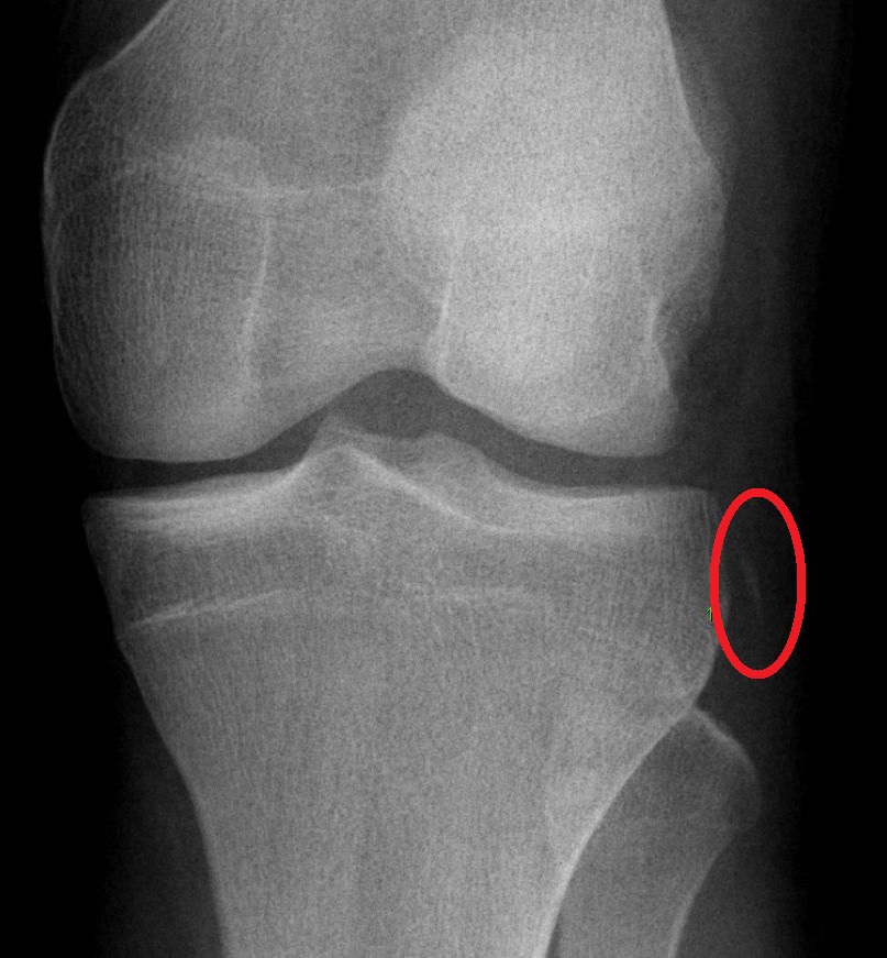
X-rayAnteroposterior knee X-ray: a small bone fragment avulsed from the lateral tibial plateau (Segond sign, circled), an indirect radiographic sign of anterior cruciate ligament rupture.
Source : Ellisbjohns, Wikimedia Commons · Domaine public
- Age 55 years or over
- Isolated tenderness of the patella
- Tenderness of the fibular head
- Inability to flex the knee to 90°
- Inability to take 4 weight-bearing steps (even limping) in the clinic or the emergency department
🧭 Clinical algorithm for assessing an acute knee sprain
From suspicion to treatment stratification: each step clears the next
A summary algorithm. Main sources: the Ottawa Knee Rules (Stiell 1996, Bachmann 2004) and the diagnostic accuracy meta-analysis for the ACL tests (Benjaminse 2006, JOSPT).
- ACL :
- Lachman test (knee at 20-30° of flexion): the most sensitive and the most useful acutely, being little affected by muscle guarding;
- Anterior drawer (at 90° of flexion): less accurate, with frequent false negatives in the acute phase;
- Pivot shift : highly specific when positive (rotational instability), often feasible only once pain is controlled or under anaesthetic.9
- PCL : posterior sag sign (knees and hips at 90°), posterior drawer. Diagnostic accuracy is good in combination.
- MCL : stress in valgus at 0° and 30° of flexion. Pain or laxity at 30° alone isolates the MCL; laxity at 0° = more extensive ligament involvement (capsule, cruciates).
- LCL and posterolateral corner : stress in varus at 0° and 30°, dial test at 30° and 90° (asymmetry of external rotation). Posterolateral corner involvement is a serious injury, frequently associated with the ACL or PCL.
- A meniscal lesion (very frequently associated with the acute ACL): McMurray and Thessaly help but have moderate accuracy;
- Patellar dislocation or subluxation: it can mimic a “pop” and rapid swelling; the patellar apprehension test is relevant;
- An osteochondral fracture in the adolescent or young adult;
- A Segond fracture on the radiograph (lateral avulsion of the tibial plateau): a near-pathognomonic sign of an ACL tear and frequently associated with a lesion of the anterolateral ligament.19
🚩 Red flags in the acute knee: urgent referral
- An obvious dislocation, or one that has reduced spontaneously , plus an absent dorsalis pedis or posterior tibial pulse → a vascular emergency : ABI < 0.9 → immediate CT angiography (injury to the popliteal artery, with a risk of amputation).18
- A common fibular nerve deficit (foot drop, weak dorsiflexion, numbness over the dorsum of the foot) after severe trauma.
- A tense haemarthrosis, disproportionate pain, paraesthesia, pallor → suspected compartment syndrome.
- An open fracture, bony deformity, shortening.
- Fever plus acute swelling with no obvious trauma → septic arthritis, to be ruled out urgently.
- A child or adolescent with haemarthrosis → look for tibial spine avulsion (the paediatric equivalent of an ACL tear) on radiograph or MRI.
⚠ Any red flag → refer to orthopaedic or vascular emergency care before any physiotherapy.
Should patients be classified, and what are the benefits?
Yes. Grading in 3 grades standardises communication, steers the initial decision and contributes to the prognosis. Grade I: a stretch with no laxity; Grade II: a partial tear, moderate laxity with a firm end point; Grade III: a complete tear, marked laxity with a soft end point.1 Important limits:- The anatomical grade does not predict functional instability. Some “coper” patients function very well with a grade III ACL; others with a less severe lesion are severely disabled.
- MRI, often seen as the gold standard, can over-diagnose clinically insignificant lesions or miss dynamic instability.
- For complex injuries (multiligament, rotational instability), dedicated classifications are needed (for example the Schenck classification for knee dislocations).
- The history (mechanism, pop, swelling within 12 h, instability) supplies most of the pre-test probability.
- Always rule out a fracture first with the Ottawa Knee Rules.
- Lachman = the most sensitive test for the ACL. Pivot shift = highly specific when positive.
- Tests by ligament: valgus/varus at 0° and 30°, posterior drawer + posterior sag for the PCL, the dial test for the posterolateral corner.
- Always look for the red flags : dislocation, a vascular or nerve deficit, compartment syndrome, septic arthritis.
Bibliography
- Sanders TL, Maradit Kremers H, Bryan AJ, et al. Incidence of Anterior Cruciate Ligament Tears and Reconstruction: A 21-Year Population-Based Study. Am J Sports Med. 2016;44(6):1502-1507. PMID 26957217.
- Stiell IG, Greenberg GH, Wells GA, et al. Prospective validation of a decision rule for the use of radiography in acute knee injuries. JAMA. 1996;275(8):611-615. PMID 8594242.
- Bachmann LM, Haberzeth S, Steurer J, ter Riet G. The accuracy of the Ottawa knee rule to rule out knee fractures: a systematic review. Ann Intern Med. 2004;140(2):121-124. PMID 14734335.
- Benjaminse A, Gokeler A, van der Schans CP. Clinical diagnosis of an anterior cruciate ligament rupture: a meta-analysis. J Orthop Sports Phys Ther. 2006;36(5):267-288. PMID 16715828.
- Logerstedt DS, Scalzitti DA, Bennell KL, et al. Knee Pain and Mobility Impairments: Meniscal and Articular Cartilage Lesions Revision 2018. J Orthop Sports Phys Ther. 2018;48(2):A1-A50. PMID 29385940.
- Claes S, Vereecke E, Maes M, Victor J, Verdonk P, Bellemans J. Anatomy of the anterolateral ligament of the knee. J Anat. 2013;223(4):321-328. PMID 23906341.
- Chahla J, Murray IR, Robinson J, et al. Posterolateral corner of the knee: an expert consensus statement on diagnosis, classification, treatment, and rehabilitation. Knee Surg Sports Traumatol Arthrosc. 2019;27(8):2520-2529. PMID 30478468.
- Medina O, Arom GA, Yeranosian MG, Petrigliano FA, McAllister DR. Vascular and nerve injury after knee dislocation: a systematic review. Clin Orthop Relat Res. 2014;472(9):2621-2629. PMID 24554457.
- Finucane LM, Downie A, Mercer C, et al. International Framework for Red Flags for Potential Serious Spinal Pathologies. J Orthop Sports Phys Ther. 2020;50(7):350-372. PMID 32438853.
- Frobell RB, Roos EM, Roos HP, Ranstam J, Lohmander LS. A randomized trial of treatment for acute anterior cruciate ligament tears. N Engl J Med. 2010;363(4):331-342. PMID 20660401.
The female factor and primary ACL prevention: why and how?
⚡ Hypotheses for the female excess risk of ACL tear: modifiable and non-modifiable factors
The neuromuscular factors are the ones prevention can address
A synthesis of the families of explanatory factors. The neuromuscular family concentrates the documented preventive effect: it is what the warm-up programmes target.
Neuromuscular prevention programmes: what has been shown?
Several structured programmes (FIFA 11+ in football, the PEP program in women's football, programmes of the Knäkontroll type) combine strengthening (posterior chain, hip), neuromuscular control, plyometrics, trunk stability and technical landing work. Built into the warm-up and applied consistently, they are associated with a reduction in the incidence of ACL injuries and of non-contact lower limb injuries in pivoting sports.2,3 The effect is dose-dependent: compliance above 2 sessions a week and continuation through the season are key conditions. Insufficient coverage explains most of the “negative” trials.| Programme | Target population | Level of evidence | Expected effect |
|---|---|---|---|
| FIFA 11+ | Footballers from amateur to professional | High | ↘ non-contact lower limb injuries, ↘ ACL where compliance is 2 or more sessions a week |
| The PEP program | University women's football | Moderate-high | ↘ ACL in female athletes |
| Knäkontroll-type programmes | General pivoting sports | Moderate | ↘ knee injuries, transferable to related sports |
| Non-standardised in-house programmes | — | Low | An uncertain effect (lack of standardisation, poor adherence) |
- The female excess risk of ACL tear in pivoting sports is documented (Montalvo 2019 meta-analysis, BJSM).
- Three families of hypothesis: neuromuscular (modifiable ++), anatomical (not modifiable), hormonal (mixed data).
- The operational preventive target = the neuromuscular factors: dynamic valgus, quadriceps dominance, trunk control.
- The FIFA 11+ / PEP / Knäkontroll programmes, in the warm-up, at 2 or more sessions a week, reduce incidence.
- Compliance is the main determinant: “a good programme done badly is no programme at all”.
Bibliography
- Montalvo AM, Schneider DK, Webster KE, et al. “What's my risk of sustaining an ACL injury while playing sports?” A systematic review with meta-analysis. Br J Sports Med. 2019;53(16):1003-1012. PMID 29514822.
- Soligard T, Myklebust G, Steffen K, et al. Comprehensive warm-up programme to prevent injuries in young female footballers: cluster randomised controlled trial (FIFA 11+). BMJ. 2008;337:a2469. PMID 19066253.
- Sadoghi P, von Keudell A, Vavken P. Effectiveness of anterior cruciate ligament injury prevention training programs. J Bone Joint Surg Am. 2012;94(9):769-776. PMID 22456856.
- Hewett TE, Ford KR, Hoogenboom BJ, Myer GD. Understanding and preventing ACL injuries: current biomechanical and epidemiologic considerations - update 2010. N Am J Sports Phys Ther. 2010;5(4):234-251. PMID 21655382.
Which treatment strategies are the most effective?
Where do you start? What is the hierarchy of interventions?
Acute management has moved on from RICE. Contemporary guidance follows the acronym PEACE & LOVE (Dubois & Esculier, BJSM 2020).1🕊️ PEACE & LOVE: acute then subacute management of soft-tissue injury
PEACE: the first 1-3 days. LOVE: from the end of the acute phase.
Source: Dubois B, Esculier JF. Soft-tissue injuries simply need PEACE and LOVE. Br J Sports Med. 2020;54(2):72-73. DOI 10.1136/bjsports-2019-101253.
What is the place of exercise, and is there a superior approach?
Therapeutic exercise is the cornerstone of rehabilitation, with a high level of evidence for the ACL. There is no single “best” approach but a progressive continuum structured by functional criteria, built around four pillars:- Strengthening: focused on the quadriceps and hamstrings; open-chain (OKC) and closed-chain (CKC) exercises are complementary.
- Neuromuscular control: balance, perturbation, agility; integrating motor learning principles (external feedback, variable practice).
- Progressive plyometrics: preparing for the impact and deceleration demands of the target sport.
- Sport-specific reintegration: straight-line running, accelerations, decelerations, changes of direction, technical movements, controlled then free opposition.
| Modality | Main indication | Level of evidence | Expected effect |
|---|---|---|---|
| Therapeutic education & alliance | All phases | High | ↗ adherence, ↘ kinesiophobia |
| Prehabilitation (before ACL surgery) | Before ACL surgery | High | ↗ postoperative function, ↗ quadriceps activation |
| Quadriceps strengthening, OKC + CKC | All ACL phases | High | ↘ strength deficit, ↗ function, ↘ osteoarthritis risk |
| Neuromuscular control / agility | Intermediate phases | High | ↗ movement quality, ↘ dynamic valgus |
| Progressive plyometrics | Late phase | High | ↗ functional performance, ↗ impact tolerance |
| Manual therapy (mobility, pain) | A targeted adjunct (post-traumatic or postoperative stiffness) | Moderate in the short term | ↘ pain, ↗ range, with no long-term functional benefit on its own |
| Cryotherapy | Analgesia after trauma or surgery | Moderate | ↘ pain; a modest anti-swelling effect |
| Quadriceps NMES | Early postoperative arthrogenic inhibition | Moderate | ↗ quadriceps activation alongside voluntary work |
| Routine ultrasound / laser | — | Low | No convincing clinical benefit for ligament healing |
| ACL reconstruction | Functional instability, the pivoting athlete | Moderate | Equivalent to rehabilitation alone in some profiles (KANON); necessary where instability persists |
Manual therapies, technologies: how effective are they really?
The passive modalities have an adjunctive place, never a leading one. Manual therapy can reduce pain and improve range in the short term, particularly for post-traumatic or postoperative stiffness; it does not replace exercise. Cryotherapy has a useful analgesic effect in the acute phase. Quadriceps NMES is worthwhile early after surgery when arthrogenic muscle inhibition makes voluntary recruitment difficult. Routine ultrasound and laser are not supported by solid clinical evidence for ligament healing and should not replace an active programme.3Beyond the physical: education and psychological factors
A biopsychosocial approach is indispensable. Kinesiophobia (fear of movement) is very frequent after ACL reconstruction. It is measurable with the ACL-RSI scale (Webster, Feller, Lambros 2008, Phys Ther Sport), now the reference scale for psychological readiness to return to sport, and with the Tampa Scale for Kinesiophobia (TSK).4 Effective strategies: education about the prognosis, shared goal-setting (shared decision-making), graded exposure to the feared sporting situations, visualisation. Optimism, the “O” of LOVE, is not wishful thinking but a recognised prognostic determinant.- Acute phase → PEACE & LOVE ; avoid systemic NSAIDs very early on.
- Exercise is the central treatment: quadriceps and hamstring strengthening, neuromuscular control, plyometrics, sport-specific work.
- The practical reference = the Dutch ACL Guideline (van Melick 2016, BJSM), with progression on functional criteria.
- Passive therapies (manual, modalities) = adjuncts in the short term, never the mainstay.
- Assess and address kinesiophobia (ACL-RSI), a central component of the long-term outcome.
Bibliography
- Dubois B, Esculier JF. Soft-tissue injuries simply need PEACE and LOVE. Br J Sports Med. 2020;54(2):72-73. DOI 10.1136/bjsports-2019-101253.
- Frobell RB, Roos HP, Roos EM, Roemer FW, Ranstam J, Lohmander LS. Treatment for acute anterior cruciate ligament tear: five year outcome of randomised trial. BMJ. 2013;346:f232. PMID 23349407.
- van Melick N, van Cingel REH, Brooijmans F, et al. Evidence-based clinical practice update: practice guidelines for anterior cruciate ligament rehabilitation based on a systematic review and multidisciplinary consensus. Br J Sports Med. 2016;50(24):1506-1515. PMID 27539507.
- Webster KE, Feller JA, Lambros C. Development and preliminary validation of a scale to measure the psychological impact of returning to sport following anterior cruciate ligament reconstruction surgery. Phys Ther Sport. 2008;9(1):9-15. PMID 19083699.
- Frobell RB, Roos EM, Roos HP, Ranstam J, Lohmander LS. A randomized trial of treatment for acute anterior cruciate ligament tears. N Engl J Med. 2010;363(4):331-342. PMID 20660401.
- Grindem H, Snyder-Mackler L, Moksnes H, Engebretsen L, Risberg MA. Simple decision rules can reduce reinjury risk by 84% after ACL reconstruction: the Delaware-Oslo ACL cohort study. Br J Sports Med. 2016;50(13):804-808. PMID 27162233.
- Wright RW, Preston E, Fleming BC, et al. A systematic review of anterior cruciate ligament reconstruction rehabilitation: part I: continuous passive motion, early weight bearing, postoperative bracing, and home-based rehabilitation. J Knee Surg. 2008;21(3):217-224. PMID 18686485.
- Ardern CL, Glasgow P, Schneiders A, et al. 2016 Consensus statement on return to sport from the First World Congress in Sports Physical Therapy, Bern. Br J Sports Med. 2016;50(14):853-864. PMID 27226389.
How do you secure lasting recovery and prevent recurrence?
How do you make the patient an active participant in their recovery through self-management?
Making the patient autonomous is a major therapeutic lever. Three routes:- Structured therapeutic education : the condition, the goals of each phase, the rationale for every exercise. It reduces anxiety and increases adherence.
- Shared decision-making : realistic goals defined together, an adjustable plan, a strengthened therapeutic alliance.
- Routine psychological assessment (ACL-RSI, TSK) with tailored interventions: visualisation, graded goal-setting, exposure to the feared situations.
When and how should a safe return to sport and activity be planned?
The return-to-sport (RTS) decision must not rest on time since surgery alone. The international consensus of Bern 2016 (Ardern et al., BJSM) proposes the StARRT (Strategic Assessment of Risk and Risk Tolerance) framework and insists on a multifactorial decision integrating tissue health, risk, and personal and contextual factors.1 The Delaware-Oslo cohort (Grindem et al., BJSM 2016) supplied one of the most striking results in the literature: in patients with an ACL reconstruction, meeting a battery of criteria (quadriceps strength, hop tests, return of sporting tasks) and a delay of more than 9 months before returning to pivoting reduce the risk of re-rupture by 84 % ; each further month beyond 9 months reduces the risk again, up to a plateau.2📊 The battery of return-to-sport criteria: an operational summary
No single criterion is enough: the decision is multifactorial
An operational summary of the 4 domains in the RTS decision after ACL reconstruction. Sources: Ardern 2016 (Bern consensus, BJSM), Grindem 2016 (Delaware-Oslo, BJSM), Wellsandt 2017 (JOSPT), Paterno 2014 (AJSM).
- Relying solely on time since surgery (“6 months and you're fine”): wrong, and dangerous.
- Relying solely on an LSI ≥ 90 %: the “healthy” leg may itself be detrained, overestimating bilateral performance (Wellsandt 2017, JOSPT).3
- Neglecting psychological preparation : a patient who is physically ready but does not feel ready is not ready.
- Underestimating context : type of sport, level of competition, team or individual sport, social support, expectations from family and club.
- The return to sport rests on multifactorial objective criteria, never on time alone.
- The Delaware-Oslo cohort (Grindem 2016): −84 % recurrence with the criteria battery + a delay ≥ 9 months.
- 4 domains to satisfy: strength, functional performance, psychological readiness, timing.
- An LSI ≥ 90 % alone is not enough (Wellsandt 2017): it can mask a bilateral deficit.
- The risk of a second tear is × 15 in the first 12 months after RTS (Paterno 2014).
Bibliography
- Ardern CL, Glasgow P, Schneiders A, et al. 2016 Consensus statement on return to sport from the First World Congress in Sports Physical Therapy, Bern. Br J Sports Med. 2016;50(14):853-864. PMID 27226389.
- Grindem H, Snyder-Mackler L, Moksnes H, Engebretsen L, Risberg MA. Simple decision rules can reduce reinjury risk by 84% after ACL reconstruction: the Delaware-Oslo ACL cohort study. Br J Sports Med. 2016;50(13):804-808. PMID 27162233.
- Wellsandt E, Failla MJ, Snyder-Mackler L. Limb Symmetry Indexes Can Overestimate Knee Function After Anterior Cruciate Ligament Injury. J Orthop Sports Phys Ther. 2017;47(5):334-338. PMID 28355978.
- Paterno MV, Rauh MJ, Schmitt LC, Ford KR, Hewett TE. Incidence of Second ACL Injuries 2 Years After Primary ACL Reconstruction and Return to Sport. Am J Sports Med. 2014;42(7):1567-1573. PMID 24753238 · PMC4205204.
- Webster KE, Feller JA, Lambros C. Development and preliminary validation of a scale to measure the psychological impact of returning to sport following anterior cruciate ligament reconstruction surgery. Phys Ther Sport. 2008;9(1):9-15. PMID 19083699.
- van Melick N, van Cingel REH, Brooijmans F, et al. Evidence-based clinical practice update: practice guidelines for anterior cruciate ligament rehabilitation. Br J Sports Med. 2016;50(24):1506-1515. PMID 27539507.
What do real clinical cases teach us about knee sprains?
A “classic” case analysed: ACL tear, from assessment to return to sport
The clinical pathway of an isolated ACL tear in a pivoting athlete illustrates how assessment, shared decision-making and criteria-based rehabilitation fit together. Initial assessment : a suggestive history (non-contact mechanism, pop, swelling < 12 h, instability), the Ottawa Knee Rules to rule out a fracture, clinical tests (Lachman, pivot shift once pain is controlled), MRI for confirmation. A systematic search for associated lesions (meniscal, osteochondral, Segond fracture). Shared decision : discuss the options with the patient (rehabilitation alone with delayed surgery as an option, KANON-style, vs reconstruction straight away), according to the profile (age, level and type of sport, goals, comorbidities, expectations). Rehabilitation by phases and criteria (reference: the Dutch ACL Guideline, van Melick 2016, BJSM):- Prehabilitation : full extension, swelling control, quadriceps activation.
- Early postoperative phase : graft protection, swelling and pain management, restoring mobility, NMES where there is arthrogenic inhibition.
- Intermediate phase : strengthening, neuromuscular control; progression towards a return to running once the strength and movement-quality criteria are met.
- Late phase and RTS : plyometrics, agility, sport-specific work; final clearance by the multifactorial battery (strength, hop tests, ACL-RSI, movement quality) + a delay ≥ 9 months.
The diagnostic challenge: the Segond fracture and the anterolateral ligament
The Segond fracture is a millimetre-sized bony avulsion on the lateral aspect of the tibial plateau, visible on the initial radiograph. Long considered a simple marker, it is today recognised as near-pathognomonic of an ACL tear and associated with a lesion of the anterolateral ligament (ALL), described anatomically by Claes et al. (J Anat 2013).1 Clinical implications :- Any small lateral avulsion of the tibial plateau should raise the suspicion of an ACL tear and prompt confirmation (MRI), without delay.
- Residual rotational instability after ACL reconstruction can be explained by an unaddressed ALL lesion; some teams offer an anterolateral augmentation procedure (tenodesis) in high-risk profiles (young, pivoting sports, hyperlaxity).
A complex case studied: knee dislocation and vascular emergency
Multiligament injuries after high-energy trauma (a road traffic accident, a motorcycle fall, high-speed sporting trauma) call for a very different approach. A knee dislocation can reduce itself before arrival in the emergency department; it has to be suspected in the face of multidirectional laxity from the outset or a large post-traumatic haemarthrosis. Priority number one: vascular screening. The systematic review of Medina et al. (Clin Orthop Relat Res 2014) synthesised the published series on vascular (popliteal artery) and nerve (common fibular nerve) injuries after knee dislocation.2🚩 Knee dislocation: the vascular and nerve emergency protocol
- Vascular examination immediately: dorsalis pedis and posterior tibial pulses, colour, warmth, capillary refill.
- Ankle-brachial index (ABI) in every case. ABI < 0.9 → urgent CT angiography; vascular opinion; revascularisation within 6-8 hours to save the limb.
- Present pulses ≠ safety : an intimal injury can present initially with a palpable pulse and thrombose later. Repeated (serial) reassessments are mandatory over the first 24-48 h.
- Neurological examination : common fibular nerve (dorsiflexion, dorsum of the foot); posterior tibial nerve.
- Assessment of the other injuries : bone, skin, ligaments.
⚠ Knee dislocation is an orthopaedic AND a vascular emergency: the priority is limb viability, not the ligament strategy.
Critique and controversy: the level of evidence of clinical cases
Clinical cases are valuable for illustrating, generating hypotheses and flagging rare presentations. They never demonstrate effectiveness: the absence of a control group, publication bias (successes are published more than failures) and regression to the mean all limit their reach. In the GRADE/Oxford CEBM hierarchy, a case report is level 5 (the weakest). When an attractive case contradicts a meta-analysis, the meta-analysis wins.📐 The hierarchy of scientific evidence: where does each type of study sit?
Strength of evidence decreasing from the top (meta-analyses of RCTs) to the bottom (isolated cases, expert opinion)
A simplified GRADE / Oxford CEBM hierarchy. Practical implication: where an attractive clinical case diverges from a meta-analysis, follow the meta-analysis. Clinical cases remain valuable for teaching and for generating hypotheses.
⭐ Key points
- A clinical case illustrates, it does not prove anything. Level of evidence 5 (CEBM).
- A Segond fracture on the initial radiograph is near-pathognomonic of an ACL tear and often associated with a lesion of the anterolateral ligament (ALL) (Claes 2013).
- Knee dislocation = an orthopaedic AND a vascular emergency: pulse examination + ABI in every case; ABI < 0.9 → CT angiography (Medina 2014).
- A pulse felt initially does not rule out an intimal injury that thromboses later: repeated reassessments over the first 24-48 hours.
- A multiligament decision is a multidisciplinary one (orthopaedics + vascular + physiotherapy), and often staged.
Bibliography
- Claes S, Vereecke E, Maes M, Victor J, Verdonk P, Bellemans J. Anatomy of the anterolateral ligament of the knee. J Anat. 2013;223(4):321-328. PMID 23906341.
- Medina O, Arom GA, Yeranosian MG, Petrigliano FA, McAllister DR. Vascular and nerve injury after knee dislocation: a systematic review. Clin Orthop Relat Res. 2014;472(9):2621-2629. PMID 24554457.
- Chahla J, Murray IR, Robinson J, et al. Posterolateral corner of the knee: an expert consensus statement on diagnosis, classification, treatment, and rehabilitation. Knee Surg Sports Traumatol Arthrosc. 2019;27(8):2520-2529. PMID 30478468.
- Frobell RB, Roos HP, Roos EM, Roemer FW, Ranstam J, Lohmander LS. Treatment for acute anterior cruciate ligament tear: five year outcome of randomised trial. BMJ. 2013;346:f232. PMID 23349407.
- Anderson AF. Transepiphyseal replacement of the anterior cruciate ligament in skeletally immature patients. A preliminary report. J Bone Joint Surg Am. 2003;85(7):1255-1263. PMID 12851350.
- Paterno MV, Rauh MJ, Schmitt LC, Ford KR, Hewett TE. Incidence of Second ACL Injuries 2 Years After Primary ACL Reconstruction and Return to Sport. Am J Sports Med. 2014;42(7):1567-1573. PMID 24753238 · PMC4205204.
- Sanders TL, Maradit Kremers H, Bryan AJ, et al. Incidence of Anterior Cruciate Ligament Tears and Reconstruction: A 21-Year Population-Based Study. Am J Sports Med. 2016;44(6):1502-1507. PMID 26957217.
How do you apply these recommendations concretely in your practice?
When and to which other health professionals should you refer?
Prompt referral is a clinical act in its own right. A proposed framework for red flags in musculoskeletal practice: Finucane et al. (JOSPT 2020).1 Emergency referral (orthopaedic/vascular emergency care) :- Knee dislocation (reduced or not), suspected injury to the popliteal artery.
- Common fibular nerve deficit (foot drop, numbness over the dorsum of the foot).
- Compartment syndrome: disproportionate pain, paraesthesia, pallor, a tense haemarthrosis.
- Open fracture, bony deformity.
- Suspected septic arthritis (fever + acute swelling with no trauma).
- Complete ACL tear in a pivoting athlete with functional instability (alongside rehabilitation).
- Grade III MCL lesion associated with an ACL tear.
- Posterolateral corner involvement, a confirmed Segond fracture.
- Tibial spine avulsion in the child or adolescent.
How do you measure outcomes and overcome the barriers to implementation?
The routine use of Patient-Reported Outcome Measures (PROMs) is now a quality standard. The validated tools for the ligamentous knee:- IKDC subjective form : symptoms, function and sporting activity; recognised responsiveness to change.
- KOOS (Knee injury and Osteoarthritis Outcome Score): 5 subscales, namely pain, symptoms, ADLs, sport and recreation, quality of life.
- Lysholm : a general functional score.
- Tegner Activity Scale : level of physical activity.
- ACL-RSI (Webster 2008): psychological readiness to return to sport.2
- Tampa Scale for Kinesiophobia (TSK-11) : fear of movement.
- Red flags of the knee: dislocation, a vascular or nerve deficit, compartment syndrome, open fracture, septic arthritis → emergency care.
- Yellow flags : kinesiophobia, fear of re-injury, catastrophising → measure them (ACL-RSI, TSK), address them, do not neglect them.
- PROMs to use: IKDC, KOOS, Lysholm, Tegner, ACL-RSI.
- A shared decision with the patient about the options (surgery, RTS timing, goals) is a pillar of adherence.
- Recognise the barriers to implementation (time, training, expectations) and work to lift them, rather than settling for knowing the science.
Bibliography
- Finucane LM, Downie A, Mercer C, et al. International Framework for Red Flags for Potential Serious Spinal Pathologies. J Orthop Sports Phys Ther. 2020;50(7):350-372. PMID 32438853.
- Webster KE, Feller JA, Lambros C. Development and preliminary validation of a scale to measure the psychological impact of returning to sport following anterior cruciate ligament reconstruction surgery. Phys Ther Sport. 2008;9(1):9-15. PMID 19083699.
- van Melick N, van Cingel REH, Brooijmans F, et al. Evidence-based clinical practice update: practice guidelines for anterior cruciate ligament rehabilitation. Br J Sports Med. 2016;50(24):1506-1515. PMID 27539507.
- Grindem H, Snyder-Mackler L, Moksnes H, Engebretsen L, Risberg MA. Simple decision rules can reduce reinjury risk by 84% after ACL reconstruction: the Delaware-Oslo ACL cohort study. Br J Sports Med. 2016;50(13):804-808. PMID 27162233.
- Ardern CL, Glasgow P, Schneiders A, et al. 2016 Consensus statement on return to sport from the First World Congress in Sports Physical Therapy, Bern. Br J Sports Med. 2016;50(14):853-864. PMID 27226389.
- Dubois B, Esculier JF. Soft-tissue injuries simply need PEACE and LOVE. Br J Sports Med. 2020;54(2):72-73. DOI 10.1136/bjsports-2019-101253.
And after this read?
This article is part of a collection of evidence-based clinical syntheses for physiotherapy. A question, a comment, a correction to suggest? Contact us through the WhatsApp button at the bottom right of the screen.

