














Online

Sports physiotherapy
Blood Flow Restriction applied to rehabilitation
Anthony Baillon
Go further
Osteochondral lesions of the knee: 24 % of healthy knees carry one. What separates a focal lesion from wear, and how long before weight-bearing again.

Faced with an incidentally discovered cartilage defect, the first question is not what to do with the lesion but whether it explains what the patient is complaining of.
24%of asymptomatic knees have a cartilage defect
What follows covers each of these points in detail, with sources. It is there if you need it.
This topic is taught in a course: Blood Flow Restriction applied to rehabilitation, online with Anthony Baillon.
1 / 10
9 article chapters · 40 min in total
Lesion or wear◔ 6 min
A cartilage defect on MRI affects 24 % of asymptomatic knees, and 43 % after the age of 40.
Injured knee◔ 4 min
After a first patellar dislocation, 43 % of knees show a lesion of the articular surfaces.
Size vs discomfort◔ 3 min
Neither size nor ICRS grade predicts the preoperative symptoms of a cartilage lesion.
Surgery◔ 7 min
Under 2 cm², microfracture is not superior to simple debridement, with more complications.
Return to weight bearing◔ 6 min
After chondrocyte implantation, full weight bearing at 6 weeks is as safe as at 8 or 11 weeks.
Return to sport◔ 3 min
Return to sport is possible for 73 % of patients, with the best rates after osteochondral grafting.
Physio's role◔ 5 min
Nobody knows what becomes of an unoperated focal lesion in ten years, for lack of natural-history data.
In practice◔ 3 min
Faced with a cartilage defect, the central question is whether it explains the patient's complaint, not what to do about it.
Common misconceptions◔ 3 min
One healthy knee in four carries a cartilage defect on MRI, a proportion that rises to 43 % after 40.
Train on this
Sports physiotherapy
Anthony Baillon
Next comes practice: the course that teaches this topic, with Anthony Baillon.
Our courses on this topic may be covered by DPC or FIFPL, the French continuing-education funding schemes, depending on your profession and situation.
Cartilage · Knee · Lower limb
A cartilage defect seen on MRI can be three very different things: an injury in a young person, wear beginning, or nothing at all. One asymptomatic knee in four carries one. What separates those three readings cannot be read on the image, and the whole of the treatment that follows depends on it.
Articular cartilage does not repair itself: it has no vessels, no nerves, and no cells that can be mobilised. That sentence underpins the whole subject, but it has produced a false belief, namely that a cartilage defect is by nature progressive and must be treated. The data say otherwise.
A cartilage defect is common in people who are perfectly well. Culvenor's meta-analysis, 63 studies and 5,397 asymptomatic and uninjured knees, measures a prevalence of cartilage defects of 24 %. And above all, it separates by age: 11 % before the age of 40, 43 % from 40 onwards2. In a 55-year-old patient, finding a cartilage defect means finding what almost one healthy knee in two carries.
The dividing line is clinical, not radiological. A focal traumatic lesion in a young person and early wear can produce the same image. What separates them is age, mechanism, timing and the number of lesions. In Widuchowski's series of 25,124 arthroscopies, 60 % of patients have a cartilage lesion, of which 67 % focal and 29 % osteoarthritic, but candidates for repair surgery (one to three focal grade III or IV lesions) account for only 7 % before the age of 40 and 9 % before 501.
Size and grade do not predict how troubled the patient is. That is a counter-intuitive result and it has been measured. Across 90 patients scheduled for cartilage surgery, there was no difference in scores between lesions under and over 2 cm², apart from one subscale, and the ICRS grade does not affect preoperative scores. These patients report symptoms worse than candidates for ligament reconstruction and close to those awaiting a joint replacement3.
The repair procedures are less clear-cut than is often said. A double-blind randomised trial published in 2025 shows that microfracture is not superior to simple debridement for femoral lesions under 2 cm²6. At fourteen or fifteen years, chondrocyte implantation and microfracture are clinically equivalent, with about a third of failures and radiographic osteoarthritis in half the survivors7.
And the time to return to weight bearing can be shortened without harm. This is the result most directly useful to the physiotherapist. Three successive randomised trials by the same team brought full weight bearing after grafting down from 11 weeks to 8, then from 8 to 6, with no deterioration of the graft on MRI and, at each step, less pain and faster function111413.
This is the question that governs everything else, and it is the one imaging does not settle. Two series, one arthroscopic and one by MRI in healthy knees, together give the markers that allow a decision.
Widuchowski and colleagues analysed every knee arthroscopy performed in their centre from 1989 to 2004, that is 25,124 procedures, classifying cartilage lesions by Outerbridge1.
Cartilage lesions are found in 60 % of patients. Distributed as follows: focal osteochondral or chondral lesion 67 %, osteoarthritis 29 %, osteochondritis dissecans 2 %, other 1 %. Outerbridge grade II is the most frequent (42 %). The dominant sites are the patellar surface (36 %) and the medial femoral condyle (34 %).
Two figures from this series deserve to be remembered above the others, because they frame the real place of repair surgery. First, 70 % of lesions are not isolated : they accompany another lesion, most often a medial meniscal tear (37 %) or an anterior cruciate ligament injury (36 %). Second, the patients who meet the criteria for repair surgery, that is one to three focal grade III or IV lesions, are only 7 % of the series before the age of 40 and 9 % before 501.
The authors themselves conclude cautiously: these patients form a heterogeneous group, and the natural history of cartilage lesions remains unknown, so that the exact number of patients who would benefit from repair cannot be established.
Where the lesions sit, and what goes with them
25,124 consecutive knee arthroscopies, 1989 to 2004
Source: Widuchowski W, Widuchowski J, Trzaska T. Knee. 2007;14(3):177-182. PMID 17428666. A single-centre arthroscopic series over 16 years: these are knees judged worth operating on, not a population sample. “Candidates for repair surgery” are defined by the authors as having one to three focal grade III or IV lesions.
Two classifications are in circulation, and it is useful to know which one you are reading. Widuchowski's series uses Outerbridge, the older one, in which grade II is the most represented (42 % of lesions)1. More recent work uses the classification of the International Cartilage Regeneration and Joint Preservation Society, ICRS, which grades from simple superficial fissuring to exposure of the subchondral bone.
These grades serve to describe a lesion and to establish an indication: repair is discussed for deep lesions, grade III or IV, not for superficial softening. But it is worth knowing from now on what they do not do, because it has been measured and the next chapter returns to it: the ICRS grade does not affect the scores patients report before surgery3. A grade is a description of a joint surface, not a prognosis of how troubled someone will be.
The decisive counterpart comes from Culvenor and colleagues, who looked for the prevalence of osteoarthritis features on MRI in asymptomatic and uninjured adults. Six databases, 63 studies included, 5,397 knees in 4,751 adults, with random-effects meta-analytic estimates2.
Overall prevalence of cartilage defects: 24 % (95 % CI 15 to 34). Stratified by age: 11 % before the age of 40 (6 to 17) and 43 % at 40 and over (29 to 57). Meniscal tears follow the same slope: 4 % before 40, 19 % after. Bone marrow lesions affect 18 % of knees, osteophytes 25 %2.
The authors' conclusion is an instruction for reading: these images must be interpreted in the context of the clinical presentation, and taken into account as such in the decision.
A cartilage defect in someone with no pain anywhere
Prevalence of cartilage defects on MRI in asymptomatic, uninjured knees, by age band
Source: Culvenor AG, Øiestad BE, Hart HF, et al. Br J Sports Med. 2019;53(20):1268-1278. PMID 29886437. The confidence intervals are wide, the heterogeneity between studies is considerable, and the estimates depend on the MRI sequences used: these are orders of magnitude, to be read as such.
From these two series, and from what the patellar dislocation cohort in the next chapter establishes, a grid can be drawn. It rests on no validated test, and that has to be said: it is a synthesis of clinical markers, not a score.
| Focal traumatic lesion | Wear beginning | |
|---|---|---|
| Age | A young person. The patellar dislocation cohort has a median age of 164 ; the patients scheduled for cartilage surgery, 333 | After 40, the age at which 43 % of HEALTHY knees already carry a defect2 |
| Onset | Dated. An identifiable event: patellar dislocation, severe sprain, impaction | Insidious, undated, often attributed after the event to something unremarkable |
| Number of lesions | One, sometimes two. The repair criteria retain one to three focal lesions1 | Diffuse, bilateral involvement, often in several compartments |
| Edges of the lesion | Sharp, healthy surrounding cartilage, often with an osteochondral fragment | Progressive thinning, blurred edges, neighbouring cartilage also altered |
| Underlying bone | Impaction oedema, sometimes an osteochondral fracture | Sclerosis, subchondral cysts, osteophytes, present in 25 % of asymptomatic people2 |
| What it changes | Repair can be discussed, and postoperative return to weight bearing becomes the subject | Management is that of osteoarthritis: exercise, load, weight. See knee osteoarthritis |
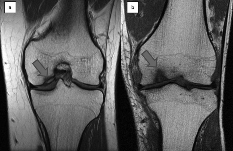
MRICoronal T1-weighted knee MRI in two patients: osteochondritis dissecans lesion of the medial femoral condyle, arrow on the subchondral lesion.
Source : Perera et al., Annals of the Royal College of Surgeons of England, 2012, figure 2 · CC BY
The best-documented mechanism is patellar dislocation, because a consecutive cohort imaged it systematically in the subacute phase. The figures that come out of it change what is done after a first episode.
Isacsson and colleagues used a consecutive cohort of 1,145 injured knees examined by MRI in a centre with a defined population catchment, to extract the primary lateral patellar dislocations. One hundred and eighty-four cases identified, MRI performed at a median of six days4.
The profile is that of an adolescent: median age 16 (interquartile range 14 to 21, range 9 to 47), 41 % women. Overall annual incidence is 14 per 100,000, with a peak between 13 and 15 at 125 per 100,000. Men are older than women at the first episode (17 against 15) and dislocate more often during sport (65 % against 40 %)4.
The result that should change practice is this: articular surface lesions are visible on MRI or found at surgery in 75 of the 184 knees, that is 43 %, and they are osteochondral fractures in 32 knees, that is 18 %. The risk does not differ significantly according to whether the dislocation happened during sport or in daily life4.
After a first patellar dislocation
184 primary dislocations in a consecutive cohort of 1,145 injured knees, MRI at a median of 6 days
Source: Isacsson A, Olsson O, Englund M, Frobell RB. Int Orthop. 2023;47(4):973-981. PMID 36749375. A consecutive cohort with a defined population catchment, which makes the incidences interpretable; the 43 % of surface lesions include surgical findings in 9 cases.
Faced with a first patellar dislocation in an adolescent, the question is not only future instability: it is whether an osteochondral fracture is present, because a loose fragment or a fixable fragment changes the decision and the timetable. Almost one dislocation in five carries one, and the clinical picture alone does not distinguish them.
Osteochondritis dissecans is the second main route to an osteochondral lesion in young people, and it follows a different logic. The review by Chau and colleagues, published in the Journal of Bone and Joint Surgery, sets out the frame: it mainly affects active children and young adults, sits at the knee, the elbow or the ankle, and can lead to early osteoarthritis5.
The decision hinges on two parameters: skeletal maturity and the stability of the lesion, the latter defined by the presence or absence of a fracture of the articular cartilage and of separation of the subchondral bone, assessed by imaging and arthroscopy. Treatment is typically non-operative for stable lesions in skeletally immature patients, and operative if conservative treatment fails or the lesion is unstable5.
The authors note that the origin remains debated (local ischaemia, aberrant endochondral ossification of a secondary subarticular physis, repeated microtrauma, genetic predisposition) and above all that clinical practice guidelines are limited by the scarcity of high-level evidence. A multicentre effort is under way to build reliable classifications and decision algorithms with prognostic value.
One link is worth making here with what the site already documents: two adolescents who developed osteochondritis dissecans following a bone bruise of a femoral condyle are reported in our article on bone bruise. Two cases do not make a frequency, but they justify monitoring a skeletally immature patient after a condylar impaction.
This is one of the most useful results on the subject, and one of those that best protect the patient from an anxiety-inducing account. The link between what the image shows and what the person experiences is weak.
Randsborg, Årøen and Owesen prospectively collected the preoperative data of 90 consecutive patients scheduled for surgery on an isolated symptomatic cartilage lesion, mean age 33.2, 69 % men, then compared them with those of patients scheduled for ligament reconstruction and for knee replacement3.
First result. There is no significant difference in scores between patients whose lesion measures less than 2 cm² and those whose lesion measures more, with one exception: the KOOS “symptoms” subscale, better in small lesions (62.8 against 51.9, p = 0.005). The correlation between lesion size and Lysholm score exists but is weak, and it disappears after adjustment for age, sex, body mass index and location3.
Second result. The ICRS grade does not affect preoperative scores. Neither depth, nor size, nor location has any notable impact on the symptoms experienced3.
Third result, and the most striking. These patients report symptoms more severe than candidates for ligament reconstruction, and approaching those of candidates for knee replacement3.
What the patient feels, and what the image shows
90 patients scheduled for cartilage surgery, compared with two other surgical populations
Source: Randsborg PH, Årøen A, Owesen C. Cartilage. 2022;13(2). PMID 35815409. A prospective series of 90 patients from one centre: the comparison with the two other surgical populations is descriptive, not matched.
Three direct consequences.
The first is a duty of caution in what is said. Announcing a size in square centimetres or an ICRS grade as though it were a prognosis has no basis: those parameters do not predict what the patient feels today. They have value for establishing a surgical indication, not for predicting an experience.
The second is a duty of recognition. The other face of the same result is that these patients are often far worse off than imagined, up to a level of symptoms close to candidates for a joint replacement. A young adult with a 1 cm² lesion can be severely troubled, and there is nothing exaggerated about it.
The third concerns follow-up. If size does not predict how troubled the patient is, then follow-up is done on functional measures and on patient-reported scores, not on repeated images.
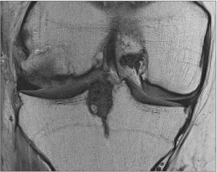
MRICoronal knee MRI: appearance of the lateral femoral condyle after autogenous osteochondral grafting covering a previous osteochondritis dissecans lesion.
Source : Yang et al., Clinics in Orthopedic Surgery, 2016, figure 3 · CC BY-NC
The physiotherapist does not set the indication, but receives these patients before and after, and is the first to be asked what the operation will bring. The recent literature is more sober than the reputation of the techniques.
This is the most important result of recent years on the subject, and it comes from a multicentre double-blind randomised trial, level 1. Randsborg and colleagues included 65 patients aged 18 to 50 with an isolated symptomatic femoral cartilage lesion under 2 cm², randomised between microfracture (31) and simple arthroscopic debridement (34), followed for two years. Mean lesion size: 1.2 ± 0.6 cm²6.
The primary outcome, the change in KOOS Quality of Life at two years, does not differ: 3.5 points (95 % CI −10.0 to 16.9), p = 0.61. No secondary outcome differs either, at any follow-up point, on a linear mixed model. The complications, however, are not symmetrical: 10 complications in 5 patients in the microfracture arm, against 2 in 2 patients in the debridement arm. The authors' conclusion, word for word: microfracture was not superior to debridement for femoral lesions under 2 cm²6.
Two reservations should accompany the result, without weakening it. A sample of 65 patients leaves a wide confidence interval, which does not exclude a moderate benefit from microfracture. And the scope is narrow: femoral lesions under 2 cm², in adults aged 18 to 50. Nothing transfers to large lesions.
Knutsen and colleagues followed 80 patients with a chronic, isolated, symptomatic femoral cartilage lesion, without generalised osteoarthritis, enrolled between January 1999 and February 2000 and randomised between autologous chondrocyte implantation and microfracture. They had published the results at 2 years and then at 5 years; in 2016 they published the follow-up at 14 to 15 years, with standardised radiographs7.
No significant clinical difference between the two groups. 17 failures in the implantation group against 13 in microfracture, and more total replacements in the implantation group (6 against 3). Patients who had not failed retain a significant improvement over their starting state, but 57 % of the survivors in the implantation group and 48 % of those in the microfracture group have radiographic osteoarthritis (Kellgren-Lawrence 2 or more), a non-significant difference7.
The authors' conclusion is unusually blunt for a surgical paper: their results raise serious concerns about the effectiveness of these procedures in delaying osteoarthritis and avoiding further surgery.
The systematic review by Mithoefer and colleagues, 28 studies and 3,122 patients, mean follow-up of 41 months with only 5 studies at 5 years or more, gives the picture: microfracture improves knee function in all studies during the first 24 months, but the data on the durability of that gain are contradictory. Defect fill on MRI is highly variable and correlates with the functional result. The limitations noted are the poorly hyaline nature of the repair tissue, the variable volume of cartilage produced, and possible functional deterioration8.
Two long-term randomised trials have since measured that deterioration. Volz and colleagues randomised 47 patients between microfracture, sutured AMIC and glued AMIC, and followed them for ten years : all three arms improve over the first two years, then the microfracture arm deteriorates progressively and significantly, while the two AMIC arms remain stable. The MOCART scores are comparable between groups9.
Gudas and colleagues randomised 60 athletes of mean age 24.3 between mosaicplasty and microfracture, with a mean follow-up of 10.4 years. Both techniques stay above the preoperative level but decline between 3 and 10 years. At ten years: 15 failures (26 %), of which 4 (14 %) after mosaicplasty and 11 (38 %) after microfracture. Maintenance of the previous level of physical activity is 75 % after mosaicplasty against 37 % after microfracture. Athletes under 25 at surgery keep better scores10.
Four randomised trials, and what they say about microfracture
Comparator, sample size, follow-up and main result
Sources: Randsborg PH, et al. Am J Sports Med. 2025;53(9):2107-2117 (PMID 40570306); Knutsen G, et al. J Bone Joint Surg Am. 2016;98(16):1332-1339 (PMID 27535435); Volz M, et al. Eur J Orthop Surg Traumatol. 2024;34(5):2429-2437 (PMID 38630297); Gudas R, et al. Am J Sports Med. 2012;40(11):2499-2508 (PMID 23024150). These four trials cover neither the same lesion sizes, nor the same populations, nor the same durations: they are read side by side, never as a meta-analysis.
Hierarchy of evidence, decision by decision
Cards stacked by level, with the study type that underpins them
Sources, in the order of the cards: Ebert 2008 (PMID 18434214), Edwards 2013 (PMID 23880403) and Ebert 2021 (PMID 33459833); Culvenor 2019 (PMID 29886437); Gudas 2012 (PMID 23024150) and Volz 2024 (PMID 38630297); Randsborg 2025 (PMID 40570306); Knutsen 2016 (PMID 27535435). The levels shown are an editorial reading of these sources, not a published formal GRADE rating.
This is the chapter where the physiotherapist has most purchase, and where the literature is, for once, clear. Three successive randomised trials shortened the time to full weight bearing without ever damaging the graft.
Ebert and colleagues open their 2012 trial with a statement less than fifteen years old: although structured postoperative rehabilitation after chondrocyte implantation is considered critical, very little data has been published on how to progressively increase weight bearing and exercise after surgery12. The protocols in use came from caution, not from measurement.
The pivotal trial randomised 62 patients after matrix-induced chondrocyte implantation at the medial or lateral femoral condyle. Both protocols protect the implant for an initial period then increase load in stages; the accelerated group reaches full weight bearing at 8 weeks, the traditional group at 1111.
At three months, the accelerated group achieves better distances on the 6-minute walk test and higher daily activity measured by accelerometry. It reports a significantly better improvement in KOOS pain at 12 weeks. And above all: no patient, whatever the protocol, had an adverse effect on the implant on the three-month MRI.
The biomechanical result is the most telling. Compared with a healthy control group, the knee adduction and flexion moments of the traditional group differ significantly from unaffected subjects, whereas those of the accelerated group do not. In other words, less time spent walking with crutches translates into a faster return to normal gait11.
Sixty-three of the 70 patients recruited were reviewed clinically at five years, 58 with high-resolution MRI. All clinical and MRI scores improve significantly over the period. The accelerated group reports significantly less frequent pain on the VAS at five years ; no other difference between the two groups, clinical or radiological12.
Two observations from this trial deserve to be remembered beyond the question of weight bearing. Patient age and defect size correlate negatively with several MRI scores at five years. And there is no significant correlation between clinical scores and MRI scores : what the image of the graft shows does not say what the patient feels, exactly as preoperatively. At five years, 94 % of patients are satisfied with the relief of their pain and 95 % with their ability to carry out their daily activities12.
The authors' conclusion is worth quoting: the accelerated protocol is safe and effective, and provides clinical results comparable if not superior throughout follow-up.
Edwards, Ackland and Ebert then tested a further shortening in the tibiofemoral joint. Twenty-eight knees assessed at twelve months, full weight bearing at 6 weeks against 8 weeks, those 8 weeks being explicitly presented as the “current best practice” arising from the earlier work14.
Both groups improve significantly on all measures. The accelerated group obtains better SF-36 physical scores at 8 weeks and a higher KOOS quality of life at 6 and 12 months. It regains full active knee extension by 4 weeks, against 12 weeks for the other group. No difference in graft quality on MRI (composite MOCART score 3.34 against 3.04), and no patient had an adverse effect on the implant up to twelve months, whatever the protocol14.
Ebert and colleagues finally followed 35 patients (37 knees) randomised between a return to full weight bearing at 6 weeks (18) and at 8 weeks (19), with a minimum follow-up of five years (5.5 to 7 years), including isokinetic strength and MRI13.
No difference between the groups, on any measure. Significant improvement in all reported scores, in 6-minute walk distance, in active flexion and extension range, and in the extensor strength symmetry index. Overall satisfaction: 87.5 % at 6 weeks and 82.4 % at 8 weeks. The composite MRI score declines non-significantly between one year and the final review, with no difference between groups. Two grafts failing on MRI, one patient referred for arthroplasty: 8.1 % failure at a minimum of five years13.
The authors conclude that a return to full weight bearing in six weeks gives comparable clinical and MRI results beyond five years without compromising the graft, and that this protocol is faster than any proposed and studied so far.
The time to weight bearing, shortened twice without harm
Three successive randomised trials after matrix-induced chondrocyte implantation, same team
Sources: Ebert JR, et al. Osteoarthritis Cartilage. 2008;16(10):1131-1140 (PMID 18434214) and Am J Sports Med. 2012;40(7):1527-1537 (PMID 22539536); Edwards PK, Ackland TR, Ebert JR. Am J Sports Med. 2013;41(10):2314-2324 (PMID 23880403); Ebert JR, et al. Knee Surg Sports Traumatol Arthrosc. 2021;29(11):3825-3833 (PMID 33459833). These trials all concern MATRIX-INDUCED CHONDROCYTE IMPLANTATION at the femoral condyle, in adults. The samples are modest (28 to 70 patients) and all come from the same team.
What they allow. After matrix-induced chondrocyte implantation at the femoral condyle in adults, a progression reaching full weight bearing in six weeks is supported by three randomised trials, with no deterioration of the graft on MRI at three months, twelve months and five years, with less pain, active extension regained earlier and gait normalised faster.
What they do not allow. Three limitations must accompany that result wherever it is used.
Two systematic reviews, sixteen years apart, give a consistent answer and a ranking between techniques that the patient has a right to know before being operated on.
Mithoefer and colleagues pooled 20 studies and 1,363 patients, mean follow-up 42 months. Return to sport is possible for 73 %, with the highest rates after osteochondral grafting. The time varies from 7 to 18 months depending on the technique. An initial return to the pre-injury level concerns 68 % of patients and does not vary significantly between techniques; the maintenance of that level over time concerns 65 %, with the best durability after chondrocyte implantation15.
The factors influencing that return are identified: the athlete's age, the duration of symptoms before surgery, the level of practice, the size of the lesion and the morphology of the repair tissue. The duration of preoperative symptoms is the only one of those factors that the care pathway has any hold on, and it is an argument for not letting the diagnosis drag.
Kunze and colleagues took the question up again across 52 studies and 2,387 patients, with random-effects meta-analyses of proportions. Overall return to sport is 80.3 % (95 % CI 73.3 to 86.5)16.
The ranking between techniques is the new result. For a return to the same level or higher : matrix-induced chondrocyte implantation OR 2.15, osteochondral grafting OR 1.83. Microfracture is the only technique associated with a return to a LOWER level, with an OR of 0.78. The fastest return is obtained after osteochondral grafting: 6.6 ± 2.6 months on average16.
The authors themselves note a bias that has to be repeated: all the professional athletes in their sample had had a microfracture, which partly confounds level of practice with technique. The association between microfracture and a return to a lower level is nevertheless also found in amateurs. The overall level of evidence of this review is 4.
Return to sport: at what rate, at what level, in how long
52 studies, 2,387 patients, meta-analysis of proportions
Source: Kunze KN, Mazzucco M, Thomas Z, et al. Am J Sports Med. 2025;53(10):2471-2482. PMID 39790040. Level of evidence 4. The 7 to 18 month range comes from Mithoefer K, et al. Am J Sports Med. 2009;37 Suppl 1:167S-176S (PMID 19861696). Bias flagged by the authors: all the professional athletes in the sample had had a microfracture, which partly confounds technique with level of practice.
Here we have to be frank about what is missing. All the rehabilitation literature cited in this article concerns the aftermath of surgery. That does not mean there is nothing to be done before, or without.
There is a striking asymmetry in this file, and the clinician feels it every day without always identifying it. The question “how do you return to weight bearing after a graft” has received three randomised trials. The question “what becomes of a focal cartilage lesion that is not operated on” has received none in this base, and the authors of the largest published arthroscopic series write in black and white that the natural history of cartilage lesions remains unknown1.
That gap has a direct consequence for what can be promised. Nobody can tell a 30-year-old patient with a symptomatic focal lesion of 1.5 cm² what will become of them in ten years if they are not operated on. We do know, on the other hand, what becomes of them if they are, and it is not triumphant: at fifteen years, about a third of failures and half the survivors with radiographic osteoarthritis, whatever the technique7.
The right way to present that to a patient is neither to dramatise nor to give false reassurance: it is to say that there are two unknowns, and that the decision is made on current trouble and on the plan, not on a prediction nobody can make.
None of the sixteen sources in this article establishes that a rehabilitation programme on its own treats a focal cartilage lesion, or that it alters its structural course. Nor is there, in this base, any natural history data on the unoperated focal lesion. That is a gap, and it must be announced rather than filled in with assertions.
Do not treat an image. That is the most direct consequence of the 24 % of cartilage defects in asymptomatic people2. A defect discovered incidentally in a patient over 40 is read as a sign of age, not as an indication.
Take the patient's trouble seriously, whatever the size of the lesion. The mirror image of the previous point: when a patient is troubled, they often are very much so, up to symptom levels close to those awaiting a joint replacement3. Playing it down because “the lesion is small” has no basis.
Treat what accompanies the lesion. Seven lesions in ten are not isolated1 : the medial meniscus and the anterior cruciate ligament accompany them one time in three each. The associated injury is often the one for which rehabilitation data exist, and often the one producing part of the trouble. Management of the reconstructed ACL is set out in our dedicated article.
Shorten the time before the decision. The duration of preoperative symptoms is one of the factors influencing return to sport15. In a young person with a focal picture and a dated mechanism, letting six months of waiting rehabilitation drag on without reassessment is a choice that has a cost.
Prepare for surgery when it is decided on. Strength, range, motor control and understanding of the postoperative timetable: the patient who arrives in theatre with a maintained quadriceps and full extension is not starting from the same place.
The postoperative phase poses a precise mechanical problem: during the six to eleven weeks of progressive loading, the quadriceps wastes, and it is exactly the quadriceps that will be needed to return. That is the situation in which strengthening under blood flow restriction finds its indication in principle, since it produces a strength gain at low load intensity. None of the sources in this article has evaluated it in this precise indication: it is reasoned transposition, and it must be presented as such.
What Ebert's trials do measure, on the other hand, deserves to be followed as a target: at five years, the knee extensor strength symmetry index improves significantly in both arms13. It is a simple, reproducible measure, and one directly steerable in rehabilitation.
This division is not a validated protocol. It is a formalisation of what Ebert's three trials describe of their two arms, completed by the criteria they measure. It applies to matrix-induced chondrocyte implantation at the femoral condyle in adults, and to that alone.
| Phase | What is at stake | What the trials measure |
|---|---|---|
| Initial protection weeks 0 to 2 | The implant is protected. Both arms of every trial go through this period; it has never been shortened | This is the only segment the trials did NOT test: they shortened the progression that follows, not the protection that precedes11 |
| Progressive loading in stages weeks 2 to 6 | Load increases in increments. It is this segment that was compressed from 11 to 8 and then to 6 weeks | Full active extension obtained by 4 weeks in the 6-week arm, against 12 weeks in the 8-week arm14 |
| Full weight bearing from week 6 | End of crutches. Strength and gait work | Better 6-minute walk distance and higher daily activity on accelerometry in the accelerated arm11 |
| Strength and symmetry months 3 to 12 | Recovery of extensor strength, a measurable and steerable target | The extensor strength symmetry index improves significantly in both arms at 5 years13 |
| Return to sport months 7 to 18 | Depending on the technique and the sport aimed at | 80.3 % overall return to sport, 6.6 months after osteochondral grafting, 7 to 18 months depending on the technique1615 |
Two things deserve emphasis in this table. The first is that the initial protection period has never been shortened : what the trials compressed is the length of the progression, not the principle of protecting the implant at the start. The second is that full active extension is the earliest and clearest marker of the benefit: four weeks against twelve is a two-month gap on a parameter that governs gait, strength and the patient's experience.
Three situations cover most of what comes into the clinic: the incidentally discovered defect, the injured knee in a young person, and the postoperative phase.
The first question is not “what do we do with this lesion” but “does this lesion explain what the patient is complaining of”. Three things point the way: age (43 % of healthy knees over 40 carry one2), whether the involvement is focal or diffuse, and the existence of a dated triggering event.
If the answer leans towards wear, management is that of osteoarthritis, described in knee osteoarthritis, and the associated meniscal question in degenerative meniscal tears. If it leans towards a focal lesion in a young person, a surgical opinion is justified, and the delay matters.
After a patellar dislocation, remember the 43 % of articular surface lesions and the 18 % of osteochondral fractures4 : an MRI in the subacute phase is not a luxury. True locking or a sensation of a loose body should raise the possibility of a free fragment and speed up the pathway.
In a skeletally immature patient, osteochondritis dissecans has its own logic: the decision is made on skeletal maturity and lesion stability, and stable lesions in immature patients are typically treated without surgery5.
The first thing to establish is which technique was performed, because that is what governs both the timetable and the sporting outlook. The second is the loading protocol prescribed, and the third is whether that protocol has any justification in the technique used.
| Technique | What the trials show | Return to sport | Evidence on the loading timetable |
|---|---|---|---|
| Matrix-induced chondrocyte implantation | Equivalent to microfracture at 15 years7. 94 to 95 % of patients satisfied at 5 years in Ebert's series12 | OR 2.15 for a return to the same level or better16 | Three randomised trials : full weight bearing at 6 weeks, safe to 5 years |
| Osteochondral grafting (mosaicplasty) | 14 % failures at 10 years against 38 % for microfracture; sporting level maintained by 75 % against 37 %10 | OR 1.83, and the FASTEST return: 6.6 months16 | No trial on return to weight bearing |
| AMIC | Stable at 10 years where microfracture deteriorates9 | Not reported separately in the available reviews | No trial on return to weight bearing |
| Microfracture | Consistent gain over the first 24 months, contradictory durability8. Not superior to debridement under 2 cm²6 | OR 0.78: the only technique associated with a return to a LOWER level16 | No trial on return to weight bearing |
First error: transferring the graft timetable to a microfracture in the belief that it rests on evidence. The three accelerated weight-bearing trials all concern matrix-induced chondrocyte implantation. The reasoning may hold, but it has to be named as reasoning.
Second error: lengthening a loading protocol “to be on the safe side”. It is the move that looks risk-free and is not: the traditional group in Ebert's trials has more pain, regains active extension eight weeks later, and keeps an abnormal gait for longer, with no gain at all on the graft1114.
Third error: promising the previous sporting level without looking at the technique. The overall rate of 80.3 % is encouraging, but it hides a ranking the patient has a right to know, and in which microfracture is the only technique associated with a return to a lower level16.
Not necessarily. One asymptomatic, never-injured knee in four carries one, and that proportion rises to 43 % after the age of 402. The image must be interpreted in the clinical context, which the authors of that meta-analysis write explicitly.
On four clinical features, not on the image alone: age, the existence of a dated triggering event, the number of lesions, and the state of the neighbouring cartilage. One to three focal grade III or IV lesions in someone under 40 account for only 7 % of a series of 25,124 arthroscopies1.
No, not measurably. Across 90 patients scheduled for surgery, there was no difference in scores between lesions under and over 2 cm² apart from one KOOS subscale, and the ICRS grade does not affect preoperative scores3.
The question is open, and a recent trial shifts it: in patients aged 18 to 50 with an isolated femoral lesion under 2 cm², microfracture did no better than simple arthroscopic debridement at two years, with more complications6. That does not say nothing should be done, it says that microfracture is not the obvious choice it was believed to be in that bracket.
Nothing shows that it does. At 14-15 years, 57 % of the survivors in the chondrocyte implantation group and 48 % in the microfracture group have radiographic osteoarthritis, and the authors write that their results raise serious concerns about the effectiveness of these procedures in delaying osteoarthritis7.
The best-supported protocol reaches full weight bearing in six weeks, in progressive stages, after matrix-induced chondrocyte implantation at the femoral condyle. Three successive randomised trials shortened that time from 11 to 8 and then from 8 to 6 weeks without ever observing deterioration of the graft on MRI111413.
No trial has tested it. The whole of Ebert's series concerns matrix-induced chondrocyte implantation. Applying the same timetable after microfracture is a defensible transposition, but it is reasoning and not evidence, and it must be presented that way.
Nothing supports that to date. The authors of the last trial write themselves that their six-week protocol is faster than any proposed and studied so far13 : there is no data below that.
Between 7 and 18 months depending on the technique, with an overall return to sport of 80.3 %1615. The fastest is obtained after osteochondral grafting, at 6.6 ± 2.6 months on average. These figures are means from series, not an individual timetable.
In an adolescent after a first episode, the figures argue for it: 43 % of those knees have an articular surface lesion and 18 % an osteochondral fracture, with MRI performed at a median of six days in the benchmark cohort4. The risk is no lower for a dislocation occurring outside sport.
We have to be honest: none of the sixteen sources in this article establishes that a rehabilitation programme treats a focal cartilage lesion. What physiotherapy can do is treat what accompanies the lesion (seven lesions in ten are not isolated1), restore strength and function, prepare for possible surgery, and not let a situation drag on in which the time before a decision matters.
Sixteen references, verified one by one through the NCBI E-utilities: a real PMID, journal, issue year, complete author list, and abstract read to check that the source does establish what is attributed to it. The working base is versioned in the repository, with the list of what it does not support.
This page deals with the focal cartilage lesion of the knee and its surgical aftermath. Diffuse wear and its conservative management are the subject of a separate article: knee osteoarthritis. The meniscal question on the same ground is dealt with in degenerative meniscal tears. The subchondral bone and post-traumatic marrow oedema are set out in bone bruise. The ligament injuries that accompany a cartilage lesion one time in three are described in knee sprains and, for the postoperative phase, in rehabilitation after ACL reconstruction. Finally, anterior knee pain, whose differential diagnosis crosses this one, is dealt with in patellofemoral pain syndrome.
Behind this article
An author who explains, a reviewer who checks.
How we write and check our content

Physiotherapist · co-founder of Physio Learning
Marked for life by his first four-hour lecture without a single image, he took a master’s in instructional design so that it would never happen to anyone again. He hunts down publication bias and unreadable slides with the same intransigence.

Head of scientific content
Physiotherapist specialising in neuro-musculoskeletal practice and holder of a master’s in public health. He checks the methodological rigour of every article: primary sources, levels of evidence, no exceptions.