


















In brief
A patient is in pain six weeks after an injury, and the radiograph is normal. This is no exaggeration: it is the typical presentation of a bone bruise, a fracture of the trabeculae of cancellous bone beneath an intact cortex, invisible on radiographs by construction and visible on MRI alone.12 At the knee, it accompanies 78 % of anterior cruciate ligament ruptures,21 and its pattern on MRI tells the mechanism of injury better than the history does. The point everyone underestimates is the time frame: the median time to disappearance measured in primary care is 42.1 weeks (close to ten months), not six.16 On weight-bearing, we have to be precise: no randomised trial, no learned society guideline, and the most authoritative summary document is rated level V.25 The only series that puts a figure on return to play covers 17 professional ice hockey players, and its result deserves to be known: a severe bruise costs as much time as an undisplaced fracture.36 And the link with post-traumatic osteoarthritis, so often stated as settled, is not: the volume and severity of the bruise predict neither the outcomes at 2 years nor those at 6 years.23
Clinical summary based on the seminal series by Yao and Lee (Radiology 1988), the prospective cohort by Boks in general practice (AJR 2007), the Costa-Paz classification and its medium-term follow-up (Arthroscopy 2001), the systematic review by Filardo covering 10,047 patients (KSSTA 2019) and the ESSKA 2025 expert review on conservative treatment: 38 references verified one by one against PubMed and CrossRef metadata.
Three figures that change management
Three separate studies, three different populations: these figures are read separately, they do not combine
Sources, in order: Filardo G et al. Knee Surg Sports Traumatol Arthrosc 2019;27(1):44-59, systematic review of 83 articles and 10,047 patients (PMID 29869683); Boks SS et al. AJR Am J Roentgenol 2007;189(3):556-562, prospective cohort of 80 patients and 157 bruises (PMID 17715100); Yao L, Lee JK. Radiology 1988;167(3):749-751, the seminal series in which all 8 patients had a normal radiograph (PMID 3363134).
Clinical summary
What to have in mind before going into detail: a real lesion that first-line imaging cannot see, a healing time far longer than the one announced to the patient, and a therapeutic literature far weaker than its reputation.
The bone bruise, also known as bone contusion and as post-traumatic bone oedema, bone marrow oedema or occult bone lesion, refers to a set of microfractures of the trabeculae of cancellous bone, accompanied by oedema and haemorrhage of the fatty marrow, with no break in the cortex.2 That last point is the whole clinical story of the subject: the plain radiograph images the cortex, and the cortex is intact. The investigation ordered first after an injury is therefore structurally incapable of showing the lesion.
Three practical consequences follow, and they are what give this article its structure.
- A normal radiograph rules nothing out. Faced with pain that persists for several weeks after an injury with “reassuring” imaging, the bone bruise is the first diagnosis to consider, ahead of concluding that there is an isolated soft-tissue lesion, and a very long way ahead of the hypothesis of symptom amplification.
- The oedema pattern is the written record of the mechanism. At the knee, the site of the bruises tells you whether the injury was a pivot, a hyperextension, a direct blow or a patellar dislocation, sometimes more reliably than the patient's own account.71011
- Weight-bearing is the real question, and it has no validated answer. No randomised trial, no formal consensus: what stands in their place is an expert opinion explicitly rated level V.25 A single retrospective series puts a figure on return to play, in professional athletes, and it shows that a severe bruise keeps a player out as long as a fracture does.36
Four secondary benchmarks, each with its source
Different populations and study designs: none of these figures follows from another
The relative risk of 6.18 measures an association at the time of injury between bone bruise and cartilage lesion, not a progression towards osteoarthritis: that distinction is the subject of the chapter devoted to post-traumatic osteoarthritis.
- What this article does not cover. The subject here is bone. The thigh muscle contusion (dead leg) and its complications, myositis ossificans included, is the subject of a dedicated article: it appears here only as a differential diagnosis after a direct blow.
- The closest neighbour. The stress fracture shares much of the MRI semiology of the bone bruise, but arises from the opposite mechanism: repeated overload versus a single injury. A whole chapter is devoted to this distinction, because confusing the two turns a training lesion into an accident, and the other way round.
Why does a normal radiograph not rule out a bone lesion?
This is the question that makes the subject a clinical one rather than a radiological one. Understanding why the radiograph fails means no longer treating it as a test that “reassures”.
The intact cortex is the reason, not the exception
The plain radiograph makes visible whatever attenuates X-rays with contrast: essentially cortical bone, dense and compact. The bone bruise, however, sits in subchondral cancellous bone, that far finer network of trabeculae, and leaves the cortex covering it perfectly continuous. There is therefore no fracture line, no break in contour, no displacement: nothing that the radiograph is able to show.23
This is not a technical shortcoming that better equipment would correct. It is a consequence of what the investigation measures. A normal radiograph after an injury means exactly one thing, there is no displaced cortical fracture , and nothing else.
The series that founded the concept: eight patients, eight normal radiographs
The original description dates from 1988. Yao and Lee, in Radiology, retrospectively reviewed the MRI scans of eight patients with a recent, symptomatic knee injury, and in whom the radiographs showed no fracture.1 T2-weighted sequences revealed irregular areas of high signal within the bone; T1 and proton-density sequences showed low-signal areas in the same places, mottled or linear. In seven of the eight patients, all those whose lesion did not result from a direct blow, the abnormalities lay in a subchondral position.
Two patients had a bone scan, which showed increased uptake at the sites identified on MRI; two patients had a follow-up MRI, at six weeks and at three months, showing complete resolution. The authors then put forward the hypothesis that remains the reference reading today: these images correspond to microscopic compression fractures of the bony trabeculae. They immediately drew the entity close to the stress fracture, a comparison whose ambiguities this field has never stopped paying for, and which on its own justifies the differential diagnosis chapter below.
The most telling figure comes from professional sport. A case series published in the American Journal of Sports Medicine brought together 31 acute foot and ankle injuries in 27 elite ice hockey players, all with no fracture visible on radiographs.38 MRI demonstrated a bone lesion in 27 of the 31 examinations, including 10 radiographically occult fractures : true fractures, invisible on the film. Direct blows, mostly from the puck, involved the medial structures in 14 of 17 cases, producing 6 fractures and 6 high-grade bone bruises. The sporting cost follows severity: players with a severe bone lesion missed on average 10.6 games against 2.4 for the others (p = 0.05).
The same mechanics hold at other joints. At the ankle, the prospective series by Pinar and colleagues covering 60 consecutive lateral sprains states explicitly that “the plain radiographs showed no bone abnormality” in every one of the patients; MRI nevertheless detected 11 bone bruises spread across 10 ankles, eight of them confined to the talus.14
What each investigation sees, and what it does not
Why the usual order of investigations produces a systematic false negative
Summary diagram built from the histological and radiological descriptions of Yao L, Lee JK. Radiology 1988;167(3):749-751 (PMID 3363134) and of Mandalia V, Fogg AJ, Chari R, Murray J, Beale A, Henson JH. Clin Radiol 2005;60(6):627-636 (PMID 16038689). Schematic representation, not to scale.
What is found when the tissue is examined
The review by Mandalia and colleagues, published in Clinical Radiology in 2005, gathers the available histological data.2 On sections of human lesions, one sees microfractures of cancellous bone and of the load-bearing trabeculae, with oedema and bleeding of the fatty marrow. In other words, the high signal on MRI is neither an artefact nor a simple “inflammation”: it corresponds to a real structural lesion of the bone, with a haemorrhagic component.
This point matters for what is said to the patient. “There is nothing wrong with you” is false. “Your bone has suffered a microscopic crushing of its internal framework, which the radiograph cannot show” is accurate, and it changes the way persistent pain is experienced.
A vocabulary problem that is not cosmetic
The same object carries half a dozen names, and that scatter has a direct consequence for reading reports. The review by the American Academy of Orthopaedic Surgeons from 2020 is explicit on this point: “bone marrow oedema” is a descriptive term denoting high-signal changes on fluid-sensitive sequences, and those changes can be attributed to a great many different underlying conditions.4
| Term | What it denotes | What it presupposes about the mechanism |
|---|---|---|
| Bone bruise bone bruise, bone contusion | The post-traumatic clinical entity: trabecular microfractures beneath an intact cortex | An injury, single and datable |
| Bone marrow oedema bone marrow edema (BME) | The MRI sign alone, with no commitment as to cause | Nothing , it is a purely descriptive term4 |
| Bone marrow lesion bone marrow lesion (BML) | A term used mainly in the osteoarthritis and MRI cohort literature | None: includes osteoarthritis, overload, trauma |
| Occult bone lesion occult bone lesion | Defined by a negative radiograph | An injury, but the definition is about the investigation, not about the lesion |
| Occult trabecular fracture occult intraosseous fracture | The historical term used by Yao and Lee (1988) | A fracture, but a microscopic one1 |
Immediate practical consequence: reading “bone marrow oedema” on a report does not mean that the patient has a bone bruise. It means there is a high signal, and it is up to the clinician to supply what the image does not contain: the history of a single, datable injury whose mechanism is compatible with the topography observed.
The opposite trap: oedema that does not hurt
The door the previous paragraph has just opened must be closed again at once. If a normal radiograph rules nothing out, an abnormal MRI is no more proof that the cause of the pain has been found.
A systematic review published in the International Journal of Sports Medicine in 2023 gathered the work covering asymptomatic bone marrow oedema in weight-bearing bones in athletes and military trainees.5 Ten articles met the criteria, totalling 444 subjects of a mean age of 28.4 years. The conclusions deserve to be quoted with all their caution: the oedema pattern is described inconsistently from one study to another, with varied classification systems; changes over longitudinal follow-up are dynamic, with both radiological progression and regression; and asymptomatic oedema might represent a natural, non-pathological response to specific biomechanical stresses.
Clinical reasoning is therefore played out in two stages, never in one: the image confirms a hypothesis built before it, it does not replace it.
Red flags in persistent post-traumatic bone pain
- Night pain that is non-mechanical, constant, progressive, independent of load : bone marrow oedema does not belong to trauma alone; infection, primary tumour and metastasis produce it too. The traumatic context must be proportionate to the lesion observed.
- Minimal or absent injury in an older, osteoporotic patient, or one on corticosteroid therapy : consider a subchondral insufficiency fracture rather than a bruise.34
- Major functional impairment from the outset, massive effusion, true locking, instability : the bruise is rarely alone; look for the ligament or meniscal injury that accompanies it.
- Pain that worsens as time passes : a bruise follows a downward slope, even a slow one. Worsening calls for the diagnosis to be reopened, not for waiting.
- Deterioration in general condition, fever, weight loss, history of cancer : cross-sectional imaging and blood tests come before any rehabilitation.
- The radiograph does not miss the bone bruise for want of quality: it images the cortex, and the cortex is intact by definition.2
- A normal radiograph report after an injury means “no displaced cortical fracture”, and strictly nothing more.
- MRI is the only investigation that shows the lesion, but a high signal is not a diagnosis : asymptomatic bone marrow oedema is common in the trained athlete.5
- What is said to the patient matters: “your bone has suffered a microscopic crushing of its internal structure” is accurate and useful; “there is nothing wrong with you” is false.
What exactly is seen on MRI, and how is it classified?
Reading the report properly means knowing the classifications hidden inside it, and knowing that the main one describes a prognosis, not just an image.
The sequences that show the lesion
The bone bruise appears as high signal on fluid-sensitive sequences (T2 with fat saturation, STIR, proton density with fat saturation) and as low signal on T1-weighted sequences, with no individualised fracture line.12 Fat signal saturation is decisive: without it, the signal of normal fatty marrow masks the oedema.
The systematic review on asymptomatic oedema confirms, as a field observation, that the sequences most used in the studies are proton density with fat saturation and T1.5
The Costa-Paz classification: three types, three prognoses
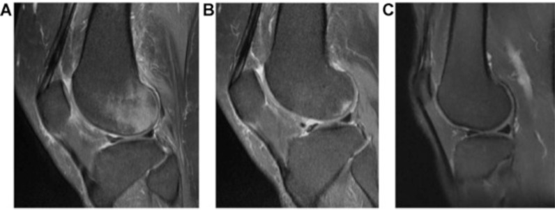
MRISuccessive sagittal MRIs of the knee: type I bone contusion, marrow hyperintensity visible on all three images after trauma.
Source : Mutlu et al., The open orthopaedics journal, 2015, figure 2 · CC BY-NC
It is the most widely used classification, and it has a rare quality: it was built with its follow-up. Costa-Paz and colleagues, in Buenos Aires, published in 2001 in Arthroscopy a cohort of 21 patients operated on for an isolated ACL rupture, all with a bone bruise on the preoperative MRI, with a second MRI obtained between 24 and 64 months after surgery (mean 34 months).18
The inclusion criteria are strict and make the cohort readable: isolated, acute ACL rupture, no new injury during follow-up, and no cartilage lesion found at arthroscopy. In other words, what is seen on the second MRI cannot be attributed to a pre-existing cartilage lesion.
| Type | MRI description | Outcome in the 2001 cohort | Implication |
|---|---|---|---|
| Type I | Diffuse marrow signal, often reticular, away from the underlying articular surface | Complete resolution of all lesions | Favourable |
| Type II | Localised signal, contiguous with the underlying articular surface | Resolution of 91 % of lesions (10 of 11) | Favourable in most cases |
| Type III | Break or depression of the normal contour of the cortical surface | Thinning and depression of the articular cartilage in every case, at 2 years of follow-up | Constant residual damage |
Across the cohort as a whole, 15 patients out of 21 (71 %) had resolution with no apparent residual change on MRI; in the other 6 (29 %), residual signs of the osteochondral lesion were visible. One point deserves emphasis because it is almost always left out when this study is cited: the authors find no correlation between the clinical scores of the patients whose lesions disappeared and those whose lesions left residual damage. The image evolves unfavourably without the patient complaining any more, a mismatch that will run through the whole of the rest of this article.
How these types are distributed in practice
A German cohort from 2023, covering 122 patients operated on for an isolated ACL rupture with no other associated injury, gives the most usable distribution: 18.9 % type I, 58.2 % type II and 14.8 % type III.9 The overall prevalence of bruising there was 91.8 %, and the mean total volume 21.84 ± 15.27 cm³ , with the lateral tibial plateau accounting on its own for 14.31 ± 9.93 cm³.
That volume figure is worth pausing on: more than 20 cm³ of oedematous bone is not a minor abnormality. It is a volume comparable to a golf ball, spread through the subchondral bone of a knee.
The problem nobody solves: four classifications for one object
A methodological limitation must be flagged here, one that weighs on the whole literature of the subject, and that the systematic review by Boks published in Radiology in 2006 documents unambiguously.17 Of 266 articles identified, only 13 met the inclusion criteria. Their quality is described as moderate. Populations were generally small and follow-up durations ranged from 1 to 73 months. Above all: four different classification systems were in use, and in two studies the bone bruise was not even defined.
This is why the published time frames vary so much from one paper to another: they are not all measuring the same thing, in the same patients, with the same stopping criteria. Any synthesis that presents “the” resolution time as an established figure over-reads the literature.
The Boks review nevertheless provides two robust observations, because they recur from one study to another. First, the clinical prognosis of bone bruises is generally good. Second, and this is the most useful result, the initial MRI appearance has prognostic value : normalisation of the image is possible and occurs most often after reticular-type lesions, whereas cartilage loss at follow-up is found mainly in the cases where cartilage damage was present from the outset, whether impaction or an osteochondral fracture.
- What predicts the outcome is not the volume of the oedema, but its position in relation to the articular surface and the integrity of the cortical contour, which is precisely what the Costa-Paz classification grades.1817
- A report that mentions a “depression of the cortical contour” is describing a type III: it is the only one of the three that calls for a surgical opinion and monitoring.
- The published time frames are not comparable with one another: four classification systems coexist, and two of the thirteen studies in the systematic review did not define the lesion.17
What does the bone oedema pattern tell us about the mechanism of injury?
This is the part of the subject with immediate clinical value: the map of bruises on a knee MRI makes it possible to reconstruct the position of the joint at the moment of the accident, and therefore to go looking for the injuries that go with it.
The pivot pattern: what an ACL rupture imprints in the bone
During a pivoting rupture of the anterior cruciate ligament, the tibia subluxes forwards and the lateral femoral condyle strikes the posterior part of the lateral tibial plateau. That collision leaves an imprint, and it is remarkably constant.
Two founding studies established it. Rosen, Jackson and Berger, in 1991, performed MRI within three weeks of rupture in 75 skeletally mature patients : occult bone lesions were documented in 64 of them, that is 85 %. Among those 64 patients, 83 % had lesions of the lateral compartment ; the lateral femoral condyle was involved in 50 % of cases and the lateral tibial plateau in 50 % as well, and 19 patients had more than one damaged area.6
Speer and colleagues, at Duke, pinned down the site the following year in 54 patients imaged within 45 days of a complete ACL rupture: 83 % (45 of 54) had a bone bruise sitting directly over the terminal sulcus of the lateral femoral condyle.7 The most intense signal was always contiguous with the subchondral plate. Above all, in 96 % (43 of 45) of the knees carrying this terminal sulcus lesion there was damage to the posterolateral compartment, combining soft-tissue lesions (the arcuate-popliteal complex) and bone lesions (the posterolateral tibial plateau).
It is this consistency that underpins the clinical use: the bruise is not an incidental piece of collateral damage, it is the signature of a mechanism, and it points to neighbouring structures that need examining.
Bone oedema patterns according to the mechanism of injury
Right knee, schematic anterior view. The intensity of the fill indicates the reported frequency of the site, not the severity of the lesion.
Sources by panel. 1 : Speer KP, Spritzer CE, Bassett FH 3rd, Feagin JA Jr, Garrett WE Jr. Am J Sports Med 1992;20(4):382-389 (PMID 1415878) and Rosen MA, Jackson DW, Berger PE. Arthroscopy 1991;7(1):45-51 (PMID 2009119). 2 : Ali AM, Pillai JK, Gulati V, Gibbons CER, Roberton BJ. Skeletal Radiol 2018;47(2):173-179, 25 patients (PMID 28856482). 3 : Dai R, Wu Y, Jiang Y et al. Bioengineering (Basel) 2023;10(12):1366, 77 ACL ruptures against 77 patellar dislocations (PMID 38135957) and Green DW, Hidalgo Perea S, Kelly AM, Potter HG. HSS J 2023;19(1):107-112 (PMID 36776513). 4 : Yao L, Lee JK. Radiology 1988;167(3):749-751 (PMID 3363134). Summary diagram, representation not anatomically to scale.
What the recent series correct: the medial compartment is not spared
The classical teaching, “the ACL bruise is lateral”, is true but incomplete, and the large contemporary series have qualified it sharply.
The Cleveland Clinic study, published in 2022 in the Orthopaedic Journal of Sports Medicine, covers 208 patients operated on for ACL injury, with MRI performed within 90 days (median: 12 days) and independent double reading according to Costa-Paz.8 The results shift the line:
Two further details give these figures their practical value. First, of the 164 medial tibial plateau bruises, 160 (98 %) involved the posterior third of the plateau, and 161 were grade 1, so discreet and easy not to mention. Second, the presence of a medial femoral condyle bruise was the only independent risk factor for a medial tibial plateau bruise (odds ratio 3.71).
The German cohort already cited gives a consistent hierarchy across its 122 patients: lateral tibial plateau 91.8 %, lateral femoral condyle 64.8 %, medial tibial plateau 49.2 %, medial femoral condyle 28.7 %.9
Frequency of bruise sites after ACL rupture, according to two cohorts
Two distinct populations: the gaps come from the inclusion criteria, not from a contradiction
Sources: Mester B, Kröpil P, Ohmann T, Schleich C, Güthoff C, Praetorius A, Dudda M, Schoepp C. Arch Orthop Trauma Surg 2023;143(10):6261-6272 (PMID 37269350); Byrd JM, Colak C, Yalcin S et al. Orthop J Sports Med 2022;10(10):23259671221120636 (PMID 36276425).
Hyperextension: an anterior pattern, and a sign that predicts the posterior cruciate
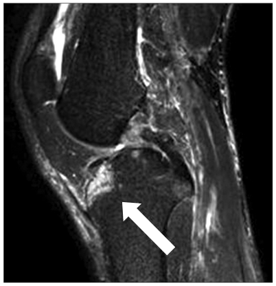
MRISagittal MRI of the knee: bone contusion (marrow oedema) of the lateral tibial plateau after a posterior cruciate ligament injury, arrow on the affected area.
Source : Lee et al., Knee surgery & related research, 2011, figure 3 · CC BY-NC
The hyperextension mechanism produces a different, more anterior imprint. The study by Ali and colleagues, covering 25 patients who had an MRI within a year of a hyperextension injury (median delay 24 days), gives bruise sites dominated by the anteromedial tibial plateau (48 %) and the anterolateral tibial plateau (44 %).10
The injury associations there are strong and directly usable:
- Oedema of the anterior tibial plateau and rupture of the posterior capsule predict a cruciate ligament injury, with respective odds ratios of 10.5 (p = 0.02) and 24.0 (p = 0.001).
- Oedema of the anterolateral tibial plateau strongly predicts an injury of the posterior cruciate ligament : OR 26.0, p = 0.003.
- The ACL injury, for its part, comes in this mechanism with a bruise pattern that is variable , so hyperextension does not produce the classical pivot signature.
- Meniscal injury bears no relation to the extent or the pattern of the bruises.
One radiographic detail is worth remembering, because it is available without MRI: in 5 of the 8 patients showing a “double sulcus” on the lateral film there was an ACL injury, and this sign was significantly associated with anteromedial “kissing” contusions (OR 7.8, p = 0.03).
Patellar dislocation: a pattern that must not be confused with that of the ACL
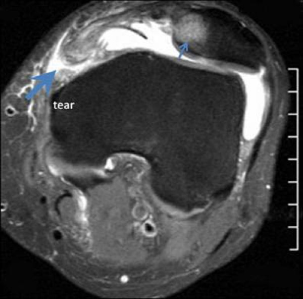
MRIAxial MRI of the knee: bone contusion of the medial patellar facet (small arrow) and tear of the medial patellofemoral ligament (large arrow) after patellar dislocation.
Source : Guerrero et al., Sports medicine, arthroscopy, rehabilitation, therapy & technology, 2009, figure 2 · CC BY
A Beijing team compared, in 2023, 77 ACL ruptures with 77 lateral patellar dislocations, all of them carrying bruises, mapping the sites in the sagittal and transverse planes.11 The authors identify four configurations after ACL rupture, with one, two, three or four bruises, the commonest being the one with three bruises, followed by the one with four. The patterns of the two conditions differ clearly, which confirms that the map of bruises does inform on the mechanism, and not simply on the intensity of the injury.
In children and adolescents, this reasoning carries even more weight. A study from the Hospital for Special Surgery, covering 62 MRI scans selected from 314 in patients aged 3 to 18, proposes six injury patterns, namely patellar dislocation, extensor mechanism overload, hyperextension, single-compartment impaction, ligament avulsion or translation, and direct contusion, with substantial inter-observer agreement (κ = 0.766).12 The commonest patterns were patellar dislocation (35 %) and extensor mechanism overload (22 %). Notably, the oedema signal was graded severe in 92 % of cases; the strength of the child's ligaments and tendons, greater than that of the epiphyseal bone, probably explains this high rate.
When the severity of the oedema informs on laxity
An Italian study from 2022 measured, in 29 patients operated on for ACL injury with MRI within three months of the injury, intraoperative rotational laxity using a surgical navigation system.13 The severity of the bruise of the medial tibial plateau was correlated with rotational laxity, and the presence of a bruise at this site was associated with a lateral femoral notch sign greater than 2 mm. An extensive bicompartmental bruise came with greater rotational laxity than an isolated extensive lateral bruise.
The cohort is small, 29 patients, and that limitation must accompany the reading. But the direction of the result is consistent with the mechanical logic: the more the impact has marked both compartments, the greater the displacement that was sustained.
- The bruise pattern is the mechanical trace of the injury: lateral femoral condyle and posterolateral tibial plateau for the ACL pivot (83 % in the Speer series), anterior tibial plateaus for hyperextension, medial patellar facet and lateral femoral condyle for patellar dislocation.71011
- “Lateral bruise only” is a dated shorthand: in a series of 208 patients, 79 % also had a medial tibial plateau bruise, almost always posterior and low grade.8
- In hyperextension, oedema of the anterolateral tibial plateau should prompt an active search for an injury of the posterior cruciate ligament (OR 26.0).10
- In children, patellar dislocation is the first pattern found (35 %), ahead of extensor mechanism overload.12
How long does a bone bruise take to disappear?
The question every patient asks, and the one the literature answers worst. Not because the data are missing, but because they are not all measuring the same thing, and because the most solid figure is far higher than the one announced in the consulting room.
The reference figure: a median of 42.1 weeks
The most useful study on this point was not carried out in a specialist surgical setting, but in general practice , which brings it closer to the population physiotherapists see. Boks and colleagues, in Rotterdam, prospectively followed patients with bone bruises after knee injury on MRI, continuing the scans until the lesion disappeared or up to one year.16
In 80 patients, 157 bruises were analysed. The main result is clear: the estimated median healing time is 42.1 weeks. That is a little over nine and a half months.
The determinants identified by survival analysis and logistic regression are few but consistent:
- Healing is lengthened in patients carrying a greater number of bruises, and in the presence of osteoarthritis.
- Resolution of individual lesions is delayed by osteoarthritis and by older age.
- The reticular lesions are less likely to persist at six months than the other types.
- None of the other variables tested, namely sex, obesity, occupational load and sporting load, has prognostic value.
This last point deserves emphasis because it contradicts a widespread intuition: neither weight nor the patient's level of occupational or sporting activity predicted the speed of healing in this cohort.
Why the published figures range from three weeks to two years
The scatter is real and it should be set out rather than hidden behind an average. It has three distinct causes, and mixing them produces exactly the contradictions one reads everywhere.
| Source | Population | What is measured | Result |
|---|---|---|---|
| Yao 19881 | 8 injured knees, normal radiographs | Disappearance on MRI, 2 patients rescanned | Complete resolution at 6 weeks and 3 months |
| Boks 200716 | 80 general practice patients, 157 bruises | Disappearance on MRI (follow-up to 1 year) | Median 42.1 weeks |
| Boks 200617 | Systematic review, 13 studies retained | Heterogeneous follow-up, 4 classifications | Follow-up from 1 to 73 months ; clinical prognosis good overall |
| Costa-Paz 200118 | 21 reconstructed ACLs, no cartilage lesion | Persistence on MRI at 24-64 months | Type I: 100 % resolved · Type II: 91 % · Type III: constant residual damage |
| Stirling 202519 | 100 ACL ruptures, MRI within 6 weeks | Automated volumetry at 1 year | 92.68 % of lesions resolved, volume reduction of 96.13 % (p < 0.001) |
| Kim 201920 | 76 patients, 102 sites, foot and ankle | Duration of PAIN, not of the image | Type I 12.15 ± 2.17 wks · Type II 14.5 ± 2.15 · Type III 21.0 ± 3.8 |
The three causes of scatter appear clearly in this table.
First cause: we are not measuring the same thing. Boks measures the disappearance of the image, Kim measures the disappearance of the pain. These are two different curves, and the second is markedly faster than the first. This matters greatly in practice: a patient can be asymptomatic while the MRI remains abnormal, and the reverse is equally true.
Second cause: the population differs. A general practice cohort (Boks) includes older patients, sometimes with osteoarthritis, two factors identified as lengthening healing. A cohort of young athletes operated on for ACL injury (Stirling, aged 14 to 55) recovers faster.
Third cause: the stopping criterion. A study that ends follow-up at one year cannot report a time frame longer than one year. That is a design constraint, not a biological fact.
Two distinct curves: pain and image do not disappear together
Benchmarks from different studies, placed on a common time scale; this is not a follow-up of the same cohort
Sources: Kim HJ, Lee KB. J Korean Foot Ankle Soc 2019;23(4):183-188, 76 patients and 102 bruise sites (DOI 10.14193/jkfas.2019.23.4.183); Boks SS et al. AJR Am J Roentgenol 2007;189(3):556-562 (PMID 17715100). These two series concern different joints and different populations : the timeline sets them side by side to show the gap between two measurement criteria, it does not describe the course of a single patient.
The most recent data, and the most reassuring
A Canadian cohort published in 2025 in the Journal of Orthopaedic Research provides the most modern measurement: 100 participants aged 14 to 55, all with an ACL rupture confirmed on MRI, imaged within six weeks (mean 30 days), with lesion volumes quantified by a validated machine learning method.19
Bone marrow lesions were present in 95 % of the participants, mainly at the lateral femoral condyle and the lateral tibial plateau. Men had volumes greater by 33 % than those of women, including after adjustment for body mass index, and volumes were higher where there was an associated depression fracture.
At one year, 92.68 % of the lesions had resolved in the non-operated participants, with a volume reduction of 96.13 %. KOOS scores were comparable between the operated and non-operated groups, apart from slightly better pain scores in the non-operated group.
Two lessons follow. The first is frankly reassuring: the vast majority of bruises disappear within a year, whatever the treatment of the ligament. The second is subtler: the authors conclude that bone marrow lesions are only modestly linked to early symptoms; oedema volume was negatively associated with the initial KOOS Symptoms score, but the follow-up scores were mainly predicted by the baseline pain and sport scores, not by the imaging.
- The median time to disappearance on MRI is 42.1 weeks in general practice, and announcing six weeks invites a pointless catch-up consultation.16
- The pain disappears well before the image : 12 to 21 weeks depending on the type in Kim's foot and ankle series, against more than nine months for MRI.20
- In young patients, the one-year prognosis is excellent: 92.68 % of lesions resolved, with or without ligament surgery.19
- What lengthens healing: the number of bruises, pre-existing osteoarthritis , and age. Neither obesity nor occupational load nor sporting load had prognostic value.16
- Do not repeat the MRI in order to decide on return to activity : an image that is still abnormal in a pain-free patient is the expected situation, not a contraindication.
How can a bone bruise be distinguished from what resembles it?
High marrow signal is a sign shared by diseases whose management has nothing in common. This chapter deals first with the costliest confusion: the one with stress fracture.
Bone bruise and stress fracture: the same image, opposite mechanisms
This is the most important distinction in this article, and it is not settled on imaging. Both entities produce bone marrow oedema on the same sequences; Yao and Lee were already drawing the two together in 1988.1 What separates them is the mechanism, and therefore the management.
| Criterion | Bone bruise | Stress fracture (fatigue fracture) |
|---|---|---|
| Mechanism | A single injury, datable, often violent: pivot, hyperextension, direct blow, dislocation | Repeated overload : cyclical stresses exceeding the adaptive capacity of the bone, with no identifiable injury |
| The patient's account | “I can tell you the day and the movement” | “It came on gradually, as I increased my training” |
| Background | Anyone, including sedentary people; depends on the accident | Endurance athlete, military personnel, abrupt return to training, relative energy deficiency |
| Topography | Subchondral, mapped onto the geometry of the joint impact | Known stress sites: femoral neck, tibia, navicular, metatarsals, sesamoids |
| What makes it worse | Time works in favour of healing if load is adapted | Continued training drives the lesion on towards a complete fracture |
| What is at stake if it is missed | Underestimating the duration, returning too early, causing needless worry | Risk of complete fracture, particularly at high-risk sites |
| Management | Adapt load to the symptom, with no routine follow-up imaging | Offloading and stratification by risk site; aetiological work-up (energy, bone density) |
The practical rule fits in one sentence: faced with bone marrow oedema, the diagnosis is made on the history, not on MRI. A single, datable injury points to a bruise; a gradual onset against a background of increasing load points to a bone stress injury, and calls for a different approach, the one detailed in our article on stress fracture in the athlete, with its stratification by high-risk and low-risk site and its Fredericson MRI classification.
- The opposite trap exists and it is more dangerous: taking for a bone bruise what is in fact a stress injury. A patient who reports a “minor injury” occurring during a period of unusual load may have both, and it is the logic of the stress fracture that must take precedence, because that is the one that can go on to complicate.
The other causes of bone marrow oedema
The AAOS review is a reminder: bone marrow oedema is a descriptive term compatible with many conditions.4 The traumatic context is what allows a decision, and it must be proportionate.
| Condition | What suggests it | What sets it apart |
|---|---|---|
| Subchondral insufficiency fracture (formerly “spontaneous osteonecrosis of the knee”) | Older, often osteoporotic patient, acute knee pain with no injury or after a minimal injury | The patient background and the absence of a proportionate injury. The nomenclature has changed: recent histological data dispute the term osteonecrosis.35 Frequently associated with a meniscal root tear |
| Transient bone marrow oedema syndrome / transient osteoporosis | Hip or knee pain of subacute onset, with no injury; pregnancy, middle-aged patient | Spontaneously resolving over several months; the oedema is diffuse and not mapped onto an impact geometry |
| Osteochondritis dissecans | Adolescent or young adult, mechanical pain, sometimes locking | An individualised osteochondral fragment. May follow a bruise: see the clinical cases below31 |
| Complex regional pain syndrome | Disproportionate pain, vasomotor, sudomotor and trophic changes | The diagnosis is clinical (Budapest criteria). MRI may show early bone marrow oedema but does not contribute to the diagnosis: see our article on CRPS |
| Osteoarthritis and degenerative bone marrow lesions | Older patient, chronic pain, joint space narrowing on radiographs | Non-traumatic context, associated remodelling. See knee osteoarthritis |
| Inflammatory arthritis, spondyloarthritis | Inflammatory pain, waking in the second half of the night, prolonged morning stiffness | Patient background, blood tests, axial or multiple peripheral involvement |
| Bone infection, tumour | Constant, non-mechanical pain, deterioration in general condition | Red flags: cross-sectional imaging and specialist opinion without delay |
| Muscle contusion (dead leg) | Direct blow to a muscle, haematoma, loss of range | A lesion of the soft tissues, and the bone may be undamaged. Covered in the dedicated article : the two can coexist after the same blow |
A decision tree for persistent post-traumatic pain
What to do when pain persists after an injury with a normal radiograph
The tree presupposes that red flags have already been ruled out
Summary tree built from the data in this article: Boks 2006 on the prognostic value of the initial appearance (PMID 16452394), Sohn 2024 on the frequency of associated injuries (PMID 38790382) and Karimi 2023 on the frequency of asymptomatic oedema (PMID 37263276). An educational proposal and not a formal guideline: no learned society has published an algorithm on this subject.
The bruise is rarely alone
This is why an MRI showing an isolated bruise should be read again carefully. The most complete meta-analysis on the subject, published in 2024, brought together 22 studies and 2,891 patients with an ACL rupture.22 Its results are mixed, and it is that contrast which makes them useful:
What the presence of a bone bruise predicts, and what it does not
Relative risks from a meta-analysis of 22 studies and 2,891 patients with an ACL rupture
Source: Sohn S, AlShammari SM, Lee JH, Kim MS. Bone Bruises and Concomitant Meniscus and Cartilage Damage in Anterior Cruciate Ligament Injuries: A Systematic Review and Meta-Analysis. Bioengineering (Basel) 2024;11(5):515. PMID 38790382. These relative risks describe lesions found concomitantly at the time of the injury, not a later evolution.
The authors conclude that the bruises sit most often in the lateral compartment and that they are linked to associated lesions of the lateral meniscus and cartilage. The absence of a link with the medial meniscus is just as instructive: it is a reminder that the bruise marks an impact geometry, and that the structures it points to are those that lay on the trajectory.
- Bruise and stress fracture have the same image and opposite mechanisms : it is the history that decides, never MRI alone.
- Faced with bone marrow oedema with no proportionate injury, think of subchondral insufficiency fracture in the older patient, transient oedema syndrome in the middle-aged adult, and rule out infection and tumour.
- A bruise after ACL rupture multiplies by 6.18 the probability of an associated cartilage lesion and by 2.71 that of a lateral meniscus tear, but it does not predict medial meniscus damage.22
Does a bone bruise really herald osteoarthritis?
This is the most widespread claim on the subject, and the least solid. It deserves to be examined piece by piece, because what is said to the patient depends on it directly, and because unwarranted anxiety carries a real clinical cost.
The common claim, and where it comes from
One regularly reads that the bone bruise is a predictor of post-traumatic osteoarthritis. The idea is attractive: a lesion of the subchondral bone, an overlying cartilage impacted by the same mechanism, and a joint that degenerates fifteen years later. The reasoning holds together. The problem is that it has not been demonstrated, and that the most rigorous work points rather the other way.
Three questions that the literature very often runs together must first be separated:
| Question | What the data say | Strength |
|---|---|---|
| 1. Is the bruise associated with cartilage lesions at the time of injury? | Yes, clearly: RR 6.18 in a meta-analysis of 22 studies and 2,891 patients22 | Established |
| 2. Does a type III bruise leave visible residual cartilage damage? | Yes in the only cohort that followed it up: all type III lesions at 2 years, in a sample of 21 patients18 | Single cohort, small sample |
| 3. Does the bruise itself cause osteoarthritis, or predict its occurrence? | Not demonstrated. Volume and severity predict neither the outcomes at 2 years nor those at 6 years23 | Not established |
The study that cools the hypothesis
The most direct work on the third question was published in Cartilage in 2017.23 In 81 patients, the volume and severity of the bruise were measured on the preoperative MRI in the four regions of the knee, using the Costa-Paz classification and a modified version of the Roemer and Bohndorf technique. KOOS and IKDC scores were collected before surgery, then at 2 years and at 6 years.
All 81 patients had a bruise in at least one region, and 70 of them (86 %) had one in at least two. The distribution confirms the earlier series: lateral tibial plateau 94 %, lateral femoral condyle 81 %, medial tibial plateau 57 %, medial femoral condyle 25 %.
The main result is unambiguous: neither the volume nor the severity of the bruise was associated with poorer postoperative outcomes, at 2 years or at 6 years.
Only one association emerged, and it is instructive: the subgroup of 17 patients presenting both a bone bruise and an associated local cartilage lesion had 3.4 times the risk of being symptomatic at 6 years compared with those without cartilage damage (p = 0.04).
What the largest systematic review says, exactly
The systematic review by Filardo and colleagues, published in KSSTA in 2019, is the broadest synthesis in the field: 415 references identified, 83 articles analysed, 10,047 patients.21 It is frequently cited as establishing the link with osteoarthritis. It is worth going back to the exact wording used by its authors.
They note that the bruise has a high prevalence, 78 % in the most recent publications , with distinct patterns according to mechanism. Then, on prognosis, they write that the presence and persistence of the bruise “have been correlated with more severe joint damage that may affect the degenerative progression of the whole joint, with recent data suggesting possible effects on long-term clinical outcome”.
“Correlated”, “may affect”, “suggesting”, “possible”: four markers of uncertainty in a single sentence, from the pens of authors who have read 83 articles. That is the real level of certainty in this field, and reporting it otherwise is over-reading.
The same authors go on to conclude that long-term prospective studies are needed to gain a better understanding of the natural history of the bone bruise, and to identify prognostic factors and therapeutic targets.
The confounder that is systematically forgotten
Here is the point that, on its own, explains why question 3 remains open.
The bone bruise is a marker of the energy of the injury. The more violent the impact, the more extensive the oedema, and the more the other structures have been damaged: cruciate ligament, menisci, cartilage, capsule. And those are precisely the lesions known to lead to post-traumatic osteoarthritis.
Observing that knees with an extensive bruise develop more osteoarthritis therefore does not say that the bruise is the cause. It may simply mean that they sustained a more severe injury. Separating the two would require comparing knees with an equivalent injury differing only in the bruise, which no study has done.
Two series support this reading. The German cohort of 122 patients shows that functional scores, quality of life and isokinetic strength improve significantly between the preoperative assessment and one year, independently of the distribution and volume of the bruises.9 The Canadian cohort of 2025, for its part, concludes that bone marrow lesions are “ modestly linked to early symptoms” and that long-term differences are “ limited ” according to surgical status.19
What remains true, and must not be thrown out with the rest
Refusing over-interpretation does not mean denying any link at all. Three facts hold.
Firstly, the Costa-Paz type III, the one that breaks or depresses the cortical contour, left residual cartilage damage in every one of the patients concerned at two years of follow-up, in a cohort that had nevertheless been selected for the absence of initial arthroscopic cartilage damage.18 The sample is small, but the result is consistent with the mechanism.
Secondly, the Boks systematic review establishes that cartilage loss at follow-up occurs mainly where cartilage damage was present from the outset, whether impaction or an osteochondral fracture.17 That is the same message as Lattermann's, formulated fifteen years earlier.
Thirdly, a Japanese cross-sectional study of 161 patients with knee pain, excluding trauma and excluding arthritis, shows that the presence of a bone bruise is associated with femorotibial osteoarthritis and with joint effusion, with a positive correlation between the bruise grade and the size of the effusion.24 But this result has to be read for what it is: a cross-sectional study, which photographs an association at a given moment and cannot establish any chronology. In an osteoarthritic population, moreover, bone marrow oedema is more likely to be a consequence of mechanical overload than a cause.
- What can be said to the patient: “your bruise is going to heal, and in the available cohorts its extent did not predict your outcomes at 2 or at 6 years”.23
- What must not be said: “you will get osteoarthritis because of this bruise”. No data support it, and the anxiety it creates carries a real cost for return to activity.
- What has to be monitored: not the bruise, but the associated cartilage lesion , which is what multiplies by 3.4 the risk of being symptomatic at 6 years, and which is what justifies follow-up.23
- The only exception: the type III, with a break or depression of the cortical contour, which calls for monitoring and a specialist opinion.18
What is to be done about weight-bearing, and on what evidence?
This is the question every patient and every physiotherapist asks, and it is the one with no validated answer. This chapter sets out what the literature supports, what it does not support, and why the void is here the main piece of information.
What exists, and what does not
Before setting out the modalities, the state of the evidence has to be framed precisely, because this is where the firmest overstatements are read, in both directions.
What does not exist : no randomised trial on return to activity after a bone bruise, no learned society guideline, no formal consensus on return criteria. A PubMed search crossing bone bruise and return to sport in the titles returns no result, and there is no randomised trial on the bone bruise itself either.
What does exist, and what it would be dishonest to pass over in silence, is a retrospective series with a grading system dedicated to return to play, published in Skeletal Radiology in 2024.36 It is small and covers a very particular population, but it is directly on the subject, and its main result is striking.
The only series that puts a figure on return to play, and what it finds
Belair and colleagues, at Thomas Jefferson University, retrospectively reviewed the traumatic lower limb bone lesions occurring over ten years in professional ice hockey players: 28 lesions in 17 elite athletes, with an MRI obtained on average within two days of the injury.36
They propose a grading of bone bruises at three levels, mild (grade 1), moderate (grade 2) and severe (grade 3), with grade 3 subdivided according to the presence or absence of a cortical fracture. The mean times to return observed:
Mean time to return to play by severity, in 17 professional ice hockey players
Retrospective series of 28 lesions over ten years, MRI about 2 days after the injury
Source: Belair JA, Jung J, Desai V, Morrison WB, DeLuca PF, Zoga AC. Bone bruise vs. non-displaced fracture on MRI: a novel grading system for predicting return-to-play. Skeletal Radiol 2024;53(5):947-955. PMID 37993556. Retrospective series of 28 lesions in 17 professional ice hockey players : these times in DAYS belong to professional sport, with its resources and its pressure to return. They do not transfer as they stand to a private practice caseload, and they say nothing about the healing of the bone; the image itself takes months.
Two lessons, not to be confused with one another.
The first is solid and directly useful: a severe bruise costs as much time as an undisplaced fracture. The absence of a fracture line on MRI is therefore not a reason for reassurance about the time frame. It is the exact mirror of the message of the chapter on radiography.
The same authors developed this reasoning in a review published the same year, which runs through the spectrum of acute bone injuries in the athlete, from bruise to undisplaced fracture, and insists on one practical point: describing the injury pattern precisely on MRI serves first of all to communicate with the medical staff, because it is that description which will carry the decision on return to play.37
The second lesson is a warning about scope: these times are counted in days because these are professional ice hockey players, in whom return to play is decided on pain and function, with daily supervision, and not on imaging, which stays abnormal far longer. The contrast between 2.8 to 21.4 days to return to play and a median of 42.1 weeks to disappearance on MRI16 is the clearest demonstration in the whole of this article: return to play is not decided on the image.
The fact remains that any protocol presented as validated, whether “three weeks off load then gradual return” or “return when the oedema disappears”, is still an extrapolation. It may be reasonable; it must be presented as such.
The reference document is a level V expert opinion
The most recent and most authoritative synthesis on the conservative treatment of bone marrow lesions of the knee was published in 2025 in the Journal of Experimental Orthopaedics, the ESSKA journal, coordinated by Andriolo and Filardo, with contributions from international experts.25
Its scope is broader than the bruise alone: it covers bone marrow lesions whatever their origin, namely traumatic bruises and fractures, the aftermath of cartilage surgery, osteoarthritis, transient oedema syndromes, subchondral insufficiency fractures and spontaneous osteonecrosis. Its conclusions deserve to be reported as they stand:
- Some positive results have been documented for unloading braces, for extracorporeal shockwave therapy, hyperbaric oxygen therapy, for pulsed electromagnetic fields and for bisphosphonates.
- But the analysis of the literature documents a limited number of publications dealing specifically with the knee, with even less data once the different causes of bone marrow lesion are separated.
- The open questions concern the duration of treatment, the stage of the lesion and the overlap with concomitant therapies.
- The level of evidence of the article is explicitly stated: level V, expert opinion.
In other words: the modalities listed are not wrong, but the document that brings them together is, by its own authors' admission, the lowest level of evidence in the hierarchy.
Modalities and level of evidence
Level of evidence by modality, following GRADE logic
Stacked cards, from the most solid evidence to the weakest. The grading covers bone marrow lesions of the knee, all causes combined, for want of data specific to the isolated post-traumatic bruise.
Sources by line. Load and information : Boks 2007 (PMID 17715100), Stirling 2025 (PMID 40974550), Kim 2019 (DOI 10.14193/jkfas.2019.23.4.183). Shockwave therapy : Gao F et al. BMC Musculoskelet Disord 2015;16:379 (PMID 26637992), Sansone V, Romeo P, Lavanga V. Med Princ Pract 2017;26(1):23-29 (PMID 27784022). Braces, PEMF, oxygen therapy : Andriolo L et al. J Exp Orthop 2025;12(2):e70151, level V (PMID 40191034). Iloprost : Zippelius T et al. J Pers Med 2022;12(11):1757 (PMID 36573724). Bisphosphonates : Anzillotti G et al. J Clin Med 2024;13(13):3799 (PMID 38999364). Subchondroplasty : Klincke V et al. Knee 2026;61:104441 (PMID 41950892). The grading expresses the judgement of the authors of this synthesis in the light of the study designs and the indirectness of the populations; it comes from no formal guideline, none having been published on this subject.
| Modality | What was measured | Population studied | Level |
|---|---|---|---|
| Load adapted to the symptom | No dedicated trial. Indirect convergence: 92.68 % resolution at 1 year in the non-operated group19 | ACL ruptures, aged 14 to 55 | Moderate (indirect) |
| Grading of return to play | Retrospective series, 28 lesions: 2.8 / 4.5 / 18.3 / 21.4 days by grade; severe bruise vs undisplaced fracture p = 0.32736 | 17 professional ice hockey players , a very particular population | Low (retrospective, small n, indirect population) |
| Information on the time frame | Median 42.1 weeks for the image16 ; 12 to 21 weeks for pain20 | General practice; foot and ankle | Moderate |
| Shockwave therapy | Randomised trial, n = 40: better change in VAS, WOMAC and SF-36 at 1, 3 and 6 months; MRI regression 95 % against 65 % at 6 months (p = 0.018)27 | PRIMARY bone marrow oedema syndrome of the knee, not post-traumatic | Low (indirect population) |
| Shockwave therapy (continued) | Comparative study, non-randomised, n = 86: reduction in oedema area of 86 % against 41 %28 | Oedema of the medial compartment of the knee | Low (no randomisation) |
| Unloading brace, PEMF, hyperbaric oxygen therapy | Positive results reported, with no pooled quantification25 | Bone marrow lesions of the knee, causes mixed | Very low (level V) |
| Iloprost | Review and meta-analysis29 | Proximal femur only | Very low for the knee |
| Bisphosphonates | Meta-analysis of 15 studies including 7 randomised trials: neither improvement in clinical scores nor reduction in size against placebo; adverse effects not significantly more frequent26 | Bone marrow lesions of the knee | Evidence of absence of effect |
| Subchondroplasty | Meta-analysis of 12 studies, 829 patients: improvement in pain and function, but 22.4 % arthroplasty at 2 years and 6.7 % complications30 | Knee osteoarthritis , a different indication | Outside the indication here |
Should the limb be offloaded? What can reasonably be argued
The question of offloading arises mainly for type III lesions, and by analogy with subchondral insufficiency fracture, where early offloading is classically recommended to prevent progression, a position defended in particular in a case reported in 2024, which insists on the importance of early diagnosis and of an early period of non-weight-bearing.34
But we have to be clear about the nature of that argument: it is an analogy with another condition, resting on a single case report. It is not evidence applicable to the post-traumatic bruise of the young patient, which we otherwise know heals in more than nine cases out of ten within a year with no particular treatment.19
A defensible position, presented as such, fits in three lines:
- Types I and II with no associated injury : pain-guided weight-bearing, with no routine offloading. Nothing justifies immobilising a lesion whose spontaneous prognosis is excellent.
- Type III, or depression of the cortical contour : specialist opinion before deciding on load. It is the only type for which residual cartilage damage has been found consistently.18
- Associated ligament or meniscal injury : it is that injury which dictates the loading protocol, not the bruise. The bruise does not change the conduct of postoperative rehabilitation of the anterior cruciate ligament or that of a knee sprain.
What the physiotherapist actually has to do
The absence of a validated specific treatment does not mean the absence of care. It shifts the work towards what is demonstrated or reasonably grounded.
- Reframe the complaint. A patient who is in pain three months after an injury with a normal radiograph has often been left to their own devices. Naming the lesion, explaining why the radiograph did not show it, and giving the real order of magnitude of the time frames is a therapeutic act in itself.
- Steer load by the symptom, not by the image. Since pain settles well before the MRI does, waiting for the imaging to normalise before returning to activity would mean immobilising asymptomatic patients for months.
- Treat the associated injuries. That is where most of the functional prognosis lies: ligament, meniscus, cartilage.
- Maintain everything that does not depend on loading the injured joint. Strength of the contralateral limb, aerobic capacity, non-weight-bearing work: the paediatric case cited below, where the child resumed activity through swimming and pool walking, illustrates this logic well.32
- Watch the trajectory. Pain that worsens as time passes is not a bruise doing badly: it is a diagnosis to be reopened.
- No randomised trial and no formal consensus on return to activity ; the only series that puts a figure on it is retrospective, in 17 professional ice hockey players.36 Any protocol presented as validated remains an extrapolation.
- A severe bruise costs as much time as an undisplaced fracture (18.3 against 21.4 days, p = 0.327): the absence of a fracture line is no reassurance about the time frame.36
- The most authoritative summary document on conservative treatment is explicitly rated level V, expert opinion, and itself stresses how few publications there are.25
- Bisphosphonates do no better than placebo, neither on clinical scores nor on lesion size: it is the most solid piece of data in this chapter, and it is negative.26
- Shockwave therapy has the best available results, but on primary oedema syndromes, not on post-traumatic bruises.2728
- Steer return to activity by pain and function, never by a follow-up MRI.
What do published clinical cases teach us?
Four real, published and referenced observations. Each illustrates a point that the cohorts average out: the lived time frame, the rare complication, the non-sporting patient and the logic of offloading.
An 11-year-old boy, a normal radiograph, six months on crutches
This case, published in Cureus in 2026, is the most faithful description of the typical pathway.32
An 11-year-old boy, with no notable history, fell while playing and injured his right foot. A week later, the pain persisted and walking remained difficult; he consulted a trauma specialist. The plain radiograph showed no abnormality and the injury was managed as a sprain.
With symptoms persisting after two weeks, an MRI was performed: it revealed bone marrow oedema of the right foot. Treatment combined anti-inflammatory drugs, complete offloading and physiotherapy including magnetotherapy. The child used crutches for six months. The follow-up MRI at five months showed resolution of the oedema.
The rest of the pathway is of particular interest for rehabilitation: the child first went back to swimming and pool walking, which allowed him gradually to overcome his fear of weight-bearing, before moving on to walking with a single crutch and then without any aid.
The authors conclude that a high index of clinical suspicion matters when pain persists and walking remains uncomfortable after an injury, and that MRI is the investigation of choice to confirm the diagnosis. They themselves add the necessary caveat: as this is a single case, the course observed may reflect the natural history rather than the effect of treatment.
- This case contains the four stages of the typical pathway: a normal radiograph → the sprain label → persistence → the MRI that settles it.
- The fear of weight-bearing emerges as an obstacle in its own right, treated by returning to activity in water. It is a rehabilitation lever that transfers directly.
Two adolescents in whom the bruise left an osteochondritis dissecans
This case series published in KSSTA in 2013 documents the unfavourable course that is feared.31 Shea and colleagues report two skeletally immature boys who developed a symptomatic osteochondritis dissecans lesion following a bone bruise of a femoral condyle.
Osteochondritis dissecans is regarded as an acquired osteochondral disorder of probably multifactorial origin, trauma, whether acute or repetitive, having long been proposed as a possible cause. These two observations suggest that an acute traumatic event can lead to the development of an osteochondritis dissecans of the knee.
Two reservations must accompany this reading, and the authors do not hide them: these are two cases, and a case series does not allow a frequency to be estimated. What this publication establishes is that the course is possible , not that it is frequent. It justifies vigilance in the child and the adolescent, in whom the epiphyseal bone is the weak link, not an alarmist message in the adult.
A 33-year-old woman postpartum: when the patient background changes everything
This case, published in the World Journal of Emergency Medicine in 2024, is a reminder that the post-traumatic bone bruise is not the only reading of a bone marrow oedema, even after a proven injury.33
A 33-year-old woman sprained her ankle in May 2020, with a documented lesion of the anterior talofibular ligament on imaging in August 2020. She gave birth by caesarean section in October 2020, and her ankle pain then worsened. An MRI performed in November 2020, about a month after delivery, revealed extensive bone marrow oedema of the distal tibia and the talus.
The work-up found raised alkaline phosphatase and low vitamin D. Treatment combined calcitonin and calcium supplementation. Improvement was gradual, with a marked reduction in symptoms in July 2021, and a follow-up MRI in March 2023 confirmed complete recovery, that is about two and a half years after the initial worsening.
The lesson is twofold. First, a recent injury does not preclude looking for a metabolic factor when the course is atypical in its duration or its intensity. Second, this case illustrates the long tail of the distribution of time frames, the one that cohorts stopped at a year cannot see.
A subchondral insufficiency fracture treated with early offloading
This case, published in Radiology Case Reports in 2024, belongs to the differential diagnosis but sheds direct light on the question of load.34
The authors report a case of subchondral insufficiency fracture of the medial femoral condyle at an early stage. They recall that the term “spontaneous osteonecrosis of the knee” has been replaced by that of subchondral insufficiency fracture in the recent pathological and radiological literature, a change of nomenclature corroborated by a 2025 scoping review on the histopathological assessment of this entity.35
They stress that the plain radiograph only allows the diagnosis to be confirmed at late stages, MRI often being necessary, and they insist on the importance of early diagnosis and of an early period of non-weight-bearing to prevent progression of the disease. When it does progress, surgery often becomes necessary.
It is from this reasoning, and from it alone, that the idea of offloading a bone oedema comes. It is solid in that condition, in that setting. Transferring it as it stands to the post-traumatic bruise of the young patient amounts to borrowing the conclusion of one disease in order to treat another.
- The typical pathway is documented: a normal radiograph, the sprain label, persistence, then the MRI that settles it, sometimes several weeks later.32
- Progression to an osteochondritis dissecans is possible in the skeletally immature patient: two published cases, not a frequency.31
- An abnormally long course justifies looking for a metabolic factor , vitamin D and alkaline phosphatase, even after a genuine injury.33
- The offloading argument comes from the subchondral insufficiency fracture, a different disease in a different patient, and the borrowing must be declared.34
What difference does the site make, away from the knee?
The knee concentrates the literature, but the bone bruise is not confined to it. Two sites are worth knowing because they change management: the ankle and the growing skeleton.
The ankle: one sprain in six hides a bruise
The series by Pinar and colleagues, published in KSSTA in 1997, remains the reference.14 Sixty consecutive patients with a lateral ankle sprain had an MRI, supplemented by magnetic resonance arthrography after gadolinium injection. All presented within three weeks of the sprain and, a crucial point, the plain radiographs showed no bone abnormality.
Eleven bone bruises were detected across ten ankles. Eight of them lay in the talus. One ankle with complete ruptures of the anterior talofibular ligament and the calcaneofibular ligament had two lesions, in the talus and the navicular; another, also with complete ruptures of both ligaments, had a lesion of the calcaneus.
Two gradients emerge from this series and are directly usable:
The clinical reading is simple: the more severe the sprain and the more often it is repeated, the higher the probability of associated bone damage. In a patient whose lateral ankle sprain drags on beyond the usual time frames, especially where it is a recurrence with damage to both ligaments, a bruise of the talar dome is one of the hypotheses to be formulated.
An imaging essay published in Insights into Imaging in 2020 completes the picture by detailing the traumatic and non-traumatic aspects of bone marrow oedema at the ankle, a useful read for interpreting reports from this region, where non-traumatic causes are numerous.15
- At the ankle, the bone bruise is present in 16 % of isolated anterior talofibular lesions and 50 % of two-ligament lesions, and it sits mainly in the talus.14
- It has been discussed as a possible precursor of an osteochondral lesion of the talus , a further reason not to treat a sprain that fails to heal as trivial.
The growing skeleton: the bone gives way before the ligament
In children and adolescents, the balance of strength is reversed. The study from the Hospital for Special Surgery already cited puts it clearly: the strength of the child's ligaments and tendons, relative to that of the epiphyseal bone, probably contributes to the high rate of bone marrow oedema patterns seen on MRI in symptomatic paediatric patients.12
Across the 62 MRI scans analysed, 40 boys and 22 girls with a mean age of 12.2 years, the oedema signal was graded severe in 92 % of cases on fat-saturated sequences. The commonest patterns were patellar dislocation (35 %) and extensor mechanism overload (22 %).
Three practical consequences follow in the child. “Severe” oedema is the rule there and must not be over-read as a marker of severity. Patellar dislocation should be considered first when faced with a knee oedema pattern in a child, even before the ligament pivot. And monitoring for a possible osteochondritis dissecans is legitimate in the skeletally immature patient.31
How is this applied concretely in practice?
What is done, in order, when a patient presents with pain that persists after an injury and imaging that has shown nothing.
Management in six stages
| Stage | What is done | What is avoided |
|---|---|---|
| 1. Take the history again | Date the injury, reconstruct the movement, specify the mechanism. A single, datable injury points to a bruise; a gradually increasing load points to a stress injury | Concluding on imaging alone |
| 2. Rule out red flags | Non-mechanical pain, constant night pain, deterioration in general condition, injury disproportionate to the patient | Starting rehabilitation before ruling out infection, tumour and insufficiency fracture |
| 3. Look for associated injuries | Ligament and meniscal tests, looking for effusion, locking and instability. They weigh far more on the prognosis than the bruise itself23 | Treating the bruise as an isolated lesion without having checked |
| 4. Set the indication for MRI | When pain persists beyond six weeks, or straight away where there are signs of an associated injury | Ordering an MRI to “see where the oedema has got to” in a patient who is getting better |
| 5. Explain the real time frames | Pain: several months depending on the type. Image: a median of 42.1 weeks. The gap between the two is expected1620 | Announcing “six weeks” and setting up a consultation full of worry |
| 6. Steer load by the symptom | Progression guided by pain and function, maintenance of everything that does not load the lesion, work in water if fear of loading dominates32 | Waiting for the MRI to normalise before allowing a return to activity |
What is said to the patient, word for word
What is said matters particularly here, because the patient often arrives feeling that they have not been believed. Four formulations, each backed by a piece of data from this article:
- On the radiograph: “Your radiograph was normal, and that is expected: it shows the outer shell of the bone, which is intact. The lesion is inside, in the cancellous part.”
- On the nature of the lesion: “This is not nothing. The trabeculae that form the internal framework of the bone were crushed by the impact. It is a real lesion, it is simply invisible on the X-ray.”
- On the time frame: “The pain often takes three to five months to disappear, and the image takes longer still. That gap is normal: you will be well before your MRI has gone back to normal.”
- On osteoarthritis: “In the follow-up studies, the extent of this type of lesion did not predict the state of the knee at two years or at six years. What we monitor is not the bruise, it is the ligament and cartilage lesions that sometimes go with it.”23
The five costliest mistakes
| Mistake | Consequence | What to do instead |
|---|---|---|
| Concluding “nothing on the X-ray, so nothing serious” | Patient sent away with no explanation, pain put down to exaggeration | Acknowledge the limits of the investigation and state the hypothesis |
| Announcing a time frame of a few weeks | Loss of trust at two months, catch-up consultations | Give the real, measured order of magnitude16 |
| Waiting for a normal MRI before allowing a return to activity | Prolonged immobilisation of an asymptomatic patient | Steer by pain and function |
| Treating oedema seen on an MRI with no clinical hypothesis | Treatment of a possibly asymptomatic oedema, common in the trained athlete5 | Set the image against the history and the examination |
| Announcing a risk of osteoarthritis | Kinesiophobia, giving up sport, over-medicalisation | Stick to what the cohorts show23 |
Frequently asked questions
The questions patients ask, and those colleagues ask.
Can a bone bruise be seen on a later follow-up radiograph?
No, not as such. The lesion sits in cancellous bone beneath an intact cortex, and the radiograph has no access to it, whatever the interval.2 If, on the other hand, the lesion in fact belonged to the fracture spectrum (an occult line, an insufficiency fracture), a late film may show a healing reaction. What is seen then is not the bruise, it is the fracture that had been missed.
Should MRI be repeated to check that healing has taken place?
In the vast majority of cases, no. The median time to disappearance of the image is 42.1 weeks,16 whereas pain settles within 12 to 21 weeks depending on the type.20 A follow-up MRI in a patient who is doing well will therefore often show an image that is still abnormal, with no practical consequence other than causing alarm. A follow-up MRI is justified when the clinical course is abnormal : worsening pain, a prolonged plateau, or an initially type III lesion.
Can you run with a bone bruise?
No randomised trial answers this question. The only series that puts a figure on return to play covers professional ice hockey players and gives 2.8 to 21.4 days depending on severity36 , time frames that belong to elite sport and do not transfer as they stand. What can be defended: progression is steered by pain and function, load is reintroduced gradually, and pain that increases during or after exercise, or that persists the next day, signals that the step was too high. Where there is an associated ligament injury, it is that injury which sets the timetable.
Does a bone bruise hurt for a long time?
Longer than is generally announced. In the foot and ankle series by Kim and Lee, pain lasted on average 12.15 weeks for a type I, 14.5 weeks for a type II and 21.0 weeks for a type III.20 A bruise that still hurts at three months is in no way abnormal.
Does bone oedema on MRI necessarily mean that there has been an injury?
No. “Bone marrow oedema” is a purely descriptive term compatible with many causes.4 A systematic review covering 444 athletes and military trainees has even documented the existence of asymptomatic bone marrow oedema in weight-bearing bones, possibly a physiological response to training load.5 It is the history that makes the diagnosis, not the image.
Do food supplements, calcium or vitamin D speed up healing?
No data show this for a post-traumatic bruise in a patient with no underlying disorder. In the case of the postpartum patient, supplementation followed a documented deficiency , namely low vitamin D and raised alkaline phosphatase.33 Correcting a proven deficiency is justified; supplementing routinely is not.
Are crutches needed?
Not routinely. For types I and II with no associated injury, pain-guided weight-bearing is the reasonable course, given an excellent spontaneous prognosis: more than nine lesions out of ten resolved at one year.19 Offloading is open to discussion for type III and in at-risk patients, by analogy with subchondral insufficiency fracture,34 and that analogy must be presented as such.
Is shockwave therapy indicated?
The best available data concern primary bone marrow oedema syndromes of the knee, not post-traumatic bruises: one randomised trial of 40 patients27 and one non-randomised comparative study of 86 patients.28 The ESSKA 2025 expert review lists them among the modalities with positive results, while stressing how few publications exist and its own level V.25 This is therefore a defensible option, not a standard.
References
Thirty-eight references, each verified on 15 August 2026 against PubMed metadata via the NCBI E-utilities (journal, year, volume, pagination, author list and DOI) or against CrossRef for the source not indexed in PubMed. The figures cited come from the abstracts or the full texts, never from secondary sources.
Fundamentals, natural history and terminology
- Yao L, Lee JK. Occult intraosseous fracture: detection with MR imaging. Radiology 1988;167(3):749-51.PMID 3363134 ·DOI
- Mandalia V, Fogg AJ, Chari R, Murray J, Beale A, Henson JH. Bone bruising of the knee. Clin Radiol 2005;60(6):627-36.PMID 16038689 ·DOI
- Mandalia V, Henson JH. Traumatic bone bruising — a review article. Eur J Radiol 2008;67(1):54-61.PMID 18534802 ·DOI
- Akhavan S, Martinkovich SC, Kasik C, DeMeo PJ. Bone Marrow Edema, Clinical Significance, and Treatment Options: A Review. J Am Acad Orthop Surg 2020;28(20):e888-e899.PMID 32701688 ·DOI
- Karimi A, El-Abtah M, Sinkler M, Faraji N, Voos J, Harlow E, Miskovsky S. Asymptomatic Bone Marrow Edema in Weight-bearing Bones in Athletes and Military Trainees: A Systematic Literature Review. Int J Sports Med 2023;44(10):683-91.PMID 37263276 ·DOI
Mechanism, injury patterns and sites
- Rosen MA, Jackson DW, Berger PE. Occult osseous lesions documented by magnetic resonance imaging associated with anterior cruciate ligament ruptures. Arthroscopy 1991;7(1):45-51.PMID 2009119 ·DOI
- Speer KP, Spritzer CE, Bassett FH 3rd, Feagin JA Jr, Garrett WE Jr. Osseous injury associated with acute tears of the anterior cruciate ligament. Am J Sports Med 1992;20(4):382-9.PMID 1415878 ·DOI
- Byrd JM, Colak C, Yalcin S, Winalski C, Briskin I, Farrow LD, Jones MH, Miniaci AA, Parker RD, Rosneck JT, Saluan PM, Strnad GJ, Spindler KP. Posteromedial Tibial Bone Bruise After Anterior Cruciate Ligament Injury: An MRI Study of Bone Bruise Patterns in 208 Patients. Orthop J Sports Med 2022;10(10):23259671221120636.PMID 36276425 ·DOI
- Mester B, Kröpil P, Ohmann T, Schleich C, Güthoff C, Praetorius A, Dudda M, Schoepp C. The influence of distribution, severity and volume of posttraumatic bone bruise on functional outcome after ACL reconstruction for isolated ACL injuries. Arch Orthop Trauma Surg 2023;143(10):6261-72.PMID 37269350 ·DOI
- Ali AM, Pillai JK, Gulati V, Gibbons CER, Roberton BJ. Hyperextension injuries of the knee: do patterns of bone bruising predict soft tissue injury? Skeletal Radiol 2018;47(2):173-9.PMID 28856482 ·DOI
- Dai R, Wu Y, Jiang Y, Huang H, Yan W, Shi H, Meng Q, Ren S, Ao Y. Comparison of Bone Bruise Pattern Epidemiology between Anterior Cruciate Ligament Rupture and Patellar Dislocation Patients — Implications of Injury Mechanism. Bioengineering (Basel) 2023;10(12):1366.PMID 38135957 ·DOI · an erratum was published in 2025 (PMID 40564492)
- Green DW, Hidalgo Perea S, Kelly AM, Potter HG. Bone Marrow Edema Injury Patterns in the Pediatric Knee: An MRI Study. HSS J 2023;19(1):107-12.PMID 36776513 ·DOI
- Agostinone P, Di Paolo S, Lucidi GA, Dal Fabbro G, Grassi A, Zaffagnini S. Severe bicompartmental bone bruise is associated with rotatory instability in anterior cruciate ligament injury. Knee Surg Sports Traumatol Arthrosc 2022;30(5):1725-32.PMID 34491380 ·DOI
- Pinar H, Akseki D, Kovanlikaya I, Araç S, Bozkurt M. Bone bruises detected by magnetic resonance imaging following lateral ankle sprains. Knee Surg Sports Traumatol Arthrosc 1997;5(2):113-7.PMID 9228318 ·DOI
- Szaro P, Geijer M, Solidakis N. Traumatic and non-traumatic bone marrow edema in ankle MRI: a pictorial essay. Insights Imaging 2020;11(1):97.PMID 32804284 ·DOI
Resolution times and classification
- Boks SS, Vroegindeweij D, Koes BW, Bernsen RMD, Hunink MGM, Bierma-Zeinstra SMA. MRI follow-up of posttraumatic bone bruises of the knee in general practice. AJR Am J Roentgenol 2007;189(3):556-62.PMID 17715100 ·DOI
- Boks SS, Vroegindeweij D, Koes BW, Hunink MGM, Bierma-Zeinstra SMA. Follow-up of occult bone lesions detected at MR imaging: systematic review. Radiology 2006;238(3):853-62.PMID 16452394 ·DOI
- Costa-Paz M, Muscolo DL, Ayerza M, Makino A, Aponte-Tinao L. Magnetic resonance imaging follow-up study of bone bruises associated with anterior cruciate ligament ruptures. Arthroscopy 2001;17(5):445-9.PMID 11337710 ·DOI
- Stirling CE, Pavlovic N, Manske SL, Walker REA, Boyd SK. Longitudinal Progression of Traumatic Bone Marrow Lesions Following Anterior Cruciate Ligament Injury: Associations With Knee Pain and Concomitant Injuries. J Orthop Res 2025;43(12):2178-87.PMID 40974550 ·DOI
- Kim HJ, Lee KB. Improvement of Pain according to Magnetic Resonance Imaging Classification in Bone Contusion around Foot and Ankle. J Korean Foot Ankle Soc 2019;23(4):183-8.DOI 10.14193/jkfas.2019.23.4.183 · source not indexed in PubMed, verified against CrossRef
Associated injuries, prognosis and post-traumatic osteoarthritis
- Filardo G, Andriolo L, di Laura Frattura G, Napoli F, Zaffagnini S, Candrian C. Bone bruise in anterior cruciate ligament rupture entails a more severe joint damage affecting joint degenerative progression. Knee Surg Sports Traumatol Arthrosc 2019;27(1):44-59.PMID 29869683 ·DOI
- Sohn S, AlShammari SM, Lee JH, Kim MS. Bone Bruises and Concomitant Meniscus and Cartilage Damage in Anterior Cruciate Ligament Injuries: A Systematic Review and Meta-Analysis. Bioengineering (Basel) 2024;11(5):515.PMID 38790382 ·DOI
- Lattermann C, Jacobs CA, Reinke EK, Scaramuzza EA, Huston LJ, Dunn WR, Spindler KP. Are Bone Bruise Characteristics and Articular Cartilage Pathology Associated with Inferior Outcomes 2 and 6 Years After Anterior Cruciate Ligament Reconstruction? Cartilage 2017;8(2):139-45.PMID 28345404 ·DOI
- Oda H, Igarashi M, Sase H, Sase T, Yamamoto S. Bone bruise in magnetic resonance imaging strongly correlates with the production of joint effusion and with knee osteoarthritis. J Orthop Sci 2008;13(1):7-15.PMID 18274849 ·DOI
Treatment
- Andriolo L, Sangiorgio A, Berruto M, Madry H, Peretti GM, Varenna M, Yiftah B, Zaffagnini S, Filardo G. Conservative treatments of bone marrow lesions. J Exp Orthop 2025;12(2):e70151. Level of evidence V, expert opinion.PMID 40191034 ·DOI
- Anzillotti G, Öttl FC, Franceschi C, Conte P, Bertolino EM, Lipina M, Lychagin A, Kon E, Di Matteo B. No Significant Differences between Bisphosphonates and Placebo for the Treatment of Bone Marrow Lesions of the Knee: A Systematic Review and Meta-Analysis of Randomized Controlled Trials. J Clin Med 2024;13(13):3799.PMID 38999364 ·DOI
- Gao F, Sun W, Li Z, Guo W, Wang W, Cheng L, Yue D, Zhang N, Savarin A. Extracorporeal shock wave therapy in the treatment of primary bone marrow edema syndrome of the knee: a prospective randomised controlled study. BMC Musculoskelet Disord 2015;16:379.PMID 26637992 ·DOI
- Sansone V, Romeo P, Lavanga V. Extracorporeal Shock Wave Therapy Is Effective in the Treatment of Bone Marrow Edema of the Medial Compartment of the Knee: A Comparative Study. Med Princ Pract 2017;26(1):23-9.PMID 27784022 ·DOI
- Zippelius T, Strube P, Rohe S, Schlattmann P, Dobrindt O, Caffard T, Awan Malik H, Lindemann C, Matziolis G, Böhle S. The Use of Iloprost in the Treatment of Bone Marrow Edema Syndrome of the Proximal Femur: A Review and Meta-Analysis. J Pers Med 2022;12(11):1757.PMID 36573724 ·DOI
- Klincke V, Demoor B, Dehouwer F, Tampere T, Van Onsem S, Vermue H. Subchondroplasty for bone-marrow lesions in the osteoarthritic knee improves pain and function: a systematic review and meta-analysis. Knee 2026;61:104441.PMID 41950892 ·DOI
Published clinical cases and differential diagnosis
- Shea KG, Jacobs JC Jr, Grimm NL, Pfeiffer RP. Osteochondritis dissecans development after bone contusion of the knee in the skeletally immature: a case series. Knee Surg Sports Traumatol Arthrosc 2013;21(2):403-7.PMID 22481267 ·DOI
- Armas Alvarez AL, Osorio Manyari AA. Post-traumatic Bone Marrow Edema of the Foot in a Child: A Case Report. Cureus 2026;18(5):e108548.PMID 42266291 ·DOI
- Zhang P, Zhang J, Chen P, Lin J, Guo J, Liu W, Luo Z, Wang Q, Zhang X. Bone marrow edema in a postpartum female following ankle sprain: a case report. World J Emerg Med 2024;15(4):325-7.PMID 39050218 ·DOI
- Maris A, Al-Khudairi R, Maslaris A, Vasiliadis AV. Subchondral insufficiency fracture of the medial femoral condyle treated conservatively with early non-weightbearing. Radiol Case Rep 2024;19(10):4158-62.PMID 39101013 ·DOI
- Za P, Ambrosio L, Vasta S, Russo F, Papalia GF, Vadalà G, Papalia R. Histopathological evaluation of spontaneous osteonecrosis of the knee: time to reconsider history and nomenclature — a scoping review. Musculoskelet Surg 2025;109(3):233-40.PMID 39616588 ·DOI
Return to sport and radiographically occult bone lesions
- Belair JA, Jung J, Desai V, Morrison WB, DeLuca PF, Zoga AC. Bone bruise vs. non-displaced fracture on MRI: a novel grading system for predicting return-to-play. Skeletal Radiol 2024;53(5):947-55.PMID 37993556 ·DOI
- Belair JA, Zoga AC. Bone Bruise versus Fracture on MRI and the Relevance to Return to Play. Semin Musculoskelet Radiol 2024;28(2):139-45.PMID 38484766 ·DOI
- Baker JC, Hoover EG, Hillen TJ, Smith MV, Wright RW, Rubin DA. Subradiographic Foot and Ankle Fractures and Bone Contusions Detected by MRI in Elite Ice Hockey Players. Am J Sports Med 2016;44(5):1317-23.PMID 26888876 ·DOI
References and verification
The 38 references in this article were verified one by one on 15 August 2026 against PubMed metadata (NCBI E-utilities), covering journal, year, volume, pagination, author list and DOI, with the exception of reference 20, which is not indexed in PubMed and was verified against CrossRef. The figures cited come from the abstracts or the full texts, never from secondary sources.
Further reading in the review
- Avascular necrosis of the femoral head : the other reading of a bone marrow oedema, when the hip is the culprit
- Articular cartilage lesions of the knee : what becomes of the bone when impaction has carried away the overlying cartilage
- Stress fracture in the athlete : the same MRI image, the opposite mechanism, the closest neighbour to this subject
- Postoperative ACL rehabilitation : the injury that accompanies 78 % of knee bruises
- Muscle contusion (dead leg) : the other lesion of a direct blow, and its complications
- Knee sprains : the ligament assessment that must accompany every bruise
- Lateral ankle sprain : 16 to 50 % of associated bone bruises depending on severity
- Complex regional pain syndrome : the other cause of disproportionate pain after an injury
- Knee osteoarthritis : bone marrow lesions in a non-traumatic context

