















Clinical synthesis based on the most recent meta-analyses and international consensus statements: Budapest criteria, Cochrane 2022, Pain Reports 2023, Lancet Neurology 2024.
Clinical summary
- The term CRPS refers to chronic pain that is out of proportion to a triggering event, usually traumatic. CRPS-I (90% of cases) without identifiable nerve lesion, CRPS-II (causalgia) with a confirmed nerve lesion.
- Annual incidence estimated at 5.5 / 100,000 (Sandroni 2003 USA) up to 26 / 100,000 (de Mos 2007 Netherlands). Female to male ratio of 3 to 4 to 1, peaking between 50 and 70 years.
- Diagnosis is clinical and rests on the Budapest criteria (Harden 2010, validated by the IASP): sensitivity 99%, specificity 79%. Additional investigations serve only to rule out differential diagnoses.
- The pathophysiology combines neurogenic inflammation, dysautonomia, central sensitisation and, in a subset of patients, autoantibodies targeting autonomic receptors (Ferraro Lancet Neurol 2024).
- The most frequent triggering factor is a wrist fracture (~40% of cases). Intense early pain (VAS ≥ 5 in the 1st week after fracture) is a powerful predictor (Moseley 2014, n=1549).
- The recognised subtypes (Knudsen Pain Reports 2023) are: warm / cold, inflammatory, dystonic, familial, paediatric and centralised. This stratification opens the way to targeted treatments.
- Management is multimodal, early and active. First line: pain neurophysiology education, then graded motor imagery (GMI) and graded exposure (PEPT).
- The Cochrane review Smart-Ferraro 2022 (CD010853.pub3) confirms a probable benefit of GMI and mirror therapy on pain and function, but with a low to very low quality of evidence.
- The passive therapies (TENS, ultrasound, manual therapy on its own) have a low level of evidence in CRPS and must never form the core of treatment: only adjuncts.
- Patient empowerment through education, daily GMI, pacing and stress management is the key to lasting recovery and to preventing relapse.
- The return to sport is guided by functional criteria (strength, mobility, confidence) and not by a calendar. A pain flare lasting > 24 h calls for an immediate adjustment of load.
- The psychological factors (kinesiophobia, catastrophising, hypervigilance) must be identified and addressed systematically (Halicka 2020).
- The differential diagnoses that must absolutely be known: deep vein thrombosis, infective cellulitis, compartment syndrome, peripheral neuropathy, occult fracture.
- The red flags (fever, signs of infection, progressive neurological deficit) call for immediate medical referral before any physiotherapy.
- Measuring outcomes with validated PROMs (BPI, PCS, TSK-11, EQ-5D) is a quality standard for evaluating the effectiveness of treatment.
Contents
- What are the fundamentals to know about complex regional pain syndrome (CRPS / algodystrophy)?
- How do you assess and diagnose complex regional pain syndrome (CRPS / algodystrophy) with certainty?
- What are the most effective treatment strategies for complex regional pain syndrome (CRPS / algodystrophy)?
- How do you secure lasting recovery and prevent recurrence of complex regional pain syndrome (CRPS)?
- What do real clinical cases teach us about complex regional pain syndrome (CRPS / algodystrophy)?
- How do you apply these recommendations concretely in your practice?
What are the fundamentals to know about complex regional pain syndrome (CRPS / algodystrophy)?
How is this condition defined, who is affected and what are the risk factors?
Clinical diagnosis rests on the Budapest criteria, validated by the IASP and recognised as the international reference since 2010.⁴ To make the diagnosis, the patient must present:- There must be continuous pain out of proportion to the triggering event.
- At least one symptom in 3 of the 4 categories below (reported by the patient):
- 🔹 Sensory : hyperaesthesia, allodynia.
- 🔹 Vasomotor : temperature asymmetry, changes in or asymmetry of skin colour.
- 🔹 Sudomotor / oedema : oedema, asymmetry or change in sweating.
- 🔹 Motor / trophic : reduced range of movement, weakness, tremor, dystonia, changes in hair / nails / skin.
- At least one sign in 2 categories demonstrated on physical examination.
- Absence of any other more likely diagnosis.
📊 Incidence of CRPS according to the reference population-based studies
Comparison of Sandroni 2003 (USA, hospital database) vs de Mos 2007 (Netherlands, primary care)
Sources: Sandroni P et al. Pain. 2003;103(1-2):199-207 (PMID 12749974); de Mos M et al. Pain. 2007;129(1):12-20 (PMID 17084977). The gap is mainly explained by the mode of recruitment (hospital vs primary care).
- 📌 Distal radius (wrist) fracture : the most frequent triggering event, around 40% of upper limb CRPS.⁹
- 📌 Female sex : OR ≈ 3 to 4 vs men, independently of age.⁸,²¹
- 📌 Prolonged immobilisation of the limb (cast, splint).⁹
- 📌 Intense early pain : a VAS score ≥ 5 in the first week after fracture significantly multiplies the risk of CRPS at 4 months (prospective study Moseley 2014, n = 1,549 patients, overall incidence 3.8%).¹⁰
- 📌 Family history: a Dutch study (de Rooij 2009) suggests a hereditary component in ~10% of cases, with an earlier onset.²²
What happens in the body and how does CRPS evolve naturally?
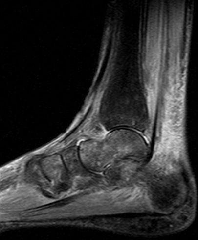
MRISagittal STIR MRI of the foot showing skin and subcutaneous oedema associated with diffuse bone marrow oedema of the calcaneus, talus and distal tibia, characteristic of type I CRPS.
Source : Poll et al., Diabetology and metabolic syndrome, 2010, figure 2 · CC BY
- 🔥 Neurogenic inflammation : excessive release of pro-inflammatory neuropeptides (substance P, CGRP) by nerve endings, causing vasodilatation, increased vascular permeability (oedema) and sensitisation of nociceptors. This component predominates in the acute phase, particularly in the « warm » form with a red, hot, oedematous limb.¹³,¹⁵
- 🧠 Dysregulation of the autonomic nervous system : sympathetic dysfunction causing the changes in colour, temperature and sweating. This component explains the « cold » presentations with a cold, cyanotic limb.⁶,¹³
- ⚡ Peripheral and central sensitisation : maladaptive neuroplasticity at the level of the peripheral nociceptors, the spinal cord (sensitisation of dorsal horn neurones) and the brain (cortical reorganisation, disturbed representation of the limb). It explains allodynia, hyperalgesia and the disturbance of body perception documented by Lewis 2007.¹⁷,²³
- 🛡️ Autoimmune component : presence of functional autoantibodies targeting β2 adrenergic and M2 muscarinic receptors in ~30% of CRPS patients. This avenue, developed by Goebel's team, opens therapeutic prospects (IVIG) but remains to be confirmed.¹⁵
- ✅ There is complete or partial resolution in the majority of patients within the 12-18 months following diagnosis, particularly where multimodal management is early.
- ⚠️ There is progression to chronicity in 15-35% of patients, with persistent pain, stiffness, muscle wasting and significant functional disability.²⁴
- 📉 The poor prognostic factors include: severe initial symptoms, presence of dystonia, delayed diagnosis, unfavourable psychological profile (high catastrophising, marked anxiety).²⁵
🚩 Red flags in the presence of a picture suggestive of CRPS
- Fever + local inflammatory signs → suspicion of deep infection (cellulitis, osteomyelitis, abscess)
- Unilateral calf swelling with asymmetrical pain → suspicion of deep vein thrombosis (D-dimers, Doppler ultrasound)
- Progressive neurological deficit (paraesthesiae in a systematised territory, motor deficit of a specific nerve) → suspicion of peripheral nerve lesion or compression
- Disproportionate pain + tense oedema + compartment tension after trauma → suspicion of compartment syndrome (surgical emergency)
- Deterioration in general condition, weight loss, night pain waking the patient → oncological work-up
- Absent distal pulses, pallor, pain at rest → suspicion of arterial ischaemia
⚠️ Any red flag calls for prompt medical referral BEFORE settling on the diagnosis of CRPS, which remains a diagnosis of exclusion.
Key points
- CRPS is chronic pain out of proportion to an initial injury, most often a wrist fracture.
- Diagnosis rests on the Budapest clinical criteria (Harden 2010), validated by the IASP: sensitivity 99%, specificity 79%.
- The women between 50 and 70 years are the most affected (F:M ratio 3 to 4 :1).
- The pathophysiology combines neurogenic inflammation, autonomic dysregulation, central sensitisation and possibly an autoimmune component.
- A rating of VAS ≥ 5 in the first week after wrist fracture is a major red flag (Moseley 2014).
- The course is variable: the majority improve but 15-35% become chronic. Early and multimodal management is the best predictor of a favourable outcome.
Bibliography
- Ferraro MC, O'Connell NE, Sommer C, Goebel A, Bultitude JH, Cashin AG, Moseley GL, McAuley JH. Complex regional pain syndrome: advances in epidemiology, pathophysiology, diagnosis, and treatment. Lancet Neurol. 2024;23(5):522-533. doi:10.1016/S1474-4422(24)00076-0.
- Bruehl S. Complex regional pain syndrome. BMJ. 2015;351:h2730. PMID 26224572.
- Goh EL, Chidambaram S, Ma D. Complex regional pain syndrome: a recent update. Burns Trauma. 2017;5:2. PMC 5244710.
- Harden RN, Bruehl S, Perez RSGM, et al. Validation of proposed diagnostic criteria (the "Budapest Criteria") for Complex Regional Pain Syndrome. Pain. 2010;150(2):268-274. PMID 20493633.
- Goebel A, Barker C, Turner-Stokes L, et al. Complex regional pain syndrome in adults: UK guidelines for diagnosis, referral and management in primary and secondary care (2nd ed). Royal College of Physicians; 2018.
- Bruehl S, Maihöfner C, Stanton-Hicks M, et al. Complex regional pain syndrome: evidence for warm and cold subtypes in a large prospective clinical sample. Pain. 2016;157(8):1674-1681. PMID 27023422.
- Shim H, Rose J, Halle S, Shekane P. Complex regional pain syndrome: a narrative review for the practising clinician. Br J Anaesth. 2019;123(2):e424-e433. PMID 31056241.
- Sandroni P, Benrud-Larson LM, McClelland RL, Low PA. Complex regional pain syndrome type I: incidence and prevalence in Olmsted County, a population-based study. Pain. 2003;103(1-2):199-207. PMID 12749974.
- Beerthuizen A, Stronks DL, Van't Spijker A, et al. Demographic and medical parameters in the development of complex regional pain syndrome type 1 (CRPS1): prospective study on 596 patients with a fracture. Pain. 2012;153(6):1187-1192. PMID 22386473.
- Moseley GL, Herbert RD, Parsons T, Lucas S, van Hilten JJ, Brooke J. Intense pain soon after wrist fracture strongly predicts who will develop complex regional pain syndrome: prospective cohort study. J Pain. 2014;15(1):16-23. PMID 24268113.
- Marinus J, Moseley GL, Birklein F, et al. Clinical features and pathophysiology of complex regional pain syndrome. Lancet Neurol. 2011;10(7):637-648. PMID 21683929.
- Birklein F, Schlereth T. Complex regional pain syndrome — significant progress in understanding. Pain. 2015;156(Suppl 1):S94-S103. PMID 25789441.
- Parkitny L, McAuley JH, Di Pietro F, et al. Inflammation in complex regional pain syndrome: a systematic review and meta-analysis. Neurology. 2013;80(1):106-117. PMID 23267031.
- Birklein F, Ajit SK, Goebel A, Perez RSGM, Sommer C. Complex regional pain syndrome — phenotypic characteristics and potential biomarkers. Nat Rev Neurol. 2018;14(5):272-284. PMID 29545626.
- Harden RN, McCabe CS, Goebel A, et al. Complex Regional Pain Syndrome: Practical Diagnostic and Treatment Guidelines, 5th Edition. Pain Med. 2022;23(Suppl 1):S1-S53. Pain Med 2022 5th ed.
- Lewis JS, Kersten P, McCabe CS, McPherson KM, Blake DR. Body perception disturbance: a contribution to pain in complex regional pain syndrome (CRPS). Pain. 2007;133(1-3):111-119. PMID 17509761.
- Halicka M, Vittersø AD, Proulx MJ, Bultitude JH. Neuropsychological changes in complex regional pain syndrome (CRPS). Behav Neurol. 2020;2020:4561831. PMC 7152960.
- Goebel A, Birklein F, Brunner F, et al. The Valencia consensus-based adaptation of the IASP complex regional pain syndrome diagnostic criteria. Pain. 2021;162(9):2346-2348. PMID 33729210.
- Wertli MM, Bachmann LM, Schecter Weiner S, Brunner F. Prognostic factors in complex regional pain syndrome 1: a systematic review. J Rehabil Med. 2013;45(3):225-231. PMID 23389624.
- Goebel A, Blaes F, Bruehl S, et al. Standards for the diagnosis and management of complex regional pain syndrome: Results of a European Pain Federation task force. Eur J Pain. 2019;23(4):641-651. PMC 6593444.
- de Mos M, de Bruijn AGJ, Huygen FJPM, Dieleman JP, Stricker BHCh, Sturkenboom MCJM. The incidence of complex regional pain syndrome: a population-based study. Pain. 2007;129(1):12-20. PMID 17084977.
- de Rooij AM, de Mos M, Sturkenboom MCJM, et al. Familial occurrence of complex regional pain syndrome. Eur J Pain. 2009;13(2):171-177. PMID 18514555.
- Mancini F, Wang AP, Schira MM, et al. Fine-Grained Mapping of Cortical Somatotopies in Chronic Complex Regional Pain Syndrome. J Neurosci. 2019;39(46):9185-9196. PMID 31570533.
- Bean DJ, Johnson MH, Heiss-Dunlop W, Kydd RR. Do psychological factors influence recovery from complex regional pain syndrome type 1? A prospective study. Pain. 2015;156(11):2310-2318. PMID 26133727.
- Bean DJ, Johnson MH, Heiss-Dunlop W, Kydd RR. Extent of recovery in the first 12 months of complex regional pain syndrome type-1: A prospective study. Eur J Pain. 2016;20(6):884-894. PMID 26524108.
- Knudsen LF, Santoro L, Bruehl S, Harden RN, Brunner F. Subtypes of complex regional pain syndrome — a systematic review of the literature. Pain Rep. 2023;8(6):e1111. PMID 38027463.
How do you assess and diagnose complex regional pain syndrome (CRPS / algodystrophy) with certainty?
Which questions should you ask to understand the patient and their history?
The structured history must systematically explore:- The triggering event : trauma (type, date, severity), surgery, prolonged immobilisation, minor event. Around 10% of cases have no clearly identified event.³
- The pain : type (burning, crushing, throbbing), intensity (VAS), course, distribution (it does not respect a systematised nerve territory, unlike a radiculopathy). The disproportion with the initial event is central.²,⁴
- Abnormal sensory symptoms : allodynia (pain provoked by a normally non-painful stimulus: the brush of a sheet, the touch of clothing), hyperalgesia (exaggerated response to a painful stimulus), spontaneous paraesthesiae.⁶
- Vasomotor symptoms : colour asymmetry (redness, cyanosis, pallor), temperature asymmetry (limb warm or cold compared with the unaffected side).⁴,⁷
- Sudomotor symptoms / oedema : oedema, excessive sweating or anhidrosis, changes in skin texture.⁴
- Motor and trophic symptoms : loss of strength, stiffness, tremor, dystonia (fixed abnormal postures), changes in hair (hypertrichosis / alopecia), in nails (accelerated / slowed growth, brittleness) and in skin (shiny appearance, thinning).⁴,⁸
- Functional and psychosocial impact : effect on activities of daily living, work, sleep and quality of life. Assess catastrophising (PCS), kinesiophobia (TSK-11), anxiety and depression (HADS).⁹
Which clinical tests should be performed and which other conditions must be ruled out?
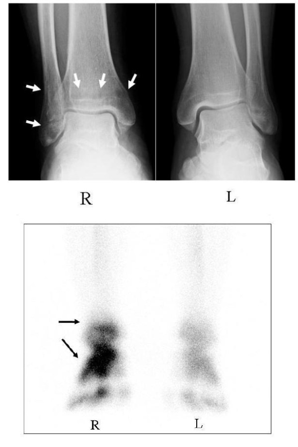
Bone scanRadiograph and bone scintigraphy of both ankles before treatment, showing demineralisation of the right ankle affected by CRPS and marked increased uptake at the same site.
Source : Abe et al., Journal of medical case reports, 2011, figure 1 · CC BY
🩺 Budapest criteria for the clinical diagnosis of CRPS
Four cumulative conditions: Harden 2010 (IASP validation), sensitivity 99%, specificity 79%
Source: Harden RN, Bruehl S, Perez RSGM, et al. Pain. 2010;150(2):268-274 (PMID 20493633). Sensitivity 99%, specificity 79% in clinical practice.
| Differential diagnosis | Clinical clues | Test used to rule it out |
|---|---|---|
| Deep vein thrombosis | Unilateral oedema, calf pain, Homans' sign | D-dimers, venous Doppler ultrasound |
| Infective cellulitis | Fever, portal of entry, leucocytosis, raised CRP | Full blood count, CRP, blood cultures if fever |
| Occult / non-united fracture | Localised pain, recent history of trauma | Plain radiograph, CT or targeted MRI |
| Peripheral neuropathy | Distribution within a systematised nerve territory | EMG / nerve conduction studies |
| Compartment syndrome (chronic) | Pain on exertion, feeling of tightness, post-traumatic | Intracompartmental pressure measurement |
| Inflammatory / rheumatic arthritis | Symmetrical involvement, morning stiffness, autoimmune context | Inflammatory markers, RF / anti-CCP, joint ultrasound |
| Vascular disease (ischaemia, Raynaud) | Pallor, distal trophic changes, changes with cold | Ankle-brachial pressure index, capillaroscopy |
- 📷 Plain radiographs : may show patchy regional bone demineralisation (Sudeck) beyond 3 to 6 weeks of evolution: a sign of low sensitivity and no specificity.
- 🔬 Three-phase bone scintigraphy : a finding of diffuse increased uptake in the late phase is suggestive but remains non-specific.⁶,¹²
- 🧲 MRI : may show bone marrow oedema in the early phase; useful above all to rule out an alternative diagnosis.
- 🩸 Laboratory tests : full blood count, CRP, ESR, rheumatological work-up: only to rule out another cause.
Should CRPS patients be classified and stratified, and for what benefit?
The dichotomous I/II classification remains fundamental:¹³- 🔸 CRPS type I (classic algodystrophy) : without identifiable nerve lesion. ~90% of cases. Often after fracture, after immobilisation, after surgery.
- 🔸 CRPS type II (causalgia) : with a confirmed peripheral nerve lesion. More often after a nerve wound, after nerve surgery, after a penetrating injury.
| Subtype | Clinical features | Therapeutic implications | Evidence |
|---|---|---|---|
| Warm CRPS | Warm, red, oedematous limb, increased sweating: predominantly neurogenic inflammation | Favour anti-inflammatories, graded exposure, monitoring | Moderate (Bruehl 2016) |
| Cold CRPS | Cold, cyanotic limb, little oedema: predominantly dysautonomic / neuropathic | Poorer response to anti-inflammatories; consider neuromodulation | Moderate (Bruehl 2016) |
| Centralised | Marked central sensitisation, disturbed body perception | GMI, mirror therapy, pain education as first line | Low (Lewis 2007) |
| Dystonic | Postures fixed in flexion, tremors, involuntary movements | Multidisciplinary approach; neurological referral; intrathecal baclofen exceptionally | Low (van Rijn 2009) |
| Familial | Family history, earlier onset, sometimes bilateral forms | Genetic information; no specific change to the protocol | Low (de Rooij 2009) |
| Paediatric | Preferential involvement of the lower limb, major psychological component, excellent response to intensive rehabilitation | Multidisciplinary day-hospital programmes (Logan 2012) | Moderate |
Key points
- The diagnosis of CRPS is strictly clinical and rests on the Budapest criteria (Harden 2010).
- Sensitivity around 99 %, specificity around 79 % : the exclusion step is therefore essential.
- The additional investigations never confirm the diagnosis; they serve only to rule out the differential diagnoses.
- The classification I/II (without / with nerve lesion) remains fundamental.
- The phenotyping into subtypes (warm / cold / centralised / dystonic / familial / paediatric) opens the way to stratified medicine.
Bibliography
- Goebel A, Barker C, Turner-Stokes L, et al. Complex regional pain syndrome in adults: UK guidelines for diagnosis, referral and management (2nd ed). Royal College of Physicians; 2018.
- Bruehl S. Complex regional pain syndrome. BMJ. 2015;351:h2730. PMID 26224572.
- Birklein F, Ajit SK, Goebel A, Perez RSGM, Sommer C. Complex regional pain syndrome — phenotypic characteristics and potential biomarkers. Nat Rev Neurol. 2018;14(5):272-284. PMID 29545626.
- Harden RN, Bruehl S, Stanton-Hicks M, Wilson PR. Proposed new diagnostic criteria for complex regional pain syndrome. Pain Med. 2007;8(4):326-331. PMID 17610454.
- Stanton-Hicks M, Jänig W, Hassenbusch S, et al. Reflex sympathetic dystrophy: changing concepts and taxonomy. Pain. 1995;63(1):127-133. PMID 8577483.
- Smart KM, Ferraro MC, Wand BM, O'Connell NE. Physiotherapy for pain and disability in adults with complex regional pain syndrome (CRPS) types I and II. Cochrane Database Syst Rev. 2022;5(5):CD010853. PMID 35579382.
- Bruehl S, Maihöfner C, Stanton-Hicks M, et al. Complex regional pain syndrome: evidence for warm and cold subtypes. Pain. 2016;157(8):1674-1681. PMID 27023422.
- Marinus J, Moseley GL, Birklein F, et al. Clinical features and pathophysiology of complex regional pain syndrome. Lancet Neurol. 2011;10(7):637-648. PMID 21683929.
- Bean DJ, Johnson MH, Heiss-Dunlop W, Kydd RR. Do psychological factors influence recovery from complex regional pain syndrome type 1? A prospective study. Pain. 2015;156(11):2310-2318. PMID 26133727.
- Harden RN, Bruehl S, Perez RSGM, et al. Validation of proposed diagnostic criteria (the "Budapest Criteria") for Complex Regional Pain Syndrome. Pain. 2010;150(2):268-274. PMID 20493633.
- Shim H, Rose J, Halle S, Shekane P. Complex regional pain syndrome: a narrative review for the practising clinician. Br J Anaesth. 2019;123(2):e424-e433. PMID 31056241.
- Wertli MM, Brunner F, Steurer J, Held U. Usefulness of bone scintigraphy for the diagnosis of Complex Regional Pain Syndrome 1: A systematic review and Bayesian meta-analysis. PLoS One. 2017;12(3):e0173688. PMID 28301606.
- Stanton-Hicks M. Complex regional pain syndrome (type I, RSD; type II, causalgia): controversies. Clin J Pain. 2000;16(2 Suppl):S33-S40. PMID 10870738.
- Knudsen LF, Santoro L, Bruehl S, Harden RN, Brunner F. Subtypes of complex regional pain syndrome — a systematic review of the literature. Pain Rep. 2023;8(6):e1111. PMID 38027463.
- Brunner F, Heitz CAM, Kissling R, Held U. Complex regional pain syndrome: clinical phenotypes and classification algorithm. Neurology. 2020;94(4):e357-e367. Neurology 2020.
- Ferraro MC, O'Connell NE, Sommer C, et al. Complex regional pain syndrome: advances in epidemiology, pathophysiology, diagnosis, and treatment. Lancet Neurol. 2024;23(5):522-533. doi:10.1016/S1474-4422(24)00076-0.
- Goebel A, Birklein F, Brunner F, et al. The Valencia consensus-based adaptation of the IASP complex regional pain syndrome diagnostic criteria. Pain. 2021;162(9):2346-2348. PMID 33729210.
What are the most effective treatment strategies for complex regional pain syndrome (CRPS / algodystrophy)?
Where do you start? What is the recommended hierarchy of interventions?
According to the reference guidelines (UK RCP Goebel 2018,⁴ 5th practical edition Harden Pain Medicine 2022,⁵ European Pain Federation Goebel 2019⁶), the first-line hierarchy is:- 🎓 Therapeutic education in pain neurophysiology (PNE / "Explain Pain") : reconceptualising pain as an alarm signal and not as an indicator of active tissue damage.⁷,⁸
- 🪞 Graded motor imagery (GMI) : 3 phases: laterality recognition, explicit motor imagery, mirror therapy.⁹,¹⁰
- 📈 Graded exposure to pain and to movement (PEPT, Pain Exposure Physical Therapy): progressive, structured exposure to sensory and motor stimuli.¹¹,¹²
- 🏃 Progressive, functional physical activity : restoring mobility, motor control, strength and endurance.¹
- 💊 Pharmacological adjuncts as medically prescribed.⁵
📐 GRADE pyramid: levels of evidence for the rehabilitation interventions in CRPS
Cochrane synthesis Smart-Ferraro 2022 (CD010853.pub3) + Méndez-Rebolledo 2017 + guidelines
Synthesis based on: Smart KM, Ferraro MC, Wand BM, O'Connell NE. Cochrane Database Syst Rev. 2022;5:CD010853 (PMID 35579382); Méndez-Rebolledo G et al. J Back Musculoskelet Rehabil. 2017;30(3):441-449 (PMID 27858687); Harden RN et al. Pain Med. 2022;23(Suppl 1):S1-S53, 5th practical edition.
What is the place of exercise and is there a superior approach?
Exercise is a pillar of CRPS rehabilitation. The question is not « which exercise? » but « how to dose and progress? ». There is no single superior exercise protocol ; individualisation remains the rule.¹ The best supported approach is graded exposure. The Pain Exposure Physical Therapy (PEPT) offers a rehabilitation programme in which the patient performs functional exercises within the limits of their tolerance, with progression of load. The randomised trial by Barnhoorn BMJ Open 2015 (n = 56, CRPS-I) shows a significant improvement in function and in pain at 9 months in the PEPT group vs conventional treatment.¹¹ Mirror therapy, a component of GMI, is one of the most studied interventions. The systematic review Méndez-Rebolledo 2017 (6 RCTs, n = 171 CRPS-I) rates the studies as of moderate to high quality and confirms a significant reduction in pain and an improvement in function.¹⁰ The seminal RCT Cacchio 2009 in 48 patients after stroke demonstrates the superiority of mirror therapy over a placebo.¹³Manual therapies and technologies: what is their real effectiveness in CRPS?
The evidence supporting manual therapies as monotherapy for CRPS is very limited.¹⁴ They can be used as a targeted adjunct to treat specific impairments (stiffness, contractures), but must not form the core of treatment. As for the passive technologies :- ⚡ TENS : the Cochrane review Gibson 2019 concludes that the evidence is of very low quality and allows no firm recommendation for CRPS.¹⁵
- 🔆 Ultrasound, low-level laser therapy (LLLT), magnetotherapy : no robust data specific to CRPS.¹
- 💉 Medical interventions (sympathetic blocks, neuromodulation, bisphosphonates in the early phase): these fall to the pain specialist and the pain physician.⁵,⁶
Beyond the physical: how do you educate the patient and address psychological factors?
CRPS is a paradigmatic example of a biopsychosocial condition.¹⁶ Addressing psychological factors is not optional: it is an essential component of treatment. Pain neurophysiology education (PNE) is the first step. It makes it possible to:- 💡 Demystify pain by explaining it as an alarm signal from the brain and not as an indicator of active tissue damage.
- 💡 Reduce the perceived threat, which directly lessens the fear and the anxiety linked to movement.
- 💡 Validate the patient's experience and build a solid therapeutic alliance.¹⁷
- 😰 The kinesiophobia (fear of movement): to be targeted through graded exposure and the rebuilding of confidence.⁸,¹⁸
- 💭 The catastrophising : to be approached through cognitive restructuring and targeted education.¹⁶
- 👁️ Hypervigilance : to be managed through distraction techniques and attentional refocusing.¹
Key points
- Management is multimodal, early and active : pain education, GMI/mirror therapy, graded exposure, functional exercise.
- Pain neurophysiology education ("Explain Pain") is the non-negotiable foundation.
- The GMI and the mirror therapy are first-line interventions with a probable benefit (low to moderate quality, Cochrane 2022).
- The PEPT is a valid option (RCT Barnhoorn 2015); individualisation takes precedence over the standardised protocol.
- The passive therapies have a low level of evidence and remain adjuncts.
- The identification and management of psychological factors are essential.
Bibliography
- Ferraro MC, O'Connell NE, Sommer C, et al. Complex regional pain syndrome: advances in epidemiology, pathophysiology, diagnosis, and treatment. Lancet Neurol. 2024;23(5):522-533. doi:10.1016/S1474-4422(24)00076-0.
- Smart KM, Ferraro MC, Wand BM, O'Connell NE. Physiotherapy for pain and disability in adults with complex regional pain syndrome (CRPS) types I and II. Cochrane Database Syst Rev. 2022;5(5):CD010853. PMID 35579382.
- O'Connell NE, Wand BM, McAuley J, Marston L, Moseley GL. Interventions for treating pain and disability in adults with complex regional pain syndrome — an overview of systematic reviews. Cochrane Database Syst Rev. 2013;(4):CD009416. PMID 23633371.
- Goebel A, Barker C, Turner-Stokes L, et al. Complex regional pain syndrome in adults: UK guidelines (2nd ed). Royal College of Physicians; 2018.
- Harden RN, McCabe CS, Goebel A, et al. Complex Regional Pain Syndrome: Practical Diagnostic and Treatment Guidelines, 5th Edition. Pain Med. 2022;23(Suppl 1):S1-S53. Pain Med 2022 5th ed.
- Goebel A, Blaes F, Bruehl S, et al. Standards for the diagnosis and management of complex regional pain syndrome: Results of a European Pain Federation task force. Eur J Pain. 2019;23(4):641-651. PMC 6593444.
- Louw A, Zimney K, Puentedura EJ, Diener I. The efficacy of pain neuroscience education on musculoskeletal pain: A systematic review of the literature. Physiother Theory Pract. 2016;32(5):332-355. PMID 27351541.
- Moseley GL, Butler DS. Fifteen Years of Explaining Pain: The Past, Present, and Future. J Pain. 2015;16(9):807-813. PMID 26051220.
- Moseley GL. Graded motor imagery for pathologic pain: a randomized controlled trial. Neurology. 2006;67(12):2129-2134. PMID 17082465.
- Méndez-Rebolledo G, Gatica-Rojas V, Torres-Cueco R, Albornoz-Verdugo M, Guzmán-Muñoz E. Update on the effects of graded motor imagery and mirror therapy on complex regional pain syndrome type 1: A systematic review. J Back Musculoskelet Rehabil. 2017;30(3):441-449. PMID 27858687.
- Barnhoorn KJ, van de Meent H, van Dongen RT, et al. Pain exposure physical therapy (PEPT) compared to conventional treatment in complex regional pain syndrome type 1: a randomised controlled trial. BMJ Open. 2015;5(12):e008283. PMC 4679993.
- Barnhoorn KJ, Staal JB, van Dongen RTM, et al. Pain Exposure Physical Therapy versus conventional treatment — a cost-effectiveness analysis alongside a RCT. Clin Rehabil. 2018;32(6):790-798. PMID 29430970.
- Cacchio A, De Blasis E, De Blasis V, Santilli V, Spacca G. Mirror therapy in complex regional pain syndrome type 1 of the upper limb in stroke patients. Neurorehabil Neural Repair. 2009;23(8):792-799. PMID 19465507.
- Smart KM, Wand BM, O'Connell NE. Physiotherapy for pain and disability in adults with complex regional pain syndrome (CRPS) types I and II. Cochrane Database Syst Rev. 2016;(2):CD010853. PMC 9112661.
- Gibson W, Wand BM, O'Connell NE. Transcutaneous electrical nerve stimulation (TENS) for neuropathic pain in adults. Cochrane Database Syst Rev. 2017;9(9):CD011976. PMID 28922669.
- Bean DJ, Johnson MH, Heiss-Dunlop W, Kydd RR. Do psychological factors influence recovery from complex regional pain syndrome type 1? A prospective study. Pain. 2015;156(11):2310-2318. PMID 26133727.
- Llewellyn A, McCabe CS, Hibberd Y, et al. Are you better? A multi-centre study of patient-defined recovery from Complex Regional Pain Syndrome. Eur J Pain. 2018;22(3):551-564. PMID 29194871.
- Halicka M, Vittersø AD, Proulx MJ, Bultitude JH. Neuropsychological changes in complex regional pain syndrome (CRPS). Behav Neurol. 2020;2020:4561831. PMC 7152960.
How do you secure lasting recovery and prevent recurrence of complex regional pain syndrome (CRPS)?
How do you make the patient an active participant in their own recovery through self-management?
Empowerment is the cornerstone of the long-term management of CRPS. Four complementary pillars:🎯 The 4 pillars of lasting self-management in CRPS
Integrated approach based on the Goebel 2018 and Harden 2022 guidelines and on the "Explain Pain" principles
Summary of the principles of active CRPS management according to the international guidelines (Goebel UK RCP 2018; Harden Pain Med 2022; Ferraro Lancet Neurol 2024).
- 📑 Phase 1: laterality recognition : smartphone applications (Recognise) or printed cards with left/right hands and feet to identify.
- 🧠 Phase 2: explicit motor imagery : imagining movements of the affected limb without performing them.
- 🪞 Phase 3: mirror therapy : visual illusion of normal, painless movement.
When and how should a safe return to sport and to activities be planned?
The return to sporting or occupational activities after CRPS must never be based on a fixed calendar, but on reaching precise functional criteria.⁹ 💪| Domain | Minimum criterion | Measurement tool |
|---|---|---|
| Pain | Resting VAS ≤ 3; no prolonged increase > 24h after exertion | Visual analogue scale, pain diary |
| Mobility | Recovery of at least 80% of the contralateral range | Comparative goniometry |
| Strength | At least 80% of contralateral strength | Dynamometry (Jamar grip, isokinetic) |
| Motor control | Ability to perform the specific movements without apprehension | DASH, PRWE, LEFS, Y-balance, hop tests |
| Confidence / kinesiophobia | TSK-11 < 17; high PSEQ | TSK-11, PSEQ, IPAQ |
| Load tolerance | 3 modified sessions without a flare > 24h | Training diary |
🚩 Warning signs that call for stopping the progression and reassessing
- Pain flare > 24 h after an effort tolerated until then → adjust the load downwards immediately
- Reappearance of vasomotor/sudomotor signs (oedema, colour change, thermal asymmetry) → suspect a relapse
- Motor regression (loss of range, weakness) → reassess the programme and consider a medical opinion
- Appearance of dystonia (fixed postures, involuntary tremors) → neurological referral
- Marked worsening of catastrophising or of kinesiophobia → referral to a specialist psychologist
- Involvement of another limb → systematic diagnostic reassessment
Key points
- Patient empowerment is the key: pain education (PNE), daily GMI, pacing, graded exposure.
- Pain in CRPS is more a problem of « software » (nervous system) than of « hardware » (tissues).
- The return to sport is guided by functional criteria : never by a fixed calendar.
- The progression must be slow, gradual and monitored ; a flare > 24 h calls for an adjustment.
- Identify quickly the warning signs and refer on where necessary.
Bibliography
- Louw A, Zimney K, Puentedura EJ, Diener I. The efficacy of pain neuroscience education on musculoskeletal pain: A systematic review of the literature. Physiother Theory Pract. 2016;32(5):332-355. PMID 27351541.
- Moseley GL, Butler DS. Fifteen Years of Explaining Pain. J Pain. 2015;16(9):807-813. PMID 26051220.
- Moseley GL. Graded motor imagery for pathologic pain: a randomized controlled trial. Neurology. 2006;67(12):2129-2134. PMID 17082465.
- Méndez-Rebolledo G, Gatica-Rojas V, Torres-Cueco R, Albornoz-Verdugo M, Guzmán-Muñoz E. Update on the effects of graded motor imagery and mirror therapy on complex regional pain syndrome type 1: A systematic review. J Back Musculoskelet Rehabil. 2017;30(3):441-449. PMID 27858687.
- Antcliff D, Keenan AM, Keeley P, Woby S, McGowan L. Engaging stakeholders to refine an activity pacing framework for chronic pain/fatigue: A nominal group technique. Musculoskeletal Care. 2019;17(4):354-362. PMID 31750627.
- Andrews NE, Strong J, Meredith PJ. Activity pacing, avoidance, endurance, and associations with patient functioning in chronic pain. Arch Phys Med Rehabil. 2012;93(11):2109-2121.e7. PMID 22728699.
- de Jong JR, Vlaeyen JW, Onghena P, Cuypers C, den Hollander M, Ruijgrok J. Reduction of pain-related fear in complex regional pain syndrome type I: the application of graded exposure in vivo. Pain. 2005;116(3):264-275. PMID 15964686.
- den Hollander M, Goossens M, de Jong J, et al. Expose or protect? A randomized controlled trial of exposure in vivo vs pain-contingent treatment as usual in patients with complex regional pain syndrome type 1. Pain. 2016;157(10):2318-2329. PMID 27429174.
- Harden RN, McCabe CS, Goebel A, et al. Complex Regional Pain Syndrome: Practical Diagnostic and Treatment Guidelines, 5th Edition. Pain Med. 2022;23(Suppl 1):S1-S53. Pain Med 2022 5th ed.
- Goebel A, Barker C, Turner-Stokes L, et al. Complex regional pain syndrome in adults: UK guidelines (2nd ed). Royal College of Physicians; 2018.
- Smart KM, Ferraro MC, Wand BM, O'Connell NE. Physiotherapy for pain and disability in adults with complex regional pain syndrome (CRPS) types I and II. Cochrane Database Syst Rev. 2022;5(5):CD010853. PMID 35579382.
- Bean DJ, Johnson MH, Heiss-Dunlop W, Kydd RR. Do psychological factors influence recovery from complex regional pain syndrome type 1? A prospective study. Pain. 2015;156(11):2310-2318. PMID 26133727.
- Halicka M, Vittersø AD, Proulx MJ, Bultitude JH. Neuropsychological changes in complex regional pain syndrome (CRPS). Behav Neurol. 2020;2020:4561831. PMC 7152960.
- Ferraro MC, O'Connell NE, Sommer C, et al. Complex regional pain syndrome: advances in epidemiology, pathophysiology, diagnosis, and treatment. Lancet Neurol. 2024;23(5):522-533. doi:10.1016/S1474-4422(24)00076-0.
What do real clinical cases teach us about complex regional pain syndrome (CRPS / algodystrophy)?
Analysis of a « classic » case: from assessment to resolution
The randomised trial by Cacchio et al. (Neurorehabilitation and Neural Repair 2009) illustrates multimodal management in 48 patients with upper limb CRPS-I occurring after a stroke.¹ The protocol, run over 4 weeks with follow-up at 6 months, combined:- 🪞 Daily mirror therapy (30 min, 5 days/week): performing movements of the unaffected limb reflected visually.
- 💆 Conventional rehabilitation : passive/active assisted exercises, gentle joint mobilisation, progressive tactile desensitisation.
- 🎓 Therapeutic education : explanation of the nature of CRPS and of the aim of mirror therapy.
- ✅ Significant reduction in pain (VAS) in the mirror therapy group vs control.
- ✅ Functional improvement of the upper limb.
- ✅ Effect maintained at 6 months of follow-up.
🧩 Cacchio 2009 case: multimodal protocol for post-stroke CRPS-I
RCT n = 48: components and aggregated levels of evidence
Source: Cacchio A et al. Neurorehabil Neural Repair. 2009;23(8):792-799 (PMID 19465507).
The diagnostic challenge: when CRPS mimics another condition
One of the greatest lessons from clinical cases is the capacity of CRPS to mimic other conditions. ⚠️ The « warm » form (warm, red, oedematous limb) can be confused with:- 🔍 Infective cellulitis : redness, heat, oedema; distinguished by the absence of a portal of entry, the absence of fever and of an inflammatory response, and the absence of any response to antibiotics.²
- 🔍 Deep vein thrombosis : unilateral oedema of the lower limb after surgery or immobilisation; venous Doppler ultrasound + D-dimers to rule it out.²
- 🔍 Septic arthritis / osteomyelitis : to be ruled out if fever + inflammatory markers + portal of entry.
- 🔍 Arterial ischaemia : pallor, pain at rest, absent distal pulses; ankle-brachial pressure index and arterial Doppler ultrasound.
- 🔍 Raynaud's disease or vasospastic phenomenon: paroxysmal episodes modulated by cold, with bilateral and symmetrical involvement.
- 🔍 Focal peripheral neuropathy : distribution within a systematised nerve territory; EMG is diagnostic.
Study of a complex case: paediatric CRPS and dystonic forms
Paediatric CRPS is rare but well described (review Weissmann & Uziel 2016).³ Features:- 👧 Female predominance is marked (~80-85%), with a peak in adolescence (12-15 years).
- 🦶 Preferential involvement of the lower limb.
- 🧠 Major psychological component : frequent association with anxiety, perfectionism, family and school conflicts.
- ✅ Excellent response to intensive multidisciplinary rehabilitation (Logan 2012, n=56 in day hospital).⁴
- 🧠 Involves central mechanisms (altered spinal and brainstem inhibition).
- 💉 Responds modestly to pharmacological treatments (intrathecal baclofen in severe cases).
- 🚫 Lends itself poorly to standard GMI protocols once the dystonia is established.
Critique and controversy: the value and the limits of clinical cases
🤔 Clinical cases are a powerful teaching tool but must be interpreted with caution.📐 Hierarchy of scientific evidence
Strength of evidence decreasing from the top (meta-analyses) towards the bottom (isolated cases)
Simplified GRADE / Oxford CEBM hierarchy. Where a seductive clinical case and a meta-analysis diverge, the decision must follow the meta-analysis.
⭐ Key points
- The RCT Cacchio 2009 illustrates the effectiveness of mirror therapy in post-stroke CRPS-I (effect maintained at 6 months).
- Differential diagnoses to know: DVT, infective cellulitis, osteomyelitis, arterial ischaemia, focal peripheral neuropathy, occult fracture.
- The paediatric form of CRPS (Logan 2012, Weissmann 2016): adolescent female predominance, lower limb, excellent response to intensive multidisciplinary rehabilitation.
- The dystonic forms (van Rijn 2011) require a multidisciplinary approach; standard GMI may be less effective.
- ⚠️ A case report = level 5. It illustrates, it never demonstrates . Where there is divergence, follow the meta-analyses (1a).
Bibliography
- Cacchio A, De Blasis E, De Blasis V, Santilli V, Spacca G. Mirror therapy in complex regional pain syndrome type 1 of the upper limb in stroke patients. Neurorehabil Neural Repair. 2009;23(8):792-799. PMID 19465507.
- Shim H, Rose J, Halle S, Shekane P. Complex regional pain syndrome: a narrative review for the practising clinician. Br J Anaesth. 2019;123(2):e424-e433. PMID 31056241.
- Weissmann R, Uziel Y. Pediatric complex regional pain syndrome: a review. Pediatr Rheumatol Online J. 2016;14(1):29. PMC 4850724.
- Logan DE, Carpino EA, Chiang G, et al. A day-hospital approach to treatment of pediatric complex regional pain syndrome: initial functional outcomes. Clin J Pain. 2012;28(9):766-774. PMID 22688602.
- van Rijn MA, Marinus J, Putter H, Bosselaar SR, Moseley GL, van Hilten JJ. Spreading of complex regional pain syndrome: not a random process. J Neural Transm (Vienna). 2011;118(9):1301-1309. PMID 21331457.
- de Rooij AM, de Mos M, Sturkenboom MCJM, et al. Familial occurrence of complex regional pain syndrome. Eur J Pain. 2009;13(2):171-177. PMID 18514555.
- Nissen T, Wynn R. The clinical case report: a review of its merits and limitations. BMC Res Notes. 2014;7:264. PMID 24758689.
- Knudsen LF, Santoro L, Bruehl S, Harden RN, Brunner F. Subtypes of complex regional pain syndrome — a systematic review of the literature. Pain Rep. 2023;8(6):e1111. PMID 38027463.
- Bruehl S, Maihöfner C, Stanton-Hicks M, et al. Complex regional pain syndrome: evidence for warm and cold subtypes. Pain. 2016;157(8):1674-1681. PMID 27023422.
- Brunner F, Heitz CAM, Kissling R, Held U. Complex regional pain syndrome: clinical phenotypes and classification algorithm. Neurology. 2020;94(4):e357-e367. Neurology 2020.
- Smart KM, Ferraro MC, Wand BM, O'Connell NE. Physiotherapy for pain and disability in adults with complex regional pain syndrome (CRPS) types I and II. Cochrane Database Syst Rev. 2022;5(5):CD010853. PMID 35579382.
- Méndez-Rebolledo G et al. Update on the effects of graded motor imagery and mirror therapy on complex regional pain syndrome type 1: A systematic review. J Back Musculoskelet Rehabil. 2017;30(3):441-449. PMID 27858687.
- Sandroni P, Benrud-Larson LM, McClelland RL, Low PA. Complex regional pain syndrome type I: incidence and prevalence in Olmsted County. Pain. 2003;103(1-2):199-207. PMID 12749974.
- Lewis JS, Kersten P, McCabe CS, McPherson KM, Blake DR. Body perception disturbance: a contribution to pain in complex regional pain syndrome (CRPS). Pain. 2007;133(1-3):111-119. PMID 17509761.
How do you apply these recommendations concretely in your practice?
When and to which other health professionals should you refer?
One of the physiotherapist's fundamental skills is recognising the limits of their scope of practice. 🚩 Identifying the red flags is the first step. The Cook 2018 review (BJSM) stresses that, taken in isolation, most red flags have poor diagnostic accuracy ; it is the combination of several signs that should raise the alarm.¹ The Finucane 2020 framework (JOSPT) provides an applicable reference grid.²🚩 Red flags in the presence of a picture suggestive of CRPS
- Fever + local inflammatory signs + portal of entry or recent wound → deep infection (cellulitis, osteomyelitis, abscess): laboratory work-up, urgent medical opinion
- Unilateral calf oedema + asymmetrical pain + thromboembolic risk factors → DVT (D-dimers, venous Doppler ultrasound)
- Progressive neurological deficit (motor deficit of a specific nerve, systematised paraesthesiae, sphincter disturbance) → nerve or spinal cord lesion, or compression
- Disproportionate pain + tense oedema + compartment tension after trauma → compartment syndrome (surgical emergency)
- Absent distal pulses, pallor, pain at rest → acute or severe chronic arterial ischaemia
- Deterioration in general condition + weight loss + night pain → oncological work-up
- Recent history of trauma with inability to bear weight → occult fracture (radiograph, CT or MRI)
- Migrating CRPS or spread to the other side → reassess the diagnosis systematically
⚠️ Any red flag calls for prompt medical referral BEFORE continuing physiotherapy.
| Situation | Recommended referral | Timeframe |
|---|---|---|
| Red flag identified | General practitioner or emergency department depending on severity | Immediate / 24-48 h |
| Diagnostic doubt about CRPS | General practitioner + pain specialist | Within 1-2 weeks |
| No improvement at 8-12 weeks | CETD / chronic pain centre | 1 month |
| Severe kinesiophobia / catastrophising / depression | Psychologist specialising in pain | 1 month |
| Established dystonic form | Neurologist + CETD | Within 2-4 weeks |
| Paediatric CRPS | Paediatrician + multidisciplinary paediatric pain team | Within 2-4 weeks |
| Severe « cold » form / marked dysautonomia | Pain physician: neurostimulation may be considered | Within 1 month |
How do you measure outcomes and overcome the barriers to implementation?
The systematic use of PROMs has become a standard for assessing the impact of treatments.⁵ 📊| PROM / tool | Domain assessed | Relevance in CRPS |
|---|---|---|
| BPI (Brief Pain Inventory) | Pain intensity + interference | Reference measure for chronic pain |
| VAS / NRS | Pain intensity (0-10) | Simple, quick, usable in routine practice |
| PCS (Pain Catastrophizing Scale) | Cognitive catastrophising | Major yellow flag; prerequisite for graded exposure |
| TSK-11 | Fear of movement | Clinical threshold < 17 favourable |
| PSEQ | Self-efficacy in the face of pain | Marker of prognosis and of active engagement |
| EQ-5D-5L | Generic quality of life | Comparable with other conditions |
| DASH / Quick-DASH | Upper limb function | Sensitive to functional change |
| PRWE | Wrist and hand function | Particularly relevant given how often CRPS follows a wrist fracture |
| LEFS | Lower limb function | Sensitive to functional gains |
| HADS | Anxiety and depression | Screening; points towards joint management with a psychologist if the thresholds are met |
- 📖 Adopt and adapt recent clinical practice guidelines (UK RCP 2018, Pain Med 2022, Lancet Neurol 2024).
- 🎓 Invest in continuing education (SFETD, IASP, EFIC, journal clubs).
- 📱 Use validated smartphone applications (Recognise for GMI) and digital PROMs.
- 🤝 Integrate shared decision-making (Shared Decision-Making).¹⁰
- 👥 Foster a supportive organisational culture (leadership, protected time, sharing of protocols).
Critique and controversy: the gap between theory and reality on the ground
There is a persistent gap between knowledge and practice (evidence-practice gap), and it remains a frustrating reality. Three major criticisms: First, the practice guidelines sometimes lack flexibility in the face of patients with several conditions or atypical presentations.⁹ There is constant tension between standardisation and individualisation. Second, the PROMs can drift into an administrative formality if the results are not used to talk with the patient and adjust treatment.⁵ There is a risk of « treating the score » rather than « caring for the person ». Third, most of the research on the barriers to implementation identifies the same obstacles without proposing or testing pragmatic solutions.⁷⭐ Key points
- Referral is a key skill: red flags (infection, DVT, neurological deficit, compartment syndrome, occult fracture) → prompt medical assessment.
- The yellow flags (catastrophising, severe kinesiophobia, depression) → collaboration with a specialist psychologist or a CETD.
- The interprofessional collaboration (pain specialist, psychologist, occupational therapist, neurologist) is essential for complex cases.
- The systematic use of validated PROMs (BPI, VAS, PCS, TSK-11, PSEQ, EQ-5D, DASH/PRWE/LEFS, HADS) is a quality standard.
- The barriers to implementation call for multifaceted strategies and continuous commitment.
- The practice of shared decision-making improves adherence and patient satisfaction.
Bibliography
- Cook CE, George SZ, Reiman MP. Red flag screening for low back pain: nothing to see here, move along: a narrative review. Br J Sports Med. 2018;52(8):493-496. PMID 28923844.
- Finucane LM, Downie A, Mercer C, et al. International Framework for Red Flags for Potential Serious Spinal Pathologies. J Orthop Sports Phys Ther. 2020;50(7):350-372. PMID 32438853.
- Bean DJ, Johnson MH, Heiss-Dunlop W, Kydd RR. Do psychological factors influence recovery from complex regional pain syndrome type 1? A prospective study. Pain. 2015;156(11):2310-2318. PMID 26133727.
- Goebel A, Barker C, Turner-Stokes L, et al. Complex regional pain syndrome in adults: UK guidelines (2nd ed). Royal College of Physicians; 2018.
- Kyte DG, Calvert M, van der Wees PJ, ten Hove R, Tolan S, Hill JC. An introduction to patient-reported outcome measures (PROMs) in physiotherapy. Physiotherapy. 2015;101(2):119-125. PMID 25620440.
- Grieve S, Perez RSGM, Birklein F, et al. Recommendations for a first Core Outcome Measurement set for complex regional PAin syndrome Clinical sTudies (COMPACT). Pain. 2017;158(6):1083-1090. PMID 28178071.
- Scurlock-Evans L, Upton P, Upton D. Evidence-based practice in physiotherapy: a systematic review of barriers, enablers and interventions. Physiotherapy. 2014;100(3):208-219. PMID 24780633.
- da Silva TM, Costa LCM, Garcia AN, Costa LOP. What do physical therapists think about evidence-based practice? A systematic review. Man Ther. 2015;20(3):388-401. PMID 25458142.
- Ferraro MC, O'Connell NE, Sommer C, et al. Complex regional pain syndrome: advances in epidemiology, pathophysiology, diagnosis, and treatment. Lancet Neurol. 2024;23(5):522-533. doi:10.1016/S1474-4422(24)00076-0.
- Stiggelbout AM, Pieterse AH, De Haes JCJM. Shared decision making: concepts, evidence, and practice. Patient Educ Couns. 2015;98(10):1172-1179. PMID 26215573.
- Harden RN, McCabe CS, Goebel A, et al. Complex Regional Pain Syndrome: Practical Diagnostic and Treatment Guidelines, 5th Edition. Pain Med. 2022;23(Suppl 1):S1-S53. Pain Med 2022 5th ed.
- Smart KM, Ferraro MC, Wand BM, O'Connell NE. Physiotherapy for pain and disability in adults with complex regional pain syndrome (CRPS) types I and II. Cochrane Database Syst Rev. 2022;5(5):CD010853. PMID 35579382.
And after this article?
This article is part of a collection of evidence-based clinical syntheses. A question, a comment, a correction to suggest? Contact us directly through the WhatsApp button at the bottom right of the screen.

