















Clinical synthesis based on the most recent international guidelines and meta-analyses: updated Dutch guideline 11, JOSPT Clinical Practice Guideline 23, Ottawa ankle rules 910 and PEACE & LOVE 16. Every reference has been individually verified on PubMed.
📝 In brief: clinical summary
- Lateral ankle sprain is one of the most common musculoskeletal injuries: around 2 million acute sprains occur each year in the United States, with a sports incidence of about 0.93 per 1000 athlete-exposures 1.
- The injury follows a predictable anatomical order in inversion: the anterior talofibular ligament (ATFL) is the first, and often the only, structure affected (85% of ligament injuries), followed by the calcaneofibular ligament (CFL, 35%), the posterior talofibular ligament being involved only in rare cases (12%) 5.
- The Ottawa ankle rules rule out a fracture with a sensitivity approaching 100% and reduce radiography use by 30 to 40%; they must precede functional management 10.
- Ligament severity is assessed most reliably by a delayed examination 4-5 days after the injury; the anterior drawer test is specific (87%) but poorly sensitive (54%), whereas ATFL palpation is highly sensitive (95-100%) but poorly specific: combining the two optimises diagnosis 1112.
- Functional treatment takes precedence over immobilisation: the patient with an acute lateral ligament rupture benefits most from taping or a brace combined with a supervised exercise programme, preferred over passive modalities 11.
- The sprain is not benign: up to 70% of individuals develop chronic instability or residual disability, 12 to 47% of sprains are recurrences, and a previous sprain multiplies the risk of another by about 3.5 1.
- To prevent recurrence, proprioceptive training significantly reduces the risk of sprain (risk ratio 0.59), an effect that is stronger in individuals with a previous sprain (0.49), and braces are supported by strong evidence 2115.
🦶 What are the fundamentals to know about ankle sprain?
Lateral ankle sprain is one of the most common musculoskeletal injuries in clinical practice. Understanding its definition, the anatomy of the lateral ligament complex, the inversion injury mechanism, its epidemiological scale and its classification is the essential foundation for rigorous management. This section sets out those fundamentals, from the elementary anatomical movement through to the associated injuries that must never be missed.
🦶 Which ligaments are injured in an inversion sprain?
The anterior talofibular ligament (ATFL) is the first, and often the only, structure affected; the injury then extends to the CFL and, rarely, to the PTFL.
Frequency of injury for each ligament of the lateral collateral complex. Source: Li et al., 2019 (PMID 30923576).
📊 An incidence almost twice as high in women
Ankle sprain rate reported per 1000 athlete-exposures, by sex.
Sports incidence by sex. Peak in girls aged 10-14 and boys aged 15-19. Source: Herzog et al., 2019 (PMID 31135209).
Key points
- Lateral ankle sprain affects the lateral collateral ligament complex, with the anterior talofibular ligament (ATFL) as the first, and sometimes only, ligament injured in the inversion mechanism 5.
- The ATFL is injured in 85 % of ligament injuries, the CFL in 35% and the PTFL in 12% 5.
- Around 2 million acute sprains occur each year in the United States; it is one of the most common musculoskeletal injuries 1.
- The grade I/II/III classification rests on the progressive involvement of the ATFL then the CFL 6.
- This is not a benign injury: up to 70 % of individuals develop chronic instability or retain residual disability 1.
Definition: what exactly are we talking about?
Lateral ankle sprain refers to an injury of the lateral collateral ligament complex occurring during a forced movement, most often in inversion. Depending on the severity of the load, the damage ranges from simple fibrillar stretching, without macroscopic rupture, to complete rupture of one or more ligament bands. This graded injury pattern lies at the heart of the clinical classification detailed below.
It is essential to distinguish from the outset the lateral sprain (by far the most common) from the syndesmotic or “high” ankle sprain, which involves the structures joining tibia and fibula above the joint. The latter is a genuine diagnostic and therapeutic dilemma, because sprains with normal radiographic bone and joint relationships are far harder to assess in terms of severity and rest on subjective clinical signs 14. It must be looked for in every lateral ankle sprain.
Anatomy of the lateral ligament complex: three bands, distinct roles
The lateral collateral ligament of the ankle is made up of three bands, knowledge of which underpins the whole clinical reading of the sprain:
- The anterior talofibular ligament (ATFL): the most anterior and the weakest.
- The calcaneofibular ligament (CFL): the middle band, joining the fibula to the calcaneus.
- The posterior talofibular ligament (PTFL): the most posterior and the strongest.
Each band has its own stabilising function. The functional role of the ATFL is to restrain both plantar flexion and inversion, which directly explains its vulnerability to an injury mechanism combining inversion and plantar flexion 5. The CFL, for its part, is the main ligamentous stabiliser of the ankle against forced inversion; its action is position-dependent: it provides stability of the talocrural joint mainly in plantar flexion, and of the subtalar joint mainly in dorsiflexion 5. This dual function, both articular and positional, makes the CFL a key structure when the inversion load continues beyond isolated ATFL involvement.
| Band | Main function | Frequency of involvement | Position in the injury order |
|---|---|---|---|
| ATFL | Restrains plantar flexion and inversion | 85% of injuries | 1st ligament injured (often the only one) |
| CFL | Main stabiliser against forced inversion; position-dependent role (talocrural in plantar flexion, subtalar in dorsiflexion) | 35% of injuries | 2nd ligament injured |
| PTFL | The strongest of the three bands | 12% of injuries | Rarely injured |
Frequencies from Li L 2019.
The injury order in inversion: a predictable sequence
The dominant injury mechanism is inversion, often combined with plantar flexion. During this movement the lateral complex fails in a remarkably predictable order. The ATFL is typically the first, and often the only, ligament injured, followed by the CFL, and only in rare cases the PTFL 5. This sequence explains why clinical examination focuses first and foremost on the anterior band.
The figures reflect this hierarchy: the ATFL is the ligament injured in 85 % of all ligament injuries in ankle sprain, while the CFL is involved in 35 % of cases and the PTFL in only 12 % 5. In other words, CFL involvement already signals a more severe sprain, since it implies that the load has exceeded the resistance of the ATFL alone. The vulnerability of the ATFL follows logically from its function: by restraining both plantar flexion and inversion, it is mechanically the first to be loaded when these two movements combine excessively 5.
The ATFL is the first, and often the only, ligament injured in inversion, followed by the CFL, and only in rare cases the PTFL.
Epidemiology: a massively common injury
The scale of the phenomenon alone justifies sustained clinical attention. In the United States, around 2 million acute ankle sprains occur each year, making it one of the most common musculoskeletal injuries 1.
In sport, the incidence is estimated at 0.93 per 1000 athlete-exposures (meta-analysis), whereas in the general population emergency department data suggest an incidence of 2 to 7 acute sprains per 1000 person-years 1. The incidence is not uniform across the sexes: it is higher in women than in men (13.6 versus 6.9 per 1000 exposures), peaking in girls aged 10 to 14 and in boys aged 15 to 19 1.
The most recent surveillance data confirm and update this picture. From 2010 to 2024, around 7.4 million ankle sprains presented to US emergency departments, an incidence of 1.53 per 1000 person-years 4. Notably, this incidence has declined, from 2.12 per 1000 in 2010 to 1.19 in 2024. It remains highest in adolescents aged 15 to 19 (2.60 per 1000), in women and in athletes. Sport accounts for 33.2 % of all sprains, with basketball alone responsible for 15.8 % of cases 4.
Far from a benign injury: the long-term burden
The sheer frequency of ankle sprain long earned it a reputation as a trivial injury. The data clearly contradict that picture. Up to 70 % of people who have sustained an acute sprain may develop chronic ankle instability (CAI) over a short period, and an equally high proportion may retain residual physical disability 1.
Recurrence is equally prominent: the proportion of recurrent sprains is estimated at between 12% and 47%, and a previous sprain multiplies by about 3.5 the risk of sustaining another 1. A systematic review provides strong evidence that a previous lateral sprain increases the risk of a subsequent lateral sprain, with a relative risk ranging from 1.29 to 6.06 across studies 2.
As for chronic instability, a systematic review establishes a prevalence of 25 % (range 7-53%) in the general population, rising to 46 % (range 9-76%) in people with a previous sprain 3. These figures alone justify active management from the acute phase onwards, rather than simple symptomatic treatment.
Classification into grades I, II and III
The severity of a lateral sprain is expressed in a three-grade classification, based on the progressive, hierarchical involvement of the ATFL then the CFL, consistent with the injury order described above 6 :
| Grade | ATFL involvement | CFL involvement | Injury pattern |
|---|---|---|---|
| Grade I | Incomplete tear (ligament stretched, no macroscopic rupture) | Intact | Isolated stretching of the anterior band |
| Grade II | Complete rupture | Incomplete tear | ATFL rupture + early CFL involvement |
| Grade III | Complete rupture | Complete rupture | Complete rupture of both bands |
After Gaddi D 2022.
Grade I therefore corresponds to an incomplete tear of the ATFL alone, the ligament being stretched without macroscopic rupture. Grade II combines a complete rupture of the ATFL with an incomplete tear of the CFL. Grade III, the most severe, involves complete rupture of both the ATFL and the CFL 6. This progression illustrates perfectly why CFL involvement marks a threshold of severity: it signals that the load has passed the first line of ligamentous defence.
An essential practical point follows from this classification: the severity of ligament injury is assessed more reliably by a delayed physical examination, performed 4 to 5 days after the injury, once the initial oedema and pain have subsided, rather than in the immediate acute phase 11. Definitive grading is therefore best made, or confirmed, at a distance from the acute episode.
Associated injuries not to be missed
Reducing lateral sprain to a purely ligamentous injury would risk overlooking associated lesions with very different consequences. Several must be sought systematically.
Fracture of the base of the 5th metatarsal
This fracture is frequently associated with an injury of the lateral collateral ligament of the ankle. In a series of 61 fractures, associated lateral ligament injury accounted for 63.93 % of fractures of the base of the 5th metatarsal, the most frequent causes being precisely sprains and falls 7. The shared inversion mechanism makes this association all the more predictable, and justifies systematic palpation of the base of the 5th metatarsal.
Osteochondral and syndesmotic injuries
Modern imaging sheds light on how common associated lesions are. In a prospective cohort of 171 acute ankle injuries in athletes assessed by 3-T MRI, a lateral ligament injury was present in 73 % of cases, but an anterior syndesmosis injury was also found in 38 % of cases, and the overall prevalence of osteochondral/cartilage lesions (talar dome) reached 14 % 8. These cartilage and syndesmosis lesions can determine the prognosis well beyond simple ligament healing.
Syndesmotic sprain (high ankle sprain)
As mentioned in the introduction, the high sprain is the differential diagnosis never to be missed. Harder to assess because it produces no bony or articular radiographic abnormality, it rests on subjective clinical signs and represents a genuine diagnostic and therapeutic dilemma 14. Looking for it is an integral part of examining every lateral sprain.
Key points: associated injuries
- Fracture of the base of the 5th metatarsal : associated lateral ligament injury in 63.93% of cases of this fracture 7 → systematically palpate the base of the 5th metatarsal.
- Osteochondral lesions of the talar dome: 14% of acute sprains in athletes on MRI 8.
- Syndesmotic sprain as an associated injury: 38% of acute injuries on MRI 8, to be sought in every lateral sprain 14.
What to take away from the fundamentals
Lateral ankle sprain is an injury that is at once commonplace in its frequency, around 2 million cases a year in the United States 1, and far from benign in its consequences, with up to 70% chronic instability or residual disability and a recurrence risk multiplied by about 3.5 after a first episode 1. Reading it rests on precise anatomy (ATFL, CFL, PTFL), a predictable injury order in inversion, a three-grade classification built on successive involvement of the ATFL then the CFL 6, and constant vigilance for associated injuries: fracture of the base of the 5th metatarsal, osteochondral lesions and syndesmotic sprain. These fundamentals directly determine the quality of the clinical examination and of the treatment choices addressed in the following sections.
🔍 How can an ankle sprain be assessed and diagnosed with certainty?
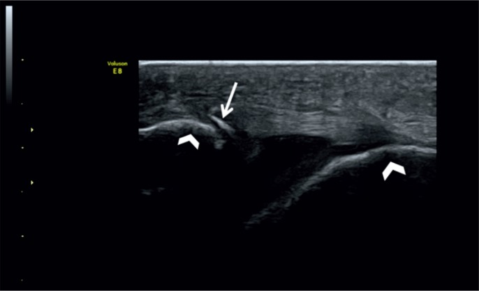
UltrasoundLongitudinal ankle ultrasound: thickened anterior talofibular ligament, with a small bone fragment avulsed from its fibular enthesis (arrow), between the two bony insertions (arrowheads).
Source : Szczepaniak et al., Journal of Ultrasonography, 2015, figure 8 · CC BY-NC-ND
Diagnosing a lateral ankle sprain is not simply a matter of noting a painful, swollen ankle after a “wrong move”. The clinical stake is twofold: on the one hand ruling out a fracture that would call for different management, on the other precisely characterising the ligament injury (which ligament, which grade), because that is what determines prognosis and rehabilitation. Such rigour is all the more justified in that lateral ankle sprain is one of the most common musculoskeletal injuries: around 2 million acute sprains occur each year in the United States 1, and analysis of US emergency department attendances between 2010 and 2024 counts nearly 7.4 million sprains, an incidence of 1.53 per 1000 person-years, highest in adolescents aged 15-19 (2.60/1000) and during sport, which accounts for 33.2% of cases, 15.8% of them in basketball alone 4. Far from benign, the sprain exposes up to 70% of individuals to chronic instability or residual disability 1 : the initial diagnosis is therefore not a formality, it is the first link in a trajectory.
🩻 The Ottawa ankle rules: when to request a radiograph?
Validated by a meta-analysis of 27 studies (15,581 patients), the Ottawa ankle rules rule out a fracture with a sensitivity approaching 100%.
A negative bony palpation and weight-bearing test allows radiography to be deferred. Source: Bachmann et al., BMJ 2003 (PMID 12595378).
🔬 Value of clinical tests and imaging
No single test is perfect: ATFL palpation “casts a wide net” (highly sensitive), the anterior drawer confirms (specific), ultrasound settles the question.
Reported sensitivity / specificity. The specificity of ultrasound is not shown here. Sources: Netterström-Wedin 2022 (PMID 34286639); Colò 2023 (PMID 37510068).
Key points
- Diagnostic reasoning follows a sequence: history and mechanism → Ottawa ankle rules to triage the indication for radiography → clinical examination, more reliable when delayed to day 4-5.
- The Ottawa ankle rules rule out a fracture with a sensitivity approaching 100% and reduce radiography use by 30 to 40% 10.
- The anterior drawer test is specific (87%) but poorly sensitive (54%): it confirms an injury, it does not rule one out 12.
- The palpation of the ATFL is highly sensitive (95-100%): if painless, it helps rule out an injury; combined with the anterior drawer, it optimises accuracy 12.
- Do not miss the differential diagnoses : fracture of the base of the 5th metatarsal and above all syndesmotic (high) sprain, a genuine diagnostic trap 14.
History and injury mechanism: the first piece of the puzzle
It all starts with the patient's account. The typical mechanism of lateral sprain combines inversion and plantar flexion of the foot, often on landing from a jump, stepping on uneven ground or changing direction. This combination is anatomically significant: the anterior talofibular ligament (ATFL) has precisely the role of restraining both plantar flexion and inversion, which explains its vulnerability to this mechanism 5. Understanding the injuring movement therefore points immediately to the structure most likely to be injured.
In an inversion sprain, ligament involvement follows a predictable order: the ATFL is typically the first, and often the only, ligament injured, followed by the calcaneofibular ligament (CFL), and only in rare cases the posterior talofibular ligament (PTFL) 5. This hierarchy is reflected in the injury frequencies: the ATFL is involved in 85% of ligament injuries, the CFL in 35% and the PTFL in 12% 5. The CFL, for its part, is the main stabiliser against forced inversion, with a position-dependent role: talocrural stability mainly in plantar flexion, subtalar stability mainly in dorsiflexion 5. These notions guide the examiner: isolated ATFL involvement does not mean the same thing as combined ATFL + CFL involvement.
The history must also establish several decisive elements:
- The sensation of a crack or of the ankle “giving out” at the moment of injury.
- The ability to bear weight immediately afterwards, or the inability to put the foot to the ground: information that feeds directly into the Ottawa ankle rules.
- The time course of the oedema : very rapid swelling and bruising suggest a more severe injury.
- Past history : a previous sprain multiplies the risk of another by about 3.5 1, and a systematic review confirms that a previous lateral sprain strongly increases the risk of a new sprain 2. A patient who “regularly turns their ankle” should raise the suspicion of underlying chronic instability, whose prevalence reaches 46% in individuals with a previous sprain 3.
The Ottawa ankle rules: is a radiograph needed?
The central question in the acute phase is simple: does this injured ankle conceal a fracture? Radiographing everyone would be both costly and needlessly irradiating; radiographing no one would risk missing fractures. The Ottawa ankle rules answer this dilemma by providing a validated, reproducible triage tool.
According to these rules, an ankle radiograph is indicated only in the presence of malleolar or midfoot pain combined with one of the following signs 9 :
- Bone tenderness localised to the posterior edge or tip of a malleolus (lateral or medial);
- Bone tenderness at the base of the 5th metatarsal ;
- Bone tenderness over the navicular bone ;
- An inability to bear weight (take a few steps) both at the time of injury and at examination.
The performance of this tool is remarkable. In the original study, the Ottawa ankle rules were 100% sensitive for detecting malleolar fractures and reduced radiography requests by 36% 9. A meta-analysis of 27 studies and 15,581 patients confirms their validity: sensitivity approaching 100% for ruling out an ankle or midfoot fracture, with modest specificity; applying them would reduce the number of unnecessary radiographs by 30 to 40%, and a negative test leaves a probability of fracture below 1.4% 10.
The clinical reading is therefore clear: these rules serve above all to rule out a fracture (high sensitivity), not to confirm one (modest specificity, hence a few radiographs “for nothing” but few missed fractures). In practice, the Ottawa bone and joint examination must precede any functional management: it is a safety prerequisite 10.
Why delay the clinical examination to day 4-5?
An often underestimated point: in the immediate acute phase, oedema, haematoma and pain make ligament examination unreliable. A swollen, guarded ankle can neither be finely palpated nor tested for laxity. That is why the severity of ligament injury is assessed more reliably by a delayed physical examination, performed 4 to 5 days after the injury, once the initial oedema and pain have subsided, rather than in the immediate acute phase 11.
The severity of ligament injury is assessed most reliably by a delayed physical examination, 4 to 5 days after the injury 11.
In practical terms, this outlines a two-stage diagnostic strategy. In the acute phase, the aim is to apply the Ottawa ankle rules to rule out a fracture and to begin protection and oedema control. At day 4-5, a repeat clinical examination allows precise grading of the ligament injury and guides rehabilitation. This timing avoids two pitfalls: overestimating a lesion on an acutely painful ankle, or conversely underestimating an instability masked by initial muscle guarding.
Clinical tests: anterior drawer, talar tilt and palpation
Once the most acute phase has passed, ligament examination relies on a combination of manoeuvres whose measurement properties must be known if they are to be interpreted correctly.
The anterior drawer test looks for excessive anterior translation of the talus within the mortise, indicating an ATFL injury. Its performance is now well characterised: low sensitivity (54%; 95% CI 35-71%) but good specificity (87%; 95% CI 63-96%) for ligament injury 12. In practical terms: when it is positive, it helps to confirm (rule in) an injury; when it is negative, it does not allow one to rule it out. A negative anterior drawer is therefore no ground for reassurance.
The palpation of the ATFL has the opposite profile: highly sensitive (95-100%) but poorly specific (0-32%) 12. Painless palpation therefore helps to rule out an ATFL injury, but painful palpation is not enough to confirm one (many false positives). It is from this complementarity that the best strategy emerges: combining palpation (sensitive) + anterior drawer (specific) optimises diagnostic accuracy 12: one to avoid missing the injury, the other to confirm it.
The talar tilt (inversion stress test) explores forced inversion and therefore the CFL side of lateral stability. Its rationale rests on the role of the CFL, the main ligamentous stabiliser against forced inversion, whose action depends on ankle position 5. Increased tilt points to combined ATFL + CFL involvement, corresponding to the more severe grades. As with any laxity manoeuvre, it is best interpreted in comparison with the uninjured side and on an ankle that is no longer swollen (delayed examination).
| Test | Structure examined | Sensitivity | Specificity | Clinical use |
|---|---|---|---|---|
| ATFL palpation | ATFL | 95-100 % | 0-32 % | Rules out an injury if painless (rule out) |
| Anterior drawer | ATFL | 54 % | 87 % | Confirms an injury if positive (rule in) |
| Talar tilt | CFL / inversion | Laxity manoeuvre, to be compared with the uninjured side | Points to combined ATFL + CFL involvement | |
These tests allow the picture to be linked to the three-grade classification of lateral sprain 6 :
- Grade I : incomplete tear of the ATFL (ligament merely stretched, without macroscopic rupture).
- Grade II : complete rupture of the ATFL + incomplete tear of the CFL.
- Grade III : complete rupture of both the ATFL and the CFL.
The place of second-line imaging: ultrasound and MRI
The diagnosis of lateral sprain remains above all clinical; cross-sectional imaging is not routine. Radiography, as we have seen, is justified only on the Ottawa criteria and targets bone, not ligament. When precise ligament characterisation is needed (doubt about a complete rupture, poor progress, elite sport, preoperative work-up), two examinations dominate.
The ultrasound examination is an accessible and dynamic alternative to MRI for assessing the lateral collateral ligament. It is highly sensitive for diagnosing an ATFL rupture (sensitivity ~97%), superior to MRI (~87%) in this specific indication 13. Its dynamic nature (the ability to test the ligament in real time, comparing with the uninjured side) and its availability make it a relevant first-line examination when ligament imaging is required.
The MRI scan retains major value for the work-up of associated injuries, which are often clinically invisible. In a prospective cohort of 171 acute ankle injuries in athletes assessed by 3-T MRI, a lateral ligament injury was present in 73% of cases, an anterior syndesmosis injury in 38%, and the overall prevalence of osteochondral/cartilage lesions of the talar dome reached 14% 8. These figures are a reminder that a “simple” sprain can conceal a syndesmotic or osteochondral injury that will change management and prognosis. MRI is therefore particularly justified when progress does not follow the expected trajectory.
Differential diagnoses not to be missed
Asserting “lateral sprain” presupposes that a number of look-alike conditions requiring different management have been ruled out. Three pitfalls deserve particular vigilance.
1. Fracture of the base of the 5th metatarsal. It shares the injury mechanism of the sprain and lies in the area covered by the Ottawa ankle rules. The link is close: in a series of 61 fractures, associated lateral ligament injury accounted for 63.93% of fractures of the base of the 5th metatarsal, the most frequent causes being sprains and falls 7. In other words, sprain and fracture of the base of the 5th metatarsal often coexist, hence the importance of specifically palpating that base, as the Ottawa ankle rules require.
2. Syndesmotic sprain (“high ankle sprain”). This is the most treacherous differential. These high sprains come with no bony or articular radiographic abnormality, so that they are far harder to assess in terms of severity and their diagnosis rests on subjective clinical signs: constituting a genuine diagnostic and therapeutic dilemma 14. It must be looked for systematically in every lateral ankle sprain, all the more so as its frequency is not negligible: the Baltes (2025) MRI cohort found anterior syndesmosis involvement in 38% of acute injuries in athletes. Missing a syndesmotic sprain means risking inappropriate treatment and prolonged recovery.
3. Underlying chronic ankle instability (CAI). Faced with a patient who sprains repeatedly, this is no longer a simple acute accident but an underlying predisposition. The International Ankle Consortium gives an operational definition: at least one significant lateral sprain sustained at least 12 months earlier, repeated episodes of giving way and/or a feeling of instability, the most recent injury more than 3 months old, with at least 2 episodes of giving way in the preceding 6 months, documented by a validated questionnaire (CAIT < 24, IdFAI > 11, or AII) 20. Recognising CAI changes the strategy: management stops being purely “acute” and becomes long-term neuromuscular reconditioning.
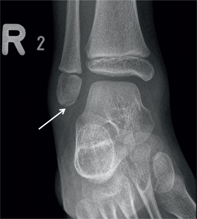
X-rayAnkle X-ray, mortise view: small bone fragment avulsed from the tip of the lateral malleolus (arrow), an avulsion fracture typical of a lateral ankle sprain.
Source : Szczepaniak et al., Journal of Ultrasonography, 2015, figure 3 · CC BY-NC-ND
Key points: the diagnostic approach in practice
- Take a history of the mechanism (inversion + plantar flexion) and past history 1.
- Apply the Ottawa ankle rules in the acute phase to triage the indication for radiography 10.
- Re-examine at day 4-5 to grade the ligament injury reliably 11.
- Combine palpation (sensitive) and anterior drawer (specific) for the ATFL, and test the talar tilt for the CFL 125.
- Reserve imaging for situations where it helps: ultrasound (ATFL ~97%) or MRI for associated injuries 138.
- Always rule out fracture of the base of the 5th metatarsal, syndesmotic sprain and chronic instability 71420.
Ultimately, “diagnosing with certainty” a lateral ankle sprain depends less on any single examination than on a reasoned sequence : a history that reconstructs the mechanism and the background, validated decision rules for radiography, a ligament examination performed at the right moment and interpreted according to the specific properties of each test, targeted rather than routine imaging, and an active search for differentials. It is this rigour that avoids the two symmetrical errors, trivialising a severe injury or over-treating a simple sprain, and lays the groundwork for effective functional management.
🛡️ What are the risk factors and how can a first sprain be prevented?
Lateral ankle sprain is one of the most common musculoskeletal injuries: an estimated 2 million acute sprains occur each year in the United States 1. This apparent banality masks a major public health and sporting issue, because a first sprain is never trivial: all too often it opens the door to a vicious circle of recurrence and chronic instability. Understanding who gets injured, why, and above all how to reduce the risk is therefore essential, both for the physiotherapist building a prevention programme and for the informed patient or athlete who wants lasting protection for their ankles.
Key points
- The most powerful risk factor for a lateral sprain is having already had one: a previous sprain multiplies the risk by about 3.5 1 and the evidence of an excess risk after a first lateral sprain is strong 2.
- Playing sport accounts for a large share of these injuries: sport represents 33.2% of all sprains seen in US emergency departments, 15.8% for basketball alone 4.
- The most exposed populations are adolescents (15-19 years) and women, in whom the incidence is higher than in men 1.
- Prevention rests above all on neuromuscular and proprioceptive training, which significantly reduces the incidence of sprains 21.
- Ankle braces are supported by strong evidence for preventing recurrence, but their place in primary prevention in the uninjured individual remains more limited 1511.
Risk factors: who gets injured, and why?
The risk factors for lateral ankle sprain are classically divided into intrinsic factors (specific to the individual: previous sprain, age, sex, neuromuscular control) and extrinsic factors (related to activity and environment: type of sport, surface, footwear). Recent epidemiological data make it possible to rank these factors and to identify the priority levers for action.
Previous sprain: the major risk factor
If only one determinant were to be remembered, it would be a previous sprain. Herzog (2019) reports that people who have already sustained an ankle sprain have a risk about 3.5 times higher of sustaining another. This relationship is solidly documented: a systematic review found a significant excess risk of lateral sprain in individuals with a previous sprain in 10 of 15 studies, with a relative risk ranging from 1.29 to 6.06 2.
This excess risk is no statistical accident: it reflects the functional sequelae left by a first sprain that was poorly or insufficiently managed. Up to 70% of people who have sustained an acute sprain may develop chronic ankle instability (CAI) over a short period, or retain residual physical disability 1. Logically enough, the proportion of recurrent sprains is estimated at between 12% and 47% 1. And the weight of a previous sprain on chronic instability is telling: the prevalence of CAI is 25% in the general population (range 7-53%), but rises to 46 % (range 9-76%) in people who have already had a sprain 3.
The best prevention of a “next” sprain begins with active, complete management of the first: a previous sprain is a risk factor, but it is also the only one on which well-conducted rehabilitation can act directly.
Sport, age and sex
Sport is the great supplier of sprains. In athletic populations the incidence is estimated at 0.93 per 1000 athlete-exposures 1, while emergency department data in the general population suggest 2 to 7 acute sprains per 1000 person-years. Above all, the most recent analysis of US emergency department attendances (2010-2024) confirms that sport is the origin of 33.2% of all sprains, with basketball alone accounting for 15.8% of cases 4. Pivoting, jumping and contact sports, with their unbalanced landings and rapid changes of direction, are particularly exposed to the inversion mechanism.
Age and sex clearly modulate this risk. Over the period 2010-2024, the incidence peaks in adolescents aged 15-19 (2.60/1000 person-years) and remains highest in women and in athletes 4. This excess risk in women is confirmed by Herzog (2019), who reports a higher incidence in women than in men (13.6 versus 6.9/1000 exposures), with a peak in girls aged 10-14 and in boys aged 15-19. Of note, in terms of trends, the overall incidence of sprains in emergency departments declined from 2.12 in 2010 to 1.19 in 2024 4, which may reflect changes in sporting practice, in prevention or in healthcare use.
| Risk factor | Magnitude of risk | Source |
|---|---|---|
| Previous sprain | Risk × ~3.5; RR 1.29 to 6.06; CAI prevalence rising to 46% | Herzog 2019; Wikstrom 2021; Lin 2021 |
| Sports participation | 33.2% of sprains (15.8% in basketball); 0.93/1000 athlete-exposures | Tyler 2025; Herzog 2019 |
| Adolescence (15-19 years) | Highest incidence: 2.60/1000 person-years | Tyler 2025 |
| Female sex | 13.6 vs 6.9/1000 exposures; peak at 10-14 years in girls | Herzog 2019; Tyler 2025 |
Primary prevention: neuromuscular and proprioceptive training first and foremost
Faced with these risk factors, what strategy should be adopted to avoid the first sprain? The answer best supported by current data is neuromuscular and proprioceptive training. This type of training aims to improve sensorimotor control of the ankle (the perception of its position in space, the reaction speed of the stabilising muscles and dynamic postural control), all qualities that allow a person to “catch” a loss of balance before it turns into a sprain.
The level of evidence is solid. A meta-analysis of 7 randomised controlled trials (3,726 participants) shows that proprioceptive training significantly reduces the incidence of ankle sprains, the effect remaining significant in individuals with a previous sprain 17. These results are confirmed and updated by Wang (2023): proprioceptive training reduces the risk of a lateral sprain compared with the control group (RR = 0.59; p < 0,001), with an even more marked preventive effect in individuals with a previous sprain (RR = 0.49; p = 0.02). That work explicitly recommends proprioceptive training to prevent lateral sprain, particularly in people who already have a history of sprain.
One important nuance should be stressed for clinical interpretation: most of this evidence concerns mixed or at-risk populations (athletes, individuals with a previous sprain), and the effect is particularly demonstrated in prevention of recurrence. In their synthesis, Doherty (2017) attributes a level of moderate evidence to neuromuscular training for preventing sprain recurrence. Proprioceptive training nevertheless retains value in primary prevention, notably when built into the warm-ups and physical preparation programmes of young sporting populations, in whom the incidence is highest.
The place of braces in prevention
Ankle braces (semi-rigid ankle supports) are the second great pillar of prevention. Their rationale is mechanical: to limit the range of inversion and provide external proprioceptive feedback, thereby reducing the likelihood of exceeding the injury threshold of the anterior talofibular ligament.
The level of evidence in favour of braces is robust, but mainly in the setting of prevention of recurrence. Doherty (2017) grants strong evidence to wearing a brace to prevent sprain recurrence, a level above that of neuromuscular training in this same indication. Vuurberg (2018) likewise concludes that, for the prevention of recurrent lateral sprains, braces should be considered an effective option.
| Preventive strategy | Level of evidence (prevention of recurrence) | Source |
|---|---|---|
| Brace | Strong evidence | Doherty 2017; Vuurberg 2018 |
| Neuromuscular training | Moderate evidence | Doherty 2017 |
| Proprioceptive training | RR 0.59 (overall); RR 0.49 in individuals with a previous sprain | Wang 2023; Schiftan 2015 |
Should the brace then be preferred over exercise, or the other way round? In athletes the answer is nuanced: one review demonstrated no difference between proprioceptive/neuromuscular training and braces in reducing the recurrence rate at 12 months 22. The available evidence (level II) does not allow one to be favoured over the other; the choice can therefore be made according to patient preference and clinician expertise. In practice the two approaches are not opposed but combined: the brace provides immediate mechanical protection, useful in particular on return to sport or during the resumption phase, while neuromuscular training builds active and lasting protection, independent of any external device.
In summary: ranking prevention
Preventing a first sprain (and, still more, its recurrence) is organised around a few principles consistent with current data:
- Identify at-risk individuals : previous sprain (the major factor, risk × 3.5), pivoting and jumping sports, adolescence, female sex. These populations are the priority targets of any preventive programme 14.
- Prioritise neuromuscular and proprioceptive training, whose effectiveness in reducing the incidence of sprains is demonstrated, particularly in individuals with a previous sprain 2117. This is the lever that builds lasting functional stability.
- Use braces as an effective option, notably in preventing recurrence and on return to activity, bearing in mind that they do not outperform training in athletes at 12 months 151122.
- Treat the first sprain actively : since a previous sprain is the main risk factor and up to 70% of individuals may develop chronic instability 13, complete functional management from the acute phase onwards is in itself an act of prevention.
In other words, preventing ankle sprain means acting on two timescales: reducing the risk in the uninjured but exposed individual through neuromuscular training, and breaking the vicious circle of recurrence in the person who has already gone over, by combining active rehabilitation with appropriate mechanical protection.
🩹 Which treatment strategies are the most effective?
Lateral ankle sprain has long been regarded as a benign injury to be left to heal on its own. Recent data flatly contradict that idea: up to 70% of individuals who sustain an acute sprain may develop chronic ankle instability (CAI) or retain residual physical disability, 12 to 47% of sprains are recurrences, and a previous sprain multiplies the risk of another by about 3.5 1. In other words, the fate of a sprain is not decided in the first few hours after the injury, but in the quality of the active management that follows. The aim of treatment is therefore not only to relieve the initial pain and oedema, but to restore the functional stability of the joint and prevent the recurrence → chronic instability spiral.
This section details the strategies whose effectiveness is best supported today: the PEACE & LOVE framework in the acute phase, the superiority of functional treatment over immobilisation, early protected loading, the measured place of cryotherapy and compression, the caution required with NSAIDs, the primacy of supervised exercise over passive modalities, the contribution of manual therapy, and finally the surgical indications, reserved for failures of conservative treatment.
Key points
- Functional treatment > immobilisation : the patient with an acute lateral ligament rupture benefits most from taping or a brace combined with an exercise programme 11.
- Supervised exercise > passive modalities : exercise programmes stimulate recovery of the functional stability of the joint 11.
- The PEACE & LOVE framework : protect without over-protecting, resume loading early, stay optimistic, keep moving, and be wary of anti-inflammatory drugs (and of ice), which may hinder healing in the long term 16.
- Caution with NSAIDs : effective on pain and oedema, but not free of complications and liable to suppress the natural healing process 11.
- Surgery as a last resort : reserved for cases that do not respond to a complete, well-conducted exercise-based treatment 11.
Acute phase: the PEACE & LOVE framework
The RICE protocol (Rest, Ice, Compression, Elevation), and then its successors POLICE and PRICE, dominated the management of soft tissue injuries for decades. In 2020 Dubois and Esculier proposed an updated framework, PEACE & LOVE, covering the whole recovery continuum: PEACE describes the first few days after the injury, LOVE the subsequent management 16. Its underlying logic: inflammation is an integral part of natural healing, and trying to suppress it systematically can work against long-term recovery.
PEACE: the first few days:
- P: Protection. Unload or restrict painful movements during the very first days, in order to limit worsening of the injury. Protection must remain brief: prolonged immobilisation is not the aim.
- E: Elevation. Raise the limb above heart level to promote drainage of interstitial fluid out of the injured region.
- A: Avoid anti-inflammatory modalities. Avoid anti-inflammatory drugs, and ice, because they may hinder long-term recovery by interfering with inflammation, a necessary step in tissue repair.
- C: Compression. Use a bandage or taping to limit intra-articular oedema and haemorrhage.
- E: Education. Inform the patient about the benefits of an active approach and steer them away from unnecessary passive treatments or superfluous imaging.
LOVE: the subsequent management:
- L: Load. Gradually resume activities and appropriate loading as soon as symptoms allow: optimal mechanical stress promotes tissue repair and remodelling.
- O: Optimism. Encourage realistic and positive expectations; psychological factors (fear, catastrophising, depression) influence prognosis.
- V: Vascularisation. Include pain-free cardiovascular activity to improve blood flow to the injured tissues and to sustain motivation.
- E: Exercise. Restore mobility, strength and proprioception through an active exercise programme, respecting the pain-free rule.
The central message of PEACE & LOVE: inflammation is not the enemy. Protect without over-protecting, then load, move and reassure, that is what distinguishes modern management from simple rest.
This framework is not a recipe of passive modalities but a philosophy: it shifts the cursor from rest towards controlled activity and invites the clinician to reconsider two historical reflexes: routine ice and early anti-inflammatory drugs.
Functional treatment versus immobilisation
The question “should a lateral ankle sprain be immobilised?” has now been settled by the international guidelines. According to the synthesis by Vuurberg et al. (2018), while brief immobilisation may be useful to relieve pain and oedema in the very first days, the patient with an acute lateral ligament rupture benefits most from the use of taping or a brace combined with an exercise programme, that is, from functional treatment, as opposed to prolonged rigid immobilisation.
The principle is that of “functional elastic support”: keeping the ankle on a safe path while allowing early movement and loading. This approach exploits the fact that ligament tissue, like other connective tissues, remodels better under appropriate mechanical load than in strict immobility.
| Approach | Principle | Place in management | Level of evidence |
|---|---|---|---|
| Prolonged immobilisation | Cast / rigid splint for several weeks | Not recommended routinely; reserved for particular situations | To be avoided routinely |
| Brief immobilisation | Relative rest for a few days | May relieve pain and oedema in the very earliest phase | Transitional adjunct |
| Functional treatment | Taping or brace + exercise programme | Reference approach for acute ligament rupture | Recommended |
The practical message is clear: the brace or the taping is not an end in itself. Its value lies in the fact that it makes early exercise possible by securing the joint, not in the fact that it replaces movement.
Early protected loading
Protected loading is the direct extension of the “Load” principle of the LOVE framework: resuming appropriate mechanical loading as soon as pain allows, rather than waiting for symptoms to disappear completely. It rests on the idea that optimal mechanical stress, neither excessive nor absent, favourably directs tissue repair and the recovery of functional stability 16.
In practical terms, this means allowing weight-bearing, guided by pain, under cover of functional support, and progressing towards normal walking and then towards more demanding activities. This early resumption of loading works together with the brace: the brace protects the joint's path while the patient reloads and remobilises. It is this combination, protection + loading + exercise, that forms the basis of modern functional treatment, not loading in isolation.
Cryotherapy and compression: a place to be qualified
Compression retains an explicit place in the PEACE framework: the “C” does indeed denote the use of a bandage or taping to limit oedema and haemorrhage 16. On this point the recommendation is stable.
The case of cryotherapy (ice), by contrast, is the one that has changed most. Where the RICE protocol made it a pillar, the PEACE framework places it among the anti-inflammatory modalities to be avoided : ice, like anti-inflammatory drugs, may interfere with the inflammatory phase necessary for healing and hinder long-term recovery 16. This does not mean that ice is formally contraindicated, it can bring transient pain relief, but that it should no longer be applied as a systematic reflex or presented as an accelerator of healing.
Compression: yes, for oedema. Ice: to be kept in proportion: an adjunct analgesic, not a treatment of the injury.
NSAIDs: effective but to be handled with care
Non-steroidal anti-inflammatory drugs illustrate a real tension in the literature, which must be presented honestly to patient and clinician alike.
On the one hand, symptomatically, the evidence is solid: the synthesis by Doherty et al. (2017) reports strong evidence in favour of NSAIDs for the treatment of acute sprain, alongside early mobilisation, for acting on pain, oedema and function. Vuurberg et al. (2018) confirm that NSAIDs may be used to reduce pain and oedema.
On the other hand, the same Vuurberg recommendation attaches an important caveat to that use: it is not free of complications and they may suppress the natural healing process 11. This is exactly the reasoning behind the “A” of PEACE: avoid anti-inflammatory modalities because inflammation contributes to repair 16.
The practical synthesis is therefore a matter of dose and purpose: NSAIDs can be useful as occasional analgesics to make early loading and exercise possible, but their routine and prolonged use is not innocuous and could come at the expense of long-term healing. The decision must be individualised, explained, and limited in time.
Supervised exercise: the priority over passive modalities
If only one principle of rehabilitation were to be remembered, it would be this one: supervised exercise programmes are to be preferred over passive modalities, because they stimulate the recovery of the functional stability of the joint 11. Exercise is not an optional add-on to analgesic modalities (ice, ultrasound, electrotherapy): it is their core, and those modalities are subordinate to it.
The overall level of evidence is consistent with this hierarchy. For the acute phase, Doherty et al. (2017) report moderate evidence in favour of exercise and manual therapy techniques for improving pain, oedema and function. Above all, it is in preventing recurrence and chronic instability that exercise comes into its own:
- A meta-analysis of 7 RCTs (3,726 participants) shows that proprioceptive training significantly reduces the incidence of ankle sprains, the effect remaining significant in individuals with a previous sprain (relative risk = 0.64; 95% CI 0.51–0.81) 17.
- Another synthesis confirms that proprioceptive training reduces the risk of lateral sprain compared with control (risk ratio 0.59; p < 0.001), with a preventive effect that is more marked in individuals with a previous lateral sprain (risk ratio 0.49; p = 0.02) 21.
- Doherty et al. (2017) report moderate evidence in favour of neuromuscular training for preventing recurrence, and stress that therapeutic exercise is supported in the prevention of chronic ankle instability (CAI).
Rehabilitation must therefore progress from range of motion and strength towards targeted proprioceptive and neuromuscular work, the condition for a lasting return to activity. The return-to-sport decision is best structured by the PAASS framework (98% panel agreement), which covers five domains: Pain, Ankle impairments (range of motion, strength/endurance/power), Athlete perception (confidence, perceived stability, psychological readiness), Sensorimotor control (proprioception, dynamic postural control/balance) and Sport/functional performance (hopping, agility, sport-specific drills, ability to complete a full training session) 18. No single objective criterion is validated, but among the functional tests, single-leg stance on a stable surface, the modified Star Excursion Balance Test, the side hop test and the figure-of-8 test appear to be the most relevant for guiding the decision 19.
Manual therapy
Manual therapy has its place in the acute-phase toolkit, as an adjunct to exercise. Doherty et al. (2017) report moderate evidence in favour of manual therapy techniques, alongside exercise, for improving pain, oedema and function. Joint mobilisations, aimed in particular at restoring physiological talocrural gliding and recovering the dorsiflexion range that is often limited after a sprain, fit logically into active management. They do not replace exercise but can facilitate it, by lifting certain mobility restrictions that hinder the resumption of loading and proprioceptive work.
Braces and prevention of recurrence
Beyond the acute phase, the brace has a dual function: it secures the initial functional treatment, then becomes a tool of prevention. For preventing recurrent lateral sprains, ankle braces should be considered an effective option 11, and Doherty et al. (2017) report strong evidence in favour of bracing to prevent recurrence.
Should the brace then be preferred, or proprioceptive/neuromuscular rehabilitation? In athletes there is no demonstrated difference between proprioceptive/neuromuscular training and braces at 12 months for reducing the recurrence rate: the choice can be made according to patient preference and clinician expertise 22. In practice the two are often combined: a brace for the period of at-risk resumption, neuromuscular training as a lasting investment.
Surgical indications: the last resort
Surgery (ligament repair or reconstruction) has no first-line place in lateral ankle sprain. The position of the guidelines is unambiguous: surgery must be reserved for cases that do not respond to a complete, well-conducted exercise-based treatment 11. In other words, it is a treatment for failure of conservative care, considered when mechanical instability persists despite active rehabilitation carried through to its end.
This principle has a direct practical consequence: the first “surgical” strategy consists precisely in making sure that the patient has received a complete, supervised and sufficiently prolonged exercise programme. A large share of operative indications disappears once functional treatment has genuinely been carried through, with targeted proprioceptive and neuromuscular work.
Key points: hierarchy of strategies
- 1. Acute phase (PEACE): protect briefly, elevate, compress, educate, and avoid the systematic anti-inflammatory reflex 16.
- 2. What follows (LOVE): load early, sustain optimism and vascularisation, make exercise the pillar of recovery 16.
- 3. Functional treatment: taping/brace + exercises, preferred over prolonged immobilisation 11.
- 4. Supervised exercise > passive modalities, with proprioceptive/neuromuscular work to prevent recurrence and CAI 11172115.
- 5. Brace for prevention (strong evidence); no demonstrated superiority of brace vs proprioception at 12 months 1522.
- 6. Surgery: only after failure of a complete, well-conducted conservative treatment 11.
🔄 How can lasting recovery be achieved and recurrence prevented?
The most delicate phase of lateral ankle sprain is not the acute pain of the first few days, but the weeks and months that follow. Ankle sprain is not a benign injury: up to 70% of people who sustain an acute sprain may develop chronic ankle instability (CAI) or retain residual physical disability, 12 to 47% of reported sprains are recurrences, and a previous sprain multiplies the risk of another by about 3.5 1. A systematic review confirms, moreover, that a previous lateral sprain strongly increases the risk of a new sprain, with a relative risk ranging from 1.29 to 6.06 across studies 2. In other words, what happens in rehabilitation largely determines the long-term prognosis.
⚠️ Ankle sprain is not a benign injury
The long-term outcome justifies complete active rehabilitation rather than simple rest.
Outcome after an acute lateral sprain. Sources: Herzog et al., 2019 (PMID 31135209).
Lasting recovery therefore amounts to more than “no longer being in pain”. It means restoring strength, range of motion, sensorimotor control and confidence, objectively validating the return to activity, and putting active secondary prevention in place. It is precisely because the risk of chronicity is high that active, exercise-based management must be started from the acute phase onwards 1.
Key points
- The long-term prognosis is decided in rehabilitation: up to 70% CAI or residual disability, 12-47% recurrence, risk ×3.5 after a previous sprain 1.
- Progression is driven by clinical and functional criteria, not by the mere passage of time.
- Proprioceptive/neuromuscular training reduces the risk of sprain (RR 0.59), with a stronger effect in individuals with a previous sprain (RR 0.49) 2117.
- Return to sport is assessed across five domains (the PAASS framework), not on a single test 18.
- Prevention of recurrence: high-level evidence for the brace, moderate evidence for neuromuscular training; the two are equivalent at 12 months in athletes 1522.
Progress by criteria, not by the calendar
Contemporary rehabilitation logic replaces chronological progression (“such a week, such an exercise”) with progression by criteria: one only moves to the next stage when the objectives of the current stage have been met. This approach fits within the PEACE & LOVE framework, which structures the management of soft tissue injuries in two phases 16. PEACE governs the first few days (Protection, Elevation, Avoid anti-inflammatory modalities, Compression, Education), while LOVE guides the rest of recovery: Load (progressive resumption of loading), Optimism (optimism), Vascularisation (cardiovascular activity) and Exercise (exercise). The founding principle is that inflammation is part of natural healing: anti-inflammatory drugs, and ice, may hinder long-term recovery 16. This is also what justifies caution with NSAIDs, which relieve pain and oedema but may suppress the natural healing process 11.
The foundation of this progression is functional treatment, to be preferred over immobilisation. While brief immobilisation may help to relieve pain and oedema, the patient with an acute lateral ligament rupture benefits most from combining taping or a brace with an exercise programme 11. Within that programme, supervised exercises are preferred over passive modalities, because they stimulate the recovery of the functional stability of the joint 11. The scientific evidence supports this hierarchy: in the acute phase there is a moderate level of evidence in favour of exercise and manual therapy techniques for improving pain, oedema and function 15.
In practical terms, progression follows stages, each of which conditions the next:
- Restore range of motion and settle symptoms : recover dorsiflexion (often limited after a sprain), reduce oedema and pain on walking.
- Restore strength and control : strengthening of the evertors and of the whole ankle complex, reintroduction of loading under control.
- Restore balance and sensorimotor control : progression from double-leg to single-leg stance, then to unstable surfaces and dynamic movement.
- Reconditioning and gradual return : jumps, changes of direction, sport-specific movements, before validating the return to sport.
None of these stages is passed on the basis of elapsed time alone: it is the achievement of objective criteria (symptoms controlled, symmetrical range of motion and strength, quality of control) that authorises progression. This rigour is all the more important in that the initial severity of the ligament injury is itself assessed more reliably by a delayed examination, 4 to 5 days after the injury, once oedema and pain have subsided 11 : regular reassessment is the rule, at the start as in the long term.
Training balance and proprioception: the heart of prevention
If a single ingredient had to be retained from ankle rehabilitation, it would be neuromuscular and proprioceptive training. A deficit in sensorimotor control is indeed a central mechanism of recurrence and chronic instability, and it is one of the few levers whose preventive effect is solidly demonstrated.
A meta-analysis of 7 randomised controlled trials including 3,726 participants showed that proprioceptive training significantly reduces the incidence of ankle sprains, the effect remaining significant in individuals who already have a previous sprain (relative risk = 0.64; 95% CI 0.51-0.81) 17. More recent work confirms and refines this result: proprioceptive training reduces the risk of lateral sprain compared with the control group (risk ratio 0.59; p < 0.001), with an even more marked preventive effect in people with a previous lateral sprain (risk ratio 0.49; p = 0.02) 21. The authors explicitly recommend proprioceptive training to prevent lateral sprain, particularly in individuals with a previous sprain, that is, precisely the population the physiotherapist manages after a first sprain.
At the level of the guidelines, these data translate into a moderate level of evidence in favour of neuromuscular training for preventing sprain recurrence 15. In practice, balance training also follows a progression by criteria:
- Stable surface, double-leg then single-leg stance, eyes open.
- Increasing the sensory difficulty : eyes closed, then dual task (cognitive or motor).
- Unstable surfaces and perturbations : cushions, wobble boards, imposed loss of balance.
- Dynamic integration : landing from jumps, changes of direction, movements specific to the sport or activity.
This training is not reserved for athletes: it is the mechanism by which the ankle “relearns” how to stabilise itself and react to loss of balance, which benefits any patient keen to avoid a recurrence.
Return to sport: five domains to validate, not a date
The timing of the return to sporting activity is one of the most consequential decisions of all, because a premature return leads directly to recurrence. Yet there is no single validated objective criterion for return to sport after a lateral sprain 19. That is why the decision must rest on a multidimensional assessment rather than on an isolated test or a set delay.
The reference framework is the PAASS model, arising from an international consensus with 98% agreement among the expert panel 18. It structures the return-to-sport decision around five domains:
| PAASS domain | What is assessed |
|---|---|
| P: Pain | Absence or control of pain during the targeted activities and movements. |
| A: Ankle impairments | Range of motion; muscle strength, endurance and power. |
| A: Athlete perception | Confidence, perceived stability, psychological readiness. |
| S: Sensorimotor control | Proprioception; dynamic postural control and balance. |
| S: Sport/functional performance | Hopping and jumping and agility; sport-specific drills; ability to complete a full training session. |
The psychological dimension, athlete perception, deserves particular attention: confidence and perceived stability are components of the decision in their own right, just as much as strength or range of motion 18. A patient who “no longer trusts their ankle” is not ready, even if their physical tests are satisfactory.
To objectify the sensorimotor and functional domains, several functional tests appear to be the most relevant for guiding the decision: single-leg stance on a stable surface, the modified Star Excursion Balance Test, the side hop test and the figure-of-8 test 19. These tests, easy to perform in the clinic, allow the injured side to be compared with the uninjured side and progress to be tracked over time. They do not replace the PAASS framework but feed into it: they objectively document postural control and performance, two of the five domains to be validated.
There is no single objective criterion for return to sport after a lateral sprain: the decision is built on five domains and a set of functional tests, never on a single measure.
Chronic ankle instability (CAI): recognising and managing it
Chronic ankle instability (CAI, chronic ankle instability) is the major complication of lateral sprain. It combines repeated episodes of giving way with a feeling of instability, and has lasting functional consequences. Its frequency alone justifies the intensity of prevention: up to 70% of people who sustain an acute sprain may develop CAI within a short period after the initial injury 1. A systematic review gives a more nuanced picture of its prevalence: 25% in the general population (range 7-53%), but 46% (range 9-76%) in people with a previous sprain 3. In other words, nearly one in two people who have already had a sprain has chronic instability.
Defining CAI by operational criteria. To avoid vague diagnoses, the International Ankle Consortium has established precise criteria 20. CAI is defined by:
- at least one significant lateral sprain, sustained at least 12 months earlier;
- repeated episodes of giving way and/or a feeling of instability;
- a most recent injury dating back more than 3 months;
- at least 2 episodes of giving way in the preceding 6 months;
- documentation by a validated questionnaire: CAIT < 24, IdFAI > 11, or AII 20.
These criteria serve two purposes: they identify the patients who need targeted management, and they are a reminder that “felt” instability must be objectified with standardised tools. The CAIT and IdFAI questionnaires, quick to administer, have their full place here in follow-up.
Managing CAI. Treatment remains above all functional and exercise-based. Therapeutic exercise and braces are supported in the prevention of chronic ankle instability 15, and neuromuscular/proprioceptive training, whose preventive effectiveness is demonstrated including in individuals with a previous sprain 1721, is its mainstay. Surgery comes only second: it must be reserved for cases that do not respond to a complete, well-conducted exercise-based treatment 11. In practical terms, this means that a comprehensive, good-quality exercise programme must always precede any surgical discussion.
Secondary prevention: brace, exercise, or both?
Once recovery is under way, the question becomes: how can the risk of recurrence be reduced for the long term? Two strategies dominate, the brace and neuromuscular training, and the literature allows them to be ranked.
In terms of evidence, prevention of recurrence rests on solid data: there is strong evidence in favour of bracing and moderate evidence in favour of neuromuscular training for preventing sprain recurrence 15. For the prevention of recurrent lateral sprains, ankle braces should therefore be considered an effective option 11.
| Strategy | Level of evidence (prevention of recurrence) | Key points |
|---|---|---|
| Brace | High-level evidence 15 | An effective option to consider for preventing recurrence 11 ; useful in particular when resuming sport. |
| Neuromuscular / proprioceptive training | Moderate evidence 15 | Reduces the risk of sprain (RR 0.59) and more so in individuals with a previous sprain (RR 0.49) 2117. |
Should a choice then be made between the two? The answer is nuanced. In athletes, one review found no difference between proprioceptive/neuromuscular training and braces in reducing the recurrence rate at 12 months: the current evidence (level II) does not favour one over the other, and the choice can be made according to patient preference and clinician expertise 22. In practice, this opens the way to a combined, individualised strategy: the brace offers immediate mechanical protection, particularly reassuring when resuming activity and for high-risk individuals, while exercise builds active, lasting protection by correcting the underlying sensorimotor deficit. The two are not opposed: they complement each other.
This combined logic in fact mirrors the reference treatment of the sprain itself, in which the patient benefits most from combining taping or a brace with an exercise programme 11. The message for the patient is clear: wearing a brace does not remove the need to work the ankle, and working the ankle does not forbid mechanical protection in risky situations.
Key points: a plan for lasting prevention
- Do not stop at the absence of pain : restore strength, range of motion, sensorimotor control and confidence before considering recovery complete.
- Make proprioceptive training the backbone of the programme: it is the best-demonstrated preventive lever, especially after a first episode 2117.
- Validate the return to sport on the five PAASS domains and a set of functional tests, never on a date 1819.
- Screen for CAI with the International Ankle Consortium criteria and the CAIT/IdFAI questionnaires 20 ; treat it first with exercise, surgery being only a fallback in case of failure 11.
- Combine brace (high-level evidence) and exercise (moderate evidence) according to patient preference: at 12 months, neither has demonstrated superiority over the other in athletes 1522.
📋 What do concrete case studies teach us?
Epidemiological data and guidelines set out principles, but it is at the patient's side that they take on meaning. To illustrate clinical reasoning, here are two cases that are fictional and purely educational : they belong to no real patient and contain no observed data. Every decision taken in them follows strictly from the principles drawn from the literature summarised in this article; no figure, no result is invented beyond what the studies allow. The aim is not to tell a story, but to make the logic visible: how, at each step, one decides what to look for, what to rule out and what to propose.
A case study proves nothing; it shows how to apply what has been proved.
Case study no. 1: Acute grade II sprain in an athlete
Imagine an amateur basketball player who turns their ankle on landing from a jump, foot rolling inwards. The mechanism described, inversion combined with plantar flexion, is precisely the one that loads the anterior talofibular ligament (ATFL), whose functional role is to restrain both plantar flexion and inversion 5. This context is in no way exceptional: sport accounts for 33.2% of the sprains presenting to US emergency departments over 2010-2024, with basketball alone accounting for 15.8% of cases, and a peak incidence in 15-19 year-olds 4. In other words, this profile is one of the most typical of the condition.
Step 1: The acute phase: first rule out a fracture
The first clinical reflex is not to grade the sprain, but to rule out a fracture. That is the role of the Ottawa ankle rules: a radiograph is indicated only in the presence of malleolar or midfoot pain combined with localised bone tenderness (posterior edge or tip of a malleolus, base of the 5th metatarsal, navicular bone) or with an inability to bear weight 9. These rules have a sensitivity approaching 100% for ruling out an ankle or midfoot fracture and reduce radiography use by 30 to 40%; a negative test leaves a probability of fracture below 1.4% 10. In our athlete, if bony palpation is painless at the Ottawa points and weight-bearing remains possible, radiography is not required.
Reasoning must nevertheless stay alert to a common trap: fracture of the base of the 5th metatarsal is frequently associated with an injury of the lateral collateral ligament. In a series of 61 fractures, associated lateral ligament injury accounted for 63.93% of fractures of the base of the 5th metatarsal, sprains and falls being the most frequent causes 7. Palpation of this landmark, explicitly included in the Ottawa ankle rules, is therefore no formality.
Step 2: Do not grade too early
The temptation is to assess ligament severity straight away. Yet the literature calls for patience: severity is assessed more reliably by a delayed physical examination, performed 4 to 5 days after the injury, once the initial oedema and pain have subsided, rather than in the immediate acute phase 11. In the acute phase, haematoma and muscle guarding distort the appreciation of laxity. The astute clinician documents the mechanism, applies Ottawa, sets up first-line management, and reschedules the structured examination.
Step 3: The delayed examination: combining a sensitive test and a specific test
Five days later, the examination can be carried out reliably. Diagnostic reasoning here relies on the complementarity of two tests with opposite properties:
| Test | Property | What it allows | Limitation |
|---|---|---|---|
| ATFL palpation | Highly sensitive (95-100%), poorly specific (0-32%) | Painless palpation helps to rule out an ATFL injury | Painful palpation is not enough to confirm one |
| Anterior drawer | Poorly sensitive (54%), good specificity (87 %) | A positive test helps to confirm (rule in) the injury | A negative test does not allow it to be ruled out |
Combining the two, palpation (sensitive) then anterior drawer (specific), optimises diagnostic accuracy 12. In our basketball player, painful ATFL palpation together with a positive anterior drawer points to a confirmed lateral ligament injury. The anatomical classification then makes it possible to place the severity: grade II corresponds to a complete rupture of the ATFL combined with an incomplete tear of the calcaneofibular ligament (CFL), to be distinguished from grade I (ligament merely stretched, without rupture) and grade III (complete rupture of both ATFL and CFL) 6. This injury progression follows a predictable order: the ATFL is injured first, it is the ligament involved in 85% of ligament injuries in sprains, followed by the CFL (involved in 35% of cases), the posterior talofibular ligament being affected only rarely (12%) 5.
Step 4: Look for what must not be missed
A “simple” grade II does not remove the need to look for associated injuries, some of which are deceptive. Syndesmotic sprain (high sprain) is a differential diagnosis never to be overlooked: harder to assess because it produces no bony or articular radiographic abnormality, it rests on subjective clinical signs and constitutes a genuine diagnostic and therapeutic dilemma; it must be looked for in every lateral sprain 14. This vigilance is not theoretical: in a prospective cohort of 171 acute ankle injuries in athletes assessed by 3-T MRI, a lateral ligament injury was present in 73% of cases, but an anterior syndesmosis injury in 38% and an osteochondral lesion of the talar dome in 14% 8. In an athlete, these hidden lesions can explain a poor course if they are not identified.
If diagnostic doubt persists about the integrity of the ATFL, second-line imaging favours ultrasound, which is highly sensitive for diagnosing an ATFL rupture (sensitivity ~97%), superior to MRI (~87%) in this specific indication, while being more accessible and dynamic 13. MRI keeps its place for osteochondral and syndesmotic lesions.
Step 5: Treatment: functional rather than immobilisation
The management of a grade II follows a clear principle: functional treatment takes precedence over prolonged immobilisation. Brief immobilisation may help to relieve pain and oedema, but the patient with an acute lateral ligament rupture benefits most from combining taping or a brace with an exercise programme 11. Supervised exercise programmes are preferred over passive modalities, because they stimulate the recovery of the functional stability of the joint 11. For the acute phase, there is strong evidence in favour of NSAIDs and early mobilisation, and moderate evidence for exercise and manual therapy on pain, oedema and function 15.
The question of anti-inflammatory drugs deserves nuance. NSAIDs can reduce pain and oedema, but their use is not free of complications and they may suppress the natural healing process, hence a recommendation of caution 11. That is also the message of the PEACE & LOVE framework: PEACE (Protection, Elevation, Avoid anti-inflammatory modalities, Compression, Education) then LOVE (Load, Optimism, Vascularisation, Exercise), inflammation being part of natural healing that ice and anti-inflammatory drugs may hinder in the long term 16.
Step 6: Deciding on return to sport
In an athlete, resumption is not decreed on elapsed time or the absence of pain alone. There is no single validated objective criterion for return to sport after a lateral sprain 19. The decision must rest on five domains: the PAASS framework (98% panel agreement): Pain, Ankle impairments (range of motion, strength, endurance, power), Athlete perception (confidence, perceived stability, psychological readiness), Sensorimotor control (proprioception, dynamic postural control, balance) and Sport/functional performance (hopping, agility, sport-specific drills, ability to complete a full training session) 18. Among the functional tests, single-leg stance on a stable surface, the modified Star Excursion Balance Test, the side hop test and the figure-of-8 test appear to be the most relevant for guiding the decision 19.
A clinician who allows a return simply because “the ankle no longer hurts” is applying a single criterion: precisely the one the literature judges insufficient. Complete reasoning checks the five PAASS domains before handing the pitch back to the player.
Key points: Case no. 1 (grade II, athlete)
- Ottawa first : rule out fracture (sensitivity ~100%) before anything else, not forgetting the base of the 5th metatarsal 97.
- Grade later : reliable examination at 4-5 days, once the oedema has settled 11.
- Combine the tests : palpation (sensitive, rule out) + anterior drawer (specific, rule in) 12.
- Treat functionally : taping/brace + supervised exercises, caution with NSAIDs and ice 1116.
- Resumption = the 5 PAASS domains, not just the absence of pain 1819.
Case study no. 2: Chronic ankle instability (CAI)
A second fictional scenario: a person who sustained a first lateral sprain well over a year earlier and who presents with repeated episodes of giving way and a persistent feeling of instability, particularly on uneven ground. This picture is not rare: up to 70% of people who have sustained an acute sprain may develop chronic ankle instability (CAI) within a short period, and up to 70% may retain residual physical disability 1. The sprain is therefore not the benign injury it is thought to be.
Step 1: Diagnose CAI with criteria, not impressions
CAI is not a vague feeling; it meets operational criteria set by the International Ankle Consortium: at least one significant lateral sprain sustained at least 12 months earlier, repeated episodes of giving way and/or a feeling of instability, the most recent injury more than 3 months old, with at least 2 episodes of giving way in the preceding 6 months, documented by a validated questionnaire (CAIT < 24, IdFAI > 11, or AII) 20. In our patient, ticking off these criteria turns a subjective complaint into a structured diagnosis and guides what to do next.
The scale of the phenomenon justifies this rigour. In a systematic review, the prevalence of chronic ankle instability is 25% (range 7-53%); it reaches 46% in people with a previous sprain (range 9-76%) 3. A previous sprain is therefore a strong signal, to be sought systematically in the history.
Step 2: Understand why recurrence breeds recurrence
The reason for consultation is part of a documented circle. The proportion of recurrent sprains is estimated at between 12% and 47%, and a previous sprain multiplies the risk of sustaining another by about 3.5 1. A systematic review shows strong evidence that a previous lateral sprain increases the risk of a subsequent sprain, with a relative risk of 1.29 to 6.06 across studies 2. Clinical reasoning takes this on board: in this patient the aim is not only to relieve symptoms, but to break the recurrence loop, which shifts the centre of gravity of management towards active prevention.
In the unstable patient, what is at stake is no longer yesterday's sprain, but tomorrow's.
Step 3: Therapeutic reasoning: preventing the next sprain
The management of CAI and the prevention of recurrence rest on two pillars whose evidence is well established:
| Lever | Level of evidence | What the literature says |
|---|---|---|
| Brace / bracing | High-level evidence | Strong evidence for the prevention of recurrence; an effective option to consider; supported for the prevention of CAI 1511 |
| Neuromuscular / proprioceptive training | Moderate evidence | Moderate evidence for preventing recurrence and CAI; reduces the incidence of sprains, with the effect maintained in individuals with a previous sprain (RR = 0.64) 1517 |
The most relevant argument for our patient is that the preventive effect of proprioception is demonstrated precisely in people with a previous sprain. A meta-analysis of 7 RCTs (3,726 participants) shows that proprioceptive training significantly reduces the incidence of sprains, the effect remaining significant in individuals with a previous sprain (relative risk = 0.64; 95% CI 0.51-0.81) 17. Another study confirms the reduction in risk (risk ratio 0.59, p < 0.001), with an even more marked preventive effect in individuals with a previous sprain (risk ratio 0.49, p = 0.02), explicitly recommending proprioceptive training in these people 21.
Step 4: Brace or rehabilitation? A shared decision
Faced with these two levers, what reasoning should be adopted? Neither has demonstrated superiority: for reducing the recurrence rate in athletes, there is no demonstrated difference between proprioceptive/neuromuscular training and braces at 12 months; the choice can be made according to patient preference and clinician expertise 22. In practical terms, the decision is built with the patient: a brace to secure a rapid return to activity, a supervised neuromuscular programme to restore sensorimotor control for the long term, often both, depending on their goals and adherence. The clinician who imposes a single lever “because they believe in it” departs from what the evidence says: the two are equivalent, and patient preference is a legitimate decision criterion.
Step 5: When to consider surgery?
Finally, in an unstable patient who remains symptomatic, the surgical question may arise, but it comes last. Surgery must be reserved for cases that do not respond to a complete, well-conducted exercise-based treatment 11. In other words, as long as a structured, comprehensive functional programme has not been carried through to its end, CAI is first and foremost a matter for rehabilitation. The chronological reasoning, exhaust conservative care before considering invasive care, protects the patient from premature surgery.
Key points: Case no. 2 (chronic instability)
- Diagnose with criteria : International Ankle Consortium criteria + a validated questionnaire (CAIT < 24, IdFAI > 11) 20.
- Target the recurrence loop : a previous sprain = ×3.5 the risk; 46% CAI in individuals with a previous sprain 13.
- Two evidence-based levers : bracing (high-level evidence) and neuromuscular/proprioceptive training (moderate evidence), with neither demonstrated superior to the other at 12 months 1522.
- Proprioception above all if there is a previous sprain : RR 0.64 17 and 0.49 21 in individuals with a previous sprain.
- Surgery as a last resort, after failure of a complete, well-conducted functional treatment 11.
What these two trajectories teach
Set side by side, these two fictional cases illuminate the same logic applied to two timescales. The athlete in the acute phase is a reminder that the right initial action (rule out fracture, delay grading, combine a sensitive test with a specific test, treat functionally), shapes the outcome. The unstable patient is a reminder of what is at stake when the first sprain has been underestimated: up to 70% possible CAI or residual disability, 12 to 47% recurrence, a risk multiplied by ~3.5 1. It is this perspective that justifies active management from the acute phase onwards: the best treatment for chronic instability remains the prevention carried out on the very first sprain.
| Dimension | Case 1: Acute grade II (athlete) | Case 2: Chronic instability |
|---|---|---|
| Initial priority | Rule out fracture (Ottawa), grade at 4-5 days | Make the CAI diagnosis on validated criteria |
| Key examination | Palpation (sensitive) + anterior drawer (specific) | Validated questionnaire (CAIT/IdFAI) + history of giving way |
| Therapeutic aim | Functional healing, restoration of function | Break the recurrence loop, restore sensorimotor control |
| Mainstay treatment | Taping/brace + supervised exercises | Bracing and/or neuromuscular training (shared decision) |
| Final decision | Return to sport on the 5 PAASS domains | Surgery reserved for failure of complete conservative care |
These two accounts are fictional, but each of their decisions refers back to a verified piece of data. That is the whole value of the case study in teaching: it does not replace evidence, it organises its application, and makes tangible, for the physiotherapist as for the informed patient, the difference between treating at random and reasoning.
🧭 How can these recommendations be applied in practice?
The epidemiological and diagnostic data set out above are only of interest if they translate into concrete actions in the clinic. Lateral ankle sprain is too often perceived as a benign injury when it is not: up to 70% of individuals may develop chronic ankle instability (CAI) or retain residual disability, 12 to 47% of sprains are recurrences, and a previous sprain multiplies the risk of another by about 3.5 1. This section offers an operational thread, from first contact to return to sport, then the key messages, the pitfalls to avoid and the situations that call for onward referral.
A decision algorithm, from initial assessment to return to sport
Management can be structured in four stages that follow on logically. Each answers a specific clinical question and conditions the next.
Step 1: Acute phase (day 0-3): rule out fracture and protect
The first reflex is not to grade the sprain, but to rule out a fracture. The Ottawa ankle rules guide the indication for radiography: a radiograph is justified only in the case of malleolar or midfoot pain combined with localised bone tenderness (posterior edge or tip of a malleolus, base of the 5th metatarsal, navicular bone) or with an inability to bear weight 9. These rules have close to 100% sensitivity for ruling out an ankle or midfoot fracture and would reduce imaging use by 30 to 40%; a negative test leaves a probability of fracture below 1.4% 10. They must therefore precede any functional management. High-level evidence
The base of the 5th metatarsal deserves systematic palpation: in a series of 61 fractures, a lateral collateral ligament injury was associated in 63.93% of fractures of the base of the 5th metatarsal, sprains and falls being the most frequent causes 7.
Once fracture has reasonably been ruled out, protection falls within the PEACE & LOVE 16 : Protection, Elevation, Avoid anti-inflammatory modalities, Compression, Education framework in the first few days, then Load, Optimism, Vascularisation, Exercise. As inflammation is an integral part of healing, anti-inflammatory drugs, and ice, may hinder long-term recovery 16. NSAIDs can reduce pain and oedema, but their use is not innocuous and they may suppress the natural healing process, hence a recommendation of caution 11.
The aim of the acute phase is not to “freeze” the ankle, but to protect it just enough to restart loading and movement as soon as possible.
Step 2: Delayed diagnostic assessment (day 4-5): grade reliably
This is a practical turning point that is often neglected: the severity of ligament injury is assessed more reliably by a delayed physical examination, performed 4 to 5 days after the injury, once the initial oedema and pain have subsided, rather than in the immediate acute phase 11. Scheduling a second assessment at day 4-5 is therefore a methodological choice, not a delay in management.
At this assessment, combining tests optimises accuracy:
- ATFL palpation : highly sensitive (95-100%) but poorly specific (0-32%). Painless palpation helps to rule out an ATFL injury; painful palpation is not enough to confirm one 12.
- Anterior drawer : low sensitivity (54%) but good specificity (87%). Useful to confirm (rule in) an injury, it does not allow one to be ruled out when negative 12.
Combining palpation (sensitive) + anterior drawer (specific) therefore performs better than either test alone 12. Recall the predictable injury order in inversion: the ATFL is the first, and often the only, ligament injured (85% of injuries), followed by the CFL (35%), the PTFL (12%) being affected only in rare cases 5. The clinical grades are built on this sequence: grade I = incomplete tear of the ATFL; grade II = complete rupture of the ATFL + incomplete tear of the CFL; grade III = complete rupture of both 6.
Second-line imaging is not routine. If doubt persists about an ATFL rupture, ultrasound is highly sensitive (~97%) and superior to MRI (~87%) in this specific indication; it is an accessible and dynamic alternative for assessing the lateral collateral ligament 13. Moderate evidence
Step 3: Active rehabilitation: functional treatment and supervised exercise
The central message of the literature is unambiguous: functional treatment is to be preferred over immobilisation. Short immobilisation may help to relieve pain and oedema, but the patient with an acute lateral ligament rupture benefits most from taping or a brace combined with an exercise programme 11. High-level evidence
Note that supervised exercise programmes are preferred over passive modalities because they stimulate the recovery of the functional stability of the joint 11. For the acute phase, there is strong evidence in favour of NSAIDs and early mobilisation, and moderate evidence for exercise and manual therapy on pain, oedema and function 15. The clinic must therefore move quickly from analgesic modalities towards progressive loading, restoration of range of motion, strengthening and sensorimotor work.
Step 4: Return-to-sport (RTS) decision: five domains, not a single test
There is no single validated objective criterion for return to sport after a lateral sprain 19. The decision must rest on five domains, the framework known as PAASS, validated with 98% panel agreement 18 :
| PAASS domain | What is assessed | Relevant tools |
|---|---|---|
| P: Pain | Absence/control of pain on exertion | Self-report, reproduction of the movement |
| A: Ankle impairments | Range of motion, strength, endurance, power | Goniometry, strength/endurance tests |
| A: Athlete perception | Confidence, perceived stability, psychological readiness | Questionnaires, interview |
| S: Sensorimotor control | Proprioception, dynamic postural control, balance | Single-leg stance, modified Star Excursion Balance Test |
| S: Sport/functional performance | Hopping, agility, sport-specific drills, full training session | Side hop test, figure-of-8 test |
Among the functional tests, the single-leg stance on a stable surface, the modified Star Excursion Balance Test, the side hop test and the figure-of-8 test appear to be the most relevant for guiding the decision 19. None is sufficient on its own: they fit into the five PAASS domains.
Key messages for the clinic
Here are the landmarks to keep in mind and to pass on to the patient, athlete or not.
- “Sprain” does not mean “benign”. The risk of chronic instability is real: in a systematic review, the prevalence of CAI is 25% in the general population and reaches 46% (range 9-76%) in people with a previous sprain 3. A previous lateral sprain strongly increases the risk of a subsequent sprain 2.
- Rule out the fracture first, grade afterwards (at day 4-5). Ottawa in the acute phase 10, reliable delayed clinical assessment 11.
- Move rather than immobilise. Taping/brace + supervised exercise rather than strict rest and passive modalities 1115.
- Do not press on with anti-inflammatory treatment. Ice and NSAIDs may hinder healing in the long term; use them sparingly 1611.
- Return to sport is decided on five domains, not on “it no longer hurts”. PAASS framework 18.
- Prevention is part of the treatment. Proprioceptive training reduces the risk of sprain (risk ratio 0.59; p<0.001), with an even more marked effect in individuals with a previous sprain (risk ratio 0.49; p=0.02) 21. Braces and neuromuscular training also reduce recurrence 15.
Common mistakes to avoid
| Common mistake | What the data say |
|---|---|
| Immobilising for a long time and relying on rest | The patient benefits most from taping/a brace combined with an exercise programme; supervised exercise is preferred over passive modalities 11. |
| Grading severity on day 0, at peak oedema | Ligament injury is assessed more reliably at day 4-5, once oedema and pain have subsided 11. |
| Radiographing every painful ankle “just to be safe” | The Ottawa ankle rules rule out fracture with a sensitivity approaching 100% and avoid 30-40% of unnecessary radiographs 10. |
| Concluding on a single clinical test | The anterior drawer confirms but does not rule out (Sn 54%); palpation rules out but does not confirm (Sn 95-100%, Sp 0-32%). It is their combination that counts 12. |
| Overusing NSAIDs and ice | They may suppress the natural healing process and hinder long-term recovery 1116. |
| Allowing sport as soon as the pain disappears | There is no single criterion; the decision rests on the five PAASS domains 1819. |
| Neglecting prevention once the pain has gone | Proprioceptive training remains effective in individuals with a previous sprain 21 ; the brace has a strong level of evidence in prevention 15. |
| Forgetting the diagnoses that “hide” behind a lateral sprain | Syndesmotic (high) sprain has no bony radiographic abnormality and rests on subjective clinical signs: a genuine dilemma, to be looked for systematically 14. |
When to refer on (and to whom)
First-line physiotherapy covers the vast majority of lateral sprains, but some situations call for a medical or specialist opinion.
- Suspected fracture. Faced with positive Ottawa criteria, localised bone tenderness (malleolus, base of the 5th metatarsal, navicular) or inability to bear weight, refer for a radiograph before continuing functional treatment 910. The base of the 5th metatarsal warrants particular vigilance 7.
- Suspected syndesmotic sprain (high ankle sprain). To be looked for in every lateral sprain: normal bone radiographs, a diagnosis resting on subjective clinical signs, a genuine diagnostic and therapeutic dilemma that justifies a specialist opinion 14.
- Suspected osteochondral lesion or associated injury. In a cohort of 171 acute injuries assessed by 3-T MRI, a lateral ligament injury was present in 73% of cases, an anterior syndesmosis injury in 38% and an osteochondral lesion of the talar dome in 14% 8. Persistent deep pain, locking or unexplained effusion should raise the suspicion of these lesions and prompt second-line imaging 13 or a specialist opinion.
- Failure of conservative treatment. Surgery must be reserved for cases that do not respond to a complete, well-conducted exercise-based treatment 11. In other words, one refers on for surgery only after active rehabilitation has been carried through to its end, not from the outset.
- Established chronic instability. The International Ankle Consortium defines CAI by operational criteria: at least one significant lateral sprain sustained ≥ 12 months earlier, repeated episodes of giving way and/or a feeling of instability, the most recent injury dating back > 3 months, with ≥ 2 episodes of giving way in the preceding 6 months, documented by a validated questionnaire (CAIT < 24, IdFAI > 11) 20. Spotting these criteria makes it possible to refocus management on therapeutic exercise and bracing, both supported in the prevention of CAI 15, and to refer on if needed.
Preventing recurrence: anchoring what happens after the clinic
The work does not stop when symptoms disappear. To prevent recurrence, there is strong evidence in favour of bracing and moderate evidence in favour of neuromuscular training 15. A meta-analysis of 7 RCTs (3,726 participants) shows that proprioceptive training significantly reduces the incidence of sprains, the effect remaining significant in individuals with a previous sprain (relative risk = 0.64; 95% CI 0.51-0.81) 17. In athletes, no difference has been demonstrated between proprioceptive/neuromuscular training and braces at 12 months: the choice can therefore be made according to patient preference and clinician expertise 22. Moderate to high-level evidence
Key points
- Rule out the fracture first. Ottawa ankle rules in the acute phase: sensitivity approaching 100%, 30-40% of radiographs avoided 10.
- Grade at day 4-5, not at day 0. Ligament injury is assessed more reliably once the oedema has subsided; combine palpation (sensitive) and anterior drawer (specific) 1112.
- Functional treatment > immobilisation. Taping/brace + supervised exercise, preferred over passive modalities; caution with NSAIDs and ice 111516.
- Return to sport on 5 domains (PAASS), not on a single test: pain, ankle impairments, athlete perception, sensorimotor control, performance 1819.
- Prevention is a treatment. Proprioception (RR 0.59; 0.49 with a previous sprain) and bracing against recurrence 211517.
- Refer on if: Ottawa positive, suspected syndesmotic sprain or osteochondral lesion, failure of well-conducted active rehabilitation, or established chronic instability 1481120.
References
Every reference has been verified individually on PubMed (clickable PMID), no citation “from memory”. 23 sources (2017-2026). Click a superscript note marker in the text: the reference list opens and highlights the source.
- Herzog MM, Kerr ZY, Marshall SW, Wikstrom EA (2019). J Athl Train 54(6):603-610. PMID 31135209.
- Wikstrom EA, Cain MS, Chandran A, Song K, Regan T, Migel K, Kerr ZY (2021). J Athl Train 56(6):578-585. PMID 34375983.
- Lin CI, Houtenbos S, Lu YH, Mayer F, Wippert PM (2021). J Foot Ankle Res 14(1):41. PMID 34049565.
- Tyler JR, Sandler AB, Albagli A, Gilat R, Scanaliato JP, Parnes N (2025). Orthopedics 48(6):371-377. PMID 41337550.
- Li L, et al. (2019). J Foot Ankle Res 12:16. PMID 30923576.
- Gaddi D, et al. (2022). Front Med (Lausanne) 9:868474. PMID 35872766.
- Cao H, et al. (2022). J Orthop Surg Res 17:45. PMID 35073951.
- Baltes TPA, et al. (2025). Am J Sports Med 53(9):2173-2180. PMID 40503595.
- Stiell IG, Greenberg GH, McKnight RD, et al. (1992). Ann Emerg Med 21(4):384-390. PMID 1554175.
- Bachmann LM, Kolb E, Koller MT, et al. (2003). BMJ 326(7386):417. PMID 12595378.
- Vuurberg G, Hoorntje A, Wink LM, et al. (2018). Br J Sports Med 52(15):956. PMID 29514819.
- Netterström-Wedin F, Matthews M, Bleakley C (2022). Sports Health 14(3):336-347. PMID 34286639.
- Colò G, Bignotti B, Costa G, Signori A, Tagliafico AS (2023). Diagnostics (Basel) 13(14):2324. PMID 37510068.
- Molinari A, Stolley M, Amendola A (2009). Iowa Orthop J 29:130-138. PMID 19742102.
- Doherty C, Bleakley C, Delahunt E, Holden S (2017). Br J Sports Med 51(2):113-125. PMID 28053200.
- Dubois B, Esculier JF (2020). Br J Sports Med 54(2):72-73. PMID 31377722.
- Schiftan GS, Ross LA, Hahne AJ (2015). J Sci Med Sport 18(3):238-244. PMID 24831756.
- Smith MD, et al. (2021). Br J Sports Med 55(22):1270-1276. PMID 34158354.
- Picot B, Hardy A, Terrier R, Tassignon B, Lopes R, Fourchet F (2022). Front Sports Act Living 4:902886. PMID 35721875.
- Gribble PA, Delahunt E, Bleakley CM, et al. (2014). J Athl Train 49(1):121-127. PMID 24377963.
- Wang F, Guan Y, Bamber Z, et al. (2023). Clin Rehabil 37(5):585-602. PMID 36630892.
- Burger M, Dreyer D, Fisher RL, et al. (2018). J Back Musculoskelet Rehabil 31(2):221-229. PMID 29154263.
- Martin RL, Davenport TE, Fraser JJ, et al. (2021). J Orthop Sports Phys Ther 51(4):CPG1-CPG80. PMID 33789434. doi:10.2519/jospt.2021.0302.
❓ Frequently asked questions
Which ligament is most often injured in a lateral ankle sprain?
In an inversion mechanism, the injury follows a predictable order: the anterior talofibular ligament (ATFL) is typically the first, and often the only, ligament affected, followed by the calcaneofibular ligament (CFL), and only in rare cases the posterior talofibular ligament (PTFL). In practice, the ATFL is injured in 85% of ligament injuries, the CFL in 35% and the PTFL in 12%. This vulnerability of the ATFL is explained by its role in restraining both plantar flexion and inversion 5.
Is a radiograph needed after an ankle sprain?
Not routinely. The Ottawa ankle rules guide the indication: a radiograph is necessary only in the case of malleolar or midfoot pain combined with localised bone tenderness (posterior edge or tip of a malleolus, base of the 5th metatarsal, navicular bone) or with an inability to bear weight. A meta-analysis of more than 15,000 patients confirms a sensitivity approaching 100% for ruling out a fracture, with a negative test leaving a probability of fracture below 1.4%; applying them reduces unnecessary radiographs by 30 to 40% 10.
How is the severity of the sprain assessed, and which clinical tests should be used?
The severity of ligament injury is assessed more reliably by a delayed physical examination, performed 4 to 5 days after the injury, once the initial oedema and pain have subsided 11. Among the tests, the anterior drawer has low sensitivity (54%) but good specificity (87%): it serves to confirm an injury without being able to rule one out. ATFL palpation, conversely, is highly sensitive (95-100%) but poorly specific (0-32%). Combining palpation (sensitive) with the anterior drawer (specific) therefore optimises diagnostic accuracy 12.
Should the ankle be immobilised or kept moving?
Functional treatment is to be preferred over immobilisation. While brief immobilisation may help to relieve pain and oedema, the patient with an acute lateral ligament rupture benefits most from taping or a brace combined with an exercise programme; supervised exercise programmes are preferred over passive modalities because they stimulate the recovery of the functional stability of the joint 11. There is, moreover, strong evidence in favour of early mobilisation and moderate evidence for exercise and manual therapy on pain, oedema and function 15.
What are the long-term risks after an ankle sprain?
The sprain is not a benign injury: up to 70% of people who sustain an acute sprain may develop chronic ankle instability (CAI) or retain residual physical disability, 12 to 47% of sprains are recurrences, and a previous sprain multiplies the risk of sustaining another by about 3.5 1. A systematic review places the prevalence of CAI at 25% in the general population, rising to 46% in people with a previous sprain 3. This justifies active management from the acute phase onwards.
How can recurrence be prevented, and when can sport be resumed?
For prevention, there is strong evidence in favour of bracing and moderate evidence in favour of neuromuscular training 15. A meta-analysis of 7 randomised trials (3,726 participants) shows that proprioceptive training significantly reduces the incidence of sprains, an effect that remains significant in individuals with a previous sprain (risk ratio 0.64) 17, a result confirmed by another meta-analysis (risk ratio 0.59, and 0.49 where there is a previous sprain) 21. The return-to-sport decision must rest on the five domains of the PAASS framework: pain, ankle impairments, athlete perception, sensorimotor control and sport/functional performance 18.

