

















In brief
Gout is the most common inflammatory arthritis in adults, caused by the deposition of monosodium urate crystals in the joints, secondary to chronic hyperuricaemia (above 360 µmol/L). The acute flare combines sudden pain, erythema and swelling, striking the great toe first (podagra) in 50-60% of first episodes. Definitive diagnosis rests on identifying urate crystals in the synovial fluid. Long-term treatment is urate-lowering and treat-to-target, aiming for a serum urate below 360 µmol/L (allopurinol first line); the acute flare is treated with NSAIDs, colchicine or corticosteroids. Global prevalence reached 654 cases per 100,000 in 2021.
Clinical synthesis grounded in meta-analyses, international guidelines (ACR 2020, EULAR 2016-2018, NICE NG219 2022, ACP 2017) and prospective GBD 2021 data.
Clinical synthesis
- Gout is the most common inflammatory arthritis in adults, caused by the deposition of monosodium urate (MSU) crystals in the joints, secondary to chronic hyperuricaemia (> 360 µmol/L or 6 mg/dL).
- GBD 2021 (Cui, Lancet Rheumatol 2024) confirms a global prevalence of 654 cases / 100,000 in 2021, with a projected rise of more than 70% by 2050 ; male-to-female ratio ~3-4:1.
- Key risk factors: genetic (SLC2A9, ABCG2 - Major 2018), dietary (red meat, seafood, beer, fructose - Choi 2004; Neogi 2014), comorbidities (obesity, hypertension, CKD, metabolic syndrome).
- The acute flare is mediated by the NLRP3 inflammasome and IL-1β (So & Martinon 2017). Left untreated, it progresses to chronic tophaceous gout with bone erosions.
- Definitive diagnosis rests on identifying MSU crystals in the synovial fluid (Richette 2018 EULAR). Where aspiration is not possible, an ACR/EULAR 2015 score (Neogi) ≥ 8 gives 92% sensitivity and 89% specificity.
- Serum urate may be falsely normal during the flare (uricosuric effect of cytokines - Urano 2002). Re-measure once the flare has settled.
- Modern imaging (ultrasound: double contour sign; DECT specificity ~100%) is useful in cases of doubt or difficult aspiration (Bongartz 2015).
- The cornerstone is treat-to-target urate-lowering therapy (ULT) with a target < 360 µmol/L (< 300 if tophi are present) (ACR 2020, EULAR 2016, NICE NG219 2022).
- Allopurinol = 1st line (progressive titration). Febuxostat as an alternative. Pegloticase for refractory forms.
- Acute flare: NSAIDs / colchicine / corticosteroids (depending on comorbidities). Cryotherapy is validated as an adjunct (Schlesinger 2002, RCT n=19).
- Adherence to ULT is low (~47%) (Scheepers 2018). Structured therapeutic patient education (Abhishek 2018) is the best-validated non-drug intervention.
- Strong link between gout ↔ cardiovascular events: a gout flare multiplies the risk of a CV event by 1.93 within 60 days (Cipolletta, JAMA 2022, n=62,574).
- The SGLT2 inhibitors (empagliflozin, dapagliflozin) reduce flare risk by 30 to 50%, independently of their effect on serum urate (Wei 2023, Front Endocrinol).
- Regular moderate aerobic exercise (3-5×/week) improves function and metabolic control; avoid high-impact sports during active periods.
- Red flags: fever + monoarthritis (rule out septic arthritis, a life-threatening emergency), atypical sites (spine), erosive tophi. Immediate referral.
Contents
- What are the fundamentals to know about gout (crystal arthritis)?
- How do you assess and diagnose gout with certainty?
- Which treatment strategies are the most effective?
- How do you secure lasting recovery and prevent flares?
- Why is gout a cardiovascular and renal risk marker?
- What do real clinical cases teach us?
- How do you apply these recommendations concretely in your practice?
What are the fundamentals to know about gout (crystal arthritis)?
How is this condition defined, who is affected and what are the risk factors?
Gout is a crystal arthritis defined by the deposition of monosodium urate crystals (MSU) in the joints and soft tissues, secondary to chronic hyperuricaemia.¹ The physico-chemical saturation threshold lies at around 404 µmol/L (6.8 mg/dL) at 37 °C; beyond that, crystals can precipitate.² 📊 The epidemiology of gout has been thoroughly updated by the Global Burden of Disease Study 2021 (GBD 2021, published in Lancet Rheumatology in July 2024):- Age-standardised global prevalence in 2021: 654 cases per 100,000 (vs 537 in 1990).³
- Global incidence: 109 / 100,000 / year in 2021 (vs 93 in 1990).³
- Annual incident cases: 9.4 million in 2021 (vs 4.0 million in 1990, a doubling in 30 years).³
- 2050 projection : an expected rise of more than 70% in cases, mainly in low- and middle-income countries.³
📈 Global trajectory of gout 1990-2050 (GBD 2021)
Age-standardised prevalence (per 100,000 population) and projections
Source: GBD 2021 Gout Collaborators. Lancet Rheumatol. 2024;6(8):e507-e517. PMID 38996590.
- Non-modifiable : male sex, age (incidence × with each decade), genetics (heritability ~30-60% of serum urate).⁵
- Dietary : beer, spirits, meats (offal, game, cured meats), seafood, fructose-sweetened drinks.⁶,⁷ Protective effect : low-fat dairy products, vitamin C, coffee.⁶
- Comorbidities : obesity, hypertension, CKD (stages 3-5), heart failure, metabolic syndrome, psoriasis.¹,²
- Medicines : thiazide and loop diuretics, ciclosporin, low-dose aspirin. Conversely, SGLT2 inhibitors, losartan and fenofibrate are uricosuric.¹
What happens in the body and how does gout progress naturally?
The pathophysiology of gout combines three cascading phenomena: hyperuricaemia → MSU crystal precipitation → innate inflammatory response. 🔬 Recognition of MSU crystals by phagocytes (resident macrophages, neutrophils) activates the NLRP3 inflammasome, an intracellular protein complex whose assembly triggers cleavage of pro-caspase-1 into active caspase-1.⁸ Caspase-1 then converts pro-IL-1β into bioactive IL-1β, the pivotal cytokine of the flare. The cascade ends in massive neutrophil recruitment, inflammatory mediators (TNF-α, IL-6, IL-8) and the cardinal signs: sudden-onset pain (within a few hours), intense erythema, swelling, heat.⁸,¹ The metatarsophalangeal joint of the great toe (podagra) is the initial site in around 50-60% of first flares, this preference being explained by lower local temperatures (which favour precipitation) and repeated microtrauma.¹ Other common sites: midfoot, ankle, knee, fingers. The natural history without treatment classically unfolds in 4 phases (Dalbeth 2021):- Asymptomatic hyperuricaemia : present in 8-21% of adults depending on the population, but only 10-20% of these individuals will develop clinical gout during their lifetime.²
- Recurrent acute flares : self-limiting episodes of 7-14 days without treatment, separated by symptom-free intervals that grow ever shorter.¹
- Intercritical gout : asymptomatic periods between flares in which crystals persist in the joint and sustain a subclinical inflammation that can be documented on ultrasound.¹⁰
- Chronic tophaceous gout : after 5-10 years of untreated disease, tophi appear (palpable aggregates of crystals), together with "punched-out" bone erosions and irreversible joint deformities.¹
⏳ Natural course of untreated gout
Indicative timeline after Dalbeth (Lancet 2021) and Neogi (NEJM 2011)
Early initiation of treat-to-target ULT (target < 360 µmol/L) interrupts this trajectory and allows progressive dissolution of the crystals (ACR 2020, EULAR 2016).
Key points
- Gout is defined by the deposition of monosodium urate crystals, secondary to chronic hyperuricaemia (> 360 µmol/L).
- Epidemiology 2021 (GBD): 654 / 100,000 population, projected +70% by 2050. In the US: 3.9% of adults (NHANES).
- Key risk factors: genetic (SLC2A9, ABCG2 - Major 2018), dietary (meats, seafood, beer, fructose - Choi 2004, Neogi 2014), cardio-metabolic comorbidities.
- Central mechanism: NLRP3 inflammasome → IL-1β (So & Martinon 2017). Initial site: podagra (great toe) in the majority of cases.
- Natural history in 4 phases ; early initiation of ULT allows reversible dissolution of the crystals.
Bibliography
- Dalbeth N, Gosling AL, Gaffo A, Abhishek A. Gout. Lancet. 2021;397(10287):1843-1855. PMID 33798500.
- Dalbeth N, Choi HK, Joosten LAB, Khanna PP, Matsuo H, Perez-Ruiz F, Stamp LK. Gout. Nat Rev Dis Primers. 2019;5(1):69. PMID 31558729.
- GBD 2021 Gout Collaborators. Global, regional, and national burden of gout, 1990-2020, and projections to 2050: a systematic analysis of the Global Burden of Disease Study 2021. Lancet Rheumatol. 2024;6(8):e507-e517. PMID 38996590.
- Chen-Xu M, Yokose C, Rai SK, Pillinger MH, Choi HK. Contemporary Prevalence of Gout and Hyperuricemia in the United States and Decadal Trends: The National Health and Nutrition Examination Survey, 2007-2016. Arthritis Rheumatol. 2019;71(6):991-999. PMID 30618180.
- Major TJ, Dalbeth N, Stahl EA, Merriman TR. An update on the genetics of hyperuricaemia and gout. Nat Rev Rheumatol. 2018;14(6):341-353. PMID 29740155.
- Choi HK, Atkinson K, Karlson EW, Willett W, Curhan G. Purine-rich foods, dairy and protein intake, and the risk of gout in men. N Engl J Med. 2004;350(11):1093-1103. PMID 15014182.
- Neogi T, Chen C, Niu J, Chaisson C, Hunter DJ, Zhang Y. Alcohol quantity and type on risk of recurrent gout attacks: an internet-based case-crossover study. Am J Med. 2014;127(4):311-318. PMID 24440541.
- So AK, Martinon F. Inflammation in gout: mechanisms and therapeutic targets. Nat Rev Rheumatol. 2017;13(11):639-647. PMID 28959043.
- Kuo CF, Grainge MJ, Zhang W, Doherty M. Global epidemiology of gout: prevalence, incidence and risk factors. Nat Rev Rheumatol. 2015;11(11):649-662. PMID 26150127.
- Neogi T. Clinical practice. Gout. N Engl J Med. 2011;364(5):443-452. PMID 21288096.
- Dehlin M, Jacobsson L, Roddy E. Global epidemiology of gout: prevalence, incidence, treatment patterns and risk factors. Nat Rev Rheumatol. 2020;16(7):380-390. PMID 32541923.
- Ragab G, Elshahaly M, Bardin T. Gout: An old disease in new perspective - A review. J Adv Res. 2017;8(5):495-511. PMID 28748116.
How do you assess and diagnose gout (crystal arthritis) with certainty?
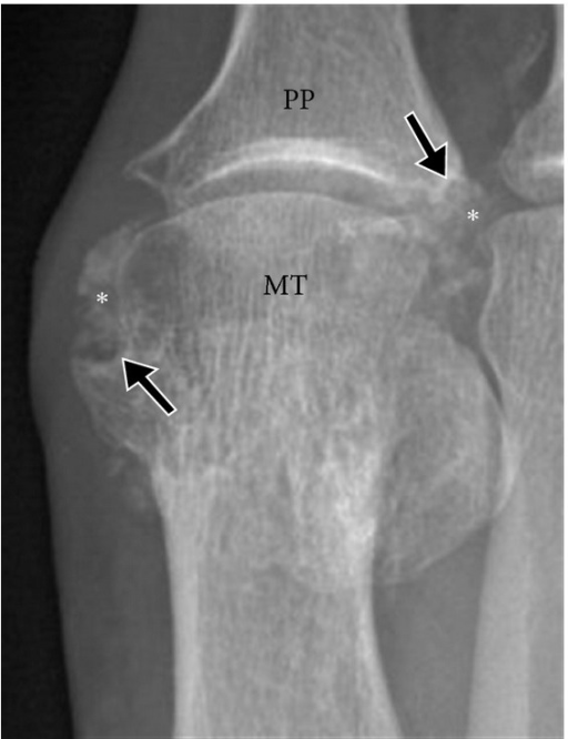
X-rayAnteroposterior X-ray of the first metatarsophalangeal joint: calcified soft tissue tophus with adjacent bone erosions, typical signs of tophaceous gout.
Source : Girish et al., Arthritis, 2013, figure 3 · CC BY
Which questions should you ask to understand the patient and their history?
🕵️♂️ The history targets several features that are highly suggestive of gout:- Kinetics of the flare : hyperacute onset over a few hours (typically at night), peak intensity < 24 h, complete spontaneous resolution in 7-14 days without treatment.³
- Topography : podagra (1st MTP) in around 50-60% of first flares; ankle, midfoot, knee in recurrences.³
- Intensity : pain that is unbearable at the mere brush of a bedsheet (the classic sign), intense erythema, major swelling, sometimes desquamation as it settles.¹
- Past history : similar episodes that resolved completely, palpable tophi (ear pinna, olecranon, Achilles tendons, fingers).⁴
- Triggering factors : recent dietary excess (banquet, meats, seafood), alcohol (beer+++), dehydration, local trauma, starting or changing a diuretic, fasting, recent surgery.⁵,⁶
- Comorbidities : hypertension, obesity, CKD, diabetes, heart failure, psoriasis. Co-prescribed diuretics.⁷
Which clinical tests should you perform and which other conditions must be ruled out?
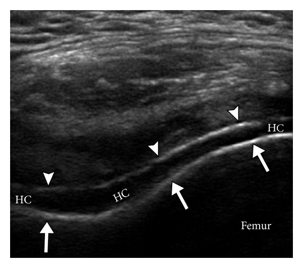
UltrasoundTransverse suprapatellar knee ultrasound: double contour sign, a hyperechoic deposit of uric acid crystals on the surface of the hyaline cartilage, characteristic of gout.
Source : Girish et al., Arthritis, 2013, figure 4 · CC BY
The place of serum urate
⚠️ A crucial paradox : during an acute flare, serum urate may be normal or low in 30-50% of patients because of the uricosuric effect of pro-inflammatory cytokines (Urano, J Rheumatol 2002).⁸ A normal serum urate therefore does not rule out the diagnosis. It should be re-measured later (2-3 weeks after complete resolution) to characterise the underlying hyperuricaemia.¹ The treatment target is then set on that measurement.Imaging: three complementary tools
| Modality | Key sign(s) | Sensitivity / Specificity | Level of evidence |
|---|---|---|---|
| Arthrocentesis + polarised microscopy | Needle-shaped MSU crystals, negative birefringence | ~100% / ~100% (gold std) | High (1a) |
| Joint ultrasound | Cartilage double contour, intra-articular "snowstorm", tophi | ~85% / ~84% (Ogdie 2015) | High (1a) |
| DECT (dual-energy CT) | Specific colour coding of urate deposits | ~87% / ~84-100% (Bongartz 2015) | Moderate (1b, limited access) |
| Plain radiography | "Punched-out" erosions (late gout) | Low in the acute phase, useful in chronic disease | Low (in the acute phase) |
🧭 Diagnostic algorithm for gout (adapted from EULAR 2018 + ACR/EULAR 2015)
A 5-step approach to reaching diagnostic certainty
Adapted from Richette P et al. Ann Rheum Dis. 2020;79(1):31-38 (EULAR 2018 diagnosis) + Neogi T et al. Arthritis Rheumatol. 2015;67(10):2557-2568 (ACR/EULAR criteria).
Critical differential diagnosis
Septic arthritis: THE diagnosis never to be missed
- Fever > 38.5 °C, chills, deterioration in general condition, laboratory signs of sepsis
- Portal of entry for infection (skin, dental, urinary, catheter)
- Immunosuppression: diabetes, corticosteroids, biologics, HIV, haemodialysis
- Synovial fluid: leucocytes > 50,000/mm³, neutrophil predominance, Gram + or culture +
- ⚠️ Coexisting gout + septic arthritis is possible (~5% of cases) - treat both if in doubt
- → Any febrile monoarthritis = urgent arthrocentesis + IV ceftriaxone ± vancomycin depending on the context
- Calcium pyrophosphate arthritis (CPPD, formerly pseudogout) : rhomboid crystals, weakly positive birefringence; tends to affect the wrist and knee; older patients. Ultrasound: fine intracartilaginous deposits.⁹
- Cellulitis : poorly demarcated confluent erythema, lymphangitic streaking, leucocytosis; it can coexist with gout (friction from the inflammatory oedema brings on a secondary cellulitis).¹⁰
- Early rheumatoid arthritis : symmetrical oligo/polyarthritis, morning stiffness > 1 h, RF/anti-CCP+. But oligoarticular forms are possible.
- Spondyloarthritis : enthesitis, axial involvement, skin/nail psoriasis.
Should patients with gout be classified, and for what benefit?
The ACR/EULAR 2015 classification criteria (Neogi) came out of the SUGAR project (Study for Updated Gout Classification Criteria) and rest on an additive scoring system.² They do not replace the clinical diagnosis but standardise it for research, and they offer a robust framework in cases of doubt. Entry criterion : at least one episode of peripheral arthritis, bursitis or tendinitis. If MSU crystals are present in the target joint or in a tophus → the diagnosis is established without calculating a score. Otherwise, calculate the score (weighted domains):- Pattern of joint involvement : 1st MTP (+2 pts), midfoot/ankle without 1st MTP (+1)
- Features of the flare : erythema (+1), severe pain (+1), resolution in ≤ 14 days (+1) - up to +3 if typical and repeated
- Clinical tophus : present (+4)
- Serum urate : 6-<8 mg/dL (+2), 8-<10 (+3), ≥10 mg/dL (+4); <4 mg/dL (-4)
- Imaging : ultrasound double contour sign (+4), positive DECT (+4), erosion on radiograph (+4)
- 📚 Standardising research populations so that studies can be compared
- 🎯 Supporting diagnosis in cases of doubt (particularly when arthrocentesis is impossible or inconclusive)
- 📝 Making communication between clinicians easier with consensus terminology (Bursill 2019, G-CAN)¹²
- 💊 Objectively justifying the start of lifelong ULT
Key points
- ✅ Gold standard : negatively birefringent MSU crystals in the synovial fluid (Richette 2018 EULAR).
- ⚠️ Serum urate paradox : it can be normal during a flare in 30-50% of cases (Urano 2002). Re-measure later.
- 📸 Ultrasound (double contour sign) and DECT : non-invasive alternatives with high specificity (Bongartz 2015).
- ⚖️ ACR/EULAR 2015 score ≥ 8: sensitivity 92%, specificity 89% (Abhishek 2020).
- 🚨 Always rule out septic arthritis in any febrile monoarthritis (a life-threatening emergency).
Bibliography
- Richette P, Doherty M, Pascual E, et al. 2018 updated European League Against Rheumatism evidence-based recommendations for the diagnosis of gout. Ann Rheum Dis. 2020;79(1):31-38. doi:10.1136/annrheumdis-2019-215315.
- Neogi T, Jansen TLTA, Dalbeth N, et al. 2015 Gout Classification Criteria: An American College of Rheumatology/European League Against Rheumatism Collaborative Initiative. Arthritis Rheumatol. 2015;67(10):2557-2568. PMID 26352873.
- Dalbeth N, Gosling AL, Gaffo A, Abhishek A. Gout. Lancet. 2021;397(10287):1843-1855. PMID 33798500.
- Clebak KT, Morrison A, Croad JR. Gout: Rapid Evidence Review. Am Fam Physician. 2020;102(9):533-538. PMID 33118789.
- Choi HK, Atkinson K, Karlson EW, Willett W, Curhan G. Purine-rich foods, dairy and protein intake, and the risk of gout in men. N Engl J Med. 2004;350(11):1093-1103. PMID 15014182.
- Neogi T, Chen C, Niu J, et al. Alcohol quantity and type on risk of recurrent gout attacks: an internet-based case-crossover study. Am J Med. 2014;127(4):311-318. PMID 24440541.
- FitzGerald JD, Dalbeth N, Mikuls T, et al. 2020 American College of Rheumatology Guideline for the Management of Gout. Arthritis Care Res (Hoboken). 2020;72(6):744-760. PMID 32391934.
- Urano W, Yamanaka H, Tsutani H, et al. The inflammatory process in the mechanism of decreased serum uric acid concentrations during acute gouty arthritis. J Rheumatol. 2002;29(9):1950-3. PMID 12233891.
- Pascart T, Lioté F. Gout: state of the art after a decade of developments. Rheumatology (Oxford). 2019;58(1):27-44. PMID 29547895.
- Edwards NL. Gout: Pearls of Clinical Practice. J Am Acad Orthop Surg. 2019;27(20):e893-e901. PMID 31568002.
- Richette P, Doherty M, Pascual E, et al. 2018 updated European League Against Rheumatism evidence-based recommendations for the diagnosis of gout. Ann Rheum Dis. 2020;79(1):31-38. PMID 31167758.
- Bursill D, Taylor WJ, Terkeltaub R, et al. Gout, Hyperuricemia, and Crystal-Associated Disease Network Consensus Statement Regarding Labels and Definitions for Disease Elements in Gout. Arthritis Care Res (Hoboken). 2019;71(3):427-434. PMID 29799677.
- Bongartz T, Glazebrook KN, Kavros SJ, et al. Dual-energy CT for the diagnosis of gout: an accuracy and diagnostic yield study. Ann Rheum Dis. 2015;74(6):1072-1077. PMID 24671771.
- Ogdie A, Taylor WJ, Weatherall M, et al. Imaging modalities for the classification of gout: systematic literature review and meta-analysis. Ann Rheum Dis. 2015;74(10):1868-1874. PMID 24915980.
Which treatment strategies are the most effective for gout (crystal arthritis)?
Where do you start? What is the recommended hierarchy of interventions?
The management of gout rests on two complementary strands: controlling the acute flare and preventing recurrences with treat-to-target urate-lowering therapy. The three major international guidelines converge on the general strategy:¹⁻³- ACR 2020 (FitzGerald, Arthritis Care Res): conditional and strong recommendations using the GRADE system.¹
- EULAR 2016 (Richette, Ann Rheum Dis 2017): 11 recommendations covering ULT, the acute flare and education.²
- NICE NG219 2022 (United Kingdom): a pragmatic guideline for primary care, the most recent update.³
Acute flare: 1st line
According to NICE NG219 (2022) and ACR 2020, the choice rests on comorbidities:- NSAIDs (ibuprofen 600-800 mg x3-4/day, naproxen 500 mg x2/day) if there is no severe renal failure, no ulcer and no uncontrolled heart failure
- Colchicine low dose (1 mg then 0.5 mg 1 h later, max 1.5 mg on day 1) - the lighter regimen (Terkeltaub) is just as effective and better tolerated⁴
- Corticosteroids oral (prednisolone 30-35 mg/day for 5 days) or intra-articular - first choice in severe renal failure or with several comorbidities¹,³
- Anti-IL-1 (canakinumab, anakinra) - 2nd/3rd line for refractory patients or those with multiple contraindications¹
Treat-to-target urate-lowering therapy (ULT)
The cornerstone of long-term treatment. Strong indications (ACR 2020 + EULAR 2016):¹,²- ≥ 2 flares per year
- Presence of tophi (clinical or on imaging)
- Radiographic erosions attributed to gout
- CKD stage ≥ 3
- Previous uric acid stones
- Conditional recommendation : ULT after the 1st flare if comorbidities are present (NICE 2022 leans this way)³
⚕️ Hierarchy of ULT (ACR 2020, EULAR 2016, NICE 2022)
Sequential strategy adapted to eGFR and tolerance
Synthesis: FitzGerald 2020 (ACR), Richette 2017 (EULAR), NICE NG219 2022. High GRADE level of evidence for allopurinol 1st line.
Non-pharmacological measures
Consistent recommendations:¹⁻³,⁶- Weight loss if BMI ≥ 25 (lowers serum urate - meta-analysis)
- Limit alcohol (beer, spirits), fructose, red meat/offal, seafood
- Encourage: low-fat dairy products, coffee, vitamin C, cherries (a modest but consistent effect)⁶
- Adequate hydration (~2 L/day)
- Regular aerobic physical activity
What is the place of exercise, and is any one approach superior?
🏋️♂️ Physical activity has a twofold benefit in the patient with gout: a direct impact on the associated cardio-metabolic risk factors (BMI, blood glucose, lipid profile) and an indirect impact on serum urate through weight loss. Level of evidence : the data specific to gout remain moderate to low. The recommendations rest mainly on:- The general rheumatology guidelines (EULAR exercise recommendations 2018)
- The ancillary study of the CARES cohort (Vargas-Santos, Ann Rheum Dis 2022): an inverse association between regular physical activity and mortality in patients with gout (adjusted HR 0.70 [0.55-0.89] for activity ≥ 4 h/week)⁷
- NHANES accelerometry data: volume of physical activity inversely correlated with the incidence of gout⁸
- Moderate aerobic exercise : 150 min/week (brisk walking, cycling, swimming) - equivalent to the WHO adult target
- Strength training : 2x/week, the main muscle groups
- Flexibility / mobility : maintained between flares
- ⚠️ Hydration increased around exercise (dehydration = an acute rise in serum urate)
- ⚠️ Avoid high-impact sports for 2-4 weeks after a flare in a weight-bearing joint
Manual therapies and technologies: how effective are they really?
🧊 In gout, cryotherapy is the only physical modality with positive direct evidence. The pioneering RCT by Schlesinger (J Rheumatol 2002, n = 19, topical ice 30 min x4/day alongside medical treatment) showed a significantly greater reduction in pain (−7.75 cm vs −4.42 cm on a 10 cm VAS) at 1 week.⁹ ACR 2020 conditionally recommends topical ice as an adjunct.¹| Physical modality | Level of evidence | Recommendation |
|---|---|---|
| Cryotherapy (topical ice) | Moderate (1 RCT n=19, conditional ACR) | Adjunct for acute flare pain |
| Joint manual therapy | Very low / absent in gout | ⚠️ Contraindicated during a flare. Between flares: caution, no specific evidence |
| Therapeutic ultrasound | Very low (animal models) | Not recommended in practice |
| TENS electrotherapy | Low (extrapolated from general pain) | Possibly useful as a non-specific analgesic adjunct |
| Low-level laser therapy (LLLT) | Low / almost absent in gout | No recommendation |
Beyond the physical: how do you educate the patient and address psychological factors?
🧠 The education of the patient (therapeutic patient education) is probably the highest-yield non-drug intervention in gout. The landmark narrative review by Abhishek & Doherty (Rheumatology Oxford 2018) identifies structured patient education as a major determinant of adherence to ULT, and therefore of long-term control.¹⁰ The problem is well known: the Scheepers meta-analysis (n = 137,699 patients, 22 studies) shows an average adherence to ULT of only 47% (95% CI 42-52%), one of the lowest among chronic treatments.¹¹ The variation with the method of measurement illustrates the difficulty: 42% (claims), 71% (pill count), 66% (self-report), 63% (interview).📉 Adherence to urate-lowering therapy (ULT) - Scheepers 2018 meta-analysis
Adherence rates by method of measurement (n = 137,699 patients)
Source: Scheepers LE et al. Semin Arthritis Rheum. 2018;47(5):689-702. Adherence varies widely with the method : the pharmacy estimate (the most objective) is the lowest.
- Explain the chronic nature of the disease ("the crystals do not disappear between flares")
- Explain the distinction between treating the flare (short) and ULT (long, lifelong)
- Present the serum urate target and its clinical value (crystals dissolve if it is maintained)
- Personalised dietary advice (without guilt-inducing rigidity)
- A flare action plan with rescue NSAID/colchicine
- Explicit discussion of comorbidities (hypertension, CKD, CVD, metabolic syndrome)
- Support for self-monitoring (diary, apps)
Key points
- ⚕️ Treat-to-target ULT (target < 360 µmol/L) = the cornerstone (ACR 2020, EULAR 2016, NICE 2022). Allopurinol 1st line.
- 🧊 Cryotherapy : the only physical modality with direct evidence (Schlesinger 2002, conditional ACR recommendation).
- 🏃 Moderate aerobic exercise on a regular basis (150 min/week): a favourable indirect impact, with moderate condition-specific data (Vargas-Santos 2022).
- 📚 Therapeutic patient education : the highest-yield non-drug intervention (Abhishek 2018), against an average ULT adherence of 47% (Scheepers 2018).
- ⚠️ Manual therapy, ultrasound, LLLT: very low or absent evidence in gout. Focus physiotherapy effort on exercise + patient education.
Bibliography
- FitzGerald JD, Dalbeth N, Mikuls T, et al. 2020 American College of Rheumatology Guideline for the Management of Gout. Arthritis Care Res (Hoboken). 2020;72(6):744-760. PMID 32391934.
- Richette P, Doherty M, Pascual E, et al. 2016 updated EULAR evidence-based recommendations for the management of gout. Ann Rheum Dis. 2017;76(1):29-42. PMID 27457514.
- National Institute for Health and Care Excellence. Gout: diagnosis and management. NICE guideline NG219. Published 9 June 2022. www.nice.org.uk/guidance/ng219.
- Terkeltaub RA, Furst DE, Bennett K, Kook KA, Crockett RS, Davis MW. High versus low dosing of oral colchicine for early acute gout flare: Twenty-four-hour outcome of the first multicenter, randomized, double-blind, placebo-controlled, parallel-group, dose-comparison colchicine study. Arthritis Rheum. 2010;62(4):1060-1068. PMID 20131255.
- Perez-Ruiz F, Dalbeth N, Bardin T. A review of uric acid, crystal deposition disease, and gout. Adv Ther. 2015;32(1):31-41. PMID 25533440.
- Qaseem A, Harris RP, Forciea MA. Management of Acute and Recurrent Gout: A Clinical Practice Guideline From the American College of Physicians. Ann Intern Med. 2017;166(1):58-68. PMID 27802508.
- Vargas-Santos AB, Peloquin CE, Zhang Y, Neogi T. Association of physical activity with mortality in gout: an ancillary study of the CARES trial. Ann Rheum Dis. 2022;81(5):747-750. PMID 35185007.
- Smith ID, Ross LM, Gabaldon JR, et al. The Relation of Accelerometer-Measured Physical Activity and Serum Uric Acid Using the National Health and Nutrition Survey (NHANES) 2003-2004. Front Sports Act Living. 2021;3:775398. PMID 35098119.
- Schlesinger N, Detry MA, Holland BK, et al. Local ice therapy during bouts of acute gouty arthritis. J Rheumatol. 2002;29(2):331-334. PMID 11838852.
- Abhishek A, Doherty M. Education and non-pharmacological approaches for gout. Rheumatology (Oxford). 2018;57(suppl_1):i51-i58. PMID 29272507.
- Scheepers LEJM, van Onna M, Stehouwer CDA, Singh JA, Arts ICW, Boonen A. Medication adherence among patients with gout: A systematic review and meta-analysis. Semin Arthritis Rheum. 2018;47(5):689-702. PMID 29198878.
- Spencer K, Carr A, Doherty M. Patient and provider barriers to effective management of gout in general practice: a qualitative study. Ann Rheum Dis. 2012;71(9):1490-1495. PMID 22440822.
- Stewart S, Tallon A, Taylor WJ, Gaffo A, Dalbeth N. How flare prevention outcomes are reported in gout studies: A systematic review and content analysis of randomized controlled trials. Semin Arthritis Rheum. 2020;50(2):303-313. PMID 31796212.
- Doherty M, Jenkins W, Richardson H, et al. Efficacy and cost-effectiveness of nurse-led care involving education and engagement of patients and a treat-to-target urate-lowering strategy versus usual care for gout: a randomised controlled trial. Lancet. 2018;392(10156):1403-1412. PMID 30343856.
How do you secure lasting recovery and prevent recurrences of gout (crystal arthritis)?
How do you make the patient an active partner in their recovery through self-management?
🧘♂️ Self-management is defined by the WHO as the patient's ability to manage the symptoms, the treatment, the physical and psychological consequences, and the lifestyle changes inherent to a chronic disease. The pivotal clinical trial by Doherty (Lancet 2018) (a 2-year randomised study, n = 517 patients with gout followed in primary care) showed the clear superiority of a structured nurse-led intervention (education + treat-to-target + adherence support) over usual care:⁴- Serum urate target reached (< 360 µmol/L) at 2 years: 95% vs 30% (absolute difference: +65 points)
- Presence of tophi: −10.4% vs −0.8%
- Few flares in the past 12 months: 87% vs 71%
- Favourable cost-effectiveness (economic analysis: ICER < £5,000 / QALY)
- Adherence to ULT : daily doses, no stopping at the first improvement. Diary or apps
- Biochemical targets : serum urate every 3-6 months until the target is reached, then yearly
- Flare action plan : rescue NSAID or colchicine kept at home
- Lifestyle : balanced diet, alcohol, hydration, weight, physical activity
- Monitoring of comorbidities : blood pressure, blood glucose, creatinine, lipids at the frequency set by the GP
Validated dietary changes
| Food / Drink | Effect on risk | Recommendation | Source |
|---|---|---|---|
| Beer | RR ↑↑↑ (purine + ethanol effect) | Avoid / limit +++ | Choi 2004, Neogi 2014 |
| Spirits | RR ↑↑ | Limit | Neogi 2014 |
| Wine (moderate) | Weak / variable effect | Sensible moderation | Neogi 2014 |
| Red meat, offal, game | RR 1.41 (Q5 vs Q1) | Limit | Choi 2004 (HPFS) |
| Seafood | RR 1.51 (Q5 vs Q1) | Limit | Choi 2004 |
| Fructose / sugary soft drinks | RR increased, dose-dependent | Avoid +++ | Choi 2008 |
| Low-fat dairy products | RR ↓ | Encourage | Choi 2004 |
| Coffee | RR ↓, dose-dependent | No need to avoid | Choi 2007 |
| Vitamin C | RR ↓ slightly | Intake through fruit and vegetables | Choi 2009 |
| Cherries | Modest reduction in flares | Optional (modest effect) | Zhang 2012 |
Weight loss and hydration
The Nielsen meta-analysis (Ann Rheum Dis 2017) confirms that weight loss (diet, exercise or bariatric surgery) significantly lowers serum urate, with a dose-response effect.⁵ A loss of 5-10 kg may be enough to avoid starting ULT in some overweight patients. Hydration (~2 L/day, more in hot weather, with exercise or with a tendency to stones) remains recommended even though the direct evidence is limited.When and how do you plan a safe return to sport and activity?
🏃♀️ Physical activity is beneficial but must be planned so that it does not trigger another flare. The recommendations are based on physiological principles (no specific RCTs):Prerequisites for a structured return
- Biochemical control : serum urate stable < 360 µmol/L for at least 4-6 weeks (the ACR/EULAR goal reached)¹,²
- No acute symptoms : no inflammatory joint signs (heat, redness, swelling) for at least 2-3 weeks
- Patient understanding : the reason for the gradual approach, the importance of hydration around exercise
Progression in 3 phases
- Phase 1 (weeks 1-4) : activities with low impact. Brisk walking 20-30 min, swimming, cycling. Frequency 3-5x/week. Moderate intensity (HR 60-70% of HRmax, or the talk test).
- Phase 2 (weeks 4-8) : gradual increase in duration (+5 min/week) and intensity (HR 70-80%). Strength training introduced 2x/week (body weight, resistance bands).
- Phase 3 (beyond 8 weeks) : careful reintroduction of impact sports or rapid changes of direction (running, tennis, basketball). Monitor symptoms 24-48 h after exercise.
Key precautions
- 💧 Strict hydration : 500 mL 2 h before, 200 mL every 20 min during, and 500-750 mL after (dehydration concentrates urate)
- 👟 Suitable footwear : enough cushioning, correct size (repeated microtrauma at the 1st ray = a local trigger)
- 🌡️ Climate : avoid exercise in low temperatures (which favours peripheral crystal precipitation) or in very hot weather (dehydration)
- 🚫 Avoid acute post-exercise purine peaks (alcohol, cured meats): the anabolic effect of muscle nucleotides during recovery
A special case: very intense physical activity
A prospective UK Biobank accelerometry study (Hu, medRxiv 2025) suggests that a high volume of physical activity is inversely associated with the incidence of gout, independently of the pattern (continuous vs intermittent).⁶ For the patient already diagnosed, keeping up regular moderate physical activity is clearly preferable to being sedentary.Key points
- 🎯 Lasting prevention = keeping serum urate stable < 360 µmol/L with well-followed ULT (key evidence: Doherty Lancet 2018).
- 📚 Structured therapeutic patient education ("nurse-led" type) = the strongest evidence of effectiveness (95% vs 30% reaching target at 2 years).
- 🥩 Diet : avoid beer, fructose, red meat, seafood; encourage low-fat dairy and coffee. A moderate but consistent effect.
- ⚖️ Weight loss of 5-10 kg: a significant impact on serum urate (Nielsen 2017).
- 🏃 Return to sport in 3 progressive phases after stable biochemical control, with strict hydration.
Bibliography
- FitzGerald JD, Dalbeth N, Mikuls T, et al. 2020 American College of Rheumatology Guideline for the Management of Gout. Arthritis Care Res (Hoboken). 2020;72(6):744-760. PMID 32391934.
- Richette P, Doherty M, Pascual E, et al. 2016 updated EULAR evidence-based recommendations for the management of gout. Ann Rheum Dis. 2017;76(1):29-42. PMID 27457514.
- National Institute for Health and Care Excellence. Gout: diagnosis and management. NICE guideline NG219. Published 9 June 2022. www.nice.org.uk/guidance/ng219.
- Doherty M, Jenkins W, Richardson H, et al. Efficacy and cost-effectiveness of nurse-led care involving education and engagement of patients and a treat-to-target urate-lowering strategy versus usual care for gout: a randomised controlled trial. Lancet. 2018;392(10156):1403-1412. PMID 30343856.
- Nielsen SM, Bartels EM, Henriksen M, et al. Weight loss for overweight and obese individuals with gout: a systematic review of longitudinal studies. Ann Rheum Dis. 2017;76(11):1870-1882. PMID 28866649.
- Choi HK, Liu S, Curhan G. Intake of purine-rich foods, protein, and dairy products and relationship to serum levels of uric acid: the Third National Health and Nutrition Examination Survey. Arthritis Rheum. 2005;52(1):283-289. PMID 15641075.
- Choi HK, Curhan G. Soft drinks, fructose consumption, and the risk of gout in men: prospective cohort study. BMJ. 2008;336(7639):309-312. PMID 18244959.
- Choi HK, Willett W, Curhan G. Coffee consumption and risk of incident gout in men: a prospective study. Arthritis Rheum. 2007;56(6):2049-2055. PMID 17530645.
- Zhang Y, Neogi T, Chen C, Chaisson C, Hunter DJ, Choi HK. Cherry consumption and decreased risk of recurrent gout attacks. Arthritis Rheum. 2012;64(12):4004-4011. PMID 23023818.
- Neogi T, Chen C, Niu J, et al. Alcohol quantity and type on risk of recurrent gout attacks: an internet-based case-crossover study. Am J Med. 2014;127(4):311-318. PMID 24440541.
- Abhishek A, Doherty M. Education and non-pharmacological approaches for gout. Rheumatology (Oxford). 2018;57(suppl_1):i51-i58. PMID 29272507.
- Vargas-Santos AB, Peloquin CE, Zhang Y, Neogi T. Association of physical activity with mortality in gout: an ancillary study of the CARES trial. Ann Rheum Dis. 2022;81(5):747-750. PMID 35185007.
Why is gout a cardiovascular and renal risk marker?
What is the link between a gout flare and a cardiovascular event?
🫀 The pivotal study Cipolletta (JAMA 2022) definitively established the temporal relationship between a gout flare and a major adverse cardiovascular event (MACE).¹ This case-control + case-crossover study, based on the CPRD cohort (Clinical Practice Research Datalink, United Kingdom), analysed 62,574 patients with gout, of whom 10,475 had a CV event. The results are unequivocal:- Increased risk of a CV event (MI, stroke) OR 1.93 (95% CI 1.57-2.38) in the 0-60 days following a flare
- Risk still raised OR 1.57 (95% CI 1.26-1.96) at 61-120 days
- Peak incidence at 30 days post-flare
- Effect maintained after adjustment for traditional CV risk factors
⏱️ Transient excess cardiovascular risk after a gout flare
Cipolletta JAMA 2022 study - n = 62,574 patients with gout
Source: Cipolletta E et al. JAMA. 2022;328(5):440-450. PMID 35916846. The risk returns to baseline beyond 180 days, suggesting a therapeutic window of opportunity around the flare.
How do you adapt treatment when cardio-renal comorbidity is present?
The presence of CKD, of hypertension, of heart failure or of a history of cardiovascular disease substantially changes the choice of treatment.Acute flare: choosing according to comorbidities
| Comorbidity | 1st line of choice | To avoid |
|---|---|---|
| Moderate-severe CKD (eGFR < 30) | Oral or intra-articular corticosteroids | NSAIDs (renal risk + hyperkalaemia), high-dose colchicine |
| Uncontrolled heart failure | Corticosteroids (oral or intra-artic.) | NSAIDs (sodium retention, decompensation) |
| History of GI ulcer / multiple antiplatelet drugs | Low-dose colchicine + PPI, corticosteroids | NSAIDs |
| Poorly controlled diabetes | Short-course NSAIDs, colchicine | Prolonged courses of corticosteroids |
| Older patient on many medicines | IA corticosteroids (1 joint), adjusted colchicine | Long-term NSAIDs |
ULT: choice according to eGFR
- Allopurinol remains the 1st line even in CKD, with a start low - go slow (50 mg/day if eGFR < 30, then slow titration) approach. HLA-B*58:01 testing in Asian populations (Han, Korean, Thai) before starting, to prevent DRESS syndrome (FDA 2010).¹
- Febuxostat : an alternative when allopurinol is not tolerated or not effective. Note: the CARES 2018 trial (White, NEJM) had suggested excess CV mortality vs allopurinol in patients at high CV risk, but the FAST 2020 trial (Mackenzie, Lancet) refuted this. Caution is still advised in patients with a history of major CV disease.
- Probenecid : only if eGFR > 50 mL/min, and no uric acid stones.
- Pegloticase : refractory tophaceous disease, infusion every two weeks. EULAR 2024 meta-analysis: 47% reach remission at 52 weeks when it is combined with methotrexate.
SGLT2 inhibitors: a paradigm shift
💊 The sodium-glucose co-transporter 2 (SGLT2) inhibitors (empagliflozin, dapagliflozin, canagliflozin), first developed for diabetes and since become standard in heart failure and CKD, have a major additional effect on gout:- A reduction of 30 to 50% in the risk of a gout flare (2023-2024 meta-analyses)³,⁴
- A 30-50 µmol/L reduction in serum urate (partial uricosuric effect)
- A suggested anti-inflammatory effect independent of the fall in serum urate
- CV and renal benefit in their own right (EMPA-REG, DAPA-CKD, CANVAS, EMPA-KIDNEY trials)
Practical implications for exercise prescription and referral
🏃 For the physiotherapist, the cardio-renal dimension changes practice:Systematic assessment before an exercise programme
- CV history / risk factors (hypertension, dyslipidaemia, smoking, diabetes)
- Resting blood pressure and heart rate
- Knowledge of renal function (estimated GFR)
- A full list of medicines (β-blockers change the target heart rate)
- A submaximal functional test if capacity is thought to be limited (6MWT, sit-to-stand)
Red flags specific to the cardio-renal profile
- Chest pain on exertion or unusual → refer to cardiac emergency care
- Disproportionate breathlessness on exertion, orthopnoea, rapid weight gain → cardiac decompensation
- Recent bilateral lower-limb oedema, oliguria → progressing CKD
- Uncontrolled hypertension (> 160/100 mmHg at rest) → postpone intense exercise, medical review
- Recent gout flare < 60 days : treat as a period of raised CV risk → moderate the intensity, increase monitoring
When to refer actively
Collaboration with the cardiologist/nephrologist/GP should be routine in any patient with gout who has ≥ 1 of:- Confirmed hypertension > 140/90 mmHg not yet managed
- Estimated GFR < 60 mL/min/1.73 m²
- History of MI, stroke, peripheral arterial disease, heart failure
- Poorly controlled type 2 diabetes (HbA1c > 7%)
- BMI > 30 kg/m² with no dietetic follow-up
- Treatments that do not match the comorbidities (e.g. NSAIDs in a patient with stage 3 CKD)
Key points
- 🫀 A gout flare is associated with a transient excess risk of a CV event: OR 1.93 within 60 days (Cipolletta JAMA 2022, n = 62,574).
- 🩺 Gout is intertwined with hypertension, CKD, metabolic syndrome and heart failure, so management is inseparable.
- 💊 The choice of flare treatment (NSAIDs / colchicine / corticosteroids) depends on the comorbidities ; avoid NSAIDs in severe CKD and heart failure.
- 🆕 The SGLT2 inhibitors reduce flare risk by 30-50% - to be considered in patients with diabetes, heart failure or CKD.
- 🚨 Specific red flags (chest pain, breathlessness, uncontrolled hypertension) → medical referral without delay. The post-flare period (60 days) = heightened CV vigilance.
Bibliography
- Cipolletta E, Tata LJ, Nakafero G, Avery AJ, Mamas MA, Abhishek A. Association Between Gout Flare and Subsequent Cardiovascular Events Among Patients With Gout. JAMA. 2022;328(5):440-450. PMID 35916846.
- Anderson JL, Knowlton KU. Cardiovascular Events and Gout Flares. JAMA. 2022;328(5):425-426. PMID 35916865.
- Yokose C, McCormick N, Lu N, et al. Sodium-glucose cotransporter-2 inhibitors use and the risk of gout: a systematic review and meta-analysis. Front Endocrinol. 2023;14:1158153. PMID 37287656.
- McCormick N, Yokose C, Wei J, et al. Comparative Effectiveness of Sodium-Glucose Cotransporter-2 Inhibitors for Recurrent Gout Flares and Gout-Primary Emergency Department Visits and Hospitalizations : A General Population Cohort Study. Ann Intern Med. 2023;176(8):1067-1080. PMID 37487215.
- FitzGerald JD, Dalbeth N, Mikuls T, et al. 2020 American College of Rheumatology Guideline for the Management of Gout. Arthritis Care Res (Hoboken). 2020;72(6):744-760. PMID 32391934.
- Richette P, Doherty M, Pascual E, et al. 2016 updated EULAR evidence-based recommendations for the management of gout. Ann Rheum Dis. 2017;76(1):29-42. PMID 27457514.
- White WB, Saag KG, Becker MA, et al. Cardiovascular Safety of Febuxostat or Allopurinol in Patients with Gout. N Engl J Med. 2018;378(13):1200-1210. PMID 29527974.
- Mackenzie IS, Ford I, Nuki G, et al. Long-term cardiovascular safety of febuxostat compared with allopurinol in patients with gout (FAST): a multicentre, prospective, randomised, open-label, non-inferiority trial. Lancet. 2020;396(10264):1745-1757. PMID 33181081.
- Singh JA, Cleveland JD. Gout and the risk of myocardial infarction in older adults: a study of Medicare recipients. Arthritis Res Ther. 2018;20(1):109. PMID 29859125.
- Clarson LE, Hider SL, Belcher J, Heneghan C, Roddy E, Mallen CD. Increased risk of vascular disease associated with gout: a retrospective, matched cohort study in the UK clinical practice research datalink. Ann Rheum Dis. 2015;74(4):642-7. PMID 25165032.
- Stamp LK, Chapman PT. Gout and its comorbidities: implications for therapy. Rheumatology (Oxford). 2013;52(1):34-44. PMID 22949727.
What do real clinical cases teach us about gout (crystal arthritis)?
Analysis of a "classic" case: from assessment to resolution.
🦶 In practice, podagra remains the teaching case of gout. The typical profile as described in the everyday practice literature (Clebak, Am Fam Physician 2020):¹- A middle-aged or older man, BMI ≥ 27
- Sudden onset at night (between 11 pm and 3 am)
- Peak pain within a few hours (unbearable under the bedsheet)
- 1st metatarsophalangeal joint, unilateral, in > 50% of first flares
- Intense inflammatory erythema, swelling, local heat, sometimes desquamation as it settles
- Common history: hypertension, excess weight, diuretic use, a rich meal the day before
- Confirm the diagnosis (ideally by arthrocentesis, otherwise the ACR/EULAR 2015 score + a serum urate measured later)
- Treat the flare: NSAIDs, colchicine or a corticosteroid depending on comorbidities
- Assess cardio-renal comorbidities
- Discuss starting ULT (a conditional recommendation at the 1st flare, strong thereafter)
- A patient education plan + follow-up at 2-4 weeks
The diagnostic challenge: when gout mimics another condition.
🧐 Gout is nicknamed the "great imitator". Four critical situations published in the literature illustrate the traps:1. Imitating or coexisting with septic arthritis
The coexistence of gout + septic arthritis is documented in around 1.5-5% of cases of crystal monoarthritis (PMC9092107 review, Bouchemma 2022 and earlier literature).⁵ An alarming picture: fever, leucocytosis, extreme pain, but also visible MSU crystals. The trap: settling for the diagnosis of gout while an infection coexists. Any febrile monoarthritis warrants joint aspiration with routine bacteriology, and if doubt persists, empirical anti-infective treatment until the final culture. A typical published case (Yu, PMC9092107) describes a 75-year-old patient with a gout flare of the knee (MSU in the synovial fluid) that was complicated by a Staphylococcus aureus bacteraemia diagnosed after the event: the coexistence of the two conditions required combined therapy.⁵2. Imitating cellulitis
The Journal of Urgent Care Medicine review (2023) frequently highlights this confusion: extensive erythema, disproportionate pain, initial suspicion of a skin infection leading to ineffective antibiotic treatment. Moving to imaging (joint ultrasound with the double contour sign, or suggestive particulate content) allows the diagnosis to be corrected.3. Imitating an inflammatory polyarthritis
Oligo- or polyarticular gout (particularly in older women on diuretics) can mimic early rheumatoid arthritis: symmetrical involvement of the fingers, chronic swelling, sometimes a "nodular" pseudo-arthritis. The diagnosis is corrected by imaging (urate-specific signs on ultrasound/DECT) and the absence of RF/anti-CCP.4. Imitating a tendinopathy / bursitis
The "gouty stenosing tenosynovitis" (gouty tenosynovitis) can present as a trigger finger (case study by Lee, J Hand Surg Asian-Pac Vol 2020), the first manifestation of tophaceous gout in some patients.⁶ Ultrasound imaging reveals the intratendinous urate deposits. The diagnosis is confirmed by aspiration or biopsy.⚠ Red flags in any acute monoarthritis
- Fever > 38.5 °C + monoarthritis: septic arthritis until proven otherwise → urgent arthrocentesis + bacteriology
- Immunosuppressed patient (corticosteroids, biologics, diabetes, haemodialysis): infection risk ↑↑
- An identified portal of entry for infection (catheter, wound, dental work)
- Spinal involvement with a progressive neurological deficit → urgent MRI (spinal abscess, tumour, spinal gout)
- Laboratory sepsis (CRP > 200, raised PCT, lactate > 2.5 mmol/L)
- Haemodynamic instability, signs of organ failure
Study of a complex case
Spinal gout: rare but serious
🦴 Although gout usually affects the peripheral joints, cases of spinal gout (epidural tophus) are reported regularly. The systematic review Nazwar (J Craniovert Junct Spine 2024) collected 88 cases published between 2013 and 2023:⁷- 89.8% men, mean age 51.9 years (range 16-87)
- Presentation: neck or low back pain (78.4%), lower-limb weakness (37.5%)
- Commonest site: lumbar spine (62.5%)
- Diagnosis often made after surgery for a suspected disc herniation or tumour
- Modern DECT now allows a specific non-invasive diagnosis, avoiding unnecessary surgery
Chronic destructive tophaceous gout
Severe tophaceous forms, now less common thanks to better management, are still seen in patients with poor adherence to ULT or diagnosed late. The case review by Sun (BMC Musculoskeletal Disord) describes a patient with pseudo-rheumatoid gouty polyarthritis, severe hand deformities and extensive bone erosions, which called for a combination of pegloticase + surgical excision of the tophi.⁸ These cases are a reminder that early and sustained management would have avoided this course, and that today aggressive treatment options (pegloticase, anti-IL-1) can still produce spectacular recoveries.The trap of a normal serum urate during a flare
As noted in chapter 2, 30-50% of patients have a normal or low serum urate during an acute flare (Urano 2002).⁹ A typical published case is that of a patient with recurrent knee arthritis and a serum urate repeatedly at 6.2 mg/dL during flares: the diagnosis was finally made on ultrasound (double contour) and then confirmed by aspiration once the flare had settled. Lesson : do not exclude gout on a "normal" serum urate in the middle of a flare.Critique and controversy: the paradox of the "gold standard" and the new technologies
Analysis of the cases reveals a permanent tension:- On the one hand, arthrocentesis remains the only way to obtain a definite diagnosis and to rule out septic arthritis: a strong EULAR 2018 recommendation.¹⁰
- On the other hand, in real practice it is underused in primary care (lack of training, time and equipment), which leads to diagnostic errors.¹¹
- The new technologies (DECT, high-quality ultrasound) offer non-invasive alternatives with high specificity, but access to them remains uneven geographically and economically.
- The pragmatic role of ultrasound (more widely available than DECT, and now well validated) is gaining importance as a useful compromise.
Key points
- 🦶 In practice, podagra remains the textbook picture of the first gout flare in a middle-aged man with cardio-metabolic risk factors.
- 🚨 The coexistence of gout + septic arthritis is possible (~1.5-5%): any febrile monoarthritis = aspiration + bacteriology.
- 🧐 Gout can mimic cellulitis, rheumatoid arthritis or a tendinopathy. Imaging (ultrasound, DECT) is valuable when the presentation is atypical.
- 🦴 Atypical forms (spinal gout, 88 cases, Nazwar 2024) are rare but serious: consider them in atypical low back pain in a hyperuricaemic patient.
- ⚠️ A normal serum urate during a flare does not exclude gout (30-50% of cases - Urano 2002). Re-measure later.
Bibliography
- Clebak KT, Morrison A, Croad JR. Gout: Rapid Evidence Review. Am Fam Physician. 2020;102(9):533-538. PMID 33118789.
- FitzGerald JD, Dalbeth N, Mikuls T, et al. 2020 American College of Rheumatology Guideline for the Management of Gout. Arthritis Care Res (Hoboken). 2020;72(6):744-760. PMID 32391934.
- National Institute for Health and Care Excellence. Gout: diagnosis and management. NICE NG219. June 2022. www.nice.org.uk/guidance/ng219.
- Doherty M, Jenkins W, Richardson H, et al. Efficacy and cost-effectiveness of nurse-led care involving education and engagement of patients and a treat-to-target urate-lowering strategy versus usual care for gout: a randomised controlled trial. Lancet. 2018;392(10156):1403-1412. PMID 30343856.
- Yu KH, Luo SF, Liou LB, et al. Concomitant septic and gouty arthritis--an analysis of 30 cases. Rheumatology (Oxford). 2003;42(9):1062-6. PMID 12730521.
- Doucet V, McLeod GJ, Petropolis CJ. Gouty Stenosing Tenosynovitis: Trigger Finger as a First Presentation of Tophaceous Gout. Plast Reconstr Surg Glob Open. 2020;8(8):e3055. PMID 32983801.
- Nazwar TA, Bal'afif F, Wardhana DW, Panjaitan C. Understanding spinal gout: A comprehensive study of 88 cases and their clinical implications. J Craniovertebr Junction Spine. 2024;15(2):133-140. PMID 38957764.
- Aati O, Taylor WJ, Horne A, Dalbeth N. Toward development of a Tophus Impact Questionnaire: a qualitative study exploring the experience of people with tophaceous gout. J Clin Rheumatol. 2014;20(5):251-5. PMID 25036566.
- Urano W, Yamanaka H, Tsutani H, et al. The inflammatory process in the mechanism of decreased serum uric acid concentrations during acute gouty arthritis. J Rheumatol. 2002;29(9):1950-3. PMID 12233891.
- Richette P, Doherty M, Pascual E, et al. 2018 updated European League Against Rheumatism evidence-based recommendations for the diagnosis of gout. Ann Rheum Dis. 2020;79(1):31-38. doi:10.1136/annrheumdis-2019-215315.
- Spencer K, Carr A, Doherty M. Patient and provider barriers to effective management of gout in general practice: a qualitative study. Ann Rheum Dis. 2012;71(9):1490-1495. PMID 22440822.
- Bongartz T, Glazebrook KN, Kavros SJ, et al. Dual-energy CT for the diagnosis of gout: an accuracy and diagnostic yield study. Ann Rheum Dis. 2015;74(6):1072-1077. PMID 24671771.
How do you apply these recommendations concretely in your practice?
When and to which other health professionals should you refer?
🧭 The physiotherapist, often in the front line, must know how to refer proactively. Interprofessional collaboration improves outcomes and patient satisfaction (Cochrane review, Reeves 2017).³Identifying red flags
Red flags specific to gout or to monoarthritis
- Fever > 38.5 °C + monoarthritis : suspected septic arthritis → immediate referral to emergency care
- Spinal involvement with neurological deficits (progressive paraesthesia, weakness, sphincter disturbance) → urgent MRI
- Laboratory sepsis (chills, deterioration in general condition, hypotension) → emergency services
- Chest pain on exertion or unusual in a patient with gout (excess post-flare CV risk, Cipolletta 2022) → urgent cardiology
- Ulcerated tophus with purulent discharge → infection → referring doctor + antibiotics
- Uncontrolled tophaceous gout despite correctly titrated ULT → rheumatologist to discuss pegloticase
- Confirmed hypertension > 160/100 mmHg untreated → GP promptly
- CKD stage ≥ 4 (eGFR < 30): adapt exercise and refer to the nephrologist/GP
Referrals to consider as a matter of routine
- GP / rheumatologist : starting or reviewing ULT, reaching the biochemical target, managing comorbidities
- Cardiologist : CV history, recent post-flare period, multiple uncontrolled CV risk factors
- Nephrologist : CKD stage ≥ 3b, recurrent uric acid stones
- Dietitian-nutritionist : excess weight, metabolic syndrome, support with weight loss
- Clinical pharmacist : optimising adherence, medication review for polypharmacy
- Psychologist : kinesiophobia, anxiety about recurrence, depressive disorders associated with chronic pain
- Podiatrist / orthotist : foot deformities, suitable footwear after an episode of podagra
Shared decision-making
🤝 The concept of shared decision-making (Hoffmann 2014, JAMA) is central: inform the patient of the options and the benefits/risks, and build the treatment plan together.⁴ This approach particularly improves adherence in chronic diseases that require lifelong treatment, such as gout.How do you measure outcomes and overcome the barriers to implementation?
📈 Outcome assessment uses objective measures + PROMs (Patient-Reported Outcome Measures).Key objective measures in gout
- Serum urate : target < 360 µmol/L (< 6 mg/dL), < 300 if tophi are present. Measured every 3-6 months until the target is reached, then yearly
- Number of flares per year (patient diary)
- Measurement of tophi : number, site, size (palpation, ultrasound, photography)
- DECT : volumetric quantification of urate deposits (research / complex cases)
- Joint function : range of motion, VAS pain, functional tests
- Cardio-renal parameters : blood pressure, eGFR, blood glucose, lipids
Gout-specific PROMs
- Gout Impact Scale (GIS) - Hirsch 2008: 24 items in 5 subscales, internationally validated⁵
- Gout Activity Score (GAS) - Scirè 2017: a composite measuring disease activity⁶
- HAQ-DI (Health Assessment Questionnaire - Disability Index): overall functional impact
- EQ-5D-5L : health-related quality of life
- Brief Pain Inventory : characterisation and impact of pain
- Pain VAS : simple follow-up in the clinic
The GRADE pyramid of evidence in gout
🏛️ GRADE pyramid - level of evidence for interventions in gout
Hierarchical synthesis (stacked cards, read from the top down)
Hierarchical synthesis. Horizontal card format stacked (rather than an SVG triangle) for readability.
Barriers to implementation, and solutions
The Spencer 2012 meta-review and later qualitative work identify the key barriers:⁷- For the patient : mistaken beliefs ("it is my fault"), seeing gout as an intermittent acute illness, fear of ULT side effects, social stigma
- For the clinician : lack of time in the consultation, underestimation of the CV impact, uncertainty about allopurinol titration, insufficient training in arthrocentesis
- Systemic : professional silos, records that are not shared, payment models that do not reward coordination
- Care coordination / nurse-led care : strong evidence (Doherty Lancet 2018) - 95% vs 30% reaching target
- A clinical pharmacist within the team: improved adherence (Mikuls 2019)
- Decision-support tools (NICE 2022 offers visual aids)
- Targeted continuing education (courses, MOOCs, learned societies)
- Integration of PROMs into the patient's electronic health record
- An interdisciplinary team approach
Key points
- 🚨 Red flags (fever + monoarthritis, neurological deficits, laboratory sepsis) → urgent medical referral.
- 🤝 Interprofessional collaboration (Reeves 2017 Cochrane) improves outcomes: GP, rheumatologist, cardiologist, nephrologist, pharmacist.
- 📈 Key measures : serum urate, number of flares, measurement of tophi, PROMs (GIS, HAQ-DI), cardio-renal parameters.
- 🏛️ GRADE pyramid : treat-to-target ULT = high, cryotherapy = low, manual therapy/ultrasound/LLLT in gout = very low.
- 💡 Implementation solutions : nurse-led care (Doherty Lancet 2018: 95% vs 30% reaching target at 2 years), a clinical pharmacist, PROMs integrated into the electronic health record.
Bibliography
- Scheepers LEJM, van Onna M, Stehouwer CDA, Singh JA, Arts ICW, Boonen A. Medication adherence among patients with gout: A systematic review and meta-analysis. Semin Arthritis Rheum. 2018;47(5):689-702. PMID 29198878.
- Doherty M, Jenkins W, Richardson H, et al. Efficacy and cost-effectiveness of nurse-led care involving education and engagement of patients and a treat-to-target urate-lowering strategy versus usual care for gout: a randomised controlled trial. Lancet. 2018;392(10156):1403-1412. PMID 30343856.
- Reeves S, Pelone F, Harrison R, Goldman J, Zwarenstein M. Interprofessional collaboration to improve professional practice and healthcare outcomes. Cochrane Database Syst Rev. 2017;6(6):CD000072. PMID 28639262.
- Hoffmann TC, Montori VM, Del Mar C. The connection between evidence-based medicine and shared decision making. JAMA. 2014;312(13):1295-1296. PMID 25268434.
- Hirsch JD, Lee SJ, Terkeltaub R, et al. Evaluation of an instrument assessing influence of Gout on health-related quality of life. J Rheumatol. 2008;35(12):2406-14. PMID 18925685.
- Scirè CA, Carrara G, Viroli C, et al. Development and First Validation of a Disease Activity Score for Gout. Arthritis Care Res (Hoboken). 2016;68(10):1530-1537. PMID 26815286.
- Spencer K, Carr A, Doherty M. Patient and provider barriers to effective management of gout in general practice: a qualitative study. Ann Rheum Dis. 2012;71(9):1490-1495. PMID 22440822.
- Finucane LM, Downie A, Mercer C, et al. International Framework for Red Flags for Potential Serious Spinal Pathologies. J Orthop Sports Phys Ther. 2020;50(7):350-372. PMID 32438853.
- Cipolletta E, Tata LJ, Nakafero G, Avery AJ, Mamas MA, Abhishek A. Association Between Gout Flare and Subsequent Cardiovascular Events Among Patients With Gout. JAMA. 2022;328(5):440-450. PMID 35916846.
- Mikuls TR, Cheetham TC, Levy GD, et al. Adherence and Outcomes with Urate-Lowering Therapy: A Site-Randomized Trial. Am J Med. 2019;132(3):354-361. PMID 30503879.
- FitzGerald JD, Dalbeth N, Mikuls T, et al. 2020 American College of Rheumatology Guideline for the Management of Gout. Arthritis Care Res (Hoboken). 2020;72(6):744-760. PMID 32391934.
- National Institute for Health and Care Excellence. Gout: diagnosis and management. NICE NG219. Published 9 June 2022. www.nice.org.uk/guidance/ng219.
And after this read?
This article is part of a collection of evidence-based clinical syntheses. A question, some feedback, a correction to suggest? Contact us directly via the WhatsApp button at the bottom right of the screen.

