














On-siteDPCFIFPL

Pain management
Managing complex pain
Anthony Halimi
Go further
The hypermobility spectrum: the 2017 nomenclature, what the Beighton score does not tell you, and how to dose exercise on the real level of evidence.
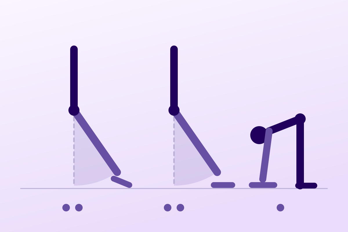
Hypermobility is not a disease but a spectrum: a simple trait in most, it becomes HSD or hEDS in a minority, with exercise then dosed with care.
90%of those with generalised joint hypermobility remain asymptomatic
What follows covers each of these points in detail, with sources. It is there if you need it.
This topic is taught in a course: Managing complex pain, on-site with Anthony Halimi.
1 / 11
10 article chapters · 42 min in total
In brief◔ 2 min
Joint hypermobility is not a disease: it is a trait, present in a large share of the population.
Nomenclature◔ 5 min
Knowing how to name what is in front of you, with the words the literature has used since 2017.
Beighton score◔ 6 min
The Beighton is the test everyone knows, and that almost nobody interprets correctly.
Other assessments◔ 3 min
Once the score is recorded, most of the assessment remains to be done: it is what follows that changes what you do.
Comorbidities◔ 5 min
A well-dosed exercise programme can fail entirely for reasons that are not joint-related.
Dosing exercise◔ 7 min
Well dosed, exercise transforms; badly dosed, it costs months.
Level of evidence◔ 5 min
This chapter forces an admission of the thinness of the foundations we work on.
Clinical cases◔ 3 min
Three real, identifiable cases each show one facet of the problem, for better or worse.
In practice◔ 3 min
An operational summary of the five chapters, in the real order of a consultation.
Questions◔ 3 min
No data justifies running a different programme depending on which side of the HSD/hEDS boundary the patient is on.
Train on this
Pain management
Anthony Halimi
Next comes practice: the course that teaches this topic, with Anthony Halimi.
Our courses on this topic may be covered by DPC or FIFPL, the French continuing-education funding schemes, depending on your profession and situation.
A joint that goes too far is not a disease. Between ordinary hypermobility and hypermobile Ehlers-Danlos syndrome lies a spectrum that the 2017 international classification redrew, and that half the online content still describes with the vocabulary of before. This article sets out the current nomenclature, says what the Beighton score measures and what it does not tell you, and deals with the question physiotherapists actually ask: how do you load without triggering a flare?
Three figures that sum up the article. Prevalence depends first of all on the definition used3 ; the 2017 diagnostic criteria exclude the great majority of patients previously labelled5 ; and the pain feared during progressive strengthening did not materialise in the only trial that measured it session by session13.
Joint hypermobility is not a disease. It is a trait, present in a large share of the population depending on how it is measured, and asymptomatic most of the time. It becomes a matter for care only when it comes with symptoms.
The 2017 international classification redrew the spectrum into three zones : asymptomatic hypermobility, hypermobility spectrum disorder (HSD) and hypermobile Ehlers-Danlos syndrome (hEDS), the last with its own strict criteria1. The old vocabulary, “benign joint hypermobility syndrome”, “Ehlers-Danlos type III”, is no longer current. No revision has appeared to date; an update is announced for the end of 2026.
HSD and hEDS are managed in the same way. The boundary between them is administrative and genetic, not rehabilitative: nothing in the trials justifies treating a patient differently according to whether or not they cross the threshold of the 2017 criteria6.
The Beighton score is reliable but narrow. Its reproducibility between examiners is good8 ; its capacity to represent hypermobility of the whole body is not, since it tests neither the shoulder, nor the hip, nor the ankle7. It varies with age and sex, and it is regularly over-scored by untrained examiners5.
Exercise is the core treatment, and it tolerates load. The best trial available did not find high load superior to low load on function at one year12, but it showed that it did not hurt more13. Excessive caution therefore costs more than it protects.
The overall level of evidence remains modest, and that has to be said. The systematic reviews gather about ten trials, a few hundred participants, and judge a minority of them to be at low risk of bias10. We are working on thin foundations.
The comorbidities are not incidental, they govern the session. Chronic fatigue in nearly one patient in two, digestive symptoms in two in three, orthostatic intolerance in more than one in three16. A programme designed without them will fail for reasons that have nothing to do with the joints.
This is the first value of this article, and probably the most useful in the clinic: knowing how to name what is in front of you, with the words the literature has used since 2017, and knowing that those words are going to change.
Until 2017, the reference nosology was that of Villefranche, published in 1998, which distinguished six subtypes of Ehlers-Danlos. Type III, known as “hypermobile”, sat alongside a competing rheumatological diagnosis, “benign joint hypermobility syndrome”, defined by the Brighton criteria. Two medical communities were therefore describing much the same patients with two distinct vocabularies, and the qualifier “benign” maintained the idea that there was not much to be done.
The International Consortium on the Ehlers-Danlos Syndromes reshuffled the cards in 2017. Its classification recognises thirteen subtypes, and sets out a principle that structures everything else: for twelve of them, the definitive diagnosis rests on molecular confirmation, that is, the identification of a causal genetic variant. hEDS is the only subtype with no known genetic marker, and therefore the only one whose diagnosis remains purely clinical1. That exception is not an administrative detail: it explains both the severity of the clinical criteria adopted, since the absence of a test had to be compensated for, and the lasting discomfort they cause.
In the same move, the category of hypermobility spectrum disorders (HSD) was created to take in the symptomatic patients who do not meet the criteria for hEDS. It is not a second-class category: it is the recognition that between asymptomatic hypermobility and hEDS there is a large, suffering population that needs care.
| Category | Definition | Symptoms | What the physiotherapist does |
|---|---|---|---|
| Asymptomatic joint hypermobility | Ranges beyond the norm, isolated (one joint), peripheral, or generalised (GJH). No complaint. | None. It is a trait, sometimes an advantage in dance, gymnastics or music. | Nothing to treat. Mention it without alarming them; do not turn a trait into a diagnosis. |
| Hypermobility spectrum disorder (HSD) | Hypermobility and attributable musculoskeletal symptoms, without meeting the criteria for hEDS and after excluding the other connective tissue disorders. | Pain, instability, subluxations, repeated sprains, fatigue. Divided into four forms according to distribution (generalised, peripheral, localised, historical). | Management identical to that of hEDS. It is the commonest category in the clinic. |
| Hypermobile Ehlers-Danlos syndrome (hEDS) | All three criteria must be met: generalised hypermobility, and a combination of systemic features or family history, and exclusion of alternative diagnoses. | Those of HSD, plus systemic involvement: skin, comorbidities, first-degree family history. | Identical to HSD in rehabilitation terms, but requires coordination with clinical genetics and the other specialties. |
| Other Ehlers-Danlos subtypes (12) | Diagnosis confirmed by a causal genetic variant. | Variable depending on the subtype. The vascular type carries a risk of arterial and intestinal rupture. | Not covered by this framework. A doubt about a vascular type calls for a specialist opinion before any management. |
The spectrum according to the 2017 classification. The proportion of about 10 % of symptomatic carriers comes from the systematic review by Blajwajs et al. on 107 studies3 ; the architecture of the categories follows Malfait et al. 20171.
The diagnosis of hEDS requires three criteria to be met simultaneously. The first is generalised joint hypermobility, measured by the Beighton score with thresholds that vary with age and puberty. The second combines systemic features, soft or slightly hyperextensible skin, unexplained striae, hernias, prolapse, dental involvement and many others, a first-degree family history, and musculoskeletal complications. The third is an exclusion: no other heritable connective tissue disorder, no autoimmune disease, no acquired cause of laxity.
These criteria were designed to improve specificity, and they succeeded. The problem is what they produced in practice. At the GoodHope clinic in Toronto, a retrospective review of 131 patients carrying a diagnosis of hEDS made under Villefranche showed that only 15 % (20 patients) met the 2017 criteria. More troubling still, most of the features supposed to distinguish hEDS were not significantly more frequent in those who crossed the threshold than in those who failed, and the usual comorbidities were distributed about equally in the two groups5. In other words: the boundary drawn separates poorly two populations that resemble each other.
An Italian study of 327 patients from 213 families reached a convergent conclusion and put it bluntly: the 2017 criteria should be relaxed so as to include some of the patients now classified as HSD, failing which diagnostic accuracy and quality of care suffer6.
Since 2024 the Ehlers-Danlos Society has been running an initiative called “Road to 2026”, intended to revise the classification, the diagnostic criteria and the care pathways. It draws on an international Delphi study, a real-world clinical practice study launched at the end of 2025, and the HEDGE genetic study, the largest ever conducted on hEDS. Publication of the new criteria is announced for the end of 2026.
At the time of writing, August 2026, no revision has appeared : the 2017 criteria remain the current reference, and all the work cited here rests on them. When the revision appears, the diagnostic part of this article will have to be reread. The rehabilitation part will not move: it does not depend on the threshold.
It is the test everyone knows and almost nobody interprets correctly. It deserves better than a figure out of nine copied into the assessment.
The Beighton score counts nine points, spread over five manoeuvres: passive extension of the fifth finger beyond 90°, passive apposition of the thumb to the forearm, hyperextension of the elbow beyond 10°, hyperextension of the knee beyond 10°, each scored right and left, that is eight points, and forward flexion of the trunk with the palms flat on the floor, knees straight, for the ninth.
You have to know where it comes from to understand its limits. It was designed as an epidemiological tool, intended to screen for hypermobility in large populations quickly and cheaply. Its later adoption as an individual diagnostic instrument was not intended by its designers, and it is from that slide that most of the misunderstandings come7.
The composition of the score, and its blind spot. The concentration of items on the upper limb and the absence of the large joints are the central argument of the review by Malek et al., who conclude from it that the score alone allows neither generalised hypermobility to be established nor ruled out7.
That is the distinction that settles most of the debates about this test, and it deserves to be set out properly, because two systematic reviews published in the same year appear to contradict each other while they are not talking about the same thing.
Reliability is good. A systematic review of 24 studies covering 1,333 patients, scored by examiners of varying experience, concludes that the Beighton score shows substantial to excellent inter- and intra-examiner reproducibility. Intra-examiner reliability was reported as excellent in all the studies using intraclass correlation coefficients8. Concretely: if two physiotherapists score the same patient, they most often agree.
Validity, on the other hand, is disputed. Agreeing does not guarantee measuring the right thing. The review by Malek et al. notes that the joints in the score are mostly in the upper limb, that they ignore major joints, and above all that one finding recurs consistently in the literature: the score fails to identify hypermobility present in the joints it does not test. It therefore works neither as a direct measure, nor as a reliable indirect indicator of generalised hypermobility. The authors explicitly call for a change in clinical thinking: the score must not serve as the main tool for distinguishing localised from generalised hypermobility, nor be used alone to rule out its presence7.
The Beighton score is not a biological constant. Laxity decreases with age, which means that a score of 5 does not have the same meaning at 12 and at 55, since a patient who is negative today may have been frankly hypermobile in their youth, hence the “historical” category of HSD and the existence of a five-item retrospective questionnaire used alongside it.
The meta-analysis by Sobhani-Eraghi et al., covering twenty studies and more than 21,000 children and adolescents, finds an overall prevalence of 34.1 % (95 % CI: 33.3-34.8), with 32.5 % in girls against 18.1 % in boys. Its authors themselves report considerable heterogeneity between studies and statistically significant publication bias: the overall figure is to be handled with care, but the gap between the sexes and the decrease with age are consistent across the literature4.
In young adults, the study by Reuter and Fichthorn in 654 American students using the threshold of ≥ 5 adopted by the 2017 criteria finds 12.5 % with generalised hypermobility. Notably and counter-intuitively, women did not have a higher overall rate than men, and scores did not differ by ethnic origin in that sample, even though women were more often hypermobile in the spine and in the right knee and elbow9.
Prevalences by source. Range 2-57 %: Blajwajs 2023, systematic review of 107 studies, thresholds from 4 to 73. Children and adolescents, girls and boys: Sobhani-Eraghi 2020, meta-analysis (major heterogeneity and publication bias flagged by the authors)4. Students: Reuter 2019, threshold ≥ 5 of the 2017 criteria9.
It is the most directly actionable result in this whole chapter, and it concerns our own practice. In the Toronto cohort, when the Beighton score had been scored by the primary care doctor and then rescored by a practitioner specialised in EDS, it was higher with the former in 81 % of cases (74 out of 91). And generalised hypermobility was ultimately confirmed in only 46 % of patients (51 out of 111) carrying a previous diagnosis of hEDS5.
The lesson is not that generalists score badly, but that this test looks simpler than it is. The sources of error are known: passive confused with active, absence of stabilisation, rough goniometry on the 10° thresholds, a prior warm-up that wins a few degrees, and above all the examiner's expectation seeking to confirm a hypothesis. A score dashed off at the end of an assessment is worth less than no score at all, because it will be taken over as it stands by the next person.
A high Beighton score is not a diagnosis, and a low score rules nothing out. The patient who subluxes their shoulder three times a week and scores 3 out of 9 fully warrants management for instability: the score simply does not test their shoulder. Conversely, the dancer at 8 out of 9 with no complaint has nothing to treat, and announcing a “syndrome” to her creates a problem she did not have.
Once the score is recorded, the bulk of the assessment remains to be done. Here is what, in practice, changes what you do.
Since the Beighton score leaves out the shoulder, the hip, the ankle, the wrist, the cervical spine and the temporomandibular joint, those joints are tested separately, particularly the ones the patient complains about. The shoulder deserves special attention: it is the site most studied in hypermobility rehabilitation trials, and multidirectional instability is frequent there14. For the lower limb, ankle instability has been the subject of recent work in dynamic imaging in hEDS patients2.
The history counts as much as the measurement. The useful questions concern subluxations and dislocations, their number, their mechanism, whether the patient reduces them themselves, repeated sprains, night pain, fatigue on waking, and what the patient has already tried. Many of these patients have several interrupted courses of care behind them.
They serve not only to tick the 2017 criteria: they point towards connective tissue involvement rather than isolated laxity. You look at the skin, its softness, texture, widened scars, striae without weight gain or pregnancy; you ask about hernias, prolapse, dental and gum history, and above all first-degree family history, which is a criterion in its own right.
One point deserves flagging because it is counter-intuitive. In the Toronto cohort, objective systemic manifestations, those the clinician observes, were found less often than the subjective manifestations reported by the patient, in both groups5. That does not invalidate the patient's account; it is a reminder that self-report and examination do not measure the same thing, and that confusing the two artificially inflates the picture.
What to do. The equivalence of management between HSD and hEDS follows the 2025 AGA practice update17 and Ritelli's 2024 finding on the artificial character of the boundary6. Targeted rather than universal screening for comorbidities follows AGA advice 217.
A programme perfectly dosed in joint terms can fail entirely for reasons that are not joint-related. It is the chapter people skip and should not.
A meta-analysis published in 2026 in Alimentary Pharmacology & Therapeutics, covering 19 studies, 17,455 hEDS/HSD patients and more than 1.6 million controls, gives the broadest picture available to date. The results are of an order of magnitude that makes it impossible to treat these comorbidities as curiosities16.
Comorbidities, pooled prevalences. Kulin et al. 2026, meta-analysis of 19 studies, 17,455 hEDS/HSD patients against 1,677,465 controls. Odds ratio for chronic digestive symptoms: 4.29 (95 % CI 3.1-6.0). The intervals are wide: POTS 21.9 % (95 % CI 5.2-59.1), fibromyalgia 27.9 % (16.0-44.0), and the authors themselves rate the level of evidence as low because of the clinical heterogeneity, calling for caution about any causal interpretation16.
This is the comorbidity that most directly changes how a session is run. The symptoms, palpitations, dizziness, a sense of impending faint, syncope, typically occur on standing and ease on sitting or lying down. They are worsened by anything that causes vasodilation: heat, effort, and digestion after a meal18.
Postural tachycardia syndrome (POTS) is its best documented form. It is defined by an increase in heart rate of more than 30 beats per minute in adults, and of more than 40 in adolescents, on standing, with no fall in blood pressure, that last point distinguishing it from orthostatic hypotension18. The relationship with hypermobility is strong in both directions: in 91 POTS patients assessed with the 2017 hEDS checklist, 31 % (28 patients) met the criteria for hEDS, and a further 24 % had generalised hypermobility without meeting the criteria19.
Be careful, however, not to turn that finding into systematic screening. The American Gastroenterological Association update is explicit on this point: the search for POTS or mast cell activation should be targeted at patients who show clinical manifestations of it, and universal screening in all hEDS/HSD patients is not supported by current data17.
In a patient who describes these symptoms, a programme designed to be done standing will fail, not for want of motivation, but because the position itself is the problem. The adaptations are simple: start lying or seated, favour lower limb work that engages the muscle pump, avoid abrupt transitions to standing, ventilate and cool the room, do not schedule the session immediately after a meal, and check hydration. Strengthening while lying down is not second-rate strengthening: it is the only practicable one to begin with.
Two patients in three report at least one chronic digestive symptom, with an odds ratio of 4.29 compared with controls. Heartburn is the commonest (34.7 %), gastro-oesophageal reflux concerns 41.3 % of patients, and 44.2 % have a disorder of gut-brain interaction, functional dysphagia being the commonest among them at 34.2 %16.
For the physiotherapist, the consequence is practical more than theoretical: strict supine lying and trunk flexion exercises may be poorly tolerated with reflux, scheduling immediately after a meal is to be avoided, and a patient who eats little and poorly will have limited resources for loaded work. The AGA update in fact recommends the reverse of the usual reflex: that gastroenterologists following disorders of gut-brain interaction ask about hypermobility and include the Beighton score in their practice17.
The hypermobile patient's pain is not only mechanical. It combines nociceptive components, the real loading on tissues that defend themselves poorly, neuropathic components, and central sensitisation mechanisms that explain the frequent disproportion between observable lesions and reported intensity20. Work conducted in adolescents with HSD or hEDS has explored these signs of central sensitisation specifically in this population21.
That dimension has a direct consequence for dosing: in a sensitised patient, the intensity of pain during exercise is a poor guide to the real tissue load. Relying on it leads to under-loading indefinitely. We return to it in the next chapter, because that is exactly the crux of the problem.
Almost one patient in two reports chronic fatigue16. It is multifactorial, poor quality sleep, night pain, dysautonomia, deconditioning, the increased energy cost of a poorly stabilised posture, and it is very often the first reason a programme is abandoned. A session that leaves the patient floored for two days will not be repeated, however good it is technically.
Most of these patients arrive with a long history. The delay in recognising the symptoms, and then the underlying hEDS or HSD, is a recurring finding in the literature, including for the dysautonomic manifestations, whose nature often takes years to be identified, which delays treatment by as much18. A recent international survey of hEDS and HSD patients documented these diagnostic difficulties, the long-term burden of comorbidities and the unmet needs22. A qualitative review devoted to the primary care pathway analysed the same difficulties from the point of view of the patient and the practitioner23.
What should be taken from this is not a matter of compassion but of clinical efficiency: a patient who has been sent away several times with “your tests are normal” arrives with justified mistrust and often with the conviction that movement damages them. The first task is sometimes to undo that conviction, and the next chapter shows that the recent data help us do so.
This is the central question of this article, the one that decides the patient's trajectory. Well dosed, exercise transforms; badly dosed, it costs months. And the most solid trial we have says something many practitioners do not believe.
The Danish trial by Liaghat et al. is the current reference on this question. One hundred adult patients recruited in primary care, with a hypermobility spectrum disorder and shoulder pain or instability for more than three months, were randomised into two groups for sixteen weeks of exercise at three sessions a week. The HEAVY group worked through full range, at high progressive load, supervised twice a week. The LIGHT group worked at low load, in neutral to mid range, with only three supervision sessions in total12.
First result, at one year: high load is not superior. The between-group difference in self-reported function measured by the WOSI was −92.9 points in favour of the high-load group, but with a 95 % confidence interval running from −257.4 to 71.5, and a p of 0.268: the result is not statistically significant. The secondary outcomes were largely inconclusive, with one exception: the patients in the high-load group improved more on the shoulder-related emotions subscale (−36.3; 95 % CI −65.4 to −7.3; p = 0.014), and the effect perceived by the patient pointed the same way on the emotions and lifestyle dimensions12.
Second result, and this is the one that changes practice: high load does not hurt more. A secondary analysis of the same trial measured pain before and after each session over sixteen weeks. Exercise-induced pain remained below 0.5 points on the numerical scale in both groups, throughout the sixteen weeks, with no difference between them. The pain trajectories were similar too. The authors conclude in so many words that these results challenge previous beliefs13.
The two results of the Danish trial. On the left, the difference in function at one year and its confidence interval12. On the right, the pain induced by the session, the mean of the three weekly sessions, which stayed below 0.5/10 for sixteen weeks in both arms13. The trace illustrates the stability reported; the week-by-week values are not published as a table.
If high load is neither superior nor more painful, what should you do? The honest answer is that the choice of load is decided on criteria other than fear: the patient's goals, their preferences, the supervision time available, and their tolerance measured rather than assumed.
Two further pieces of data help set the frame. First, the trial by Spanhove et al. in 21 hEDS/HSD patients with multidirectional shoulder instability: two different home programmes, six months, and significant improvement in both: 240 WOSI points at twelve weeks and 325 at twenty-four weeks, with no difference between the programmes. Kinesiophobia, by contrast, is the only outcome that did not improve, which led the authors to suggest that a supervised, multidisciplinary approach would be more effective on that particular dimension14. Second, an analysis of effect modifiers in the Danish trial showed that psychological factors and symptom duration are associated with the response to treatment24.
In other words: the variable that decides is probably not the number of kilos, but the support given and the patient's beliefs.
The idea of strengthening in inner range, in the portion of the range where the muscle is most shortened, is widely taught in hypermobility, with attractive reasoning: since the joint lacks a passive brake at end of range, you may as well build active control where the muscle is in a position to provide it, and avoid the extreme positions where the capsuloligamentous apparatus is most loaded.
One has to be frank about the status of this practice: no trial has directly compared inner-range strengthening with full-range strengthening in these patients. What exists is indirect, and points rather the other way: in the Danish trial, it was the group working through full range and at high load that produced neither excess pain nor any notable adverse effect1213, while the group in neutral to mid range obtained equivalent results. Inner-range work therefore remains a reasonable option, particularly useful at the start of a programme, in a very apprehensive patient or when resuming after a subluxation. It is not an obligation, and nothing authorises making it a permanent limit that would deprive the patient of the functional range they need.
A deficit in joint position sense is a documented feature of these populations, and it is one of the rare areas where the systematic reviews report a consistent signal. The review by Reychler et al., covering six randomised trials of 20 to 57 patients and programmes of four to eight weeks, finds a significant improvement in pain or in proprioception in the intervention group whatever the type of intervention, and a systematic improvement in quality of life11. The scoping review by Brittain et al., on 28 studies and 630 participants, concludes for its part that therapeutic exercise and motor control training are the effective modalities, the others resting on weak evidence15.
In practice, proprioceptive work does not have to be sophisticated: control of range without going to the end, visual feedback then removal of visual feedback, mid positions held, slow transitions, then integration into functional movements. The important point is that it combines with strengthening rather than preceding it: the two progress together.
This is where most programmes fail, and the mechanism is well identified. The patient has a good session, feels capable, does a bit more, and pays two days later. The experience repeats, and they conclude that exercise does not suit them.
Three principles limit that scenario. The first is to cap the good days : the instruction is not to exceed the planned volume even when everything is going well, which means explaining it, because it is counter-intuitive. The second is to spread rather than concentrate : three short weekly sessions, as in the trials, rather than one long session. The third is to distinguish explicitly, with the patient, overshoot pain from sensitisation pain: the first is proportionate to what was done and eases within one to two days, the second is disproportionate, diffuse, and does not follow the load. Confusing the two leads to under-loading a patient who did not need it.
The protective reflex (limited ranges, token loads, stopping at the slightest signal), looks cautious and is not. It produces deconditioning that worsens instability, it confirms to the patient that their body is fragile, and it consumes the months during which real progression would have been possible. The data from the Danish trial remove the main argument for that caution: over sixteen weeks of full-range, high-load strengthening, the pain induced by the sessions stayed below 0.5 out of 1013.
This chapter is the most uncomfortable to write, because it forces an admission of how thin the foundations are on which we work. Saying so is nevertheless the condition of honest practice.
Three recent systematic reviews make it possible to measure precisely what we have, and the picture they draw converges.
Buryk-Iggers et al. counted, up to November 2020, ten eligible studies totalling 330 participants : five randomised trials, one cohort, two single-arm interventions, one retrospective study and one feasibility study. All reported improvement in at least one physical or psychological outcome, and only one non-serious adverse effect possibly related to the intervention was reported. But of the five randomised trials, only two were judged to be of high quality and at low risk of bias on the PEDro scale, and most of the non-randomised studies were classified at critical risk of bias. Their conclusion explicitly calls for rigorous, adequately powered randomised trials10.
Reychler et al., searching up to April 2020, retained six randomised trials, with sample sizes between 20 and 57 patients, programmes of four to eight weeks and heterogeneity described as enormous between the interventions. They conclude to a benefit on proprioception and pain, while noting that robust trials are lacking11.
Brittain et al., with a search extended to April 2023, included 28 studies and 630 participants, mostly women, of mean age 26.2. They identify therapeutic exercise and motor control training as effective methods, and describe as weak the evidence supporting adaptive equipment, patient advice, manual therapy and functional training. They stress that further research is still needed to determine efficacy and above all the dosing of interventions15.
A word on what that means. Thirty-three years after the description of the hypermobility syndrome, the whole of the interventional physiotherapy literature fits into a few hundred patients, almost all studied at the shoulder, over periods of a few weeks. No high-level clinical practice guideline, of the kind we have for low back pain or tendinopathy, exists for this population. An editorial published in 2026 in the Journal of Orthopaedic & Sports Physical Therapy took note of that situation by challenging the profession directly on its ability to recognise these patients25.
Grading of the evidence. A rating derived from the three systematic reviews available (Buryk-Iggers 202210, Reychler 202111 and Brittain 202415) and from the individual trials cited in the table below. This is an editorial appraisal applying GRADE logic to the available data, and not a published formal GRADE rating: no group has carried out such an exercise on this population to date.
| Modality | Level | What underpins it | What can be said about it |
|---|---|---|---|
| Progressive shoulder strengthening | Moderate | RCT n = 100, 1-year follow-up, mixed model analysis, blinded analyst12 | High and low load give equivalent results on function. High load does not increase pain13. Generalisation to other joints not established. |
| Home exercise, multidirectional instability | Low | RCT n = 21, 6 months14 | Significant improvement in function in both arms, increasing up to 24 weeks. Sample too small to separate the programmes. No effect on kinesiophobia. |
| Proprioception and motor control | Low | 6 RCTs of 20 to 57 subjects, programmes of 4-8 weeks11 ; scoping review of 28 studies15 | Improvement in pain or proprioception whatever the type of intervention, and quality of life systematically improved. Major heterogeneity. |
| Inspiratory muscle training | Low | A single RCT in hEDS patients26 | Benefit on lung function and functional exercise capacity. A single trial, a narrow outcome. |
| Compression garments | Low | A single RCT, compression garment plus physiotherapy against physiotherapy alone27 | Improves dynamic balance in combination with physiotherapy. Pain fell in both groups, with no difference between them : it is an adjunct for balance, not an analgesic treatment. |
| Wrist orthosis against stabilising exercise | Low | RCT, 116 participants completing the intervention, 12 weeks28 | No significant difference between stabilising exercise and orthosis on activity capacity, quality of life, function, grip strength, pain or paraesthesiae. |
| Targeted against generalised rehabilitation in children | Low | RCT, 57 children aged 7 to 16, targeted (n = 30) against generalised (n = 27)29 | Significant and lasting reduction in pain when the two groups are combined, but no difference detected between them. The first randomised physiotherapy trial published on hypermobility. |
| Manual therapy | Very low | Described as weak evidence by the scoping review15 ; a case series calling for caution30 | To be used with discernment and never as the main treatment. Active exercise remains the essential element of care. |
| Adaptive equipment, advice alone | Very low | Scoping review, 28 studies15 | Weak evidence. May have a place in an overall plan, without constituting an intervention on their own. |
| Hippotherapy | Very low | A single published clinical case31 | One documented case with a favourable course. No generalisation possible from one case. |
A low level of evidence is not proof of ineffectiveness: it is uncertainty about the size of the effect. What follows from it is not to abstain, but to stay modest in what you promise, to measure in your own patient what the literature does not guarantee, and to reassess. That is particularly true here, where all the studies report improvement and where only one non-serious adverse effect has been reported across the whole corpus10.
Three real, published and identifiable cases, each showing one face of the problem: what sustained rehabilitation can achieve, what the absence of active care produces, and what caution manual therapy demands.
Published in BMJ Case Reports in 2024, this case concerns a patient in her late teens with hEDS, who had experienced a rapid functional decline to the point of depending on a wheelchair. She followed a hippotherapy programme of thirty hours. The authors report substantial improvement in fatigue and chronic pain, and a recovery of walking ability: the patient went from wheelchair dependence to walking with elbow crutches, then markedly improved the quality and speed of her gait. The authors attribute these gains to work on postural balance, motor skills, proprioception, muscle function and endurance, as well as to an effect on cognitive and emotional regulation31.
What to take from it, and what not to. A single case does not demonstrate the efficacy of hippotherapy, and it would be dishonest to present it that way. What it does illustrate solidly is that severe deconditioning in this population is not inevitable: an active, prolonged and sufficiently dosed intervention can reverse a trajectory that seemed established. The volume is the notable element here: thirty hours, when so many programmes stop at six sessions.
Published in Cureus in 2023, this case describes a 67-year-old woman with hypermobile Ehlers-Danlos, with associated osteoarthritis and anxiety, treated with opioids, oxycodone and slow-release morphine, for a decade, until a change of general practitioner led to a reassessment. Morphine equivalents were calculated and a progressive withdrawal begun in order to reduce the risk of overdose. The authors are a reminder that chronic pain in EDS has both nociceptive and neuropathic components, and that its management must rest on a global approach combining non-opioid treatments, physiotherapy and psychological support32.
What to take from it. It is the exact counter-example of the previous case, and it is more frequent in practice. Ten years of analgesic escalation reflect first of all a failure of recognition: without a clear diagnosis, pain becomes a symptom to suppress rather than a problem to treat. The physiotherapist who receives these patients often arrives after those years, and their first task is to offer a credible alternative to a strategy that has failed slowly.
A series of three cases published in 2020 in the Journal of the Canadian Chiropractic Association describes the presentation, assessment, management and course of patients with joint hypermobility syndrome. The authors note that recognising hypermobility as a significant contributing factor is often difficult, and that failing to know this framework delays both diagnosis and the setting up of effective care. Their practical conclusion is doubly explicit: manual therapy must be used with discernment, and active exercise is an essential element of care30.
What to take from it. In a patient whose joints already lack passive restraint, techniques that remove still more have no obvious rationale, and may bring a misleading immediate relief that delays active work. That does not mean forbidding all manual contact, treating secondary muscle tension has its place, but that high-velocity manipulation at end of range requires a particularly solid justification in this population.
An operational summary of the five previous chapters, in the order in which the questions actually arise in the clinic.
A useful framework for organising that assessment is the one proposed by the international group of physiotherapists convened in 2017, which adopts the International Classification of Functioning to structure the assessment around functions, activities and participation, without forgetting environmental and personal factors. That text remains, nearly ten years later, the reference statement for our profession on this subject, and it already noted that many physiotherapists knew neither the criteria, nor the prevalence, nor the usual presentation33. In children, a specific diagnostic framework was proposed in 202334.
The progression proposed. The rhythm of three weekly sessions and the horizon of sixteen weeks take up the protocol of the Danish trial12 ; the growth of benefits between the twelfth and twenty-fourth week comes from Spanhove's trial14. The rules for managing flares are an editorial proposal consistent with these data, and not a validated protocol: no trial has compared different progression rules in this population.
The content of what is said counts as much as that of the programme, because psychological factors are associated with the response to treatment24 and because kinesiophobia is the outcome that resists most in the home-based trials14. Four messages deserve to be put explicitly.
To clinical genetics or a reference centre when the picture suggests a heritable connective tissue disorder needing confirmation, and without delay for any suspicion of a vascular type. To a cardiologist or neurologist for disabling orthostatic intolerance symptoms that have never been investigated. To a gastroenterologist for chronic digestive symptoms affecting nutritional status. To chronic pain management when signs of sensitisation dominate the picture and block progression despite correct dosing.
No. The distinction between HSD and hEDS rests on criteria whose relevance is itself debated56, and no data justifies running a different programme depending on which side of the boundary the patient falls. The only investigations not to be delayed are those that rule out a vascular type or another connective tissue disorder requiring specific surveillance.
No, for two reasons. The score tests neither the shoulder, nor the hip, nor the ankle: frank hypermobility of those joints can coexist with a low score7. And laxity decreases with age, hence the existence of a “historical” category and of retrospective questionnaires for patients who were once more supple. A low score does not rule out; it invites you to look elsewhere.
That is not what the available data show. The group working through full range and at high load in the Danish trial did not have more pain than the group staying in neutral to mid range, over sixteen weeks of session-by-session follow-up13. Inner-range work keeps its value at the start of a programme and in the apprehensive patient, but no trial has compared it directly with full range, and nothing justifies making it a permanent limit.
It is not contraindicated, it is weakly supported. The largest scoping review classes it among the modalities resting on weak evidence15, and a case series concludes that it should be used with discernment, active exercise remaining the essential element of care30. Treating secondary muscle tension keeps its place; techniques that remove passive restraint from a joint that already lacks it require a solid justification.
That is the sign of an overshot dose, not of a contraindication to exercise. What to do is go back to the previous step, split the volume over more, shorter sessions, and check what, outside the session, is consuming the patient's resources: sleep, orthostatic intolerance, nutrition. Chronic fatigue concerns nearly one patient in two in this population16 : it is planned for, not overcome.
No. The American Gastroenterological Association update states that the search for POTS or mast cell activation should be targeted at patients with suggestive clinical manifestations, universal screening not being supported by current data17. Asking about dizziness on standing, on the other hand, is part of the routine assessment, because the answer changes how the session is run.
Asymptomatic hypermobility justifies no restriction. The question arises only in the presence of symptoms, and the answer is then dosing rather than stopping. In the symptomatic child, the first randomised physiotherapy trial showed a significant and lasting reduction in pain, without a targeted approach standing out from a generalised one29.
A revision is announced for the end of 2026 as part of the Ehlers-Danlos Society's “Road to 2026” initiative, supported by a Delphi study, a real-world clinical practice study and the HEDGE genetic study. At the time of writing this article, August 2026, it has not appeared: the 2017 criteria remain in force. The diagnostic part of this article will have to be reread at that point; the rehabilitation part will not.
Behind this article
An author who explains, a reviewer who checks.
How we write and check our content

Physiotherapist · co-founder of Physio Learning
Marked for life by his first four-hour lecture without a single image, he took a master’s in instructional design so that it would never happen to anyone again. He hunts down publication bias and unreadable slides with the same intransigence.

Head of scientific content
Physiotherapist specialising in neuro-musculoskeletal practice and holder of a master’s in public health. He checks the methodological rigour of every article: primary sources, levels of evidence, no exceptions.
