
















On-siteDPCFIFPL

Sports physiotherapy
From injury to return to play: structure every stage of reconditioning
Maxence Ponthus
Go further
Up to 59% of Lisfranc injuries are missed at first examination, and 31% follow a simple trip. Displacement over 2 mm warrants surgical reduction.
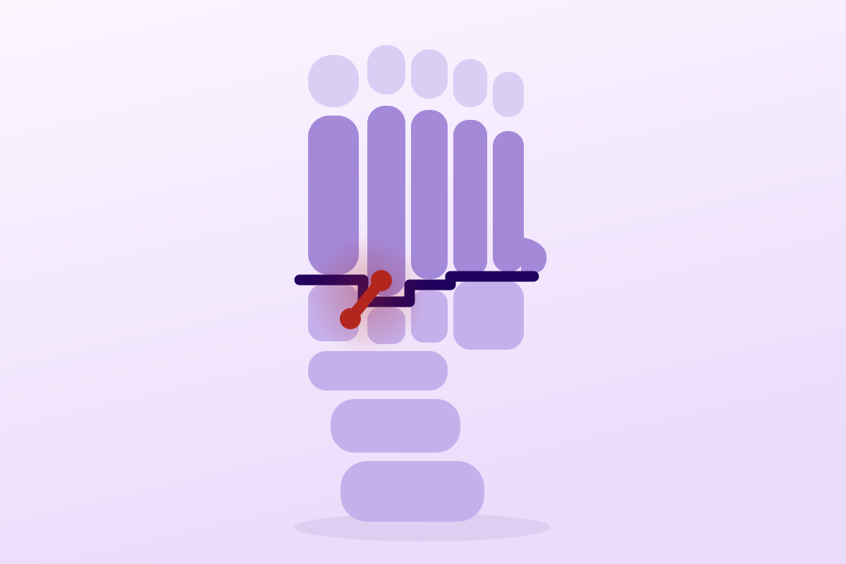
A Lisfranc injury is not an ankle sprain: the pain is in the middle of the foot, not below the malleolus.
99.4%pooled sensitivity of the Ottawa rules for detecting a foot or ankle fracture
What follows covers each of these points in detail, with sources. It is there if you need it.
This topic is taught in a course: From injury to return to play: structure every stage of reconditioning, on-site with Maxence Ponthus.
1 / 13
12 article chapters · 67 min in total
Distinguish◔ 7 min
A Lisfranc injury and an ankle sprain share three elements of the picture, which explains the confusion.
Understand◔ 4 min
Stability between M1 and M2 relies entirely on a single oblique ligament, the Lisfranc ligament.
Mechanisms◔ 4 min
The dorsal ligaments, more slender than the plantar ones, give way first.
Recognise◔ 5 min
No isolated sign makes the diagnosis; it's their accumulation that must trigger imaging.
Image◔ 8 min
MRI is clearly the best investigation for detecting ligamentous abnormalities.
Classify◔ 4 min
Three classification systems coexist, but only one answers the question that concerns the physiotherapist.
Operate◔ 6 min
The physiotherapist doesn't set the indication, but sees the patient before, between and after.
Rehabilitate◔ 11 min
There is no randomised trial of rehabilitation after Lisfranc injury.
Populations◔ 5 min
The reasonable approach is a paediatric orthopaedic surgeon's opinion for any persistent midfoot pain in a child.
Case studies◔ 4 min
Finding a diagnosis doesn't mean you can stop looking: two injuries can coexist.
In practice◔ 3 min
A finger on the dorsum of the midfoot changes everything, in the history as in the exam.
FAQ◔ 6 min
The reference protocol for a stable injury is immobilisation with non-weightbearing for six weeks.
Train on this
Sports physiotherapy
Maxence Ponthus
Next comes practice: the course that teaches this topic, with Maxence Ponthus.
Our courses on this topic may be covered by DPC or FIFPL, the French continuing-education funding schemes, depending on your profession and situation.
A patient twists their foot, it swells, they limp, they can no longer bear weight. The reflex is to call this an ankle sprain, and it is precisely that sentence which costs the prognosis. This article is about the midfoot: the tarsometatarsal joint, known as the Lisfranc joint. For the anterior talofibular ligament and the lateral ankle, see our dedicated article on lateral ankle sprain. The two look alike at first examination, are treated in opposite ways, and only one of them leaves three patients out of four with osteoarthritis ten years later when it is poorly reduced.
What to have in mind before reading the rest, if this patient walks in tomorrow morning.
The essentials in ten lines
“Lisfranc injuries are frequently missed at the first consultation, for want of demonstrative physical signs, and yet they leave significant disability.”
Wadsworth & Eadie, Journal of Orthopaedic & Sports Physical Therapy, 2005: PMID 15839309This question is not rhetorical: it is the error mechanism that produces most of the diagnostic delays, and it can be taken apart point by point.
The patient arrives with a swollen foot after a twisting injury. They limp, they point to “the ankle” with a broad gesture, they cannot put the foot down. Three elements of this picture are shared word for word with lateral ankle sprain, which is twenty to a hundred times more common. Clinical reasoning does what it always does: it keeps the most probable hypothesis. And it is right in the vast majority of cases, which is exactly what makes the error so hard to correct.
The problem is that the four features which separate the two injuries are all discreet, and that none of them leaps out unless it is looked for.
A patient almost never says “my midfoot hurts”. They say “my foot hurts” or “my ankle”, and they point to a ten-centimetre area. If the examination starts with palpation of the bundles of the lateral collateral ligament, it will almost always find something tender, because an injured, oedematous foot is diffusely painful. The test ends on a confirmation that is nothing of the sort.
The remedy is mechanical: palpate the line of the metatarsal bases systematically, from the first to the fifth ray, before even touching the malleoli. Exquisite, reproducible tenderness over the joint line between the first cuneiform and the base of the second metatarsal does not belong to the register of ankle sprain.
“I twisted my foot” covers two mechanisms that have nothing in common. In lateral ankle sprain, it is the foot that goes into inversion under a leg that carries on along its path. In Lisfranc injury it is the reverse: the forefoot is fixed to the ground, often in forced plantar flexion, and it is the body that topples or is loaded over the top. The registries find a clear predominance of low-energy mechanisms: in the Swedish fracture register, a simple trip and fall accounts for 31 % of the 2,084 injuries recorded between 2013 and 2022, and only one case in five arises from high-energy trauma (Juto 2025, PMID 39626601). In the Norwegian series, only 31 % of injuries were high-energy (Stødle 2020, PMID 31257042).
In other words: waiting for a spectacular injury before considering a Lisfranc means ruling out two thirds to four fifths of cases from the start.
The patient leaves the emergency department with a foot film and the verdict “no fracture”. The film was taken lying down, without load, because the patient could not stand. Yet it is precisely weightbearing that separates the joint surfaces and makes the diastasis visible. A normal non-weightbearing radiograph, in a patient whose midfoot is painful, is an inconclusive test, not a negative one. We return to this in detail in the imaging chapter.
The Ottawa ankle and midfoot rules are among the best validated triage tools in trauma care: the meta-analysis of 66 studies reports a pooled sensitivity of 99.4 % (95 % CI: 97.9-99.8) for a specificity of 35.3 % (95 % CI: 28.8-42.3) (Beckenkamp 2017, PMID 27884861). That sensitivity figure is reassuring, and that is where the reasoning error slips in.
These rules were built and validated to rule out a fracture. Their midfoot criterion covers the base of the fifth metatarsal and the navicular. A purely ligamentous Lisfranc injury is not a fracture, and its site is neither the navicular nor the base of the fifth. A patient can therefore satisfy the Ottawa rules, receive no radiograph, and still have an unstable midfoot injury. This is not a flaw in the rule: it is an application outside its domain of validity.
What the 99.4 % sensitivity does not say
A sensitivity measured for one diagnosis, fracture, does not carry over to another, ligamentous instability. The Ottawa rules remain excellent at what they do. They simply do not constitute a safety net for the Lisfranc complex, and never claimed to be one.
The published missed-diagnosis rates vary enormously, and that variation is itself instructive: it measures the recruitment as much as the difficulty of the diagnosis.
The attentive reader will note that Renninger finds no significant difference between high and low energy in the missed-diagnosis rate: 21 % versus 18 %. This is a counter-intuitive and important result. One might think that the discreet injuries are missed and the shattered ones spotted; in reality both are missed to much the same extent, for different reasons: the former because they are faint, the latter because they are lost in a polytrauma.
| Feature | Lisfranc injury | Lateral ankle sprain |
|---|---|---|
| Mechanism | Axial load or twist on a forefoot fixed to the ground, with the foot in plantar flexion | Inversion-supination of the foot under the leg, partly unloaded |
| Site of the pain | Line of the metatarsal bases, dorsum of the midfoot, C1-M2 joint line | Below and in front of the lateral malleolus, along the course of the anterior talofibular ligament |
| Ecchymosis | May lie on the sole of the midfoot, a highly suggestive sign | Lateral and retromalleolar, migrating towards the heel |
| Weightbearing | Often impossible from the outset, and above all impossible on the toes | Painful but often possible in grade I-II |
| Pain on abduction-pronation of the forefoot | Reproduces the midfoot pain | Reproduces nothing in particular |
| Non-weightbearing radiograph | May be strictly normal while the injury is unstable | Requested under the Ottawa rules, to rule out a fracture |
| The decisive investigation | Radiograph, bilateral and weightbearing, side by side | Clinical examination deferred to 4-5 days, imaging as per Ottawa |
| Consequence of a delay | Midfoot osteoarthritis, collapse of the arch, heavier surgery | Chronic instability, proprioceptive deficit, recurrences |
Red flags: refer without delay
Key points from this chapter
The confusion with ankle sprain does not come from a resemblance between the injuries, but from a resemblance between the presentations: swollen foot, twist, limp. Four safeguards are enough to lift it: palpate the line of the metatarsal bases before the malleoli, establish whether the forefoot was fixed to the ground, never take a non-weightbearing radiograph for a negative test, and know that the Ottawa rules do not cover ligamentous instability of the midfoot.
Understanding why an almost immobile joint becomes unstable after a discreet injury means looking at its geometry before its ligaments.
The midfoot is the region of the foot lying between the hindfoot and the metatarsals. The tarsometatarsal joint (known as the Lisfranc joint, after the military surgeon Jacques Lisfranc de Saint-Martin) is the line of union between the cuneiforms and the cuboid on one side, and the five metatarsal bases on the other. It crosses the foot obliquely, in a staircase rather than a straight line.
Functionally, three columns are described:
This difference in mobility has a direct surgical consequence, which we return to: the lateral column is not fixed like the other two, on pain of stiffness.
The base of the second metatarsal is recessed and keyed into a mortise formed by the three cuneiforms. This mortise-and-tenon arrangement is what locks the whole line: the second ray cannot move laterally as long as the mortise is intact. It also explains why the injury concentrates there, because a force that drives the first metatarsal away from the second comes up against that keying, and it is the ligamentous link, or the bone itself, that gives way.
An anatomical fact that is decisive for reading radiographs: there is no transverse intermetatarsal ligament between the base of the first and that of the second metatarsal. Stability between these two rays is provided entirely by an oblique ligament running from the first cuneiform to the base of the second metatarsal, the Lisfranc ligament proper. That is why widening between M1 and M2 on a weightbearing film signals injury to that ligament, and not a benign anatomical variant.
The systematic review of the cadaveric literature describes the Lisfranc ligamentous complex as three distinct structures: the dorsal bundle, the interosseous bundle and the plantar bundle. Across eight cadaveric studies selected from 1,204 screened articles, it is the interosseous bundle that shows the greatest thickness, the largest bony insertion area, the highest stiffness and the best resistance to failure when loaded along the axis of its fibres (Sripanich 2021, PMID 33639336).
But thickness is not function. The sequential sectioning cadaveric study is more instructive still: across sixteen lower limbs placed in a radiolucent loading frame and scanned at each stage, section of the dorsal ligament and of the interosseous ligament produces only a slight increase in the M1-M2 and C1-M2 distances. The widening worsens on sectioning the plantar ligament between the first cuneiform and the third metatarsal, and becomes maximal after sectioning the plantar ligament between the first cuneiform and the second metatarsal. The authors' conclusion is explicit: frank instability appears only with simultaneous sectioning of the Lisfranc ligament and of the two plantar ligaments (Penev 2021, PMID 31950232).
“An isolated rupture of the Lisfranc ligament, without dislocation, does not necessarily require surgical intervention.”
Penev et al., European Journal of Trauma and Emergency Surgery, 2021: PMID 31950232. A conclusion drawn from a cadaveric sequential sectioning model on sixteen specimens, and therefore to be transposed with all the caution owed to a model that is ex vivo.This plantar hierarchy explains two things in the clinic. First the diagnostic value of plantar ecchymosis: it reflects bleeding from the plantar structures, the ones whose injury tips the balance towards instability. Then the weight MRI gives to the plantar bundle in the decision, which we return to in the imaging chapter.
Key points from this chapter
The midfoot does not hold by the strength of one large ligament, but by a bony keying (the base of the second metatarsal in its mortise) locked by a ligamentous network whose thickest element is not its most decisive one. It is injury to the plantar bundles that tips the balance towards instability, and the absence of a transverse ligament between M1 and M2 that makes any measurable widening pathological by construction.
Reconstructing the movement is the first step of the examination, and often the one that orients you fastest. But the right question still has to be asked.
The foot is in plantar flexion, the forefoot resting on the ground through the metatarsal heads. An axial load travels down the limb, or a rotation is applied to the tibia while the forefoot stays fixed. The dorsal ligaments, more slender than the plantar ones, give way first; the metatarsal bases move dorsally and laterally. Depending on the intensity, the result is a simple ligamentous sprain, a bony avulsion at the ligament's insertion, or a frank dislocation.
The typical situations are not exotic:
A crush injury to the dorsum of the foot (a vehicle wheel, a heavy load falling) produces a different injury: more fractures, more columns involved, more soft-tissue damage, a risk of compartment syndrome. These are not the injuries that get missed, but the ones whose prognosis stays bleakest: in Myerson's original series, only one of the eight direct crush injuries achieved a good or excellent result (Myerson 1986, PMID 3710321).
The most useful comparison in practice comes from Renninger's series of 80 operated injuries, which sets 32 high-energy injuries against 48 low-energy ones (Renninger 2017, PMID 28693353). The differences are clear-cut:
| Characteristic | High energy (n = 32) | Low energy (n = 48) |
|---|---|---|
| Purely ligamentous injury | 16 % | 68 % |
| Associated fracture of the foot | 78 % | 4 % |
| Fracture of a metatarsal base | 84 % | 29 % |
| Displaced intra-articular fracture | 59 % | 4 % |
| Cuboid fracture | 31 % | 6 % |
| Associated fracture outside the foot | 37 % | 6 % |
| Involvement of all five rays | 23 % | 6 % |
| Mean number of rays involved | 4.1 | 2.7 |
| Diagnosis missed initially | 21 % | 18 % (difference not significant) |
The clinical reading fits in one sentence: the low-energy injury is isolated, ligamentous, limited to two or three rays, that is to say exactly the injury that shows almost nothing on a non-weightbearing radiograph. And it is the one that dominates in numbers.
One point deserves a pause. Stødle reports that unstable injuries are more common in women (p = 0.016), and identifies two further predictors of instability: the presence of intra-articular fractures of the two lateral tarsometatarsal joints (p = 0.007), and a reduced height of the second tarsometatarsal joint (p = 0.036), that is, a shallower mortise (Stødle 2020, PMID 31257042). This last point links the anatomy described above directly to clinical risk: the less deeply the keystone is set, the less it holds.
How to question the mechanism
Three questions, in this order, are enough to point the way:
Key points from this chapter
Lisfranc injury is not a major-trauma injury that occasionally spills over into sport: it is mostly the other way round. Two thirds to four fifths of cases arise from low energy, and in that register more than two thirds are purely ligamentous. The picture that causes the diagnosis to be missed is also the most common picture.
No single sign makes the diagnosis. It is their accumulation that must trigger the request for the right imaging, and you have to know which ones to look for.
Three things are visible, provided the patient is completely unshod and the two feet are compared side by side, soles included.
Swelling of the dorsum of the midfoot. It is central, in front of the instep, and not retromalleolar. Compared with the other foot, it visibly widens the region of the metatarsal bases.
Plantar ecchymosis. This is the sign that carries a name. Described in 1996 in a series of patients whose radiographs did not show the true extent of the injuries, it consists of an area of ecchymosis over the plantar aspect of the midfoot, and it reflects potentially major injury to the plantar tarsometatarsal ligaments. The authors conclude that patients carrying this sign must be assessed aggressively, with early recourse to stress films (Ross 1996, PMID 8932671).
What the plantar ecchymosis sign is not
It has never been the subject of a diagnostic accuracy study with sensitivity and specificity. It is a warning sign described in a case series, not a validated test. Its absence rules out strictly nothing, and it often appears one or two days late. To cite it otherwise is to lend it an authority it does not have.
Collapse of the medial arch. It can only be judged under load, and therefore only when the patient can put the foot down. It is a late sign, which already signals an advanced-stage injury.
The rule is to palpate the whole tarsometatarsal line, from the first to the fifth ray, before anything else. You are looking for:
They reproduce the pain by loading the ligamentous complex. None has published diagnostic values in the sources we verified; they serve to reinforce a suspicion, not to settle it.
These are the ones that sort best, once the hyperalgesic phase of the first three or four days has passed.
| Sign | What it suggests | Value and limit |
|---|---|---|
| Plantar ecchymosis of the midfoot | Injury to the plantar tarsometatarsal ligaments | Strong alert when present; absence uninformative; appears 24 to 48 h late |
| Exquisite tenderness over the C1-M2 joint line | Injury to the Lisfranc complex | The most specific site; to be sought actively, it is not pointed out spontaneously |
| Palpable gap between the bases of M1 and M2 | Established diastasis | Late sign; swelling often masks it |
| Pain on abduction-pronation of the forefoot | Tensioning of the Lisfranc ligament | Useful as an adjunct; painful and poorly reproducible in the acute phase |
| Positive piano key test | Instability of a single ray | Depends heavily on the examiner |
| Inability to hold a single-leg stance | Failure of load transmission through the midfoot | The best functional discriminator after the acute phase |
| Inability to rise onto the toes | The same, under propulsive load | A patient who runs on tiptoe is probably stable: an argument used in practice and documented in one case |
| Collapse of the arch under load | Loss of arch height, Nunley-Vertullo stage III | A sign of severity; always compared with the contralateral foot |
Red flags specific to the clinical examination
Key points from this chapter
The clinical examination of a suspected Lisfranc is not an ankle examination extended to the foot: it is an examination whose order changes. You inspect the sole, you palpate the line of the metatarsal bases before the malleoli, and you finish with loading tests (single-leg stance, heel raise) which are the most discriminating. None of these signs has a quantified diagnostic value: it is their convergence that triggers imaging, and imaging that settles the matter.
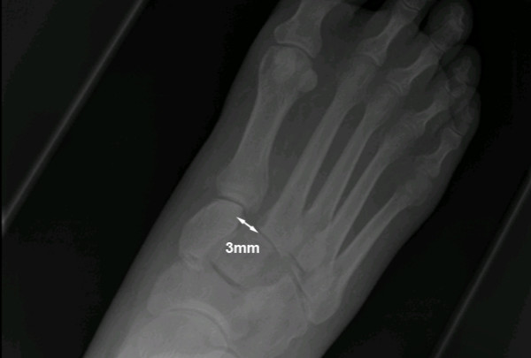
X-rayWeight-bearing anteroposterior foot X-ray: 3 mm lateral displacement of the base of the 2nd metatarsal relative to the medial cuneiform, a sign characteristic of a Lisfranc sprain.
Source : Yeoh et al., Cases Journal, 2008, figure 1 · CC BY
This is the chapter that decides. A well-chosen investigation rescues a hesitant clinical examination; a badly chosen one turns hesitation into false certainty.
This is the investigation the patient has almost always already had. Three views (anteroposterior, lateral, 30-degree oblique) taken lying down, because they could not stand. What is classically looked for is the fleck sign, a small bony avulsion in the space between M1 and M2, which signals that the Lisfranc ligament has been torn from its insertion.
The problem is not that this investigation is useless, because it detects frank dislocations and fractures. The problem is logical: without load, the joint surfaces are held in place by bony congruence alone, and a torn ligament shows no widening at all. The systematic imaging review concludes that conventional radiography can show a frank diastasis, but that weightbearing improves the ability to detect subtle injuries (Sripanich 2020, PMID 31368007).
A normal non-weightbearing radiograph in a patient whose midfoot is painful is therefore an inconclusive investigation. Putting it that way, in the report as in the conversation with the patient, stops them leaving convinced that “all is well”.
Three views of the foot, weightbearing, and above all the contralateral foot under the same conditions. It is the comparison that gives the information, because the normal distance between C1 and M2 varies widely between individuals.
The most useful Australian study on this point included 117 patients who had both bilateral weightbearing radiographs and a CT scan. The diastasis ratio, that is the measured gap on the injured side divided by the gap on the healthy side, was 1.53 (95 % CI: 1.41-1.65) in the operated patients, versus 1.11 (95 % CI: 1.07-1.16) in those treated without surgery, a highly significant difference (p < 0.001). The correlation between the diastasis measurement and the decision to operate was R = 0.576 (p < 0.001) (Kennelly 2019, PMID 30780193).
The most counter-intuitive result of this study concerns CT. When the initial weightbearing radiograph was positive, 54 % of the CT scans that followed were reported as equivocal or negative. And when the weightbearing radiograph was negative or equivocal, only 12 % of the CT scans were positive. The authors conclude that “CT offers limited benefit to the diagnosis and initial management of subtle Lisfranc injuries in the emergency setting” and argue for the bilateral weightbearing radiograph as the first-line investigation.
The practical difficulty, and how to handle it
A patient in the acute phase often cannot bear weight, and that is itself one of the signs that raise suspicion of the injury. The weightbearing radiograph then becomes impossible on the day. That is not a reason to abandon it: it is a reason to schedule it. The reasonable course is temporary immobilisation and offloading, then a weightbearing film as soon as weightbearing becomes possible again. In the Norwegian prospective weightbearing CT cohort, patients were able to tolerate full weightbearing after a median of 9 days (Poulsen 2025, PMID 41170783). That is the order of magnitude for the appointment to be set. The physiotherapist's role is often to notice that this appointment was never made.
Conventional CT, performed without load, provides what radiography does not: undisplaced fractures, minimal bony subluxations, the state of the lateral columns, the exact map before surgery. The systematic review confirms it: “CT is more useful than radiography for detecting undisplaced fractures and minimal bony subluxations” (Sripanich 2020, PMID 31368007).
What it does not give, performed without load, is stability, that is, the information that decides the treatment. That is the whole import of Kennelly's result. CT remains essential as soon as surgery is considered, and for injuries with a bony component; it does not replace the weightbearing film for the question “is this injury unstable?”.
Weightbearing CT (weightbearing CT) combines the three-dimensionality of CT with physiological loading. The systematic review of nine studies concludes that it “appears better able to detect subtle Lisfranc instability than conventional CT, presumably because it allows the injury to be assessed under load” (Talaski 2023, PMID 38027457).
The most accomplished work to date is the Oslo prospective cohort. Thirty-eight patients with undisplaced fractures (less than 2 mm) or avulsions in tarsometatarsal zone 1 to 3 had a bilateral single-leg weightbearing CT. The C1-M2 measurements, assessing the integrity of the dorsal bundle and of the interosseous bundle, were combined and compared with the healthy side to produce a difference score. Results:
What this study does not yet prove
Thirty-eight patients, a single centre, a single measurement protocol, and follow-up to twelve weeks. This is a prospective validation, which places it well above retrospective series, but the 3 mm threshold is not an established standard, and the authors themselves recall in their introduction that there is “no consensus on the optimal timing, the measurement protocol or the stability threshold” in weightbearing CT. Presenting this threshold as a settled rule would be an over-interpretation. And weightbearing CT remains a piece of equipment that is far from widespread.
MRI is, according to the systematic review, “clearly the best investigation for detecting ligamentous abnormalities”, but “its usefulness for detecting subtle Lisfranc instability requires further work” (Sripanich 2020, PMID 31368007). That nuance is the heart of the matter: seeing a torn ligament and predicting instability are not the same thing.
The study that narrowed this gap compares MRI with intraoperative findings in twenty patients (twenty-one feet), with the manual stress test under anaesthesia and surgery as the reference. Seventeen joints proved unstable, four stable. The best predictor of instability was neither the dorsal bundle nor the overall appearance of the complex, but rupture of the plantar ligament between the first cuneiform and the bases of the second and third metatarsals (pC1-M2M3), with a sensitivity of 94 %, a specificity of 75 % and a positive predictive value of 94 %. Nineteen of the twenty-one complexes were correctly classified by MRI (Raikin 2009, PMID 19339574).
Two practical lessons. First, MRI is not read by looking for “the Lisfranc ligament”: it is read by looking for the plantar bundle, consistent with what the sequential sectioning cadaveric study had established. Second, a plantar bundle of normal appearance points to a stable midfoot, and may spare a stress test under anaesthesia in a patient whose clinical picture and radiograph are equivocal, which is the authors' explicit conclusion. Conversely, rupture of the second plantar tarsometatarsal ligament, present in eighteen of the twenty-one feet, had “little clinical correlation with instability”: one more torn ligament is not one more argument.
Key points from this chapter
One investigation alone answers the question that decides the treatment, and it is not the most sophisticated: it is the bilateral weightbearing radiograph. Conventional CT maps the bone without judging stability; weightbearing CT is promising but its threshold is not established; MRI settles the matter provided the right ligament is asked of it, the C1-M2M3 plantar bundle. And when weightbearing is impossible, the weightbearing film is not cancelled: it is scheduled, around the tenth day.
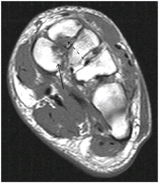
MRICoronal midfoot MRI: the plantar Lisfranc ligament and the Lisfranc ligament proper shown by arrows, at the level of the medial cuneiform fracture.
Source : Chang et al., Case Reports in Medicine, 2014, figure 2 · CC BY
Three systems coexist. They do not answer the same question, and only one answers the question that concerns the physiotherapist.
The anatomical classification, from Myerson's work on 76 fracture-dislocations, describes the direction and the extent of the displacement: involvement of all rays in the same direction (type A, homolateral), partial involvement with one or more rays spared (type B), divergent involvement where the columns separate in opposite directions (type C).
This classification has considerable historical and descriptive value, since it is the one that established that the quality of the initial reduction is the major determinant of unacceptable results, and that displacement greater than 2 mm after closed reduction mandates open reduction (Myerson 1986, PMID 3710321). It also retains prognostic value: type C is one of the three long-term risk factors for osteoarthritis identified, alongside non-anatomical reduction and smoking (Dubois-Ferrière 2016, PMID 27147683).
Its limitation is structural: it presupposes a visible displacement. It says nothing about the injury that appears only under load, that is, the one that gets missed.
Published in 2002, this classification was built for midfoot sprains in athletes, on the basis of the clinical picture, weightbearing radiographs and bone scintigraphy:
The authors report 93 % excellent results at a mean follow-up of 27 months, in fifteen athletes. This figure circulates widely; it should be quoted with its sample size (Nunley & Vertullo 2002, PMID 12435655).
The lasting contribution of this work is not the success rate, it is the demonstration that diastasis is measured by comparison with the healthy side and under load, and that loss of arch height is the severity marker that separates stage II from stage III.
A methodological detail that has aged
Nunley-Vertullo stage I rests on increased uptake on bone scintigraphy. Bone scintigraphy is no longer the fallback investigation in this setting: MRI and weightbearing CT have taken over. The logic of the three stages remains valid; the means of access to stage I has changed. Taking the classification up without saying so would implicitly recommend an investigation that has left practice.
The Bone & Joint Journal review of December 2024 states the problem head-on: “the original classification systems were anatomical, and limited as tools to guide treatment decisions”. It proposes instead a classification based on stability, with the weightbearing radiograph and CT as the reference diagnostic tools (Poutoglidou 2024, PMID 39615511).
The shift fits in one sentence: the question is not “what shape is the displacement?” but “does this midfoot hold under load?”. And the answer governs management directly:
| System | Question asked | Tool required | What it brings the physiotherapist |
|---|---|---|---|
| Hardcastle / Myerson (1986) | In which direction and on which rays did the displacement occur? | Conventional radiographs | Type C signals a higher risk of osteoarthritis, hence tailored surveillance and a tailored message to the patient |
| Nunley & Vertullo (2002) | How large is the diastasis in this athlete, and has the arch given way? | Radiographs weightbearing, side by side (+ scintigraphy at the time) | The move from stage II to stage III is read on the height of the arch: a collapse appearing during follow-up is an alert |
| Stability-based (2024) | Does this midfoot hold under load? | Weightbearing radiograph + CT, possibly weightbearing CT | It is the only one that predicts the protocol: six weeks of non-weightbearing if stable, surgery and its own timetable if unstable |
“The original classification systems were based on anatomy, and limited as tools to guide treatment.”
Poutoglidou et al., The Bone & Joint Journal, December 2024, PMID 39615511Key points from this chapter
Being able to recite types A, B and C changes nothing in management. What changes it is knowing whether the injury is stable or unstable, and that information is read neither in the operative report nor in the name of the classification, but in the behaviour of the midfoot under load. When the notes do not say, that is the question to put to the surgeon before building the protocol.
The physiotherapist does not set the indication, but sees the patient before, between, and after. Understanding what was decided, and why, conditions everything else.
The principle has been stable for forty years and has not been contradicted: a displaced injury must be reduced anatomically and held there. Myerson already stated that “displacement of more than 2 mm or a talometatarsal angle of more than 15 degrees on radiographs after closed reduction mandates open reduction” (Myerson 1986, PMID 3710321).
Fourteen years later, the Seattle series confirms this in 48 patients followed for a mean of 52 months: “the major determinant of a good result was anatomical reduction” (p = 0.05). The mean AOFAS midfoot score reached 77 points out of 100, with 25 % post-traumatic osteoarthritis, six of those patients requiring an arthrodesis (Kuo 2000, PMID 11097452). The same work notes that purely ligamentous injuries tend towards poorer results despite anatomical reduction and screw fixation, an observation that still feeds today's debate on primary arthrodesis.
The contemporary literature takes up the threshold: “residual displacement of more than 2 mm is associated with inferior outcomes and significantly reduced return-to-play rates” (Hammad 2026, PMID 41522288).
The systematic review of non-operative treatment is the most honest document on this question, because it measures failure as much as success. Across eight studies and 220 patients, mean age 39.8 years, mean follow-up 4.3 years:
The authors' conclusion deserves to be taken up as it stands: the heterogeneity is too great, a clear definition of a stable injury is lacking, and prospective work is needed. In other words, non-operative treatment is not a bad treatment: it is a treatment whose right candidates cannot yet be identified with certainty, which is what Poulsen's work on weightbearing CT is beginning to correct.
Two meta-analyses published in 2024 reach conclusions that must be read together, because they do not draw on the same material.
The first pools eighteen studies comparing primary arthrodesis and internal fixation in acute injuries. It favours arthrodesis on almost every criterion: AOFAS of 84.4 versus 75.7, VAS pain of 1.4 versus 2.0, return to previous activity in 79.2 % versus 65.7 %, midfoot osteoarthritis in 2.8 % versus 17.3 %, and above all a rate of unplanned reoperations of 14.7 % versus 38.3 % (p < 0.001) once planned metalwork removals are excluded. The pooled effect sizes confirm this: AOFAS 0.41 (95 % CI: 0.13-0.68), osteoarthritis OR 0.29 (95 % CI: 0.11-0.77), reoperations OR 0.16 (95 % CI: 0.06-0.44) (O'Connor 2024, PMID 39680239).
The second retains only the randomised trials: five trials, 241 patients, 121 arthrodeses against 120 internal fixations. It finds an advantage for arthrodesis on pain at two years (mean difference 0.89; 95 % CI: 0.18-1.59), on satisfaction (OR 10.04; 95 % CI: 1.78-56.76) and on all-cause reoperations (OR 27.31; 95 % CI: 12.72-58.63), but no difference in the AOFAS midfoot score at two years, nor in the SF-36, nor in unplanned reoperations. And the authors conclude that “these findings are not powered to confer an advantage to either approach, because of substantial heterogeneity” (Mactier 2024, PMID 39444525).
Why the gap between these two meta-analyses is not a contradiction
The first includes observational series, subject to indication bias: the same patients are not operated on by arthrodesis and by internal fixation. The second retains only randomised evidence, which neutralises that bias but cuts the sample to 241 patients. The all-cause reoperation figure that counts against internal fixation is largely expected by construction, since internal fixation often imposes a second procedure to remove metalwork, which both meta-analyses point out explicitly. That is why the comparison that counts is the one for unplanned reoperations.
Three families coexist, and the construct chosen determines the timelines the surgeon will pass on to you.
One hundred per cent return to activity: read the figure before quoting it
This 100 % relates to 35 patients drawn from 5 studies, with no comparator and no consistent definition of “activity”. It is not a success rate, it is the absence of reported failure in small series often published by the promoters of the technique. Flexible fixation is promising; it has not been shown to be superior.
The lateral column, that is the fourth and fifth tarsometatarsal joints, is mobile by nature, and fixing it rigidly produces stiffness. The recent literature recommends treating it with K-wires rather than with screws or a plate, with early removal (Hammad 2026, PMID 41522288). In practice this means that a patient whose lateral column has been wired will have an extra removal step in their timetable, often at around six to eight weeks, and that your mobility work on the lateral rays will begin after that.
Key points from this chapter
One criterion alone is solid and long-standing: any unstable injury, or one displaced by more than 2 mm, must be reduced anatomically, and the quality of that reduction weighs more than the choice of technique. On the rest, whether to fix or to fuse, screws, plate or suture button, the state of the evidence allows surgical preference, not assertion. What you must extract from the operative report is not the name of the construct: it is which columns were fixed, with what metalwork, and when it will be removed.
This is the chapter where the physiotherapist decides. First we must say honestly what it rests on: a level V expert consensus, a systematic review of postoperative protocols, and a handful of biomechanical series. There is no randomised trial of rehabilitation after Lisfranc injury.
The most structured source is the international consensus on foot and ankle trauma in sport, conducted in 2024 by a modified Delphi method over four rounds, backed by a systematic review. Its figures:
A gap between figures that must not be smoothed over
The consensus gives a return to sport at 4-6 months. The two NFL series give a median of 11.1 months (McHale 2016) and 11.0 months (Abed 2023). This is not a contradiction: the consensus describes the time to fitness after successful surgery, while the NFL series measure the observed time to return to a competitive match, season calendar included, and in contact athletes in the most exposed positions. Telling a patient 4 to 6 months without mentioning that the observed reality is often double would be a promise that will not be kept.
The reference protocol is the one adopted by the Bone & Joint Journal review: “stable injuries generally do well with non-operative treatment, most reliably by immobilisation and non-weightbearing for six weeks” (Poutoglidou 2024, PMID 39615511).
Two points that are not in the sentence but make it applicable. First, this approach presupposes that stability has been established, not presumed, that is, by a comparative weightbearing film, ideally completed by a CT. Second, it presupposes a weightbearing radiographic review later on, because late displacement is the dominant failure mode: up to 54 % in one of the series surveyed, and a surgical conversion rate of up to 56 % (Guerreiro 2023, PMID 36841140).
The only study to have quantified the late functional sequelae is a biomechanical analysis of seventeen patients operated on for a Lisfranc fracture-dislocation, reviewed on average 50.5 months after surgery. Its results guide the content of sessions better than any intuition:
The authors conclude that “sufficient rehabilitation is decisive for the clinical outcome” and support “a rehabilitation approach centred on restoring proprioception and triceps surae strength, including isometric exercises for the dorsiflexors” (Mehlhorn 2017, PMID 28043054).
The deficit that predicts the clinical outcome is not that of the triceps surae, but that of the dorsiflexors, and it persists more than four years after surgery.
Reading of the results of Mehlhorn et al., Gait & Posture, 2017: PMID 28043054. A series of 17 patients: a correlation in a sample of that size guides, it does not prove.| Phase | Landmark | Objectives and means | Criteria for moving on |
|---|---|---|---|
| 1. Protection | D0 to 4 wk | Strict offloading in a boot. Oedema drainage, scar work. Active mobilisation of the ankle and toes without loading the midfoot. Isometrics for the triceps and the dorsiflexors without weightbearing. Maintenance of the hip, the knee and the contralateral limb. Patient education on what is at stake at this stage. | Wound closed and dry. Swelling controlled. Surgical green light for weightbearing. No pain at rest. |
| 2. Progressive loading | 4 to 8 wk | Partial weightbearing in a boot, progressing in quantified load increments (scales, crutches). Relearning the roll-through of the step. Gentle mobility of the unfixed tarsometatarsal joints and of the ankle. Start of single-leg stance work with hand support. Analytical strengthening of the dorsiflexors. | Walking in the boot without a limp for 500 m. Midfoot pain below 3/10 on weightbearing and no worse the next day. Satisfactory check radiograph. |
| 3. Full loading | 8 to 12 wk | Progressive weaning from the boot to a rigid-soled shoe. Loaded strengthening of the triceps and the dorsiflexors. Double-leg then single-leg heel raises. Single-leg postural control, on stable then unstable surfaces. Cardiovascular reconditioning in relative offloading: cycling, swimming, cross-trainer. | Twenty full single-leg heel raises without pain. Single-leg stance for 30 s with eyes open and no hip strategy. Barefoot walking without a limp. No swelling the day after a session. |
| 4. Return-to-sport conditioning | 3 to 5 months | Progressive straight-line running, then accelerations, decelerations, changes of direction. Double-leg then single-leg plyometrics, with specific work on landing. Push-off strength and posterior chain endurance. Sport-specific technical skills, first controlled, then under constraint. | Single-leg hop test symmetry above 90 %. Running for 30 min without pain or swelling. No midfoot pain under maximal single-leg loading. |
| 5. Return to sport | From 4 to 6 months | Graded reintegration into training, then into contested play, then into competition. Continued strength and control work for at least six months more. | All the phase 4 criteria, surgical agreement, and a documented gradual return to the previous training load. |
How to read this grading
The grading below is ours, applied to the verified sources of this article following GRADE logic: high, moderate, low, very low. No learned society has published a clinical practice guideline on Lisfranc injury, unlike plantar fasciopathy or ankle sprain. None of these lines should therefore be quoted as “recommended by” anyone.
| Modality | Level | Reason for the grading |
|---|---|---|
| Bilateral weightbearing radiographs | Moderate | A cohort of 117 patients with a surgical endpoint, corroborated by a systematic imaging review. No randomised trial, but a consistent and large effect. |
| Anatomical reduction if > 2 mm | Moderate | Three independent series over 40 years, converging on the same prognostic determinant, with a dose-response effect on osteoarthritis. |
| Immobilisation and offloading for 6 weeks if stable | Moderate | A high-level review, but resting on heterogeneous series; the definition of “stable” remains the weak link. |
| MRI targeted on the plantar ligament | Low | A single study, 21 feet, a solid intraoperative reference but a very small sample and a population already selected for surgery. |
| Weightbearing CT, C1-M2 threshold > 3 mm | Low | Single-centre prospective validation, 38 patients, 12 weeks of follow-up. Excellent reproducibility. Threshold not confirmed elsewhere. |
| Primary arthrodesis vs internal fixation | Low | Two 2024 meta-analyses in partial disagreement: favourable when observational data are included, inconclusive on randomised evidence alone. Long term unknown. |
| Rehabilitation for strength and postural control | Low | One biomechanical series of 17 patients identifies the targets; no trial has tested the programme itself. |
| Flexible suture-button fixation | Very low | Eleven series with no comparator, samples of 35 to 216, discordant biomechanics, and an absence of complications to be interpreted with caution. |
| Arch-support foot orthosis | Very low | Used in one published case report with a favourable outcome; no comparative data. |
| Return to sport at 4-6 months | Very low | An expert consensus explicitly rated level V by its authors, at odds with the times observed in contact sport. |
| Non-operative treatment of an unstable injury | To be avoided | Late displacement up to 54 %, surgical conversion up to 56 %, osteoarthritis 5 to 38 %. |
Red flags during rehabilitation
Key points from this chapter
Three things hold, and it is worth knowing that they do not hold on the same footing. The time boundaries (partial weightbearing at around 4 weeks, full weightbearing at 8-12 weeks, sport at 4-6 months) come from a level V expert consensus, to be set against the 11 months observed in contact sport. The targets (dorsiflexors as much as triceps, single-leg stance, restoring load under the second metatarsal) are identified by measurement. And the vigilance for late displacement does not stop when the boot comes off.
Three populations in which the injury takes a particular form, and in which it is missed for three different reasons.
In athletes the injury is most often purely ligamentous, low-energy, and without displacement visible off load. This is the Nunley-Vertullo stage I or II injury. The athlete walks, limps moderately, and says “it's just a sprain”. Nunley and Vertullo already noted that half the athletes in their series had normal non-weightbearing imaging.
Two NFL series shed light on what becomes of this injury when it is managed properly, and they do not say quite the same thing:
| Criterion | McHale 2016 (2000-2010) | Abed 2023 (2009-2020) |
|---|---|---|
| Sample size | 28 athletes | 33 athletes |
| Return rate | 92.9 % (2 of 28 never played again) | 81.8 % |
| Time to return | Median 11.1 months (IQR 10.3-12.5) | Median 11.0 months (IQR 10.2-11.8) |
| Games missed | Median 8.5 regular-season games | No significant before/after difference in games played |
| Performance after return | Fall in offensive and defensive indices not significant compared with controls | Statistically significant fall in approximate value at one year (6.0 → 5.0; p = 0.022) |
| Positions most affected | 11 offensive, 17 defensive | Offensive linemen 24.2 %, running backs 21.2 % |
The convergence on timing, medians of 11.1 and 11.0 months over two different periods, is the most solid thing in these two papers. The divergence on performance after return is partly explained by the indicators used, which do not measure the same thing. What can be said without forcing it: return is the rule, it is slow, and the question of a return to the previous level remains open.
In the high-demand non-professional athlete, the American military cohort gives a useful landmark: at a mean follow-up of 3.5 years, 69 % had returned to the daily running required by their role, and 80 % had remained on active duty or completed their term of service. Two predictors stand out, and the first is directly actionable: a BMI below 30 multiplied the probability of returning to running by 13 (OR 13.14; 95 % CI: 2.50-69.19), whereas a BMI of 30 or more multiplied the risk of medical discharge by nearly 18 (OR 17.67; 95 % CI: 3.69-84.53) (Koehler 2022, PMID 32517507).
The classical dancer spends part of their time in demi-pointe and en pointe, that is, in the exact position that loads the tarsometatarsal line. The injury then takes two forms that resemble one another and are treated differently: the stress fracture of the base of the second metatarsal and the synovitis or sprain of the Lisfranc joint.
Harrington's work on eight ballet dancers sets out the problem and its solution: “diagnosis of this syndrome usually requires distinguishing synovitis of the Lisfranc joint from a stress reaction of the base of the second metatarsal. Prompt diagnosis is important because the treatment of these two conditions differs significantly and, in the case of a bone stress reaction, delay can worsen the injury”. The authors report good clinical results in this group thanks to early diagnosis and treatment, in contrast with the poor results described in the literature when management is late (Harrington 1993, PMID 8368422).
Kriz's case report shows that the two can coexist in the same patient: a 14-year-old pre-professional dancer presenting with both a stress fracture of the base of the second metatarsal and a sprain of the Lisfranc complex, two midfoot conditions “often missed or diagnosed late in these young athletes” (Kriz 2015, PMID 26045400).
The trap specific to the dancer
The pain sets in gradually, with no identifiable injury. There is therefore no accident story to question, and the hypothesis “Lisfranc injury” occurs to no one. In a dancer, a gymnast or an athlete in sports with repeated loading in plantar flexion, pain at the base of the second metatarsal lasting more than two to three weeks deserves imaging, and the imaging must look for both diagnoses.
In children and adolescents, the presence of growth plates and ligamentous laxity change the behaviour of the midfoot under load. Adult radiographic landmarks do not apply directly, and incomplete ossification of the cuneiforms makes the interpretation of gaps more delicate. The sources verified for this article do not allow quantified paediatric thresholds to be proposed, and we shall therefore propose none: the reasonable course is the opinion of a paediatric orthopaedic surgeon for any persistent midfoot pain in a child after trauma, with systematic comparison against the healthy side.
Two situations deserve to be named because they present in the clinic under the same mask and do not belong to the same register:
Key points from this chapter
Three populations, three reasons for missing the injury. In the athlete, because it looks like nothing and the non-weightbearing imaging is normal. In the dancer, because there is no accident to recount and the injury is confused with a stress fracture. In the older person, because the injury is banal and no one thinks of it. And a fourth situation, the neuropathic foot, which resembles all of that and belongs to none of it.
Four published cases, all verifiable by their identifier. They prove nothing, which is the nature of the case report, but they show four trajectories that statistics do not convey.
Source: Wadsworth DJ, Eadie NT. Journal of Orthopaedic & Sports Physical Therapy, 2005 (PMID 15839309). Published by Australian physiotherapists.
The patient: a 21-year-old professional basketball player with a recurrent ligamentous injury of the Lisfranc joint.
What was decided: conservative treatment, on two grounds: the absence of bony displacement, and the player's ability to run on tiptoe shortly after the injury. Management was explicitly global: assessment and treatment of the whole lower limb and pelvis, not of the midfoot alone.
The outcome: return to the previous level of competition at 12 weeks. At two years of follow-up, the player was still playing professionally, asymptomatic.
What the authors conclude: in patients without bony displacement and able to run on tiptoe shortly after the injury, conservative treatment may be appropriate. Its key elements are recognition of a prolonged recovery time, enough rest for soft-tissue healing, restoration of a normal gait pattern to avoid chronic overload of the injured tissues, an appropriate orthosis prescription, and proprioceptive retraining.
What this case brings, and what it does not allow
It gives a functional criterion that can be used in the clinic, running on tiptoe, and it shows that well-conducted conservative treatment can bring a professional back to their level. But this is one patient, young, an elite athlete, closely followed, with an undisplaced injury. To generalise would be to ignore the 54 % of late displacements and the 56 % of surgical conversions reported in series (Guerreiro 2023). The criterion “he runs on tiptoe” complements the imaging; it does not replace it.
Source: van Rijn J et al. Journal of Foot and Ankle Surgery, 2012 (PMID 22168954).
The patient: a 60-year-old sports instructor, the victim of an indirect injury, with no crush and no high energy.
What happened: she attended a hospital the same day, where the Lisfranc fracture-dislocation was not recognised. The diagnosis was made subsequently at another institution, which performed an immediate reduction. Internal fixation had to be deferred by two weeks because of swelling. The course: a non-weightbearing cast for six weeks, then progressive weightbearing in a walking brace, screw removal at three months.
What the authors conclude: these injuries “are missed initially in up to one third of cases”. A careful clinical examination and radiographs in three planes are necessary; CT helps when these remain inconclusive.
The lesson for the physiotherapist: age and the innocuous nature of the mechanism protect against nothing. A 60-year-old sports instructor after an indirect injury is exactly the dominant profile in the Swedish register, not an exception.
Source: Amirthalingam S et al. Journal of Orthopaedic Case Reports, 2023 (PMID 37255642, PMCID PMC10226627).
The patient: a 53-year-old man presenting with chronic pain and swelling of the foot.
The diagnosis: a homolateral Lisfranc fracture-dislocation (Hardcastle and Myerson type A), neglected for six weeks. The initial injury had been taken for a simple foot sprain.
The treatment and the outcome: open reduction and internal fixation with plates and screws, with an excellent functional outcome at six months of follow-up.
What the authors conclude: Lisfranc injuries are frequently undetected, “with estimates ranging from 20 % to 80 %”. The radiological workup must include anteroposterior, lateral and medial oblique views after a complete physical examination. And a delayed presentation can still be treated by open reduction with a good functional outcome.
A message to handle with care
This case says that a delay is not a foregone conclusion, and that is important to convey to a patient who has just learned that their diagnosis was missed. It does not say that delay is without consequence: series of neglected injuries require heavier surgery, and delay remains a factor for a poor result in the literature. The right wording is: “this delay complicates management, it does not doom the outcome”.
Source: Kriz P et al. Journal of Dance Medicine & Science, 2015 (PMID 26045400).
The patient: a 14-year-old pre-professional ballet dancer.
The diagnoses: two coexisting midfoot conditions: a stress fracture of the base of the second metatarsal and a sprain of the Lisfranc joint complex, which the authors describe as “often missed or diagnosed late in these young athletes”.
What the authors conclude: these are injuries that may be decisive for a professional dancing career, and a high index of clinical suspicion, a careful physical examination, appropriate imaging and prompt treatment are essential to the best possible outcome.
The lesson: finding one diagnosis does not entitle you to stop looking. Finding the stress fracture and being satisfied with it would have let the sprain slip through, and the other way round.
“These injuries are frequently undetected, with estimates ranging from 20 % to 80 %.”
Amirthalingam et al., Journal of Orthopaedic Case Reports, 2023: PMID 37255642. The width of that range is itself the information: there is no reliable measurement in the general population.Key points from this chapter
These four cases sketch the four possible trajectories. A successful conservative treatment in an athlete whose stability had been established. A diagnosis missed on the day in a patient with an entirely typical profile. A six-week delay made up at the cost of heavier surgery. And two coexisting diagnoses in an adolescent, either of which alone would have been enough to stop the search.
What this chapter changes, concretely, in a practice where most of what is seen is ankle sprains.
It fits in one sentence, to be applied to oneself in front of any injured foot: before concluding “ankle sprain”, palpate the line of the metatarsal bases and look at the sole. It costs fifteen seconds and covers most of the risk.
The most frequent scenario in practice is not the acute patient: it is the patient referred “for rehabilitation of an ankle sprain” two or three weeks later. Five questions are enough to reopen the case when it must be reopened:
When the arguments accumulate, the request to be made is a precise one. “The patient should perhaps have further tests” gets nowhere. The wording that gets something is the one that names the investigation and its reason:
Model letter
“Persistent midfoot pain at D[X] after an injury in [mechanism]. On examination: exquisite tenderness over the C1-M2 joint line, [plantar ecchymosis / inability to perform a single-leg heel raise / widening of the midfoot under load]. The available radiographs were taken without weightbearing. May I suggest bilateral weightbearing radiographs of the foot, side by side, looking for a C1-M2 diastasis, before rehabilitation continues.”
Three messages, to be given early, because they determine adherence over the first six weeks:
For neighbouring diagnoses and the conditions with which a painful midfoot is confused:
Key points from this chapter
The action that changes the most is neither a test nor a treatment: it is to palpate the line of the metatarsal bases before the malleoli, and to ask the patient whether their radiograph was taken standing. The rest, the letter, the timetable, the message, follows from those fifteen seconds.
Yes, on one condition: that the injury is stable, and that this stability has been established and not presumed. The reference protocol is immobilisation with non-weightbearing for six weeks (Poutoglidou 2024, PMID 39615511). Patients whose CT shows no displacement on the C1-M2 measurement have the best outcomes (Guerreiro 2023, PMID 36841140).
What does not heal without surgery is an unstable or displaced injury. The review of non-operative treatment reports, across all injury types, late displacement in up to 54 % of patients in one series and a surgical conversion rate of up to 56 %.
No, not if it was taken without weightbearing. Without load, a torn ligament produces no visible widening, because bony congruence holds the surfaces in place. The systematic imaging review concludes that weightbearing “improves the ability to detect a subtle Lisfranc injury” (Sripanich 2020, PMID 31368007).
A normal bilateral weightbearing radiograph, with perfect symmetry between the two feet, is on the other hand strong information. It does not remove the need for a review if pain persists beyond two to three weeks.
Not as a first-line test when a subtle injury is suspected. In Kennelly's series of 117 patients, when the weightbearing radiograph was positive, 54 % of the CT scans that followed were reported as equivocal or negative; and when it was negative or equivocal, only 12 % of the CT scans were positive. The authors conclude that CT “offers limited benefit” in this setting (Kennelly 2019, PMID 30780193).
CT remains essential as soon as surgery is considered, to map the fractures and the columns involved, and in any injury with a bony component.
After surgery, the landmarks from the 2024 international consensus are: weightbearing allowed at around 3.5 weeks for ligamentous injuries and 4.5 weeks for bony injuries, full weightbearing between 8 and 12 weeks for unstable ligamentous injuries (Webber 2026, PMID 41582692).
For a stable injury treated without surgery, the landmark is six weeks of non-weightbearing, followed by progressive reloading under radiographic surveillance.
These figures are landmarks drawn from a level V expert consensus. The timetable that applies is always the one the surgeon sets for that patient and that construct.
The international expert panel reached unanimous agreement on a return to sport between 4 and 6 months after surgery, and more than 90 % of athletes do return (Webber 2026, PMID 41582692).
But the times observed in professional contact sport are longer: a median of 11.1 months in 28 NFL players (McHale 2016, PMID 27166291) and 11.0 months in 33 players (Abed 2023, PMID 37056454). Announcing 4 to 6 months without mentioning that gap means promising a timetable that reality often contradicts.
The data are divided. McHale does not find a statistically significant fall in performance indices after return, nor any difference in career length compared with controls. Abed, by contrast, does find a significant fall in approximate value at one year after surgery (6.0 versus 5.0; p = 0.022).
In the high-demand non-professional athlete, the military cohort reports 69 % returning to the daily running required by the role at 3.5 years of follow-up, and 80 % remaining on active duty (Koehler 2022, PMID 32517507).
It is real and substantial. In the Geneva series of 61 patients reviewed between 2.4 and 23.9 years after surgery (mean follow-up 10.9 years), radiographic osteoarthritis was present in 72.1 % of patients, and symptomatic osteoarthritis in 54.1 %, the latter having poorer functional outcomes. Three risk factors stand out: non-anatomical reduction, the Myerson type C and smoking (Dubois-Ferrière 2016, PMID 27147683).
Smoking is the only one of these three factors over which the patient keeps any hold after surgery. That is a reason to raise it.
The question is not settled. The meta-analysis including 18 studies, among them observational series, favours primary arthrodesis on almost every criterion (O'Connor 2024, PMID 39680239). The meta-analysis restricted to the 5 randomised trials, that is 241 patients, finds no difference in the AOFAS score at two years and concludes that “these findings are not powered to confer an advantage to either approach” (Mactier 2024, PMID 39444525).
Both papers point out that the long-term fate of primary arthrodesis in the young and active patient, namely osteoarthritis of the adjacent joints, remains unknown.
Immobilise, unload, and schedule the film. Do not cancel it: postpone it with a date. In the Norwegian prospective cohort, patients were able to tolerate full weightbearing after a median of 9 days (Poulsen 2025, PMID 41170783). That is the interval around which to set the appointment.
An alternative exists when weightbearing remains impossible or the decision is urgent: MRI, explicitly requesting analysis of the plantar bundle between the first cuneiform and the bases of the second and third metatarsals, whose rupture predicts instability with a sensitivity of 94 % (Raikin 2009, PMID 19339574).
Clinically, poorly: both sit in the same place and sometimes coexist in the same patient, as shown by the case of the 14-year-old ballet dancer who had both (Kriz 2015, PMID 26045400).
Two features help. The mode of onset: sudden and traumatic for the sprain, gradual and linked to an increase in training load for the stress fracture. And the precise site: stress fracture pain lies over the shaft or the base of the metatarsal itself, sprain pain over the joint line.
In a dancer, Harrington stresses that the treatment of the two conditions “differs significantly” and that delay worsens the bone stress reaction (Harrington 1993, PMID 8368422). When in doubt, look for both.
No, but it makes management heavier. The case of an injury neglected for six weeks and treated by open reduction with internal fixation reports an excellent functional outcome at six months (Amirthalingam 2023, PMID 37255642). The British series of missed injuries even concludes that “definitive treatment by surgical fixation and anatomical reduction has more influence on functional outcome than the timing of fixation” (Singh 2021, PMID 32926323).
This is not an invitation to wait: it is an argument to give the patient who has just learned that their diagnosis was missed, and for whom anatomical reduction is still possible.
On what measurement has identified. More than four years after an operated injury, there persist a deficit in plantar flexor torque, a deficit in the torque of the dorsiflexors, which is the one that correlates with the clinical outcome, a shortened single-leg stance time, and a collapsed plantar pressure under the midfoot and the second metatarsal (Mehlhorn 2017, PMID 28043054).
The authors argue for an approach centred on restoring proprioception and triceps surae strength, including isometric work for the dorsiflexors. This is a series of 17 patients: it guides the content of sessions, it does not prescribe it.
Thirty-six references. Each was verified twice before being written: existence of the PMID through the NCBI E-utilities API, and resolution of the DOI through CrossRef with agreement of title, journal and year.
Methodological note
This article draws on no clinical practice guideline, for a simple reason: none exists for Lisfranc injury, unlike plantar fasciopathy or ankle sprain. The evidence gradings presented here are therefore ours, applied explicitly to the sources listed above, and never attributed to a learned society. The algorithms and progression tables are editorial syntheses; the figures, for their part, all carry their source at the exact place where they are written.
Where the sources contradict one another, on the missed-diagnosis rate, the time to return to sport, the advantage of primary arthrodesis, the disagreement is shown rather than resolved by choosing the most convenient figure.
Behind this article
An author who explains, a reviewer who checks.
How we write and check our content

Physiotherapist · co-founder of Physio Learning
Marked for life by his first four-hour lecture without a single image, he took a master’s in instructional design so that it would never happen to anyone again. He hunts down publication bias and unreadable slides with the same intransigence.

Head of scientific content
Physiotherapist specialising in neuro-musculoskeletal practice and holder of a master’s in public health. He checks the methodological rigour of every article: primary sources, levels of evidence, no exceptions.
