














On-siteDPCFIFPL

Sports physiotherapy
From injury to return to play: structure every stage of reconditioning
Maxence Ponthus
Go further
Chronic ankle instability: telling mechanical from functional instability, the CAIT criteria, graded rehabilitation and the surgical indications.

A certain degree of mechanical insufficiency cannot be compensated for by functional training alone: distinguishing mechanical from functional changes management.
10%prevalence of chronic instability under the strictest research criteria
What follows covers each of these points in detail, with sources. It is there if you need it.
This topic is taught in a course: From injury to return to play: structure every stage of reconditioning, on-site with Maxence Ponthus.
1 / 12
11 article chapters · 43 min in total
Understand◔ 6 min
Chronic instability describes a patient more than twelve months after their sprain, with a tendency to recurrent sprains.
Distinguish◔ 4 min
Chronic instability results from the interaction of three factors: mechanical, functional and perceived.
Diagnose◔ 6 min
No clinical test achieves both 90% sensitivity and 90% specificity.
The deficit◔ 3 min
Unstable patients show kinaesthesia deficits, in both inversion and eversion.
Rehabilitate◔ 7 min
The message is not “do proprioception”: it is far more precise than that.
Taping and bracing◔ 3 min
Bracing and training show no difference in recurrence at twelve months.
Operate◔ 2 min
Surgery should be reserved for cases that fail to respond to a treatment based on exercise, thorough and complete.
Return to sport◔ 3 min
One possible cause of the high recurrence rate is a premature return to sport, with no specific criterion to guide it.
Case studies◔ 3 min
The first case shows the effect of a complete programme; the second shows what is missed by not looking at the ankle.
In practice◔ 3 min
Without an initial CAIT and FAAM measurement, progress will remain a mere impression.
FAQ◔ 3 min
Rehabilitation improves reported function, dynamic balance and joint position sense.
Train on this
Sports physiotherapy
Maxence Ponthus
Next comes practice: the course that teaches this topic, with Maxence Ponthus.
Our courses on this topic may be covered by DPC or FIFPL, the French continuing-education funding schemes, depending on your profession and situation.
An ankle that gives way on a kerb, six months or six years after the sprain. The patient says it “lets go”, the examination sometimes finds nothing, and the physiotherapist prescribes proprioception. This page deals with what happens after the sprain, when it recurs: for the management of the acute sprain itself, from the Ottawa rules to the PEACE & LOVE protocol, it is the lateral ankle sprain that should be read. Here, one question alone governs all the rest, and it is badly framed in practice: is this instability mechanical, functional, or simply perceived?
A synthesis written from primary sources verified one by one on PubMed: identifier resolved through the E-utilities API, journal, year and author list checked, abstract read before citation. Every figure carries its source at the point where it is written. Full bibliography at the end of the article.
Chronic ankle instability in three figures
Three independent pieces of work, and one common finding: what you think you are measuring depends entirely on the grid you apply.
Sources: Koshino et al., Phys Ther Sport 2020 (PMID 32585473); Lin et al., J Foot Ankle Res 2021 (PMID 34049565); Vermorel et al., J Exp Orthop 2026 (PMID 42282263).
Before treating, you have to know what you are talking about. The term chronic instability in fact covers an entity defined by consensus, a frequency whose estimate depends on the grid used, and joint consequences that go well beyond functional trouble.
Chronic ankle instability, in the English-language literature chronic ankle instability or CAI, means the state of a patient more than twelve months from their initial lateral sprain, who has a tendency to repeated sprains, frequent episodes of giving way or a sensation of instability, and persistent symptoms: pain, swelling, limited mobility, weakness, and reduced function as reported by the patient themselves.2
Three elements of that definition deserve emphasis, because they are commonly skipped in practice. The first is the twelve-month interval : a painful, unstable ankle three months after a sprain is not chronic instability, it is a sprain still recovering. The second is repetition : a single episode of giving way is not enough. The third is the most important: the definition explicitly includes patient-reported function, which means that a questionnaire is not an administrative extra, it is a constitutive criterion.
Four figures on the unstable ankle
The burden is not read in today's pain, but in what accumulates.
Sources: Doherty et al., Sports Med 2014 (PMID 24105612); Vermorel et al., J Exp Orthop 2026 (PMID 42282263); Herrera-Pérez et al., J Clin Med 2021 (PMID 34640504); Xue et al., J Sport Health Sci 2021 (PMID 33017672).
This is the figure everyone quotes, and it is the one to handle with the greatest care. Depending on the source consulted, you read that 10 to 20 %, 20 to 30 %, or up to 40 % of sprains progress to chronic instability. Those figures do not contradict each other: they are not measuring the same thing.
The Cochrane review devoted to interventions for chronic ankle instability gives a range of 10 - 20 % after an acute sprain.21 The international consensus of ESSKA and AFAS retains 20 - 30 %.25 The reference model of Hertel and Corbett estimates that up to 40 % of people who have had a first sprain will develop chronic instability.2 And the systematic review by Lin and colleagues, which brought together nine studies and 3,804 participants, gives a prevalence of 25 % in an active population (range 7 to 53 %), which rises to 46 % in people with a history of sprain (range 9 to 76 %).4
One piece of work makes it possible to understand where that gap comes from, because it applies two grids to one and the same population. Koshino and colleagues assessed 470 Japanese university athletes. Applying the inclusion and exclusion criteria of the International Ankle Consortium, as they are designed for research, prevalence is 10.0 %. Applying only the inclusion criteria, which corresponds to the way you reason in the clinic, it is 19.8 %.5 Double, without a single subject having changed.
The gap between 10 % and 46 % does not tell of different populations. It tells of different definitions applied to the same ankles.
The same reality, five estimates
Each bar is a genuinely published figure. What separates them is not the quality of the work, it is what they count: incidence after an acute sprain, or prevalence in a given population.
Sources: de Vries et al., Cochrane Database Syst Rev 2011 (PMID 21833947); Guillo et al., Orthop Traumatol Surg Res 2013 (PMID 24268842); Hertel & Corbett, J Athl Train 2019 (PMID 31162943); Koshino et al., Phys Ther Sport 2020 (PMID 32585473); Lin et al., J Foot Ankle Res 2021 (PMID 34049565).
What is at stake is not only comfort. The lateral sprain is the commonest ankle sprain, and the meta-analysis of 181 prospective studies puts its incidence at 13.6 per 1,000 exposures in women against 6.94 in men, indoor and court sports being the most exposed.9 On that volume, the proportion that tips into chronicity represents a considerable population.
Two documented consequences justify treating it seriously. The first is intra-articular : in a series of 179 ankles operated on for chronic lateral instability, associated lesions were found in 54.7 % of cases, dominated by soft tissue impingement (43.6 %) then osteochondral lesions (15.1 %).7 It should be noted that this series describes ankles that were operated on, and therefore selected: the figure does not transfer as it stands to unstable patients as a whole.
The second is post-traumatic osteoarthritis. Ankle osteoarthritis affects about 1 % of the population, and differs from knee and hip osteoarthritis in that it is secondary to previous trauma in more than 75 % of cases. It also affects a younger, active population, with marked social and occupational consequences, and instability is among the mechanical factors clearly associated with its development.8 The link between chronic instability and osteoarthritis is also noted in the epidemiological synthesis by Herzog and colleagues, which treats the acute sprain, chronic instability and post-traumatic osteoarthritis jointly as the three stages of one trajectory.10
Chronic instability is defined by criteria, not by a complaint. Its estimated frequency varies fourfold depending on the grid applied, which means the definition must be quoted alongside the figure. And the unstable ankle is not merely a nuisance: it carries a documented risk of intra-articular lesion and of post-traumatic osteoarthritis, in a young population.
This is the chapter that governs all the others. Two patients describe exactly the same complaint, “my ankle gives way”, and warrant different management. What separates them appears neither in the history nor in the questionnaire: you have to go and look for it.
The distinction is an old one and it is robust. As early as 2002, Hertel's synthesis on the functional anatomy and pathomechanics of lateral instability held that chronic instability may arise from mechanical instability, from functional instability, or most often from a combination of the two. Mechanical instability refers to objectifiable insufficiencies: pathological laxity, arthrokinematic changes, synovial irritation, degenerative changes. Functional instability refers to insufficiencies of proprioception and neuromuscular control.11
The contemporary reading adds a third term, and it is not an academic refinement. For Wenning and Schmal, chronic instability results from the interaction of three contributing factors: mechanical instability, functional instability and perceived instability. The three are not on the same footing: the perceived component is what the patient reports, the other two are what causes it. Deciding on the most appropriate therapeutic option therefore means establishing which of the two main aetiologies, functional or mechanical, dominates the trouble experienced.6
The perceived component is not a psychological residue to be got rid of: it is what the patient reports, it is what the validated questionnaires measure, and it is what the diagnostic criteria themselves rest on. A patient can have frank laxity and feel stable; another can have no laxity at all and no longer dare step off a kerb.
The three components, and what measures them
They coexist in most patients. What the assessment must establish is which one dominates.
Source: Wenning & Schmal, Z Orthop Unfall 2023 (PMID 35158394), which lists the measurement methods for each component. The bottom line takes up their conclusion.
The most useful sentence in the recent literature on this subject is also the most uncomfortable for our practice: it is becoming increasingly clear that a certain degree of mechanical insufficiency cannot be compensated for by functional training. The point of the differential diagnosis is precisely to identify those patients and offer them the appropriate mechanical therapy: adhesive taping, an ankle brace, or surgical ligament reconstruction.6
That does not mean exercise is useless in the patient with frank laxity: conservative treatment remains the standard of care, and it includes sensorimotor training, strengthening of the periarticular muscles, balance work, and walking and running on varied surfaces. It means that faced with a patient who is not progressing despite a well-conducted programme, the hypothesis to examine first is not “they have not done enough” but “perhaps their mechanical component dominates”.
A patient who does not respond to proprioception is not necessarily a patient who has not done enough of it.
One has to be honest about the limit. Quantifying mechanical instability remains a challenge in the clinical approach to this condition. The best established procedure for diagnosing it is physical examination, but that does not allow the deficit to be quantified. The complementary options are stress ultrasound, three-dimensional stress MRI, ankle arthrometry and three-dimensional marker-based motion analysis. Diagnostic arthroscopy is regarded as the reference, but it is invasive, it is not performed for a purely diagnostic purpose, and it grades instability no better.6
The size of the gap between what the examination predicts and what the joint contains has been measured recently. In 179 patients operated on arthroscopically for chronic lateral instability between 2020 and 2024, agreement between the preoperative diagnosis, based on clinical examination and MRI, and the intraoperative findings is poor: kappa of 0.08, for absolute agreement of 24.2 %.7 Three ankles in four did not contain exactly what the examination had announced.
That figure calls for two comments. First, it concerns a population selected for surgery, in whom subtle lesions are over-represented: it does not transfer to the ordinary clinic. Second, it does not say that clinical examination is useless: it says that it is not enough to establish the lesions present, which is very different. For the physiotherapy decision, clinical examination and questionnaires remain the relevant tools, because the decision concerns rehabilitation, not the surgical procedure.
We have neither an arthrometer, nor stress ultrasound, nor an arthroscope. The question is therefore not to grade laxity to the millimetre: it is to spot the signals that tilt towards the mechanical component so as not to persist. A frankly positive anterior drawer, compared with the healthy side, on an ankle that is neither swollen nor guarding; a soft end-feel; the absence of progression on functional indicators after six to eight weeks of a well-conducted programme. Those three elements together justify a specialist opinion, not more proprioception.
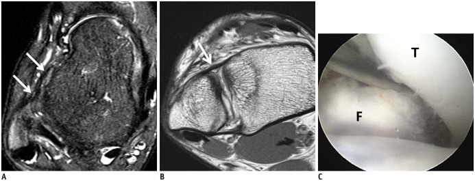
MRIAxial ankle MRI, with arthroscopic correlation: anterior talofibular ligament and anterior inferior tibiofibular ligament loose and irregular, indicating chronic instability, with widening of the distal tibiofibular joint confirmed on arthroscopy.
Source : Chun et al., Korean Journal of Radiology, 2015, figure 2 · CC BY-NC
Chronic instability is one of the few diagnoses in our field to have operational criteria published by international consensus. Applying them turns a subjective complaint into a structured picture, and makes it possible to say which patient you are dealing with.
The International Ankle Consortium published a position statement in 2014 intended to standardise patient selection in research, faced with the finding that heterogeneous inclusion criteria limited the generalisability of results.1 The text was reproduced in three journals, the Journal of Orthopaedic and Sports Physical Therapy in 2013 then the Journal of Athletic Training and the British Journal of Sports Medicine in 2014, which shows the intention to disseminate it.
Those criteria were designed for research, and that matters: applied to the letter, they exclude clinically unstable patients, which is exactly what Koshino's comparison made visible.5 Their author acknowledges it in his conclusion: standard criteria may not capture the whole clinical population. In the clinic, they therefore serve as a framework and not as a barrier.
| Criterion | Threshold used | What it rules out |
|---|---|---|
| History of a significant lateral sprain | At least one lateral sprain that caused pain, swelling and interruption of activity | The painful ankle with no initial trauma, which belongs to another diagnosis |
| Time since the first sprain | At least 12 months | Recovery still under way, which is not chronicity |
| Time since the most recent injury | More than 3 months | Acute inflammation, which distorts every examination and every functional test |
| Giving way or a sensation of instability | Repeated episodes, and at least 2 giving-way episodes in the last 6 months | The isolated incident, which does not establish a syndrome |
| Documented by a validated questionnaire | CAIT, IdFAI or Ankle Instability Instrument | The undocumented clinical impression, not followed over time |
Three instruments are recommended by the consortium to establish subjective instability: the Ankle Instability Instrument, the Cumberland Ankle Instability Tool and the Identification of Functional Ankle Instability.4 The CAIT is the most used in practice, because it is short and produces a continuous score from 0 to 30, so it can be followed over time.12
One point deserves attention because it is a source of confusion. The threshold historically quoted is 27 or less; the threshold used by the consortium in its selection criteria is 24 or less; and a recalibration published in 2014 on two datasets proposes 25 or less, with a sensitivity of 96.6 % and a specificity of 86.8 %, a positive likelihood ratio of 7.3 and a negative one of 0.04.14 Its authors explicitly recommend using that recalibrated threshold to distinguish groups with and without chronic instability. In practice, what counts most is not the threshold chosen but the fact of recording the score and repeating it : individual variation under treatment is more informative than crossing a boundary.
| Instrument | What it measures | Threshold | Note |
|---|---|---|---|
| CAIT Cumberland Ankle Instability Tool | Severity of perceived instability, score out of 30 | 24 or less (consortium) 25 or less (recalibrated 2014) | The shortest. A continuous score, so usable in follow-up |
| IdFAI Identification of Functional Ankle Instability36 | Injury history and sensation of instability | 11 or more | Includes the injury history, which the CAIT does not |
| AII Ankle Instability Instrument | Dichotomous answers on history and giving way | Depending on the version used | Retained by the consortium on the same footing as the other two |
| FAAM Foot and Ankle Ability Measure | Foot and ankle function, activities of daily living and sport subscales | No diagnostic threshold | Measures function, not instability. Validated French version |
The FAAM deserves particular mention for French-speaking practice: its French version was the subject of a validity and reliability study published in 2011, which makes it a tool that can be used directly with no in-house translation.15 It does not replace the CAIT, it complements it: the CAIT says instability, the FAAM says function, and the two do not move together.
The benchmark meta-analysis on the diagnostic value of clinical ankle tests brought together 14 studies and 6,302 observations. Its conclusion is clear: no test reaches both 90 % sensitivity and 90 % specificity. Palpation of the anterior talofibular ligament is highly sensitive, 95 to 100 %, but very unspecific, 0 to 32 %: it serves to rule out, not to confirm. The anterior drawer, across 6 studies and 885 observations, conversely has a low sensitivity of 54 % (95 % CI: 35 to 71 %) and a high specificity of 87 % (95 % CI: 63 to 96 %): it serves to confirm, not to rule out.17
The practical consequence is direct, and it is worth writing down: a negative anterior drawer does not exclude mechanical laxity. With a sensitivity of 54 %, it misses nearly one in two. Concluding “the drawer is negative, so it is functional” is a reasoning error, and it is probably the commonest error in this field.
Steering towards the dominant component
No branch ends in a single test: it is the convergence of clues that steers, and the absence of progression under treatment that decides.
Built from: Gribble et al., J Athl Train 2014 (PMID 24377963) for the criteria; Netterström-Wedin et al., Sports Health 2022 (PMID 34286639) for the diagnostic values; Wenning & Schmal, Z Orthop Unfall 2023 (PMID 35158394) for the orientation logic; Vuurberg et al., Br J Sports Med 2018 (PMID 29514819) for the place of surgery after conservative failure.
The diagnosis is ticked off: a significant sprain more than 12 months ago, last injury more than 3 months ago, at least two giving-way episodes over six months, and a documented score. A CAIT of 25 or less is the most recent validated threshold, but it is its change that informs. And above all: the anterior drawer confirms without excluding. Negative, it says nothing.
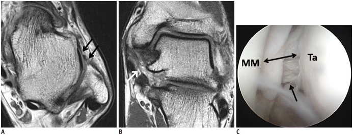
MRIAxial and oblique coronal ankle MRI, with arthroscopic correlation: chronic rupture of the anterior talofibular ligament and thinning of the superficial deltoid ligament, with widening of the medial mortise.
Source : Chun et al., Korean Journal of Radiology, 2015, figure 1 · CC BY-NC
The word “proprioception” is used so often that it ends up meaning nothing. It nevertheless covers deficits that are measured, quantified, and whose distribution is instructive: they are not where intuition places them.
The most complete meta-analysis on the subject examined 7,731 references, retained 30, and meta-analysed 21. It separates two dimensions of proprioception: kinaesthesia, that is the perception of movement, and joint position sense. Compared with healthy subjects, unstable patients show deficits in kinaesthesia in inversion (effect size 0.64) and in eversion (0.76), and deficits in active joint position sense in inversion (1.00) and in eversion (4.82). Compared with their own healthy side, the deficits exist too: kinaesthesia in inversion (0.41) and in plantarflexion (0.92), active and passive joint position sense in inversion (0.92 and 0.72).13
The most interesting result of that work is the one you do not expect: the proprioceptive deficits of the knee and shoulder of the same patients are not statistically significant. In other words, chronic ankle instability is not the local expression of a global proprioceptive deficit. It is a joint-specific deficit, consistent with damage to the mechanoreceptors and the deafferentation that follows ligament injury. That point has a direct consequence for the content of sessions: the work must be loaded on the injured ankle, not diluted in a general balance programme.
Where the deficit is measured, and by how much
Standardised effect sizes, unstable patients against healthy subjects. By convention, 0.2 is a small effect, 0.5 a medium effect, 0.8 a large effect.
Source: Xue et al., J Sport Health Sci 2021 (PMID 33017672), a meta-analysis of 21 studies. The eversion value of 4.82 comes from a small number of studies and should be read as an order of magnitude, not as a precise estimate.
Hertel and Corbett's updated model organises these observations. It describes eight interacting components: the primary tissue injury, pathomechanical impairments, sensory-perceptual impairments, motor-behavioural impairments, personal factors, environmental factors, interactions between components, and the spectrum of clinical outcomes. The whole is set against the biopsychosocial model, the notions of self-organisation and perception-action cycles from dynamical systems theory, and a patient-specific “neurosignature” derived from the neuromatrix theory of pain.2
That framework justifies going beyond the strict joint perimeter. A meta-analysis of nine randomised trials and 306 participants assessed hip strengthening in these patients: it significantly improves dynamic balance, with effect sizes of 0.72 in the anterior direction, 1.35 posterolateral and 1.66 posteromedial on the Star Excursion Balance Test, and reduces the area of the centre of pressure ellipse in static balance by 11.62 mm² on average.18
There is no contradiction with the previous paragraph. The proprioceptive deficit is local; postural control, on the other hand, is a matter of the whole chain. Loading the ankle and strengthening the hip are not two competing options, they are two levers acting on different things.
The proprioceptive deficit is measured, it concerns above all position sense in inversion, and it is confined to the injured ankle. Postural control, for its part, also depends on the hip, whose strengthening improves dynamic balance with large effect sizes. A session that does nothing but single-leg balance misses half the problem.
This is the strong point of this file. Where many of the conditions we treat rest on fragile data, chronic ankle instability has dozens of randomised trials, several network meta-analyses, and direct comparisons between modalities. The message is not “do proprioception”: it is more precise than that.
The most useful synthesis for practice was published in 2026: 58 studies, 2,097 participants, with the explicit aim of determining the relative effectiveness of each modality on each deficit. Its results read like a prescribing table.16
A network meta-analysis completes the picture on inversion muscle function, from nine studies and 366 participants. Every modality does better than no treatment, but the ranking is clear: the combination of neuromuscular training and strengthening comes top with a 99.9 % probability of being the best option, and an effect size of 2.82 (95 % CI: 1.89 to 3.74). Strengthening alone (SUCRA 64.6 %) is ahead of neuromuscular training alone (45.9 %), and neuromuscular training plus whole-body vibration brings up the rear (38.9 %).19
One counter-intuitive result deserves flagging. Another network meta-analysis, this time centred on joint position sense, places foot and ankle strengthening exercise first (SUCRA 74.6 %), ahead of static balance alone (67.9 %) and corrective exercise (56.1 %). Its authors conclude that the more complex the balance exercise becomes, the less marked the proprioceptive effect.20 That does not disqualify progression towards complex tasks, which serves other objectives, but it is an invitation not to abandon simple, loaded work too soon.
The more complex the balance exercise becomes, the less marked its effect on proprioception. Difficulty is not the goal.
A meta-analysis of 26 randomised trials and 1,032 participants, published in 2025, has the rare merit of crossing effectiveness with the characteristics of dose. Exercise significantly improves reported function: a mean gain of 4.59 points on the CAIT (95 % CI: 4.16 to 5.03), of 7.71 points on the FAAM activities of daily living (6.36 to 9.05) and of 11.86 points on the FAAM sport (7.86 to 15.85). The subgroup analyses suggest that manual therapy delivered once or twice a week for four weeks or less optimises the CAIT, while multimodal training once or twice a week over five to eight weeks produces the best effects on both FAAM subscales.22
That same meta-analysis applied the GRADE method to its results, and its conclusion is cautious: the level of certainty ranges from very low to moderate. That is important to say to the patient as to the referrer: the direction of the effect is solid, its exact size less so.
Nine randomised trials and 364 participants have been meta-analysed on joint mobilisation. The result is clear in both directions: a significant improvement in dorsiflexion range (effect size 1.02; 95 % CI: 0.41 to 1.63) and in dynamic balance (0.49; 0.06 to 0.78), but no significant improvement in patient-reported function (0.76; 95 % CI: −0.00 to 1.52).23 The practical reading is simple: mobilisation is a tool for gaining dorsiflexion, which makes sense when dorsiflexion is limited, and not a treatment for instability.
| Modality | What it acts on | What the data say | Level of evidence |
|---|---|---|---|
| Balance training | Function, dynamic balance, position sense, concentric strength | Benefits across the whole spectrum measured; improves functionality (0.81), instability (0.77) and dynamic balance (0.83) against usual exercise37 | Moderate |
| Foot and ankle strengthening | Concentric strength, function, position sense | First for position sense in network comparison (SUCRA 74.6 %) | Moderate |
| Neuromuscular and strengthening combined | Inversion muscle function | Effect size 2.82; probability of being the best option 99.9 % | Moderate |
| Hip strengthening | Dynamic and static balance | Effect sizes 0.72 to 1.66 on the SEBT; centre of pressure ellipse reduced | Low to moderate |
| Joint mobilisation | Dorsiflexion, dynamic balance | A clear effect on range and balance, not on reported function | Low |
| Bracing and taping | Recurrence, sense of security | No difference from proprioceptive training on recurrence at 12 months | Low to moderate |
| Whole-body vibration | Dynamic balance only | No demonstrated effect beyond that; last in the network comparisons | Very low |
| Eccentric strength work | Eccentric strength, force sense | No intervention has demonstrated an effect on those two parameters | None to date |
The ranking above is an editorial appraisal applying the GRADE principles, and not a reproduction of a published GRADE assessment, except for the dose row, where the 2025 meta-analysis did conduct a GRADE rating and concluded to a level of certainty of very low to moderate.22 The Cochrane review, for its part, noted as early as 2011 limitations of design, conduct and reporting in the ten trials it retained, with an unclear or high risk of bias for allocation, blinding of assessors and selective reporting of outcomes.21 That caution has not entirely gone away: the trials are more numerous, they are not all better.
The modalities ranked by level of evidence
Read as horizontal cards: the hierarchy reads from top to bottom, and each card stays readable in full.
Balance training as the base, foot and ankle strengthening, and above all the combination of the two. That is what should take up most of the session time.
Hip strengthening, bracing or taping to prevent recurrence. Useful, worth including, but not at the expense of the base.
Joint mobilisation, whose documented effect concerns dorsiflexion and dynamic balance, with no translation into patient-reported function.
Whole-body vibration, whose effect is limited to dynamic balance and which ranks last in the network comparisons.
Improvement in force sense and eccentric strength: none of the modalities tested across the 58 studies reviewed showed a significant effect. That is not a reason to do nothing, it is a reason to promise nothing.
Sources: Xu et al., J Foot Ankle Res 2026 (PMID 41771785); Zhang et al., Front Bioeng Biotechnol 2025 (PMID 41487952); Han et al., Arch Phys Med Rehabil 2022 (PMID 35550140); Hou et al., BMC Musculoskelet Disord 2026 (PMID 41942996); Kim & Moon, J Funct Morphol Kinesiol 2022 (PMID 36135424); Liu et al., BMC Sports Sci Med Rehabil 2025 (PMID 41233837), the only published GRADE rating taken up here. The ranking is an editorial appraisal applying the GRADE principles.
From these data a framework can be drawn, to be adapted to the patient and to their dominant component. The durations correspond to those that emerge from the dose analyses, that is five to eight weeks of multimodal work at one to two supervised sessions a week, completed by daily work at home.22
| Phase | Objective | Content | Criterion for moving on |
|---|---|---|---|
| 1. Load the joint weeks 1 to 2 | Restore range and strength, put simple and demanding work back in | Weight-bearing dorsiflexion, mobilisation if limited; concentric strengthening of the evertors and invertors; single-leg stance with eyes open on a stable surface, long sets | Symmetrical dorsiflexion on the lunge test; single-leg stance stable for 30 s |
| 2. Add complexity progressively weeks 3 to 5 | Dynamic balance and control of the whole limb | Modified SEBT as an exercise and as a measure; hip strengthening (abductors, lateral rotators); unstable surfaces, eyes closed, dual task | Measured progression in the three directions of the modified SEBT; no giving way during exercise |
| 3. Bring it back to the real world weeks 6 to 8 | Landing, change of direction, sport-specific or work-specific movement | Single-leg jumps and landings, side hop, figure of eight, running with cutting; added cognitive load; real playing surfaces | Return-to-sport battery, shared decision (next chapter) |
It does not promise to restore eccentric strength or force sense: no modality has demonstrated an effect on those two parameters across the 58 trials reviewed.16 Nor does it promise to remove laxity, which cannot be rehabilitated. What it does produce, and what the trials measure, is a gain in reported function, in dynamic balance and in position sense, with a reduction in giving-way episodes. That is already a lot, and it is what should be announced.
The question is asked with a moral charge that has no place. People readily contrast the patient “who takes charge of themselves” with the one “who leans on a brace”. The data do not support that hierarchy.
Three trials have compared proprioceptive and neuromuscular training head-to-head with wearing a brace to reduce the recurrence rate in athletes. The pooled data show no difference between the two strategies at twelve months. The authors conclude that the available evidence, of level II, does not favour training over bracing, and recommend choosing according to patient preference and practitioner expertise.24
The overview of systematic reviews published in the British Journal of Sports Medicine points the same way and ranks the levels: there is strong evidence for bracing and moderate evidence for neuromuscular training in preventing sprain recurrence.26 The Dutch guideline, updated in 2018, likewise retains bracing as an effective option for preventing recurrence.3
There is more than an argument of effectiveness, and this is where the chapter on the three components finds its application. Bracing and adhesive taping are explicitly placed by Wenning and Schmal among the mechanical therapies, those offered to the patient whose mechanical component dominates and which training will not compensate for.6 A brace is therefore not the consolation prize of the patient who does not train: it is the treatment suited to a component that training does not reach.
The meta-analysis devoted to the effect of external supports on ankle biomechanics in these patients adds a useful nuance. With a very low level of evidence, external supports reduce frontal plane excursion, but do not change the inversion angle at initial contact on landing. Limiting frontal excursion could reduce the risk of sprain. In running, frontal plane kinematics are not influenced. Reductions in the sagittal plane are observed on landing and in running, with a low to very low level of certainty, and their bearing on sprain risk remains undetermined.27
In other words: the brace limits what happens during the movement, not the starting position of the foot at the moment it touches the ground. That is consistent with the idea that it does not replace motor control.
The most recent meta-analysis brought together eight studies and 190 participants. Taping produces a small but significant improvement in the total Y-Balance Test score (effect size 0.269; 95 % CI: 0.031 to 0.508), in the posterolateral direction of the same test (0.385; 0.106 to 0.664) and in the anterior direction of the Star Excursion Balance Test (0.686; 0.083 to 1.289). By contrast, no significant difference appears in the total SEBT score, in its posteromedial and posterolateral directions, or in the hop test. Its authors conclude that it can be a simple, low-cost adjunct to complete rehabilitation, but must not replace conventional interventions.29
On recurrence, bracing and training are equal at twelve months, and bracing even carries the higher level of evidence. The choice is made with the patient. Kinesio taping produces a modest, directional effect on dynamic balance: it adds, it does not replace. And offering a brace to a patient whose mechanical component dominates is not giving up, it is reasoning.
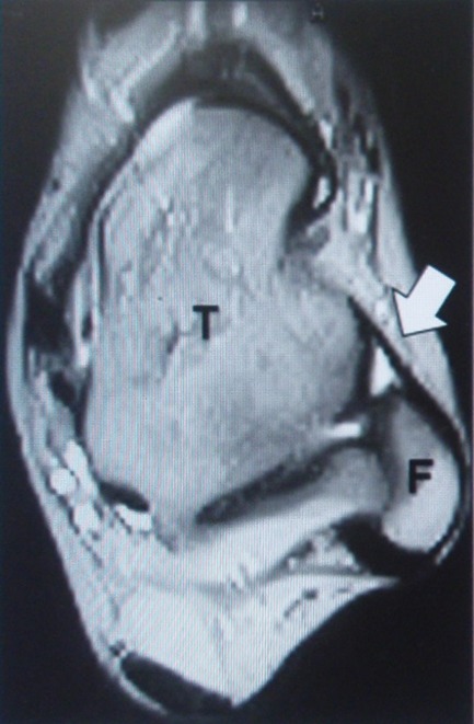
MRIAxial T2-weighted ankle MRI: thinned and remodelled anterior talofibular ligament, located between the tibia and fibula, measured before surgery for chronic ankle instability.
Source : Kanamoto et al., Bone & Joint Research, 2014, figure 1 · CC BY-NC
The position of the guidelines has been stable for ten years and comes down to one sentence. It deserves to be read carefully, because half its content concerns what must have been done beforehand.
The Dutch guideline, the most cited on this point, states that surgery should be reserved for cases that do not respond to thorough and complete exercise-based treatment.3 The two adjectives are not decorative: they set the level of demand on conservative treatment, and that is where our responsibility lies. A patient referred to the surgeon after six unsupervised sessions has not had the treatment the guideline assumes.
The international consensus of ESSKA and AFAS set out precisely to identify which patients warrant surgery, which procedure to choose, and how to treat associated conditions.25 It also points out that predicting which patient will develop instability after a sprain remains difficult, which is why preventive surgery is not possible.
The comparison between open and arthroscopic technique has been meta-analysed in 408 patients from eight comparative studies: 193 operated on open and 215 arthroscopically. The arthroscopic technique does better on the AOFAS score at six months (92.25 against 82.4) and at one year (88.6 against 80.05), on pain at six months and at one year, and on time to return to weight bearing (9.0 weeks against 14.25). By contrast, operating time, complication rate, postoperative talar tilt and anterior drawer are statistically comparable. The authors conclude that long-term trials are needed before making arthroscopy the new reference.30
The Cochrane review, on this chapter, remains the most cautious: it concludes to insufficient evidence to prefer one surgical procedure over another, while noting limitations specific to dynamic tenodesis. It does, however, provide a result directly useful to the physiotherapist: after surgical reconstruction, early functional rehabilitation is superior to six weeks of immobilisation for restoring function, with return to work two weeks earlier and return to sport three weeks earlier.21
Finally, a point that the 2026 arthroscopic series makes concrete: in patients operated on, associated intra-articular lesions are present in 54.7 % of cases, and the poor agreement between preoperative diagnosis and intraoperative findings underlines the role of arthroscopy in identifying and treating them.7 That explains why a surgical procedure that succeeded on the ligament can leave a symptomatic ankle: it did not treat the same thing.
Surgery repairs the mechanical component. It does not rehabilitate the functional component, which is still entirely there on waking.
The operated patient is not a cured patient. Their laxity is corrected, their sensorimotor deficit is not: joint position sense, dynamic balance and control of the whole limb still have to be worked on, and early mobilisation is part of the result.21 It is the same programme as in the previous chapter, applied within the surgeon's instructions.
This is the chapter where you have to accept saying what is not known. A battery of relevant tests exists, it is documented, it can be used from tomorrow. The cut-off values, on the other hand, do not exist.
A narrative review and expert opinion, conducted by French-speaking authors from the network of the French society of sports physiotherapists, set out to identify the most relevant tools for this decision. It starts from a finding: one possible cause of the high rate of recurrence and giving way is a premature return to sport, and apart from the time elapsed, there is no specific criterion to guide the clinician.28
Four functional tests emerge as the most relevant for targeting the impairments of these patients: single-leg stance on a stable surface, the modified version of the Star Excursion Balance Test, the side hop test and the figure of eight test. On the questionnaire side, the combination of the FAAM and the ALR-RSI, a return-to-sport scale after ligament reconstruction, is retained as the most relevant.
The return-to-sport battery, and its status
The tests are validated as relevant for this population. The decision thresholds, on the other hand, remain to be established: the battery documents, it does not decide on its own.
Source: Picot et al., Front Sports Act Living 2022 (PMID 35721875), a narrative review and expert opinion. The authors themselves point out that objective scientific criteria with cut-off values are still lacking.
The authors of that review are explicit: although these tests and questionnaires provide a solid base, objective scientific criteria with cut-off values are still lacking. They recommend adding an analysis of the context, in particular the characteristics of the sport played, such as fatigue and cognitive load, to better appreciate the risk of recurrence. They finally suggest assessing, in ecological conditions, the ability to control ankle inversion under load on a single leg.28
A survey of French-speaking doctors on actual return-to-play practice after a lateral sprain completes that picture and confirms the heterogeneity of practice in our language area.31 The absence of a validated threshold is therefore not a French shortcoming, it is the state of the question.
Running the battery makes sense: it objectifies, it can be repeated, it makes progress visible to the patient. Announcing a percentage of symmetry as a condition of return does not, because no threshold is validated for this population. The honest formulation is this: “here is where you are on four tests, here is where you were six weeks ago, and here is what that does not tell us.”
The two reports that follow are genuine published cases, with their identifiers. The first shows what a complete programme produces on measured indicators; the second shows what gets missed when you do not look at the ankle.
Case no. 1: four sprains in four years, and what five indicators say about the treatment32
A 25-year-old woman presents in July 2024 with swelling of the dorsum and lateral border of the foot, with pain on walking and on any movement of the ankle. The history is one of a trajectory, not of an accident: a first sprain in April 2020 coming down a ramp, a second in November 2020 on a staircase, with a sensation of instability and giving way appearing from then on, a third in July 2023 with marked ligament laxity and a positive anterior drawer, a fourth in June 2024 on a wet floor.
Examination finds lateral and dorsal swelling, a hypermobile joint with a painful soft end-feel reflecting the ligament laxity, and a bilateral high arch that the authors link to the recurrent character. The initial assessment quantifies the situation: pain at 7/10, CAIT at 15/30, FAOS functional score at 63 %, walking speed at 0.79 m/s for a cadence of 99.24 steps a minute.
The programme is deliberately multidimensional: ankle stretching, joint mobilisation, trunk strengthening, hip strengthening, strengthening of the dorsiflexors, plantarflexors, invertors and evertors, work on the intrinsic foot muscles, and dynamic balance on unstable surfaces. At the end of the course of care: pain at 1/10, CAIT at 28/30, FAOS at 89 %, walking speed at 0.90 m/s.
What this case illustrates. The gain of 13 points on the CAIT is far greater than the mean gain of 4.59 points observed in meta-analysis, 22 which is to be expected of an isolated case published for its result and should temper enthusiasm. What is transferable is the structure : trunk, hip, ankle, intrinsic foot, dynamic balance, exactly the combination that the network comparisons place at the top. The authors themselves acknowledge the absence of longer-term follow-up as the main limitation, which forbids any conclusion about durability.
Gangwani N, Patil DS, Kaur G. Cureus 2024;16(9):e68880. PMID 39376853, PMCID PMC11457927. Verbal consent obtained, reported by the authors.
Case no. 2: three non-specific low back pains whose cause was in the ankle33
Three women, followed in a Japanese peripheral nerve surgery unit, present with low back pain with no abnormality on imaging. A 32-year-old woman has had it for more than ten years, with three weeks of right buttock pain worse on spinal extension. A 59-year-old woman has repeated transient low back pain after long car journeys. A 42-year-old woman triggered acute low back pain while gardening, with numbness of the left lower limb, worsening over a month despite treatment and time off work.
The common feature appears only on examination of the lower limb: all three have chronic ankle instability, with a positive anterior drawer, and tenderness where the superficial peroneal nerve pierces the deep fascia. The authors propose a traction neuropathy of that nerve, maintained by the instability.
The results are striking in their simplicity. In the first patient, the pain on extension temporarily disappears with an ankle brace and a superficial peroneal nerve block; after ligament reconstruction and neurolysis, the low back pain disappears, with no recurrence at one year. In the second, after the nerve block, the active straight leg raise angle goes from 20° - 45° and the pain on walking from 8/10 - 3/10 ; with the braces, no further recurrence on long journeys for more than a year. In the third, a brace alone brings the pain on standing down to 1/10, the pain returning as soon as it is removed; she has been back at work for more than two years.
What this case illustrates. The point is not to claim that non-specific low back pain comes from the ankles. It is to take away one examination reflex: in a patient whose low back pain has no cause found and whose history includes repeated sprains, looking at the ankle costs thirty seconds. The decisive test in those reports was available in the clinic: put the brace on and see whether the complaint changes.
Hagiwara Y, Natsume Y, Wagatsuma T, Hasegawa T, Yoshida R. Cureus 2024;16(7):e65405. PMID 39188445, PMCID PMC11346748. A series of three cases; the authors are peripheral nerve specialists, which is a recruitment bias to keep in mind.
This chapter condenses the above into decisions. It does not replace clinical reasoning, it sets its checkpoints.
| Error | Why it is one | Instead |
|---|---|---|
| Concluding “it is functional” from a negative anterior drawer | A sensitivity of 54 %: the test misses nearly one in two | Cross-check several clues, and let yourself be corrected by the response to treatment |
| Making balance work harder and harder | The more complex the exercise becomes, the less marked the proprioceptive effect | Keep simple, loaded work, add complexity for other objectives |
| Treating the ankle alone | Hip strengthening improves dynamic balance with effect sizes of 0.72 to 1.66 | Include the hip from phase 2, without giving up local loading |
| Refusing a brace in the name of independence | On recurrence, no difference at 12 months from training, and the level of evidence favours it | Shared decision, brace and exercise together if the patient wishes |
| Announcing a return-to-sport threshold | No cut-off value is validated for this population | Document the battery, compare with the initial state, decide with the patient |
The question deserves an honest answer. Rehabilitation improves, in a documented way, reported function, dynamic balance and joint position sense, and reduces giving-way episodes. It restores neither eccentric strength nor force sense, on which no modality tested has shown an effect.16 And it does not tighten a stretched ligament. The realistic objective is an ankle that no longer gives way in everyday life, not a new ankle.
The dose analyses place the optimum around five to eight weeks of multimodal work, once or twice a week, with daily work at home.22 That is not a healing time: it is the window over which the trials measure their gains. Maintaining what has been gained means keeping up some upkeep work.
Because the instability may be functional: the ligament holds, but sensorimotor control is deficient. Deficits in joint position sense in inversion are in fact the most marked of all those measured.13 And because the anterior drawer, with a sensitivity of 54 %, misses nearly one laxity in two: “the examination finds nothing” does not mean “there is nothing”.17
No. It has its place during effort and in risky situations, and it reduces frontal excursion during landing, without correcting the position of the foot at initial contact.27 It does not remove the need for active work, and nothing suggests that it weakens the ankle: the direct comparison with training does not separate the two strategies.24
Modestly, and only in certain directions. The most recent meta-analysis finds a small improvement in the total Y-Balance Test score and in two directions, with no effect on the rest. Its authors describe it as a simple, low-cost adjunct that does not replace conventional rehabilitation.29
Partly. A prospective cohort followed 82 people after a first sprain. At two weeks, the inability to perform two landing tasks, a single-leg landing and a vertical jump with landing, predicted the outcome in 67.6 % of cases (sensitivity 83 %, specificity 55 %). At six months, the combination of deficits on the Star Excursion Balance Test in the posterior directions and of the FAAM activities of daily living score correctly classified 84.8 % of cases (sensitivity 75 %, specificity 91 %).38 The practical message is clear: the single-leg landing at two weeks and dynamic postural control at six months are worth testing, because they herald something.
Not straight away. The position of the guidelines reserves surgery for cases that do not respond to thorough and complete exercise-based treatment.3 And even after the operation, rehabilitation is still needed: surgery treats the laxity, not the sensorimotor deficit, and early functional recovery is part of the result.21
The link is documented without being quantifiable for an individual. Ankle osteoarthritis concerns about 1 % of the population and is distinctive in being secondary to trauma in more than 75 % of cases, with instability among the associated mechanical factors.8 That does not make it possible to announce a quantified risk to a given patient. It does justify treating an ankle that gives way at 25 seriously. For established osteoarthritis, see talocrural osteoarthritis.
All the references below have been verified one by one through the NLM E-utilities API: identifier resolved, authors, journal, year and pagination checked, abstract read before citation. All 38 identifiers were found; none remained untraceable.
A note on method. The levels of evidence in the table of modalities are an editorial appraisal applying the GRADE principles, and not a reproduction of a published GRADE assessment, with the exception of the dose results of reference 22, where a GRADE rating was indeed conducted and concludes to a level of certainty of very low to moderate. The clinical practice guidelines of the American Physical Therapy Association covering the lateral sprain and chronic instability jointly are also the most complete reference document in the field for physiotherapy.35 Article written on 15 August 2026.
Behind this article
An author who explains, a reviewer who checks.
How we write and check our content

Physiotherapist · co-founder of Physio Learning
Marked for life by his first four-hour lecture without a single image, he took a master’s in instructional design so that it would never happen to anyone again. He hunts down publication bias and unreadable slides with the same intransigence.

Head of scientific content
Physiotherapist specialising in neuro-musculoskeletal practice and holder of a master’s in public health. He checks the methodological rigour of every article: primary sources, levels of evidence, no exceptions.
