















In brief
Hallux valgus, commonly called a bunion, is a progressive three-dimensional deformity of the first ray combining lateral deviation of the great toe phalanx with adduction of the first metatarsal, and subluxation of the metatarsophalangeal joint. It presents as a painful medial bony prominence; diagnosis is clinical and radiographic under weight-bearing (HVA ≥ 15°, IMA ≥ 9°), with a female predominance and frequent radiographic-clinical discordance. First-line management favours footwear education, exercise, toe spacers, orthoses and night splints, with surgery reserved for documented failures. Overall prevalence in adults is 19 %.
Clinical synthesis based on the 2020-2024 meta-analyses (Hurn 2022, Cai 2023, Hernandez-Castillejo 2020, Singh 2020), the Cochrane 2024 review (Dias) and the ACFAS 2022 consensus, with systematic checking of the PubMed references and DOIs.
Clinical summary
- Hallux valgus is a three-dimensional deformity of the first ray combining lateral deviation of the phalanx with adduction of the 1st metatarsal, and progressive MTP1 subluxation.
- The Cai 2023 meta-analysis (45 studies, J Foot Ankle Res) puts overall prevalence at 19 % (95 % CI: 13-25 %), with a marked regional gradient: Oceania 29 %, Asia 22 %, Europe 18 %, North America 16 %, Africa 3 %.
- The prospective cohort study Menz 2023 (Arthritis Care Res, n=1482, ≥ 50 years, 7 years of follow-up) shows an incidence of 1 in 5 and progression in 1 in 3 cases, correlated with age, impaired physical health, foot pain and past use of constricting footwear.
- A 2025 Mendelian randomisation study identifies causal risk factors: high BMI, smoking, rheumatoid arthritis, gout, knee osteoarthritis and hip osteoarthritis.
- Female sex is associated with an adjusted OR of 2.64 (95 % CI: 2.26-3.08) and great toe pain with an OR of 3.28 (Roddy 2008).
- Diagnosis is above all clinical and radiographic under weight-bearing : a pathological HVA ≥ 15°, IMA ≥ 9°; the Manchester Scale is validated for visual grading (Menz 2005).
- An acute, hyperalgesic, erythematous pain at the MTP1 should raise the suspicion of a gout flare or septic arthritis before any treatment of the deformity.
- The radiographic-clinical discordance is major: the treatment decision centres on the pain and functional disability reported, not on the angles.
- The Hurn 2022 meta-analysis (Arthritis Care Res, 18 studies) concludes that certainty of evidence is low: non-surgical interventions reduce pain more than they reduce the angle of deformity.
- The toe-spread-out exercise (Kim 2015, n=24 RCT) significantly reduces the HVA and activates abductor hallucis after 8 weeks.
- The joint mobilisation + toe separator + exercise (Abdalbary 2018, RCT n=56) reduces the HVA by about 9° at 1 year of follow-up.
- A foot orthosis + toe spacer + night splint (Tehraninasr 2008) reduces pain and angles in the short term; the effect is not durable on the deformity.
- The Torkki 2001 JAMA trial (n=209, 2 years) confirms that surgery is superior at 1 year but that the groups converge at 2 years.
- The Cochrane 2024 review (Dias) concludes that evidence for long-term surgical superiority is weak, and reserves surgery for documented failures of conservative treatment.
- The ACFAS 2022 consensus recommends a symptom-centred rather than a radiographic-severity approach to the decision to operate.
- Postoperative recurrence correlates with a TSP > 4 (Hardy-Clapham) and with a high preoperative IMA.
- Footwear education (a wide toe box, a low heel) is the non-negotiable long-term preventive measure.
- The international red-flag framework (Finucane 2020 JOSPT) guides triage towards urgent medical referral.
Contents
- What are the fundamentals to know about hallux valgus (bunion)?
- How do you assess and diagnose hallux valgus with certainty?
- Which treatment strategies are the most effective for hallux valgus?
- How do you secure lasting recovery and prevent recurrence?
- What do real clinical cases teach us about hallux valgus?
- How do you apply these recommendations concretely in your practice?
1. What are the fundamentals to know about hallux valgus (bunion)?
1.1. How is this condition defined, who does it affect and what are the risk factors?
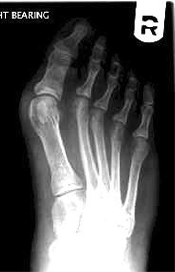
X-rayWeight-bearing dorsoplantar X-ray of the right foot: marked lateral deviation of the great toe and increased intermetatarsal angle, a deformity typical of hallux valgus.
Source : Iselin et al., BMC Musculoskeletal Disorders, 2015, figure 1 · CC BY
Hallux valgus (HV), commonly called a bunion, is a complex, progressive three-dimensional deformity of the first ray of the foot¹. It combines lateral deviation of the proximal phalanx of the great toe at the metatarsophalangeal joint (MTP1) with adduction (medial deviation) of the first metatarsal, generating the characteristic bony prominence on the medial border of the foot².
The most recent global meta-analysis (Cai et al., J Foot Ankle Res 2023, 45 studies included) puts pooled prevalence at 19 % (95 % CI: 13-25 %) in the adult population³. The reference meta-analysis Nix 2010 (21 English-language studies) already reported 23 % in adults and 35.7 % in those ≥ 65 years⁴. The regional gradient, brought out in 2023, is notable: Oceania 29 %, Asia 22 %, Europe 18 %, North America 16 %, Africa 3 %³.
Regional prevalence of hallux valgus (Cai 2023, meta-analysis of 45 studies)
Percentage of the adult population with an HV deformity
Source: Cai Y, Song Y, He M, et al. Global prevalence and incidence of hallux valgus: a systematic review and meta-analysis. J Foot Ankle Res. 2023;16(1):63. PMID 37726760.
The risk factors are multifactorial. A Mendelian randomisation study (Liu et al., 2025, 2-sample MR) made it possible for the first time to identify modifiable causal factors: smoking (lifetime smoking and smoking initiation), high BMI, rheumatoid arthritis, gout, knee osteoarthritis and hip osteoarthritis⁵,⁶. The hereditary component is strongly documented: the Framingham Foot Study (Hannan 2013, n=2,446) reports heritability of 89 % for men and 99 % for women of Caucasian ancestry⁷.
- Intrinsic factors: female sex (adjusted OR 2.64; 95 % CI 2.26-3.08 ; Roddy 2008⁸), advanced age (adjusted OR 1.61 per decade⁸), strong heritability (Hannan 2013⁷), knee osteoarthritis (OR 1.66⁸), rheumatoid arthritis (OR 2.04⁸), hypermobility of the first ray (Faber 2004 RCT n=101⁹, a debated role).
- Extrinsic causal factors: smoking and high BMI (2025 MR study⁵). Wearing constricting footwear (a narrow toe box, high heels) is associated with progression in the prospective Menz 2023 cohort (n=1 482, 7 years)²,¹⁰.
1.2. What happens in the body, and how does hallux valgus evolve naturally?
Biomechanically, hallux valgus results from progressive instability of the MTP1 joint (Perera, Mason & Stephens, JBJS Am 2011)¹. The imbalance of tendon forces (the extensor and flexor hallucis displaced laterally, the “bowstring effect”) amplifies the deviation in a vicious circle¹. The sesamoid complex subluxes, abductor hallucis loses its correct mechanical insertion and the adductor takes over.
The natural course is progressive and generally continuous. The prospective cohort Menz 2023 (n=1 482, mean age 62.9 years, 7 years of follow-up) shows:
- Cumulative incidence: 21.3 % (1 adult in 5 develops HV over 7 years)¹⁰.
- Progression in 33.7 % of existing cases (1 in 3)¹⁰.
- Predictors of progression: older age, impaired physical health (a low SF-12 PCS), foot pain at baseline and the use of constricting footwear¹⁰.
The functional consequences include transfer metatarsalgia on the central rays (failure of great toe propulsion), secondary deformities (hammer and claw toes), and an increased risk of falls in older people¹¹. The discordance between radiographic severity and symptoms is one of the major clinical paradoxes of the condition: a patient can have an HVA > 35° without pain, and the reverse (ACFAS Consensus 2022)¹².
- Hallux valgus is a 3D deformity of the first ray, not a simple bump.
- Overall prevalence: 19 % (Cai 2023, 45 studies) with a substantial regional gradient.
- Very strong heritability (89-99 % in Framingham) plus Mendelian causality confirmed for BMI, smoking, rheumatoid arthritis and gout (MR 2025).
- A marked female predominance: adjusted OR 2.64 (Roddy 2008).
- Prospective course, Menz 2023: 1 in 5 incidence, 1 in 3 progression at 7 years.
- Radiographic-clinical discordance is frequent: the decision must be centred on pain and function, not on the angles alone.
Bibliography, chapter 1
- Perera AM, Mason L, Stephens MM. The pathogenesis of hallux valgus. J Bone Joint Surg Am. 2011;93(17):1650-1661. PMID 21915581.
- Coughlin MJ, Jones CP. Hallux valgus: demographics, etiology, and radiographic assessment. Foot Ankle Int. 2007;28(7):759-777. PMID 17666168.
- Cai Y, Song Y, He M, et al. Global prevalence and incidence of hallux valgus: a systematic review and meta-analysis. J Foot Ankle Res. 2023;16(1):63. PMID 37726760.
- Nix S, Smith M, Vicenzino B. Prevalence of hallux valgus in the general population: a systematic review and meta-analysis. J Foot Ankle Res. 2010;3:21. PMID 20868524.
- Liu J, Wang Z, Xu Q, et al. The Causal Relationship Between Multiple Modifiable Risk Factors and Hallux Valgus: A Two-Sample Mendelian Randomization Study. Food Sci Nutr. 2025. DOI 10.1002/fsn3.70965.
- Zhang T, Liu Y, Wang J, et al. The causal relationship between hallux valgus and endogenous pathogenic factors: A 2-sample Mendelian randomization. Medicine (Baltimore). 2025;104(8):e41647. DOI 10.1097/MD.0000000000041647.
- Hannan MT, Menz HB, Jordan JM, Cupples LA, Cheng CH, Hsu YH. High Heritability of Hallux Valgus and Lesser Toe Deformities in Adult Men and Women. Arthritis Care Res (Hoboken). 2013;65(9):1515-1521. DOI 10.1002/acr.22040.
- Roddy E, Zhang W, Doherty M. Prevalence and associations of hallux valgus in a primary care population. Arthritis Rheum. 2008;59(6):857-862. PMID 18512715.
- Faber FW, Mulder PG, Verhaar JA. Role of first ray hypermobility in the outcome of the Hohmann and the Lapidus procedure. A prospective, randomized trial involving one hundred and one feet. J Bone Joint Surg Am. 2004;86(3):486-495. PMID 14996873.
- Menz HB, Marshall M, Thomas MJ, Rathod-Mistry T, Peat G, Roddy E. Incidence and Progression of Hallux Valgus: A Prospective Cohort Study. Arthritis Care Res (Hoboken). 2023;75(1):166-173. PMID 34268894.
- Menz HB, Auhl M, Spink MJ. Foot problems as a risk factor for falls in community-dwelling older people: A systematic review and meta-analysis. Maturitas. 2018;118:7-14. PMID 30415759.
- Meyr AJ, Doyle MD, King CM, et al. The American College of Foot and Ankle Surgeons® Clinical Consensus Statement: Hallux Valgus. J Foot Ankle Surg. 2022;61(2):369-383. PMID 34706857.
2. How do you assess and diagnose hallux valgus with certainty?
2.1. Which questions should you ask to understand the patient and their history?
The history is the cornerstone of clinical reasoning. It should map five dimensions :
- Characteristics of the pain : location (the medial prominence, the MTP1 joint itself, the sesamoids, the lateral metatarsals), intensity (NPRS 0-10 or VAS), rhythm (mechanical and worse on walking vs inflammatory and nocturnal), triggering and relieving factors. Central plantar pain marks a transfer of load to the lateral metatarsals¹.
- History of the deformity : age at onset, speed of progression, inflammatory episodes (bursitis), initial trauma. Rapid progression points to an inflammatory aetiology (rheumatoid arthritis, recurrent gout).
- Past history and comorbidities : family history (heritability up to 99 % in Caucasian women, Hannan 2013²), rheumatoid arthritis (adjusted OR 2.04³), knee osteoarthritis (OR 1.66³), gout, diabetes (an impact on postoperative healing).
- Footwear habits : type, heel height, width of the toe box, how old the current shoes are. Cited as a major factor in progression in the Menz 2023 cohort⁴.
- Functional impact and quality of life : use a validated PROM at baseline. The MOXFQ (Manchester-Oxford Foot Questionnaire, Morley 2013) offers 16 items across 3 domains with a single index score (alpha = 0.93)⁵. The FFI (Foot Function Index, Budiman-Mak 1991) remains widely used (23 items, 3 subscales)⁶.
2.2. Which clinical tests should you perform, and which other conditions should you rule out?
The examination is carried out barefoot and weight-bearing. Inspection assesses overall alignment (calcaneal valgus, a frequently associated collapse of the medial arch). Palpation looks for:
- The tender point of the medial bursitis.
- MTP1 joint pain (early osteoarthritis = hallux rigidus, Coughlin & Shurnas 2003⁷).
- Subsesamoid pain (sesamoiditis, stress fracture, osteonecrosis).
- Passive reducibility of the deviation (a “flexible” vs a “rigid” HV = a different surgical prognosis).
In any acute, hyperalgesic MTP1 episode, rule out first:
- Acute gout flare (podagra): a red, hot, swollen MTP1 with extreme pain (allodynia to the bedsheet). Confirmed by joint aspiration (negatively birefringent monosodium urate crystals). Cited as a causal factor for hallux valgus in the 2025 MR study⁸.
- Septic arthritis : fever, raised inflammatory markers (CRP, white cells), a portal of entry (a wound, a procedure). A surgical emergency.
- Metatarsal stress fracture : pain on direct pressure over the shaft, MRI if the plain radiograph is normal.
- Tumour : night pain, a palpable mass, systemic signs.
- Morton's neuroma : electric-shock pain in the 3rd-4th space, positive Mulder test (a painful click on transverse compression).
- Early hallux rigidus : restricted MTP1 dorsiflexion, pain at end range⁷.
Diagnosing hallux valgus is above all clinical. The weight-bearing radiograph (dorsoplantar and lateral views) is the reference investigation when objective quantification or surgical planning is being considered⁹,¹⁰.
2.3. Should patients with hallux valgus be classified, and what are the benefits?
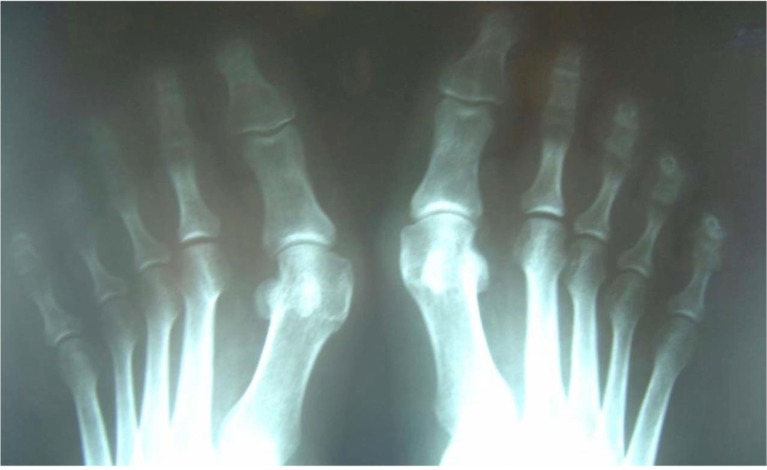
X-rayAnteroposterior X-ray of both feet: bilateral, symmetrical hallux valgus, deviation of the great toe visible on both sides.
Source : Xarchas et al., The Open Orthopaedics Journal, 2014, figure 1 · CC BY-NC
Classification has three aims: standardising communication, guiding the treatment decision and following the course. Two tools stand out in practice:
- Manchester Scale (Garrow et al. 2001; validated radiographically by Menz & Munteanu, Rheumatology 2005¹¹). Four standardised photographs grade the deformity visually (none / mild / moderate / severe). Good inter-rater reliability. The tool of choice for non-surgical follow-up.
- Weight-bearing radiographic measurements :
- HVA (Hallux Valgus Angle): the angle between the axis of the 1st metatarsal and the phalangeal axis. Normal < 15° ; mild 15-25°; moderate 25-40°; severe > 40°¹,⁹.
- IMA (InterMetatarsal Angle): the angle between the 1st and 2nd metatarsals. Normal < 9°¹,⁹.
- DMAA (Distal Metatarsal Articular Angle): the orientation of the articular surface; it influences the surgical choice.
- TSP (Tibial Sesamoid Position): the Hardy-Clapham scale 1-7; a position > 4 = a major factor in postoperative recurrence.
Radiographic classification of hallux valgus (ACFAS 2022 consensus)
Severity by the HVA and IMA angles measured under weight-bearing
Classification adapted from the ACFAS 2022 consensus (Hatch et al., JFAS. 2022;61(2):369-383). The treatment choice remains centred on symptoms rather than on the angles alone.
Critique and controversy
The radiographic-clinical discordance remains the major paradox of hallux valgus¹². A patient can have an HVA of 35° without pain, and conversely a “mild” HV can be highly symptomatic. The ACFAS 2022 consensus explicitly recommends that the treatment decision be based on the pain and functional disability reported, and not on the HVA alone¹².
The AOFAS score, historically used, has been criticised for the absence of formal validation and for excessive weighting of the clinician's judgement. Contemporary recommendations (NIH Consortium, COMET, IDEOM) favour pure PROMs such as the MOXFQ or the FAOS⁵.
Finally, the role of first ray hypermobility remains debated: the Faber 2004 RCT (n=101, 5 years) showed no difference in outcome between the Hohmann and the Lapidus procedures by preoperative mobility¹³, which questions its relevance as a decision criterion.
- Diagnosis is clinical above all, based on the history and the weight-bearing examination.
- Use a validated PROM: MOXFQ (Morley 2013) or FFI (Budiman-Mak 1991).
- Weight-bearing radiographic measurements: HVA >=15° and IMA >=9° = pathological.
- Manchester Scale validated for non-invasive visual follow-up (Menz 2005).
- Priority differential diagnoses: gout flare, septic arthritis, stress fracture, hallux rigidus, Morton's neuroma.
- The radiographic-clinical discordance calls for a decision centred on symptoms (ACFAS 2022).
Bibliography, chapter 2
- Perera AM, Mason L, Stephens MM. The pathogenesis of hallux valgus. J Bone Joint Surg Am. 2011;93(17):1650-1661. PMID 21915581.
- Hannan MT, Menz HB, Jordan JM, Cupples LA, Cheng CH, Hsu YH. High Heritability of Hallux Valgus and Lesser Toe Deformities in Adult Men and Women. Arthritis Care Res (Hoboken). 2013;65(9):1515-1521. DOI 10.1002/acr.22040.
- Roddy E, Zhang W, Doherty M. Prevalence and associations of hallux valgus in a primary care population. Arthritis Rheum. 2008;59(6):857-862. PMID 18512715.
- Menz HB, Marshall M, Thomas MJ, Rathod-Mistry T, Peat G, Roddy E. Incidence and Progression of Hallux Valgus: A Prospective Cohort Study. Arthritis Care Res (Hoboken). 2023;75(1):166-173. PMID 34268894.
- Morley D, Jenkinson C, Doll H, Lavis G, Sharp R, Cooke P, Dawson J. The Manchester-Oxford Foot Questionnaire (MOXFQ): development and validation of a summary index score. Bone Joint Res. 2013;2(4):66-69. PMID 23673374.
- Budiman-Mak E, Conrad KJ, Roach KE. The Foot Function Index: a measure of foot pain and disability. J Clin Epidemiol. 1991;44(6):561-570. PMID 2037861.
- Coughlin MJ, Shurnas PS. Hallux rigidus: demographics, etiology, and radiographic assessment. Foot Ankle Int. 2003;24(10):731-743. PMID 14587987.
- Liu J, Wang Z, Xu Q, et al. The Causal Relationship Between Multiple Modifiable Risk Factors and Hallux Valgus: A Two-Sample Mendelian Randomization Study. Food Sci Nutr. 2025. DOI 10.1002/fsn3.70965.
- Wulker N, Mittag F. The treatment of hallux valgus. Dtsch Arztebl Int. 2012;109(49):857-867. PMID 23267411.
- Coughlin MJ, Jones CP. Hallux valgus: demographics, etiology, and radiographic assessment. Foot Ankle Int. 2007;28(7):759-777. PMID 17666168.
- Menz HB, Munteanu SE. Radiographic validation of the Manchester scale for the classification of hallux valgus deformity. Rheumatology (Oxford). 2005;44(8):1061-1066. PMID 15901901.
- Meyr AJ, Doyle MD, King CM, et al. The American College of Foot and Ankle Surgeons® Clinical Consensus Statement: Hallux Valgus. J Foot Ankle Surg. 2022;61(2):369-383. PMID 34706857.
- Faber FW, Mulder PG, Verhaar JA. Role of first ray hypermobility in the outcome of the Hohmann and the Lapidus procedure. A prospective, randomized trial involving one hundred and one feet. J Bone Joint Surg Am. 2004;86(3):486-495. PMID 14996873.
3. Which treatment strategies are the most effective for hallux valgus?
3.1. Where do you start? What is the recommended hierarchy of interventions?
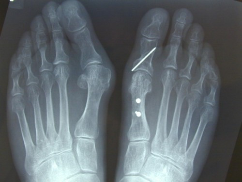
X-rayAnteroposterior X-ray of both feet: unoperated hallux valgus on one side, and the right foot corrected with scarf and Akin osteotomies, fixation screws visible, alignment of the first ray restored.
Source : Kilmartin et al., Journal of Foot and Ankle Research, 2010, figure 7 · CC BY
The recent literature converges on a ranked multimodal approach, with low to moderate certainty of evidence on the GRADE system (Hurn 2022)¹. The recommended sequence is:
- Education and behavioural advice (suitable footwear, weight management, activity modification). This is the intervention with the best benefit-to-risk ratio, and it is built into every multimodal approach¹,².
- Active exercise (the intrinsic foot muscles, abductor hallucis, Kim's 2015 toe-spread-out³, the short foot exercise).
- Passive therapies : joint mobilisations, foot orthosis, toe spacer, night splint (Tehraninasr 2008⁴).
- Adjunctive modalities : shockwave, kinesio taping: the data are limited.
- Surgery reserved for documented failures (3-6 months of well-conducted conservative treatment) with persistent pain and impaired quality of life (ACFAS 2022⁵, Cochrane Dias 2024⁶).
The pivotal trial Torkki 2001 (JAMA, n=209, 93 % women, mean age 48 years, 2 years of follow-up) compared immediate surgery / waiting with an orthosis / waiting without an orthosis⁷. At 1 year the surgical group had less pain, but at 2 years the three groups converge. The meta-analysis Hernandez-Castillejo 2020 (Acta Orthop) confirms that surgery improves quality of life in the physical domains, but with variable effect sizes⁸.
GRADE pyramid of the evidence for hallux valgus (2026 synthesis)
Level of certainty of the available data by modality
GRADE synthesis established following Schunemann 2019 (GRADE handbook). No conservative intervention reaches the HIGH level for correcting the deformity; the best levels concern epidemiological prevalence and the natural course.
3.2. What is the place of exercise, and is there a superior approach?
Therapeutic exercise is one of the best-documented modalities within the conservative approach. The SR/MA Hurn 2022 (Arthritis Care Res, 18 studies included) concludes that certainty is low, but with a favourable trend towards reducing pain more markedly than correcting the angle¹.
Three key RCTs shape practice:
- Kim et al. 2015 (J Phys Ther Sci, RCT n=24): 8 weeks of the toe-spread-out exercise combined with wearing an orthosis significantly reduces the active HVA and increases the cross-sectional area of abductor hallucis on ultrasound³. Against the orthosis alone, the difference is clinically significant (p < 0.05).
- Abdalbary 2018 (JAPMA, RCT n=56): a programme combining joint mobilisations + strengthening exercises (hallux plantarflexion + abduction + toe grip) + stretching + a toe separator, 36 sessions over 3 months. A radiographic reduction in HVA of about 9° at 12 months⁹.
- Tehraninasr 2008 (Prosthet Orthot Int, RCT n=30 women aged 19-45): an orthosis with a toe spacer vs a night splint. Pain fell in both groups; the effect on the angles was limited⁴.
3.3. Manual therapies, technologies: how effective are they really?
| Modality | Effect on pain | Effect on the HVA | GRADE level | Source |
|---|---|---|---|---|
| Education + suitable footwear | Moderate | Indirect (slows progression) | Moderate | Hurn 2022; ACFAS 2022; Menz 2023 |
| Intrinsic exercises + TSO | Significant in the short term | A reduction of 3-9° (small n) | Low | Kim 2015 RCT n=24; Abdalbary 2018 RCT n=56 |
| Mobilisations + manual therapy | Short term | Low | Low | Abdalbary 2018 (combined programme) |
| Foot orthosis + toe separator | Significant | Limited or absent | Low | Tehraninasr 2008; Hurn 2022; Torkki 2001 |
| Night splint | Symptomatic | No durable correction | Low | Tehraninasr 2008 |
| Kinesio taping | Transient analgesia | Non-corrective | Very low | Emerging, heterogeneous data |
| Osteotomy surgery | Significant at 1 year | Durable correction | Moderate | Torkki 2001 JAMA; Singh 2020; Hernandez-Castillejo 2020 |
| Minimally invasive (MIS) | Equivalent or better in the short term | Equivalent | Moderate | Singh 2020 SR/MA |
The SR/MA Singh 2020 (J Clin Orthop Trauma) shows that MIS gives less pain early on, with shorter operating and hospital times, and no significant difference in HVA, IMA, AOFAS or VAS at last follow-up¹². The Cochrane review Dias 2024 (CD013726) concludes that certainty of evidence for a long-term surgical advantage is low⁶.
3.4. Beyond the physical: how do you educate the patient and address psychological factors?
Education should cover three points:
- The nature of the condition : a progressive deformity, where the conservative aim is symptomatic and not structural (be transparent about the evidence; Hurn 2022)¹.
- The major role of footwear : a wide toe box, a heel <= 3 cm. Wearing constricting footwear is an independent factor in progression in Menz 2023 (n=1 482, 7 years)⁹.
- Shared decision-making following the Hoffmann-Montori 2014 framework: set out the options (multimodal conservative vs surgery), their evidence, and build in the patient's preferences¹⁰. Particularly crucial in the face of the radiographic-clinical discordance.
- A multimodal ranked approach: education + footwear > exercise > passive modalities > surgery where these fail.
- SR/MA Hurn 2022 ACR : conservative interventions reduce pain more than they reduce the angle.
- Exercise RCTs: Kim 2015 (toe-spread-out + orthosis) and Abdalbary 2018 (mobilisation + TS + exercise) show a modest reduction in HVA and in pain.
- Orthosis + spacer + splint: an analgesic effect but no durable correction (Tehraninasr 2008).
- Torkki 2001 JAMA : surgery superior at 1 year, but convergence at 2 years.
- Cochrane Dias 2024 : surgery remains reserved for documented failures, with low certainty of long-term evidence.
Bibliography, chapter 3
- Hurn SE, Matthews BG, Munteanu SE, Menz HB. Effectiveness of Nonsurgical Interventions for Hallux Valgus: A Systematic Review and Meta-Analysis. Arthritis Care Res (Hoboken). 2022;74(10):1676-1688. DOI 10.1002/acr.24603.
- Cai Y, Song Y, He M, et al. Global prevalence and incidence of hallux valgus. J Foot Ankle Res. 2023;16(1):63. PMID 37726760.
- Kim MH, Yi CH, Weon JH, Cynn HS, Jung DY, Kwon OY. Effect of toe-spread-out exercise on hallux valgus angle and cross-sectional area of abductor hallucis muscle in subjects with hallux valgus. J Phys Ther Sci. 2015;27(4):1019-1022. PMID 25995546.
- Tehraninasr A, Saeedi H, Forogh B, Bahramizadeh M, Keyhani MR. Effects of insole with toe-separator and night splint on patients with painful hallux valgus: A comparative study. Prosthet Orthot Int. 2008;32(1):79-83. PMID 18330806.
- Meyr AJ, Doyle MD, King CM, et al. The American College of Foot and Ankle Surgeons® Clinical Consensus Statement: Hallux Valgus. J Foot Ankle Surg. 2022;61(2):369-383. PMID 34706857.
- Dias CGP, Soares CO, Lenza M, et al. Surgical interventions for treating hallux valgus and bunions. Cochrane Database Syst Rev. 2024;7(7):CD013726. PMID 39051477.
- Torkki M, Malmivaara A, Seitsalo S, Hoikka V, Laippala P, Paavolainen P. Surgery vs orthosis vs watchful waiting for hallux valgus: a randomized controlled trial. JAMA. 2001;285(19):2474-2480. PMID 11368700.
- Hernandez-Castillejo LE, Martínez-Vizcaino V, Garrido-Miguel M, Cavero-Redondo I, Pozuelo-Carrascosa DP, Alvarez-Bueno C. Effectiveness of hallux valgus surgery on patient quality of life: a systematic review and meta-analysis. Acta Orthop. 2020;91(4):450-456. PMID 32408787.
- Abdalbary SA. Foot Mobilization and Exercise Program Combined with Toe Separator Improves Outcomes in Women with Moderate Hallux Valgus at 1-Year Follow-up: A Randomized Clinical Trial. J Am Podiatr Med Assoc. 2018;108(6):478-486. PMID 29683337.
- Hoffmann TC, Montori VM, Del Mar C. The connection between evidence-based medicine and shared decision making. JAMA. 2014;312(13):1295-1296. PMID 25268434.
- Menz HB, Marshall M, Thomas MJ, Rathod-Mistry T, Peat G, Roddy E. Incidence and Progression of Hallux Valgus. Arthritis Care Res (Hoboken). 2023;75(1):166-173. PMID 34268894.
- Singh MS, Khurana A, Kapoor D, Katekar S, Kumar A, Vishwakarma G. Minimally invasive vs open distal metatarsal osteotomy for hallux valgus - A systematic review and meta-analysis. J Clin Orthop Trauma. 2020;11(3):348-356. PMID 32405192.
4. How do you secure lasting recovery and prevent recurrence?
4.1. How do you make the patient an active participant in their recovery through self-management?
Self-management structures long-term success. It is built around five strands:
- Understanding the condition : transparent information about its progressive nature and the limits of conservative treatment (Hurn 2022: pain improves more than the angle does)¹.
- Suitable footwear : a wide, soft toe box, a low heel (<3 cm), avoiding narrow points. Cited as a modifiable factor in progression in Menz 2023².
- Home exercises : a programme drawn from Kim 2015 (toe-spread-out) and Abdalbary 2018 (mobilisation + strengthening + toe separator); 3 times a week, for at least 8 weeks before effects are seen³,⁴.
- Managing painful flares : cryotherapy, transient offloading, assessment of the triggering factors (footwear, excessive activity).
- Tracking PROMs : MOXFQ or FFI every 3 months to make progression objective⁵,⁶.
4.2. Vulnerable subgroups: older people, juveniles, rheumatoid arthritis
Three populations deserve a distinct clinical strategy.
Causal risk factors and relative risk for hallux valgus
2-sample Mendelian randomisation study (Liu 2025) and endogenous factors (Zhang 2025)
Sources: Liu et al. 2025 (2-sample Mendelian randomisation, BMI and smoking); Zhang et al. 2025 (endogenous factors: RA, gout, osteoarthritis); Roddy 2008 (female sex, community cohort n=4,249). The confidence intervals are approximated from the publications. RA and female sex show the strongest associations.
Older people (≥ 65 years): the risk of falls
The prevalence of HV in those ≥ 65 reaches 35.7 % (Nix 2010)⁷. The meta-analysis Menz, Auhl & Spink 2018 (Maturitas) shows that foot problems, including HV, significantly increase the risk of falls in community-dwelling populations⁸. HV is associated with increased fear of falling and with impaired dynamic balance. Practical implications: falls screening (Timed Up and Go, FES-I), advice on stable footwear, and particular attention to orthosis funding.
Juvenile hallux valgus (the adolescent): a specific challenge
The juvenile form has a historically high rate of postoperative recurrence because the growth plates have not yet closed. An expert review (Foot Ankle Clin, Coughlin 2007 on the juvenile form, and recent paediatric publications) recommends delaying surgery as long as possible and favouring the conservative approach (footwear, exercise, education). Where surgery is unavoidable (disabling pain, a severe progressive deformity), it should ideally be scheduled after growth has finished .
Rheumatoid arthritis: rapid deformity and complex surgery
The 2025 MR study confirms the causality of RA for HV (OR 2.04)¹³. RA patients often develop rapid, associated deformities (hammer toes, MTP subluxation), with:
- Impaired tissue quality (thin skin, fragile healing).
- Residual inflammatory activity despite biologic therapy.
- A multidisciplinary approach is mandatory (rheumatologist + physiotherapist + podiatrist + orthopaedic surgeon).
- Favour joint-preserving techniques where the disease is controlled. A review (2024, ecios.org) shows favourable results with a distal Chevron osteotomy in RA: JSSF Hallux 41 -> 88, recurrence 7.5 %¹⁴.
Red flags specific to the vulnerable subgroups
- An older person + a recent fall : assess for an occult fracture (femoral neck, wrist) and suspected metatarsal stress fracture.
- An RA patient + an acute inflammatory episode : rule out superimposed septic arthritis and skin ulceration.
- An RA patient + scheduled surgery : infection risk raised by biologic therapy (anti-TNF, anti-IL6); plan the interruption following the EULAR recommendations.
- Juvenile hallux valgus + disproportionate pain: rule out primary osteochondritis (Freiberg's of the 1st metatarsal) and bone neoplasia.
- Any patient on long-term corticosteroids + an acute MTP1 episode: joint aspiration is essential to rule out sepsis.
4.3. When and how should a safe return to sport and to activity be planned?
After surgery, the timeline depends on the technique (distal Chevron, Scarf, Lapidus, MIS), on the fixation, and on the patient. The SR/MA Hernandez-Castillejo 2020 reports significant improvements in quality of life at 6-12 months in the physical domains⁹.
A schematic progression :
- 0-6 weeks : a rigid post-op shoe, weight-bearing according to the technique, oedema control (cryotherapy + elevation). A stationary bike without resistance is possible once cleared.
- 6-12 weeks : transition to suitable normal shoes after radiographic union. Progressive strengthening, proprioceptive exercises. Low-impact activities (swimming, cycling).
- 3-6 months : resumption of impact activities on functional criteria. The median time to return to sport after a distal osteotomy is 3 to 6 months in contemporary surgical series.
Objective return-to-sport criteria :
- No pain in activities of daily living (NPRS <=2/10).
- Recovery of >=80 % of MTP1 dorsiflexion compared with the other side.
- Symmetrical intrinsic strength (heel rise test >15 repetitions).
- Satisfactory dynamic balance (Y-balance, single leg stance).
- MOXFQ or FFI in the “minimal to no discomfort”.
Critique and controversy
The standardisation of postoperative rehabilitation protocols remains a major weak point. Most trials compare surgical techniques without controlling the rehabilitation, which turns the latter into a methodological “black box” . The debate between early and delayed weight-bearing remains partly open: recent reviews tend to favour early weight-bearing where the fixation allows it, with no increase in complications.
Meanwhile, preventing long-term recurrence through conservative interventions lacks solid data beyond 12 months post-op. The prospective Menz 2023 cohort shows that 1 case in 3 progresses over 7 years, which underlines the limits of purely structural conservative means².
- Self-management: suitable footwear + home exercises + PROM tracking is the foundation.
- Older people: falls screening is the priority; HV = an independent risk factor (Menz 2018 MA).
- Juvenile HV: a prolonged conservative approach, with surgery ideally after growth has finished.
- Rheumatoid arthritis: causality confirmed by MR 2025 (OR 2.04); a multidisciplinary approach is essential.
- Return to activity: guided by objective functional criteria and not by a fixed calendar; 3-6 months on average.
- Recurrence correlates with a postoperative TSP > 4 and a high preoperative IMA.
Bibliography, chapter 4
- Hurn SE, Matthews BG, Munteanu SE, Menz HB. Effectiveness of Nonsurgical Interventions for Hallux Valgus. Arthritis Care Res (Hoboken). 2022;74(10):1676-1688. DOI 10.1002/acr.24603.
- Menz HB, Marshall M, Thomas MJ, Rathod-Mistry T, Peat G, Roddy E. Incidence and Progression of Hallux Valgus. Arthritis Care Res (Hoboken). 2023;75(1):166-173. PMID 34268894.
- Kim MH, Yi CH, Weon JH, Cynn HS, Jung DY, Kwon OY. Effect of toe-spread-out exercise on hallux valgus angle. J Phys Ther Sci. 2015;27(4):1019-1022. PMID 25995546.
- Abdalbary SA. Foot Mobilization and Exercise Program Combined with Toe Separator. J Am Podiatr Med Assoc. 2018;108(6):478-486. PMID 29683337.
- Morley D, Jenkinson C, Doll H, et al. The MOXFQ summary index score. Bone Joint Res. 2013;2(4):66-69. PMID 23673374.
- Budiman-Mak E, Conrad KJ, Roach KE. The Foot Function Index. J Clin Epidemiol. 1991;44(6):561-570. PMID 2037861.
- Liu J, Wang Z, Xu Q, et al. The Causal Relationship Between Multiple Modifiable Risk Factors and Hallux Valgus: A Two-Sample Mendelian Randomization Study. Food Sci Nutr. 2025. DOI 10.1002/fsn3.70965.
- Roddy E, Zhang W, Doherty M. Prevalence and associations of hallux valgus in a primary care population. Arthritis Rheum. 2008;59(6):857-862. PMID 18512715.
- Hernandez-Castillejo LE, et al. Effectiveness of hallux valgus surgery on patient quality of life. Acta Orthop. 2020;91(4):450-456. PMID 32408787.
- Nix S, Smith M, Vicenzino B. Prevalence of hallux valgus in the general population. J Foot Ankle Res. 2010;3:21. PMID 20868524.
- Menz HB, Auhl M, Spink MJ. Foot problems as a risk factor for falls in community-dwelling older people: A systematic review and meta-analysis. Maturitas. 2018;118:7-14. PMID 30415759.
- Zhang T, Liu Y, Wang J, et al. The causal relationship between hallux valgus and endogenous pathogenic factors. Medicine (Baltimore). 2025;104(8):e41647. DOI 10.1097/MD.0000000000041647.
- Meyr AJ, Doyle MD, King CM, et al. The American College of Foot and Ankle Surgeons® Clinical Consensus Statement: Hallux Valgus. J Foot Ankle Surg. 2022;61(2):369-383. PMID 34706857.
- Tomonaga S, Suzuki H, et al. Joint-Preserving Surgery for Hallux Valgus Deformity in Rheumatoid Arthritis. Clin Orthop Surg. 2024;16(3):483-491. DOI 10.4055/cios23184.
5. What do real clinical cases teach us about hallux valgus?
5.1. Analysis of a typical conservative case: from assessment to resolution
The most frequent profile is a woman in her fifties with a moderate deformity, mechanical pain on walking in shoes and intermittent medial bursitis. In line with the data from Roddy 2008 (women OR 2.64; age ≥ 50 OR 1.61 per decade) and with the ACFAS 2022 consensus, the typical clinical sequence breaks down as follows¹,²:
- Initial assessment : history (pain VAS 6/10, positive maternal history, wearing narrow town shoes), weight-bearing examination (bilateral HV, more marked on the right, Manchester Scale grade 2 moderate), standing radiograph (right HVA 28°, right IMA 12°, giving an ACFAS classification of mild-moderate)²,³.
- Baseline PROM : MOXFQ index = 38/100 (moderate discomfort) with the “Pain” subscale predominant⁴.
- An evidence-based multimodal plan : (a) footwear education with a demonstration of a suitable shoe (Hurn 2022 ACR⁵, Menz 2023⁶); (b) an exercise programme from Kim 2015 (toe-spread-out) + Abdalbary 2018 (mobilisation + strengthening + toe separator) over 12 weeks⁷,⁸; (c) a foot orthosis + toe spacer at rest (Tehraninasr 2008)⁹.
- Follow-up at 3 months : the MOXFQ index falls to 18/100, pain VAS to 2/10. The radiographic HVA is stable at 26°. The pain has improved more than the angle, consistent with the conclusions of Hurn 2022⁵.
- Follow-up at 12 months : the MCID is reached on the MOXFQ. The patient reports a return to daily activities with minimal discomfort. A shared decision to continue conservative management in the absence of symptomatic progression (Hoffmann 2014)¹⁰.
This profile illustrates that multimodal care + education + adherence can defer or avoid surgery in most patients with symptomatic moderate HV, in agreement with the pivotal Torkki 2001 trial (the groups converge at 2 years)¹¹.
5.2. The diagnostic challenge: when the bunion hides an acute gout flare
A patient with a known HV may present as an emergency with abrupt, intense, hyperalgesic MTP1 pain. The diagnostic trap is to attribute the flare to the medial bursitis when it is in fact an inflammatory flare of a comorbidity. The 2025 MR study confirmed precisely the causality of gout for hallux valgus (OR 1.68)¹², which explains how frequently the two conditions coexist.
The typical clinical picture of podagra :
- Abrupt onset, often at night; pain ≥ 8/10; allodynia to the touch of the bedsheet.
- A red, hot, swollen MTP1; periarthritis; complete loss of function.
- Past history: similar flares (wrist, ankle), known hyperuricaemia, excess alcohol, diuretics, renal impairment.
- Bloods: raised CRP, hyperuricaemia (imperfect sensitivity: 30 % of flares have a normal urate level).
- Confirmation: joint aspiration demonstrating negatively birefringent monosodium urate crystals under polarised light. This is the gold standard.
Traps to avoid :
- Confusing it with septic arthritis : any acute MTP1 episode in a patient who is diabetic, immunosuppressed, recently operated on or with a skin portal of entry must have infection ruled out (aspiration + Gram + culture).
- Overlooking a stress fracture in an endurance athlete with an abrupt onset and no clear trauma.
- Treating as trivial a “medial bursitis” that does not settle within 7 days on NSAIDs, and repeat the work-up.
The literature documents several real published cases where gout mimicked infection or bursitis in patients with bunions (see the gout / bunion publications on PubMed). The clinical rule: faced with any intense acute presentation of the MTP1, never stop at the visual diagnosis of a bunion¹³.
5.3. A complex case: juvenile hallux valgus and rheumatoid arthritis
Juvenile hallux valgus
A 15-year-old girl may present with a visible, painful deformity, often with a strong genetic component (mother and grandmother affected, consistent with the 99 % heritability of Hannan 2013¹⁴). The particular features of management are:
- A prolonged conservative approach : suitable footwear is non-negotiable, education about the natural progression, intrinsic exercises.
- Delay surgery as long as possible, until skeletal growth has finished (a high risk of recurrence on an immature metatarsal).
- If surgery becomes unavoidable (disabling pain, severe progression beyond an HVA of 35°), favour techniques that respect the growth plates.
- MOXFQ follow-up adapted for adolescents, with psychological support where there is anxiety or depression as a comorbidity (the cosmetic impact at this age can be significant).
Rheumatoid arthritis
The 2025 MR study confirmed the causality of RA for HV (OR 2.04)¹². A 58-year-old woman with RA diagnosed 10 years earlier, on biologic therapy (anti-TNF), may present with a rapid bilateral deformity including:
- Hammer toes from the 2nd to the 5th, MTP2-3 subluxation.
- Diffuse plantar bursitis.
- Radiographic erosions characteristic of the metatarsal heads.
- Residual inflammatory activity (DAS28 = 3.5, moderate).
Management is multidisciplinary :
- Coordination with the rheumatologist to optimise control of the underlying disease.
- A full podiatric assessment, a custom orthosis with selective offloading of the central metatarsals.
- Physiotherapy: passive mobilisations, education in therapeutic footwear (comfortable and adapted).
- If surgery: joint-preserving techniques where possible. A 2024 review shows favourable results with a distal Chevron osteotomy in RA: JSSF Hallux score 41 -> 88, recurrence 7.5 %¹⁵.
- Perioperative management of the biologic therapy (stopping it 2-4 weeks before surgery following the EULAR protocol).
Critique and controversy
Published clinical cases are by their nature biased by selection : only the exemplary cases or the traps get published, rarely the ordinary failures. The wide heterogeneity of protocols (moulded vs off-the-shelf orthosis, a supervised programme vs a sheet of paper) limits generalisation.
Controversy persists over the surgical threshold in adolescents and in older people. The ACFAS 2022 consensus explicitly recommends basing the decision on the functional complaint and not on the angles alone, but that requires structured communication with the patient (shared decision-making, Hoffmann 2014)¹⁰.
- The typical conservative case: multimodal care + adherence = symptomatic improvement even without major radiographic correction.
- Acute gout flare : must be ruled out in any acute, hyperalgesic presentation of the MTP1, even where an HV already exists.
- Juvenile hallux valgus: prolonged conservative treatment ; surgery ideally after growth has finished.
- Rheumatoid arthritis: causality confirmed by MR 2025; a mandatory multidisciplinary approach, with joint-preserving surgical techniques.
- The limit of conservative treatment: it is mostly symptomatic management, with no proven structural correction in the long term (Hurn 2022).
Bibliography, chapter 5
- Roddy E, Zhang W, Doherty M. Prevalence and associations of hallux valgus in a primary care population. Arthritis Rheum. 2008;59(6):857-862. PMID 18512715.
- Meyr AJ, Doyle MD, King CM, et al. The American College of Foot and Ankle Surgeons® Clinical Consensus Statement: Hallux Valgus. J Foot Ankle Surg. 2022;61(2):369-383. PMID 34706857.
- Menz HB, Munteanu SE. Radiographic validation of the Manchester scale. Rheumatology (Oxford). 2005;44(8):1061-1066. PMID 15901901.
- Morley D, Jenkinson C, Doll H, et al. The MOXFQ summary index. Bone Joint Res. 2013;2(4):66-69. PMID 23673374.
- Hurn SE, Matthews BG, Munteanu SE, Menz HB. Effectiveness of Nonsurgical Interventions for Hallux Valgus. Arthritis Care Res (Hoboken). 2022;74(10):1676-1688. DOI 10.1002/acr.24603.
- Menz HB, Marshall M, Thomas MJ, et al. Incidence and Progression of Hallux Valgus. Arthritis Care Res (Hoboken). 2023;75(1):166-173. PMID 34268894.
- Kim MH, Yi CH, Weon JH, et al. Effect of toe-spread-out exercise on hallux valgus angle. J Phys Ther Sci. 2015;27(4):1019-1022. PMID 25995546.
- Abdalbary SA. Foot Mobilization and Exercise Program Combined with Toe Separator. J Am Podiatr Med Assoc. 2018;108(6):478-486. PMID 29683337.
- Tehraninasr A, Saeedi H, Forogh B, Bahramizadeh M, Keyhani MR. Effects of insole with toe-separator and night splint. Prosthet Orthot Int. 2008;32(1):79-83. PMID 18330806.
- Hoffmann TC, Montori VM, Del Mar C. The connection between evidence-based medicine and shared decision making. JAMA. 2014;312(13):1295-1296. PMID 25268434.
- Torkki M, Malmivaara A, Seitsalo S, et al. Surgery vs orthosis vs watchful waiting for hallux valgus. JAMA. 2001;285(19):2474-2480. PMID 11368700.
- Liu J, Wang Z, Xu Q, et al. The Causal Relationship Between Multiple Modifiable Risk Factors and Hallux Valgus: A Two-Sample Mendelian Randomization Study. Food Sci Nutr. 2025. DOI 10.1002/fsn3.70965.
- Coughlin MJ, Shurnas PS. Hallux rigidus: demographics, etiology, and radiographic assessment. Foot Ankle Int. 2003;24(10):731-743. PMID 14587987.
- Hannan MT, Menz HB, Jordan JM, et al. High Heritability of Hallux Valgus and Lesser Toe Deformities. Arthritis Care Res (Hoboken). 2013;65(9):1515-1521. DOI 10.1002/acr.22040.
- Tomonaga S, Suzuki H, et al. Joint-Preserving Surgery for Hallux Valgus Deformity in Rheumatoid Arthritis. Clin Orthop Surg. 2024;16(3):483-491. DOI 10.4055/cios23184.
6. How do you apply these recommendations concretely in your practice?
6.1. When and to which other health professionals should you refer?
The physiotherapist in direct access must be able to recognise the situations that call for prompt or urgent referral. The reference framework is the IFOMPT International Framework for Red Flags (Finucane 2020 JOSPT, 100 experts from 19 countries)¹.
Red flags calling for URGENT medical referral
- An intense acute MTP1 episode (redness, heat, fever) -> rule out septic arthritis or a gout flare (urgent joint aspiration).
- An open wound + local or systemic signs of infection -> antibiotics + possible surgery.
- Progressive night pain with a palpable mass -> rule out a bone tumour, MRI or bone scan.
- Rapid bilateral deformity in a young patient -> rheumatology work-up (juvenile RA, spondyloarthropathy).
- Skin discolouration, distal numbness -> assess for ischaemia or a peripheral neurological syndrome.
- A history of cancer + new non-mechanical pain -> rule out bone metastasis.
Beyond the red flags, referral is warranted by the yellow flags (psychosocial factors that predict chronicity: kinesiophobia, catastrophising, depression, low self-efficacy). Collaboration with a psychologist specialising in chronic pain can be offered. The framework “Screening for Yellow Flags in Orthopaedic Physical Therapy” published in JOSPT in 2021 provides a practical outline².
The recommended interprofessional network :
- General practitioner : coordination, prescriptions, overall follow-up.
- Rheumatologist : suspected rheumatoid arthritis, gout, spondyloarthropathy.
- Foot and ankle orthopaedic surgeon : documented conservative failure, a severe symptomatic deformity.
- Podiatrist : custom orthosis, nail care, selective offloading.
- Psychologist : yellow flags, chronic pain management.
- Dietitian : excess weight (BMI confirmed as a causal risk factor by MR 2025).
6.2. How do you measure outcomes and overcome barriers to implementation?
Objective outcome assessment is indispensable for adjusting management and for demonstrating the value of care. Validated PROMs are now the patient-centred standard.
Validated PROMs for hallux valgus: choice and characteristics
A synthesis of the tools recommended by the 2020-2024 international consensus statements
A synthesis of the international consensus statements (NIH, COMET, IDEOM). The MOXFQ is recommended as first choice for clinical trials after surgery; the FFI remains a valid alternative in everyday practice. The AOFAS is now considered insufficiently validated to serve alone as an outcome measure.
A practical implementation strategy :
- Baseline + 3 months + 6 months + 12 months.
- Build the PROM into the electronic patient record.
- Feed the scores back to the patient (this strengthens self-efficacy and adherence).
- Use the MCID (Minimal Clinically Important Difference) to interpret the changes.
Barriers to EBP and the levers
The classic barriers to evidence-based practice (lack of time, access to resources, continuing education) have been widely documented in the physiotherapy literature. The high-value levers are:
- Synthesis tools : articles of this kind built on recent SRs/MAs; CATs (Critically Appraised Topics); guideline summaries.
- Easier access : a subscription to PubMed Plus, the Cochrane Library, JOSPT, BJSM, FAI.
- EBP mentors within the practice or the professional network.
- Structured shared decision-making (Hoffmann 2014³): taking 3-5 minutes to set out the options, their evidence and the patient's preferences. It significantly increases adherence.
- The IFOMPT 2020 framework¹ for red-flag triage: a memorable table, applicable in a few minutes.
Critique and controversy
The sensitivity vs specificity of red flags remains a permanent tension: too low a threshold leads to over-referral and patient anxiety, too high a threshold can let serious disease through. Clinical judgement combined with the IFOMPT framework remains the best approach available today.
The “knowing-doing gap” remains the major challenge: even with knowledge of the recommendations and access to the resources, changing practice is slow. Economic models (fee-for-service), habits and time pressure are systemic brakes that go beyond individual effort.
Finally, the industrialisation of PROMs must not replace clinical dialogue: the risk is that of “treating the questionnaire” rather than the patient. Scores are tools in the service of reasoning, not ends in themselves.
- The international red-flag framework: Finucane 2020 JOSPT (IFOMPT, 100 experts).
- Urgent referral for: an acute hyperalgesic MTP1 episode, rapid deformity in a young patient, night pain with a mass, ischaemia.
- Recommended validated PROMs: MOXFQ as first choice (Morley 2013), FFI as an alternative (Budiman-Mak 1991).
- AOFAS on its own is no longer recommended (lack of validation, clinician weighting).
- Structured shared decision-making (Hoffmann 2014): 3-5 minutes that change adherence.
- The “knowing-doing gap”: knowing the evidence is not enough, organisational levers are needed.
Bibliography, chapter 6
- Finucane LM, Downie A, Mercer C, et al. International Framework for Red Flags for Potential Serious Spinal Pathologies. J Orthop Sports Phys Ther. 2020;50(7):350-372. PMID 32438853.
- Stearns ZR, Carvalho ML, Beneciuk JM, Lentz TA. Screening for Yellow Flags in Orthopaedic Physical Therapy: A Clinical Framework. J Orthop Sports Phys Ther. 2021;51(9):459-469. PMID 34465140.
- Hoffmann TC, Montori VM, Del Mar C. The connection between evidence-based medicine and shared decision making. JAMA. 2014;312(13):1295-1296. PMID 25268434.
- Morley D, Jenkinson C, Doll H, et al. The MOXFQ summary index. Bone Joint Res. 2013;2(4):66-69. PMID 23673374.
- Budiman-Mak E, Conrad KJ, Roach KE. The Foot Function Index. J Clin Epidemiol. 1991;44(6):561-570. PMID 2037861.
- Hurn SE, Matthews BG, Munteanu SE, Menz HB. Effectiveness of Nonsurgical Interventions for Hallux Valgus. Arthritis Care Res (Hoboken). 2022;74(10):1676-1688. DOI 10.1002/acr.24603.
- Meyr AJ, Doyle MD, King CM, et al. The American College of Foot and Ankle Surgeons® Clinical Consensus Statement: Hallux Valgus. J Foot Ankle Surg. 2022;61(2):369-383. PMID 34706857.
- Dias CGP, Soares CO, Lenza M, et al. Surgical interventions for treating hallux valgus and bunions. Cochrane Database Syst Rev. 2024;7(7):CD013726. PMID 39051477.
- Menz HB, Marshall M, Thomas MJ, et al. Incidence and Progression of Hallux Valgus. Arthritis Care Res (Hoboken). 2023;75(1):166-173. PMID 34268894.
- Cai Y, Song Y, He M, et al. Global prevalence and incidence of hallux valgus. J Foot Ankle Res. 2023;16(1):63. PMID 37726760.
And after this article?
This article is part of a collection of evidence-based clinical syntheses. A question, a comment, a correction to suggest? Contact us directly through the WhatsApp button at the bottom right of the screen.

