

















In brief
Morton's neuroma, or Morton's metatarsalgia, is not a tumour but a perineural fibrosis of the common plantar digital nerve, secondary to chronic compression under the deep transverse intermetatarsal ligament, sitting above all in the 3rd intermetatarsal space. The typical pain is a burning or an electric shock in the forefoot, made worse by tight shoes and relieved by taking the shoe off and by massage; the diagnosis stays clinical, helped by Mulder's sign (highly specific) and by high-resolution ultrasound. The first line combines a change of footwear (a wide toe) and a foot orthosis with a metatarsal dome, with ultrasound-guided corticosteroid injections coming second. The female to male ratio is about 4:1.
A 2026 clinical synthesis on plantar interdigital entrapment neuropathy, based on the recent systematic reviews Pitcher 2024, Lu 2021, Matthews 2019, Bhatia 2020 and on 2023-2025 data.
Clinical synthesis
- Morton's neuroma is not a tumour but a perineural fibrosis of the common plantar digital nerve, secondary to chronic compression under the deep transverse intermetatarsal ligament.
- The main location is the 3rd intermetatarsal space (up to 60-70 % of cases), followed by the 2nd space. The female to male ratio is around 4:1, with a peak between 40 and 60 years.
- Wearing shoes with a narrow toe and high heels is the main modifiable risk factor. Foot deformities (hallux valgus, pes planus, pes cavus) and high-impact sports also contribute.
- Without a change in the mechanical stresses, the course is broadly progressive : the fibrosis organises and becomes less reversible with time.
- The typical pain is a burning or an electric shock in the forefoot, made worse by tight shoes and relieved by taking the shoe off and by massaging the interdigital space concerned.
- The Mulder sign (a palpable click on lateral compression + plantar pressure) has a very variable sensitivity (29-94 %) but a high specificity (87-100 %), so it is a confirmation test, not a screening one (Pitcher 2024).
- High-resolution ultrasound is the first-line imaging: sensitivity 90 % / specificity 88 % (Bignotti 2015). MRI is reserved for atypical cases or for excluding a stress fracture or Freiberg's disease.
- The imaging-clinical correlation is imperfect : up to 33 % of compatible images are asymptomatic (Bencardino 2000). The diagnosis must stay fundamentally clinical.
- The essential differential diagnosis covers intermetatarsal bursitis, stress fracture, MTP capsulitis or synovitis, Freiberg's disease, L5-S1 radiculopathy and tarsal tunnel syndrome.
- 1st line treatment: a change of footwear (a wide toe) + a foot orthosis with a metatarsal dome placed behind the metatarsal heads. Saygi 2005, a randomised trial: 63 % complete or partial relief at 12 months with suitable footwear and orthoses.
- 2nd line: ultrasound-guided corticosteroid injections, effective in the short and medium term (Choi 2021), with a limited risk of fat pad atrophy. Alcohol sclerosis is less solidly supported and riskier.
- Emerging options: ESWT (moderate evidence of efficacy) and above all pulsed radiofrequency (SR/MA 2025, 8 studies, 237 patients): a significant VAS reduction at the last follow-up, 47.6 % complete pain relief and 16.4 % of patients with no benefit at all.
- Surgery (neurectomy) as a last resort: success ≈ 74 % but a risk of a recurrent stump neuroma ≈ 14 % (Lu 2021, MA of 2,998 patients).
- Strengthening the intrinsic foot muscles, stretching the gastrocnemius (DiGiovanni 2002) and working the posterior chain are logical additions in physiotherapy.
- Specific red flags: a firm or painless swelling, non-mechanical night pain, a history of cancer, a wound or ulceration, fever, all call for immediate referral (Finucane 2020).
- Measure outcomes with validated PROMs (MOXFQ, FFI, AOFAS, VAS) and engage in shared decision-making (Légaré 2018) before any move to injection or surgery.
Contents
- What are the fundamentals to know about Morton's neuroma?
- How do you assess and diagnose Morton's neuroma with confidence?
- Why does Morton's neuroma mainly affect women, and what part does footwear play?
- Which treatment strategies are the most effective for Morton's neuroma?
- How do you secure a lasting recovery and prevent recurrence in Morton's neuroma?
- What do real clinical cases teach us about Morton's neuroma?
- How do you apply these recommendations concretely in your practice?
What are the fundamentals to know about Morton's neuroma (Morton's metatarsalgia)?
How is this condition defined, who is affected and what are the risk factors?
Morton's neuroma, also called Morton's metatarsalgia or, more precisely, interdigital entrapment neuropathy, is a painful mechanical condition of the forefoot. Despite its historical name, it is neither a true nerve tumour nor a neoplastic process, but a perineural fibrosis of the common plantar digital nerve secondary to chronic compression under the deep transverse intermetatarsal ligament.¹٬² The alternative terms used in the literature include « interdigital neuralgia », « interdigital nerve entrapment syndrome », « Civinini-Morton syndrome » and « intermetatarsal neuroma ».² The preferred location is, by a long way, the 3rd intermetatarsal space (between the heads of the 3rd and 4th metatarsals), followed by the 2nd space.² Involvement of the 1st and of the 4th space is exceptional.³ The local anatomy explains this predilection: at that level the common plantar digital nerve receives an anastomosis from the lateral plantar nerve, which creates an anatomical thickening vulnerable to compression.² Epidemiologically 👱♀️:- The female to male ratio is around 4:1 in the English-language clinical series.²
- The incidence peaks between 40 and 60 years, the period of peak exposure to heeled and narrow shoes combined with a loss of tissue suppleness.²٬⁴
- An incidental finding on imaging is frequent : Bencardino et al. showed, on 57 MRI examinations, that about 33 % of people with no clinical complaint had an image compatible with a Morton's neuroma.⁵
📊 Distribution of Morton's neuromas by intermetatarsal space
Data pooled from the main clinical series (Bhatia 2020, Munir 2023, Di Caprio 2018)
Sources: Bhatia M & Thomson L. J Clin Orthop Trauma. 2020; Munir U et al. StatPearls. 2023; Di Caprio F et al. Foot Ankle Surg. 2018.
- Structural foot deformities 🦶: hallux valgus (often associated), hammer toes, pes planus (flat foot) or, conversely, a markedly cavus foot. These deformities change the load distribution and increase interdigital compression.²٬⁴
- High-impact or repetitive activities 🏃♀️: running (on hard ground in particular), dance (classical dance on demi-pointe above all), jumping sports. The mechanisms invoked are repeated MTP hyperextension and microtrauma to the nerve.²
- Laxity of the transverse ligament : forefoot instability that increases the shear stresses on the digital nerve.⁴
- Obesity or excess weight : raises forefoot plantar pressures with every step and can speed up the fibrosis.⁶
⚖️ Hierarchy of the mechanical risk factors (2026 narrative review)
Approximate strength of association, the relative risk estimated against the general population (an indicative range, no formal meta-analysis available)
⚠️ Narrative estimates from clinical series and reviews, with no formal meta-analysis available for these factors. Sources: Bhatia 2020, Munir 2023, Lizano-Díez 2019.
What happens in the body and how does Morton's neuroma evolve naturally?
The pathophysiology of Morton's neuroma rests on a cycle of chronic compression and repeated microtrauma of the common plantar digital nerve, where it passes under the deep transverse intermetatarsal ligament.¹٬² The anatomical space there is very tight and any local rise in pressure starts a stereotyped pathological sequence. 🔬 That sequence covers:- Mechanical compression from MTP hyperextension, a narrow shoe, and crushing between the metatarsal heads.
- Local ischaemia : the pressure reduces the blood supply from the vasa nervorum.¹
- An inflammatory reaction with intra-epineural oedema, further increasing the volume in that confined space.²
- Perineural fibrosis : proliferation of dense connective tissue by the fibroblasts in response to the chronic trauma. It is this fibrous thickening that makes up the « neuroma ».¹٬²
- A vicious circle : the fibrosis increases the diameter of the nerve, making it more liable to compression, so the condition sustains itself.²
🔁 The pathophysiological vicious circle of Morton's neuroma
A self-sustaining loop when the initial mechanical stress is not corrected
Without a change in the initial mechanical stress, the vicious circle sustains itself and the fibrosis becomes progressively irreversible. Sources: Bhatia 2020, Munir 2023.
Key points
- Morton's neuroma is not a tumour but a perineural fibrosis of the common plantar digital nerve secondary to chronic mechanical compression.
- Main location: 3rd intermetatarsal space, ratio 4:1 female to male, peak 40-60 years.
- The dominant and modifiable risk factor: narrow high-heeled shoes (a major biomechanical impact).
- Mechanism: a vicious circle of compression → ischaemia → inflammation → fibrosis → ↑ volume → ↑ compression.
- 33 % of compatible images are asymptomatic (Bencardino 2000), so the diagnosis stays above all clinical.
- Without a change in the stresses, the course is progressive and the fibrosis becomes less reversible with time.
Bibliography
- Munir U, Tafti D, Morgan S. Morton Neuroma. In: StatPearls [Internet]. Treasure Island (FL): StatPearls Publishing; updated 2023 May. NBK470249.
- Bhatia M, Thomson L. Morton's neuroma — Current concepts review. J Clin Orthop Trauma. 2020;11(3):406-409. PMID 32405199.
- Di Caprio F, Meringolo R, Shehab Eddine M, Ponziani L. Morton's interdigital neuroma of the foot: A literature review. Foot Ankle Surg. 2018;24(2):92-98. PMID 29409221.
- Lizano-Díez X, Ginés-Cespedosa A, Alentorn-Geli E, Pérez-Prieto D. Morton's interdigital neuroma: instructional review. EFORT Open Rev. 2019;4(1):14-24. doi:10.1302/2058-5241.4.180025.
- Bencardino J, Rosenberg ZS, Beltran J, Liu X, Marty-Delfaut E. Morton's neuroma: is it always symptomatic? AJR Am J Roentgenol. 2000;175(3):649-653. PMID 10954445.
- Jain S, Mannan K. The diagnosis and management of Morton's neuroma: a literature update. Foot Ankle Spec. 2013;6(4):307-317. PMID 23811947.
- Stecco C, Fantoni I, Macchi V, et al. The role of fasciae in Civinini-Morton's syndrome. J Anat. 2015;227(5):654-664. PMID 26467241.
How do you assess and diagnose Morton's neuroma with confidence?
Which questions should you ask to understand the patient and their history?
The history aims to reconstruct the typical symptom picture of an interdigital compression neuropathy. The elements to look for actively are:- Characteristics of the pain : burning, shooting, cramping or an electric shock in the forefoot, most often in the 3rd intermetatarsal space. A highly suggestive sensation of « walking on a pebble » or on a fold in a sock.¹٬² Sometimes with paraesthesia (numbness or pins and needles) radiating towards the 3rd and 4th toes.
- Aggravating factors 👠: wearing tight shoes with a narrow toe or high heels; prolonged walking; sporting activities with repeated impact.²
- An almost pathognomonic relieving factor : taking the shoe off and massaging the forefoot bring rapid relief. That information alone points strongly to the diagnosis.²٬⁴
- Time course : an often insidious onset, symptoms intermittent at first and then more and more frequent. Assess the duration, the functional impact and the disability felt (the FFI or MOXFQ questionnaire is useful).⁵
- Relevant history : known foot deformities (hallux valgus, hammer toes), previous trauma, a standing occupation, the sport played (running or dance), footwear habits.
Which clinical tests should you perform and which other conditions should you rule out?
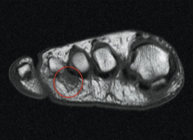
MRITransverse T1 MRI of the foot: well-defined mass in the third intermetatarsal space, circled in red, corresponding to Morton's neuroma.
Source : Torres-Claramunt et al., Indian journal of orthopaedics, 2012, figure 1 · CC BY-NC-SA
- Lateral compression test (squeeze test) : mediolateral pressure applied with one hand across the forefoot. Positive if it reproduces the neuralgic pain. Variable sensitivity, poorly specific.
- Mulder test : combines lateral compression + plantar pressure from the thumb in the interdigital space. A palpable click (sometimes audible) reproducing the pain marks a positive test. It is the best known and the most taught test.¹٬⁶
- Dorsoplantar test (« thumb-index finger squeeze »): targeted dorsoplantar compression of one intermetatarsal space, less studied but useful in addition.²
🎯 Diagnostic accuracy of the clinical tests (Pitcher 2024, systematic review)
Pooled sensitivity (Se) and specificity (Sp), with the range reported across the included studies for each test
Sources: Pitcher M et al. Foot Ankle Orthop. 2024 (Mulder); Bignotti B et al. Eur Radiol. 2015 (US vs MRI, a meta-analysis of 14 studies).
- Local conditions : intermetatarsal bursitis (often associated), metatarsal stress fracture, capsulitis or synovitis of an MTP joint, Freiberg's disease (osteonecrosis of the M2 head), flexor tenosynovitis.²٬⁶
- Conditions at a distance : L5-S1 radiculopathy, tarsal tunnel syndrome, metabolic peripheral neuropathy (diabetes).²
- Rare systemic conditions: osteomyelitis, bone tumour, soft tissue sarcoma, to be considered if there are red flags.
🚩 Red flags specific to the painful forefoot
- Non-mechanical night pain, unrelieved by taking the shoe off → suspected malignancy, infection or a metabolic cause.
- A firm, fixed, painless swelling or a progressive one → a tumour work-up (synovial sarcoma, schwannoma).
- A wound, ulceration, marked inflammatory signs → infection (osteomyelitis, abscess), to be explored urgently in the person with diabetes.
- Fever + forefoot pain → a deep infection.
- A history of cancer + focal bone pain → suspected metastasis.
- Unexplained weight loss + persistent pain → a cancer work-up.
- Stocking hypoaesthesia + diffuse pain → peripheral neuropathy (diabetes, alcohol, toxic).
⚠️ Any red flag → prompt medical referral before any physiotherapy management. Reference: Finucane LM et al. JOSPT 2020.
Should patients with Morton's neuroma be classified, and for what benefit?
There is no universally standardised classification for Morton's neuroma, but stratifying patients by severity has a major therapeutic value: avoiding overtreatment in early forms, and not delaying more intensive management in severe and disabling ones.²٬⁴ Two complementary approaches:- A clinical classification by severity :
- Mild : intermittent pain, triggered only by certain shoes, with no impact on daily life.
- Moderate : frequent pain, limitation of some activities (sport, prolonged walking), recurrence in suitable shoes.
- Severe : almost constant pain including barefoot, persistent paraesthesia, occupational or social impact, failure of well-conducted conservative treatment.
- An ultrasound classification by size : a size > 5 mm is often considered clinically significant and associated with a poorer response to conservative treatment.²٬⁸ Size alone must not dictate the strategy: an asymptomatic neuroma > 5 mm is still an asymptomatic neuroma.
Criticism and controversies on the diagnosis
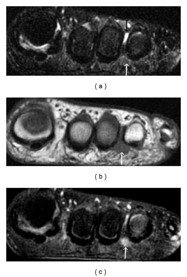
MRIFoot MRI, three coronal sequences (fat-saturated T2, T1, post-contrast fat-saturated T1): tear-drop-shaped mass in the third intermetatarsal space marked by arrows, typical of Morton's neuroma.
Source : Dong et al., Radiology research and practice, 2012, figure 22 · CC BY
Key points
- ✅ The diagnosis is fundamentally clinical : burning or electric-shock pain, made worse by tight shoes, relieved by taking the shoe off.
- ✅ The Mulder sign is specific (87-100 %) but not very sensitive (29-94 %, Pitcher 2024): a negative test does not rule out the diagnosis.
- ✅ Combining several coherent clinical elements (history + Mulder + focal palpation) remains the best diagnostic strategy.
- ✅ High-resolution ultrasound (Se 90 % / Sp 88 %) is the confirmation examination of choice, MRI being reserved for atypical cases.
- ✅ The imperfect imaging-clinical correlation means never treating an image without a symptom: ≈ 33 % of compatible images are asymptomatic (Bencardino 2000).
- ✅ The differential diagnosis (bursitis, stress fracture, Freiberg, capsulitis, radiculopathy) is obligatory before any treatment decision.
Bibliography
- Munir U, Tafti D, Morgan S. Morton Neuroma. In: StatPearls [Internet]. Treasure Island (FL): StatPearls Publishing; updated 2023 May. NBK470249.
- Bhatia M, Thomson L. Morton's neuroma — Current concepts review. J Clin Orthop Trauma. 2020;11(3):406-409. PMID 32405199.
- Pitcher M, Moulson A, Pitcher D, Herbland A, Cairns MC. Diagnostic Accuracy of Subjective Features and Physical Examination Tests for Morton Neuroma: A Systematic Review. Foot Ankle Orthop. 2024;9(4). doi:10.1177/24730114241291055.
- Lizano-Díez X, Ginés-Cespedosa A, Alentorn-Geli E, Pérez-Prieto D. Morton's interdigital neuroma: instructional review. EFORT Open Rev. 2019;4(1):14-24. doi:10.1302/2058-5241.4.180025.
- Pastides P, El-Sallakh S, Charalambides C. Morton's neuroma: A clinical versus radiological diagnosis. Foot Ankle Surg. 2012;18(1):22-24. PMID 22325999.
- Mahadevan D, Venkatesan M, Bhatt R, Bhatia M. Diagnostic accuracy of clinical tests for Morton's neuroma compared with ultrasonography. J Foot Ankle Surg. 2015;54(4):549-553. doi:10.1053/j.jfas.2014.09.021.
- Bignotti B, Signori A, Sormani MP, Molfetta L, Martinoli C, Tagliafico A. Ultrasound versus magnetic resonance imaging for Morton neuroma: systematic review and meta-analysis. Eur Radiol. 2015;25(8):2254-2262. PMID 25809742.
- Jain S, Mannan K. The diagnosis and management of Morton's neuroma: a literature update. Foot Ankle Spec. 2013;6(4):307-317. PMID 23811947.
- Bencardino J, Rosenberg ZS, Beltran J, Liu X, Marty-Delfaut E. Morton's neuroma: is it always symptomatic? AJR Am J Roentgenol. 2000;175(3):649-653. PMID 10954445.
- Finucane LM, Downie A, Mercer C, et al. International Framework for Red Flags for Potential Serious Spinal Pathologies. J Orthop Sports Phys Ther. 2020;50(7):350-372. PMID 32438853.
Why does Morton's neuroma mainly affect women, and what part does footwear play?
What is the biomechanical impact of high heels and narrow shoes?
The women's shoe with high heels and a narrow toe combines three mechanical stresses that are directly harmful to the interdigital nerve:- Hyperextension of the metatarsophalangeal (MTP) joints : an 8 cm heel imposes an MTP dorsiflexion of the order of 50-70°, which puts the common plantar digital nerve under maximum tension beneath the deep transverse intermetatarsal ligament.¹٬³
- Lateral compression from the narrow (« pointed ») toe: the metatarsal heads are pushed together, which physically narrows the space the nerve travels through.¹٬⁴
- Forward translation of body weight onto the forefoot: a 2.5 cm (1 inch) heel already raises forefoot pressure by about +22 %, and that rise is more than linear with heel height.⁵
📈 Rise in forefoot plantar pressure by heel height
An indicative estimate (against a flat foot), after the narrative reviews and baropodometric studies
⚠️ Indicative values from biomechanical reviews. For a personalised analysis: a baropodometric examination. Sources: Munir 2023, Bhatia 2020; a dose-dependent effect confirmed by biomechanical reviews on high heels.
How do you target prevention in the woman at risk?
A systematic assessment of footwear must be part of the minimum protocol of any physiotherapy consultation with a woman presenting with forefoot pain. Here is a condensed practical tool:| Criterion assessed | Recommended target | Marker of raised risk |
|---|---|---|
| Heel height | ≤ 2.5 cm (1 inch) | ≥ 5 cm, risk ×2-3; ≥ 7.5 cm, risk ×4 |
| Width of the toe box | Toes flat with no compression, able to move freely | A pointed toe or a « fashionably narrow toe » compressing the metatarsal heads |
| Depth of the toe box | Enough depth for hammer toes or hallux valgus | A flat toe preventing free MTP extension |
| Stiffness of the sole | Moderate, with the flex just at the MTP joints | A sole that is too soft (flexing at the midfoot) or too stiff throughout |
| Cushioning of the sole | Present under the forefoot (EVA foam or similar) | A hard leather-on-leather sole, hard ground (asphalt) with no cushioning |
| Midfoot hold | Lacing or a fastening that holds the base of the foot | Mules, clogs, ballet flats: the foot slides forward with every step |
🧭 An algorithm for intervening on footwear in the woman at risk
A pragmatic hierarchy of the changes to propose in the clinic
A summary algorithm for intervening on the modifiable mechanical factors. Sources: Matthews 2019, Bhatia 2020, Munir 2023.
The key message in practice
In the woman aged 40-60 with a metatarsalgia suggestive of a Morton's neuroma, the most effective, least costly and most reproducible intervention remains a change of footwear. That means:- An explicit audit of her shoe collection (at least those worn > 50 % of the time).
- An education about the mechanism, since adherence is better when the patient understands why her shoes are the cause.
- An initial trial of 6-8 weeks with suitable footwear before judging conservative treatment a failure.
- An honest, pragmatic negotiation : it is rarely realistic to impose zero heels. Set priorities: heels kept for short occasions (evenings, events), not for 8 hours of standing work.
- An ongoing reassessment of footwear whenever it recurs, even late.
Key points
- 📊 The female to male ratio of ≈ 4:1 reflects a differential exposure to footwear, not an intrinsic anatomical vulnerability.
- 👠 A high-heeled shoe with a narrow toe combines 3 stresses at once: MTP hyperextension, lateral compression, and raised forefoot pressure (+22 % from 2.5 cm, ~+100 % at 10 cm).
- 🦶 The footwear audit is the most cost-effective intervention of the physiotherapy consultation when Morton is suspected.
- 📐 The orthosis with a metatarsal dome is effective if correctly positioned (fitting + adjustment at 4-6 weeks).
- 🤝 The honest, pragmatic negotiation (heels kept for short occasions) achieves better adherence than an absolute ban.
Bibliography
- Bhatia M, Thomson L. Morton's neuroma — Current concepts review. J Clin Orthop Trauma. 2020;11(3):406-409. PMID 32405199.
- Mak MS, Chowdhury R, Johnson R. Morton's neuroma: review of anatomy, pathomechanism, and imaging. Clin Radiol. 2021;76(3):235.e15-235.e23. doi:10.1016/j.crad.2020.10.006.
- Munir U, Tafti D, Morgan S. Morton Neuroma. In: StatPearls [Internet]. Treasure Island (FL): StatPearls Publishing; updated 2023 May. NBK470249.
- Lizano-Díez X, Ginés-Cespedosa A, Alentorn-Geli E, Pérez-Prieto D. Morton's interdigital neuroma: instructional review. EFORT Open Rev. 2019;4(1):14-24. doi:10.1302/2058-5241.4.180025.
- Frey C, Thompson F, Smith J, Sanders M, Horstman H. American Orthopaedic Foot and Ankle Society women's shoe survey. Foot Ankle. 1993;14(2):78-81. PMID 8454237 [Note: a founding study on the woman-footwear impact, still widely cited].
- Saygi B, Yildirim Y, Saygi EK, Kara H, Esemenli T. Morton neuroma: comparative results of two conservative methods. Foot Ankle Int. 2005;26(7):556-559. PMID 16045848.
- Matthews BG, Hurn SE, Harding MP, Henderson RA, Lazzarini PA. The effectiveness of non-surgical interventions for common plantar digital compressive neuropathy (Morton's neuroma): a systematic review and meta-analysis. J Foot Ankle Res. 2019;12:12. PMID 30809275.
Which treatment strategies are the most effective for Morton's neuroma?
Where do you start? What is the recommended hierarchy of interventions?
The therapeutic approach must follow a clear hierarchy, validated by the main recent reviews.¹٬²٬³🔺 The treatment pyramid for Morton's neuroma (five levels)
From the most invasive (top) to the most universal (base), with an indicative % of patients concerned
A summary pyramid based on the recent systematic reviews. Sources: Matthews 2019, Bhatia 2020, Lu 2021, Choi 2021, RFA SR 2025.
- A change of footwear (a wide toe box, a low heel, forefoot cushioning), set out in the previous chapter. It is the most effective and most cost-effective intervention.¹٬⁴
- A foot orthosis with a metatarsal dome, placed behind the heads to spread them apart and unload the nerve.¹٬²٬⁵ A randomised trial (Saygi 2005, Foot Ankle Int): 63 % complete or partial relief at 12 months with suitable footwear and orthoses, against 82 % after a corticosteroid injection, with no significant difference between the two groups at one year.⁵
- Ultrasound-guided corticosteroid injections : the systematic review by Choi et al. (2021, Clin Orthop Surg) confirms maximal relief between 1 week and 3 months after the injection, with patient satisfaction in the majority on the Johnson score.⁷ Limits: the long-term effect is more debated, and there is a risk of plantar fat pad atrophy (rare but cumulative with repeated injections). Recommendation: limit to 1-3 spaced injections.²٬⁷
- Sclerosing alcohol injection : the evidence of efficacy is variable; the Pasquali 2015 study (n = 508 patients) shows acceptable satisfaction rates but far from negligible adverse effects (post-injection pain, rarely digital ischaemia).⁸ To be used with caution, by an experienced operator.
- Pulsed or continuous radiofrequency : the systematic review and meta-analysis by Llombart-Blanco et al. (2025, 8 studies, 237 patients) reports a significant reduction in pain at the last follow-up (a mean difference of 5.74 VAS points, 95 % CI 5.58 to 5.90), 47.6 % complete relief (95 % CI 25.1 to 70.0) but 16.4 % of patients with no benefit at all (95 % CI 11.9 to 20.9), for 2.1 % of complications, most of them resolving.⁹ In the sensitivity analyses, protocols of 3 cycles or fewer do better than those of more than 3 cycles, and temperatures ≥ 85 °C better than < 85 °C.⁹
- ESWT (extracorporeal shockwave therapy) : Seok et al. (2016, JAPMA) ran a placebo-controlled RCT confirming a reduction in VAS and an improvement in AOFAS at 4 weeks.¹⁰ Level of evidence: moderate, with limited data.
- Guided cryoablation : Cazzato et al. (2016, CVIR), 20 patients, 24 neuromas, 95 % satisfaction at 20 months (a pilot study).¹¹ An emerging technique, to be confirmed.
- Complete resolution of the pain: 43 % after injection, 68 % after neurolysis, 74 % after neurectomy.
- Complete satisfaction: 35 % after injection, 63 % after neurolysis, 57 % after neurectomy.
- Major complication: recurrent stump neuroma ≈ 14 % after neurectomy.
📊 Comparative efficacy: injection vs neurolysis vs neurectomy (Lu 2021, MA n=2998)
The percentage of patients with complete pain resolution and with satisfaction
Note: neurectomy carries a risk of a recurrent stump neuroma ≈ 14 % , hence the importance of exhausting conservative care first. Source: Lu VM et al. Acta Neurochir. 2021;163:531-543.
What place does exercise hold and is there a superior approach?
The literature on exercise specific to Morton's neuroma is surprisingly thin. No randomised controlled trial has directly compared exercise protocols; the current recommendations rest on solid but non-randomised biomechanical reasoning. 👟 Three aims clinically justify an exercise programme:- Stretching the gastrocnemius-soleus complex : DiGiovanni et al. (2002, JBJS) showed that isolated gastrocnemius tightness (measured by the Silfverskiöld test) is associated with raised forefoot pressures during walking.¹⁴ Stretching the calf (against a wall, off a step, the Silfverskiöld stretch) is therefore a logical addition.
- Strengthening the intrinsic foot muscles : abductor hallucis, the short flexors, the lumbricals. The « short foot » exercise (actively shortening the plantar arch without curling the toes) strengthens the active support of the transverse arch and potentially reduces the dropping of the metatarsal heads.¹٬¹⁵
- Mobility of the MTP and intermetatarsal joints : gentle joint mobilisation aimed at keeping the heads moving and avoiding locking in hyperextension. Self-mobilisation by rolling the forefoot on a massage ball is tolerated by most patients.
Manual therapy, shockwave, radiofrequency: how effective are they really?
| Modality | Proposed mechanism | Level of evidence | Practical indication |
|---|---|---|---|
| A change of footwear | Removing the mechanical stress | High (consensus, the base of the 1st line) | Always, 1st line |
| An orthosis with a metatarsal dome | Decompression of the interdigital space | Low to moderate (RCT Saygi 2005, SR Thomson 2020) | 1st line, combined with the footwear |
| Exercise (stretching + intrinsics) | Reducing the biomechanical stresses | Low (biomechanical reasoning, few RCTs) | A useful addition, with few adverse effects |
| Targeted manual therapy | Intermetatarsal mobilisation, myofascial release | Low (a rationale, few specific studies) | An adjunct in the physiotherapy clinic |
| Ultrasound-guided corticosteroid injection | A local anti-inflammatory effect | High in the short and medium term (Choi 2021) | 2nd line, ≤ 3 injections |
| Alcohol sclerosis | Chemical neurolysis | Moderate (Pasquali 2015) with adverse events not rare | Experienced operators only |
| ESWT (shockwave) | Tissue stimulation, neovascularisation | Moderate (RCT Seok 2016, limited data) | A non-invasive option, 2nd-3rd line |
| Pulsed or continuous radiofrequency | Nerve modulation, partial neurolysis | Moderate-high (SR/MA 2025, n=237, 47.6 % complete relief, 16.4 % with no benefit) | 3rd line, an alternative to surgery |
| Cryoablation | Neurolysis by cold | Low (pilot series, Cazzato 2016) | Emerging, to be confirmed |
| Surgical neurectomy | Excision of the neuroma | High (Lu 2021, 74 % success) | A last resort, with a 14 % risk of a recurrent neuroma |
Beyond the physical: how do you educate the patient and address psychological factors?
The education of the patient is the cornerstone of treatment. 🧠 The patient must understand:- The mechanical nature of the condition (nerve compression, not a tumour); defusing the word « neuroma » is in itself therapeutic.²
- The causal link footwear → condition: without that understanding, adherence to the changes is poor.¹٬²
- The realistic expectations : progressive improvement (weeks to months), no « miracle cure ». Early frustration and discouragement hold back compliance.²
- The importance of a temporary change of activity (reducing running on hard ground, alternating with cycling or swimming), followed by a progressive return.
Criticism and controversies on treatment
Three areas of debate shape current practice: First, the definition of conservative failure remains vague. The recommended durations range from 3 to 12 months across sources, and the objective criteria for moving to the 2nd line are not standardised. That leaves considerable room for clinical judgement and patient preference.²٬⁶ Secondly, the long-term benefit of corticosteroid injections is more debated than their short-term benefit. The cumulative risk of plantar fat pad atrophy, which perpetuates metatarsalgia, justifies limiting the number of injections (≤ 3) and spacing them (≥ 4-6 weeks apart).⁷ Thirdly, the evidence base for exercise is surprisingly weak. Most recommendations rest on coherent biomechanical reasoning rather than on RCTs comparing specific protocols. It is a priority field of research for physiotherapy: a structured protocol (exercise + manual therapy vs an orthosis alone) still needs validating at high quality.Key points
- 🥇 The 1st line is conservative : a change of footwear + an orthosis with a metatarsal dome, to be kept up for ≥ 3 months.
- 💉 The ultrasound-guided corticosteroid injections are effective in the short and medium term (Choi 2021), so limit them to ≤ 3 injections.
- ⚡ The radiofrequency (SR/MA 2025: 47.6 % complete relief, 16.4 % with no benefit) and the ESWT (RCT Seok 2016) are validated minimally invasive options for the 2nd-3rd line.
- 🔪 The surgery (neurectomy) offers 74 % success (Lu 2021) but carries a 14 % risk of a recurrent stump neuroma.
- 🧠 The education and the shared decision-making (Légaré 2018) are non-negotiable pillars before any invasive procedure.
- 🏋️♀️ The exercise (gastrocnemius stretching, intrinsic strengthening) has a solid rationale but a limited evidence base, so use it as an addition.
Bibliography
- Bhatia M, Thomson L. Morton's neuroma — Current concepts review. J Clin Orthop Trauma. 2020;11(3):406-409. PMID 32405199.
- Munir U, Tafti D, Morgan S. Morton Neuroma. In: StatPearls [Internet]. 2023. NBK470249.
- Matthews BG, Hurn SE, Harding MP, Henderson RA, Lazzarini PA. The effectiveness of non-surgical interventions for common plantar digital compressive neuropathy (Morton's neuroma): a systematic review and meta-analysis. J Foot Ankle Res. 2019;12:12. PMID 30809275.
- Thomson L, Aujla RS, Divall P, Bhatia M. Non-surgical treatments for Morton's neuroma: A systematic review. Foot Ankle Surg. 2020;26(7):736-743. PMID 31718949.
- Saygi B, Yildirim Y, Saygi EK, Kara H, Esemenli T. Morton neuroma: comparative results of two conservative methods. Foot Ankle Int. 2005;26(7):556-559. PMID 16045848.
- Valisena S, Petri GJ, Ferrero A. Treatment of Morton's neuroma: A systematic review. Foot Ankle Surg. 2018;24(4):271-281. PMID 29409240.
- Choi JY, Lee HI, Hong WH, Suh JS, Hur JW. Corticosteroid Injection for Morton's Interdigital Neuroma: A Systematic Review. Clin Orthop Surg. 2021;13(2):266-277. doi:10.4055/cios20256.
- Pasquali C, Vulcano E, Novario R, Varotto D, Montoli C, Volpe A. Ultrasound-guided alcohol injection for Morton's neuroma. Foot Ankle Int. 2015;36(1):55-59. PMID 25367249.
- Llombart-Blanco R, Mariscal G, Benlloch M, Barrios C, Llombart-Ais R. Systematic Review and Meta-analysis of Radiofrequency Ablation for Morton's Neuroma: Outcomes and Predictors of Success. Am J Phys Med Rehabil. 2025;104(5):465-472. PMID 39642322.
- Seok H, Kim SH, Lee SY, Park SW. Extracorporeal Shockwave Therapy in Patients with Morton's Neuroma: A Randomized, Placebo-Controlled Trial. J Am Podiatr Med Assoc. 2016;106(2):93-99. PMID 27031544.
- Cazzato RL, Garnon J, Ramamurthy N, et al. Percutaneous MR-Guided Cryoablation of Morton's Neuroma: Rationale and Technical Details After the First 20 Patients. Cardiovasc Intervent Radiol. 2016;39(10):1396-1402. PMID 27189181.
- Lu VM, Puffer RC, Everson MC, Gilder HE, Burks SS, Spinner RJ. Treating Morton's neuroma by injection, neurolysis, or neurectomy: a systematic review and meta-analysis of pain and satisfaction outcomes. Acta Neurochir (Wien). 2021;163(2):531-543. PMID 32056015.
- Zhang J, Fu Q, Zhang Y, Tu C, Zhang H. Effect of surgical approach on the treatment of Morton's neuroma: a systematic review and meta-analysis. J Foot Ankle Res. 2023;16:60. PMID 37674248.
- DiGiovanni CW, Kuo R, Tejwani N, et al. Isolated gastrocnemius tightness. J Bone Joint Surg Am. 2002;84(6):962-970. PMID 12063330.
- Lizano-Díez X, Ginés-Cespedosa A, Alentorn-Geli E, Pérez-Prieto D. Morton's interdigital neuroma: instructional review. EFORT Open Rev. 2019;4(1):14-24. doi:10.1302/2058-5241.4.180025.
- Légaré F, Adekpedjou R, Stacey D, et al. Interventions for increasing the use of shared decision making by healthcare professionals. Cochrane Database Syst Rev. 2018;7(7):CD006732. PMID 30025154.
How do you secure a lasting recovery and prevent recurrence in Morton's neuroma?
How do you make the patient an active player in their own recovery through self-management?
Self-management rests on four complementary pillars:- A lasting change of footwear 👟: this is the most cost-effective element. The practical rule is: no narrow-toed shoe and no heel > 5 cm in everyday wear. Heels stay reserved for short occasions (≤ 2-3 h now and then). The education stresses negotiation, not an absolute ban, which improves adherence.¹٬²٬³
- Continuous use of the orthosis with a metatarsal dome in the adapted footwear, for at least 3 months, then according to tolerance and recurrence. It need not be worn permanently for life, but it stays useful for prolonged standing or sporting activity.⁴
- A home exercise programme 🧘 :
- Gastrocnemius stretching (against a wall, off a step, 3×30 s, ≥ 5×/week), reference DiGiovanni 2002.⁵
- Soleus stretching (knee bent, same position).
- Intrinsic strengthening: the « short foot exercise » (3×10 repetitions per foot, ≥ 4×/week); picking up a towel with the toes; abductor hallucis against a band.¹٬⁶
- Self-mobilisation of the MTP and intermetatarsal joints by rolling on a massage ball, 1-2 min per foot, after the shower or at the end of the day.
- Weight and activity management ⚖️: a modest reduction in body weight (5-10 %) significantly lowers plantar pressures. Advise a temporary substitution of high-impact activities (running, dance, jumping) with activities that spare the forefoot (cycling, swimming, cross-trainer) during the acute and transition phases.²
When and how do you plan a safe return to sport and to activity?
The return to sport or to high-impact activity must be progressive, structured and guided by the symptoms. No single standardised protocol exists in the literature, but a consensus emerges on the principles of progression, valid for most foot and ankle conditions.²📅 A plan for a progressive return to activity, five steps
Criteria for moving to the next step: pain ≤ 2/10 during the activity, no worsening at 24-48 h
A plan adapted from the general principles of return to musculoskeletal activity (a narrative consensus). Sources: Bhatia 2020, Munir 2023.
- Step 1, controlled daily life : a prerequisite before any return to activity. The pain must be ≤ 2/10 on normal walking in suitable shoes. If that criterion is not met, the work stays centred on the 1st line (footwear + orthosis + home exercise).
- Step 2, reintroducing low-impact activities 🚴: swimming (preferred, as it puts no load on the forefoot), cycling (check the position of the foot on the pedal, avoid clip pedals that squeeze the forefoot), light cross-trainer. 2-3 sessions of 30 min a week, pain-free.
- Step 3, moderate loads : brisk walking on a treadmill or a soft trail, a more intense cross-trainer. Prerequisite: keeping to the VAS < 2/10 criterion. Always in wide shoes + the orthosis.
- Step 4, the return to impact 🏃: running is reintroduced very gradually. A walk-run programme: start with 1 min running / 4 min walking, ×5-8 repetitions, 2×/week. Prefer soft ground (a tartan track, an earth trail) before asphalt. Increase the volume before the intensity.
- Step 5, the return to performance : a progressive increase in weekly volume (the +10 % per week rule), then the introduction of intensity (intervals, fast pace). A continuous check: if the pain recurs, step back one level for 1-2 weeks before progressing again.
🚩 Warning signs during the return to sport
- A return of burning or electric-shock pain in the forefoot → go back to the previous step, check the footwear, wear the orthosis systematically.
- Pain persisting > 48 h after the activity → a signal that the load exceeds the tissue capacity; reduce the volume.
- New oedema or swelling → have a bursitis, a stress fracture or an inflammatory reaction in another joint ruled out.
- Pain at night or at rest → reconsider the diagnosis; a medical consultation to exclude an alternative condition.
- Early recurrence despite suitable footwear and an orthosis → discuss a 2nd line of treatment (injection, ESWT, radiofrequency).
Criticism and controversies on recovery
Three areas of debate persist in the literature: First, although orthoses with a metatarsal dome are widely recommended, there is no clear standardisation of their optimal design (dome size, density, precise position). Some of the variability in clinical results follows from that.²٬⁴ Clinical follow-up with adjustment at 4-6 weeks remains the best guarantee of efficacy. Secondly, the literature badly lacks return-to-sport protocols specific to Morton. The current recommendations rest on general principles of musculoskeletal progression, not on dedicated studies. The pain thresholds, transition durations and objective criteria for progressing still need validating. Thirdly, the threshold for conservative failure before considering a 2nd or 3rd line remains imprecise. The recommended durations range from 3 to 12 months across studies and schools of thought.²٬⁷ Clinical common sense prevails: a patient at a moderate to severe stage with a marked impact can justify accelerating towards an injection, without waiting 6 months of conservative care if quality of life is severely affected.Key points
- 🏠 The lasting recovery is decided in the long term through self-management : suitable footwear, wearing the orthosis, home exercises.
- 📉 The return to sport must be progressive (five steps), guided by a VAS ≤ 2/10 and by the absence of worsening at 24-48 h.
- 🚴 Favour swimming and cycling in the transition, before reintroducing impact activities (alternating walking and running).
- 👟 The audit of sports footwear is compulsory (a wide toe box, a drop ≤ 8 mm, forefoot cushioning).
- 🚦 If it recurs: step back one level, wear the orthosis permanently, and discuss a 2nd line if it recurs repeatedly despite good compliance.
Bibliography
- Bhatia M, Thomson L. Morton's neuroma — Current concepts review. J Clin Orthop Trauma. 2020;11(3):406-409. PMID 32405199.
- Munir U, Tafti D, Morgan S. Morton Neuroma. In: StatPearls [Internet]. 2023. NBK470249.
- Lizano-Díez X, Ginés-Cespedosa A, Alentorn-Geli E, Pérez-Prieto D. Morton's interdigital neuroma: instructional review. EFORT Open Rev. 2019;4(1):14-24. doi:10.1302/2058-5241.4.180025.
- Matthews BG, Hurn SE, Harding MP, Henderson RA, Lazzarini PA. The effectiveness of non-surgical interventions for common plantar digital compressive neuropathy (Morton's neuroma): a systematic review and meta-analysis. J Foot Ankle Res. 2019;12:12. PMID 30809275.
- DiGiovanni CW, Kuo R, Tejwani N, et al. Isolated gastrocnemius tightness. J Bone Joint Surg Am. 2002;84(6):962-970. PMID 12063330.
- Jain S, Mannan K. The diagnosis and management of Morton's neuroma: a literature update. Foot Ankle Spec. 2013;6(4):307-317. PMID 23811947.
- Thomson L, Aujla RS, Divall P, Bhatia M. Non-surgical treatments for Morton's neuroma: A systematic review. Foot Ankle Surg. 2020;26(7):736-743. PMID 31718949.
What do real clinical cases teach us about Morton's neuroma?
Analysis of a « classic » case: from assessment to conservative resolution
An archetypal profile (a composite of the clinical series published in Bhatia 2020 and Munir 2023): a woman in her fifties, a professional standing 8 hours a day, regularly wearing narrow-toed shoes with a medium heel (4-6 cm), consults for a burning pain in the 3rd intermetatarsal space of the right foot, present for 8 months. The pain is intermittent, triggered by her work shoes, relieved immediately by taking the shoe off. The sensation of « walking on a pebble » is reported spontaneously.¹٬² On examination:- Inspection: moderate bilateral hallux valgus, no swelling.
- Palpation: reproducible focal pain in the 3rd space, no bone pain.
- Mulder test : a palpable click, reproducing the typical pain → strongly suggestive (high specificity).³
- No red flag.
- A change of footwear : dropping the narrow-toed work shoes; buying shoes with a wide toe box + forefoot cushioning.
- A foot orthosis with a metatarsal dome placed behind heads 3 and 4 (follow-up at 4 weeks for adjustment).
- A home exercise programme : gastrocnemius stretching 3×30 s, ×5/week; short foot exercise 3×10 per foot, ×4/week; self-mobilisation with a ball at the end of the day.
- Education : an explanation of the mechanism, defusing the word « neuroma », realistic expectations (progressive improvement over 2-3 months).
The diagnostic challenge: when Morton's neuroma mimics another condition
Classic traps in practice (a synthesis of Mahadevan 2015, Pitcher 2024):- Metatarsal stress fracture : forefoot pain in a runner, similar in location. The distinction: the fracture is more painful on focal bone palpation, persists at rest, and imaging (a repeat radiograph at 2 weeks or an early MRI) confirms it.³٬⁸
- Intermetatarsal bursitis : a very close picture, sometimes associated. Ultrasound tells the two apart (a bursitis is a compressible anechoic structure; a neuroma is a non-compressible hypoechoic mass).⁴
- MTP capsulitis or synovitis : pain that is more dorsal and focal over the MTP joint, a positive drawer sign, sometimes a floating toe. To be considered for the 2nd MTP joint in particular.
- Freiberg's disease : osteonecrosis of the metatarsal head, most often M2, in the adolescent girl or young adult. Diagnostic imaging (radiograph, MRI).
- L5-S1 radiculopathy : pain referred from the lumbar spine, with a spinal examination and a straight leg raise to include.
- Peripheral diabetic neuropathy : to be considered in the patient with diabetes and diffuse stocking hypoaesthesia.
A complex case: stump recurrence and multiple forms
Two particular clinical situations deserve attention: 1. The recurrent stump neuroma after neurectomy is a dreaded but documented complication. The meta-analysis by Lu et al. (2021, Acta Neurochir, n = 2,998) reports an incidence of ≈ 14 % stump neuroma after neurectomy.¹⁰ The mechanism: the divided nerve stump regrows in a disorganised way, forming a genuine and very painful « stump neuroma », often harder to treat than the original one. Clinically: the pain returns a few months after initially satisfactory surgery, sometimes more intense, sometimes with a marked neuropathic component (allodynia, dysaesthesia). MRI or ultrasound confirms a fibrous scar mass at the operative site. Revision surgery (often by the dorsal approach, sometimes burying the stump in a muscle or a bone) is an option, but with a more modest success rate.¹⁰٬¹¹ Techniques of injecting the stump (corticosteroid, alcohol) or of pulsed radiofrequency can be tried before a further operation.- An analysis of the mechanical factors in detail : a markedly cavus or flat foot, an associated deformity, chronically unsuitable footwear.
- An ordering of priorities : treat the most symptomatic and disabling space first.
- An in-depth discussion about surgery: excising several nerves at once or in stages, the cumulative risk of complications, the loss of interdigital sensation.
Criticism: what clinical cases do and do not say
Published clinical cases are instructive but carry important limits 💡:- They often represent « interesting » cases or therapeutic successes ; the failures are under-represented. Publication bias probably overestimates the efficacy of the interventions.
- The anatomical-clinical correlation stays imperfect : Bencardino (2000) documented ≈ 33 % compatible images in asymptomatic people.¹² Finding a « neuroma » on imaging in a patient in pain does not prove causation, hence the importance of clinical and imaging coherence.
- The surgical option, despite high satisfaction rates (74 %), carries non-trivial complications (14 % recurrent stump neuroma, loss of interdigital sensation).¹⁰ The decision must always be shared and informed.
- The emerging techniques (pulsed radiofrequency, cryoablation) show promising results but often rest on small series (n=20-50). Confirmation by larger RCTs is needed.
Key points
- 📋 The typical case (a woman of 40-60, unsuitable footwear, the 3rd space, Mulder +) is generally resolved by footwear + an orthosis + ± an injection.
- 🎭 The differential diagnosis is essential: if there is no improvement in 6-8 weeks, reconsider (stress fracture, bursitis, Freiberg, capsulitis).
- 🔄 The recurrent stump neuroma affects ≈ 14 % of those operated on (Lu 2021), a strong argument for exhausting conservative care.
- 👯 The multiple or bilateral forms (< 5 %) complicate management and call for complete imaging and a more cautious surgical discussion.
- 🤝 The shared decision and transparency about the risks are non-negotiable before any surgery.
Bibliography
- Munir U, Tafti D, Morgan S. Morton Neuroma. In: StatPearls [Internet]. 2023. NBK470249.
- Bhatia M, Thomson L. Morton's neuroma — Current concepts review. J Clin Orthop Trauma. 2020;11(3):406-409. PMID 32405199.
- Pitcher M, Moulson A, Pitcher D, Herbland A, Cairns MC. Diagnostic Accuracy of Subjective Features and Physical Examination Tests for Morton Neuroma: A Systematic Review. Foot Ankle Orthop. 2024;9(4). doi:10.1177/24730114241291055.
- Bignotti B, Signori A, Sormani MP, Molfetta L, Martinoli C, Tagliafico A. Ultrasound versus magnetic resonance imaging for Morton neuroma: systematic review and meta-analysis. Eur Radiol. 2015;25(8):2254-2262. PMID 25809742.
- Matthews BG, Hurn SE, Harding MP, Henderson RA, Lazzarini PA. The effectiveness of non-surgical interventions for common plantar digital compressive neuropathy (Morton's neuroma): a systematic review and meta-analysis. J Foot Ankle Res. 2019;12:12. PMID 30809275.
- Lizano-Díez X, Ginés-Cespedosa A, Alentorn-Geli E, et al. Corticosteroid Injection for the Treatment of Morton's Neuroma: A Prospective, Double-Blinded, Randomized, Placebo-Controlled Trial. Foot Ankle Int. 2017;38(9):944-951. PMID 28617064.
- Choi JY, Lee HI, Hong WH, Suh JS, Hur JW. Corticosteroid Injection for Morton's Interdigital Neuroma: A Systematic Review. Clin Orthop Surg. 2021;13(2):266-277. doi:10.4055/cios20256.
- Mahadevan D, Venkatesan M, Bhatt R, Bhatia M. Diagnostic accuracy of clinical tests for Morton's neuroma compared with ultrasonography. J Foot Ankle Surg. 2015;54(4):549-553. doi:10.1053/j.jfas.2014.09.021.
- Post MD, Maccio JR. Mechanical diagnosis and therapy and Morton's neuroma: a case-series. J Man Manip Ther. 2020;28(1):60-67. PMID 31177965 [case series accessible in PMC].
- Lu VM, Puffer RC, Everson MC, Gilder HE, Burks SS, Spinner RJ. Treating Morton's neuroma by injection, neurolysis, or neurectomy: a systematic review and meta-analysis of pain and satisfaction outcomes. Acta Neurochir (Wien). 2021;163(2):531-543. PMID 32056015.
- Zhang J, Fu Q, Zhang Y, Tu C, Zhang H. Effect of surgical approach on the treatment of Morton's neuroma: a systematic review and meta-analysis. J Foot Ankle Res. 2023;16:60. PMID 37674248.
- Bencardino J, Rosenberg ZS, Beltran J, Liu X, Marty-Delfaut E. Morton's neuroma: is it always symptomatic? AJR Am J Roentgenol. 2000;175(3):649-653. PMID 10954445.
How do you apply these recommendations concretely in your practice?
When, and to which other health professionals, should you refer?
One of the fundamental skills of the first-line physiotherapist is the ability to recognise the situations that lie beyond their scope. Three categories of flag structure that decision: Red flags 🚩: they indicate a potentially serious condition and call for prompt medical referral. The international IFOMPT 2020 framework (Finucane et al, JOSPT) remains the reference for assessing musculoskeletal red flags.¹ The red flags specific to the painful forefoot were set out in chapter 2. No single sign has a strong predictive value: it is the combination of signs and the overall clinical context that point the way.² Yellow flags ⚠️: psychosocial factors that predict chronicity, catastrophising, kinesiophobia, mistaken beliefs about pain, depressive symptoms, low self-efficacy. In the context of Morton's neuroma these factors are frequent in working women who are exposed (standing, sport) and see their quality of life affected. A biopsychosocial approach (pain education, cognitive restructuring, encouragement to keep up an adapted activity) is essential. When yellow flags predominate and persist despite physiotherapy, refer to a psychologist trained in chronic pain or to a pain physician.³ Targeted interprofessional referral 🤝, according to the picture:| Clinical situation | Recommended referral | Why |
|---|---|---|
| A red flag (night pain, a history of cancer, fever…) | GP / emergency department | Excluding a serious condition before management |
| Persistent diagnostic doubt or failure at 6-8 weeks | Sports physician / orthopaedic foot surgeon | Further imaging, differential diagnosis |
| Ultrasound or MRI needed | Musculoskeletal radiologist | High-resolution ultrasound / targeted MRI |
| A custom orthosis to be made | Podiatrist / orthotist | A personalised adaptation of the metatarsal dome |
| A corticosteroid injection needed | Rheumatologist or interventional radiologist | An ultrasound-guided procedure for precision |
| Conservative failure ≥ 6 months, a surgical indication | Orthopaedic foot surgeon | Discussion of neurectomy / minimally invasive techniques |
| Marked kinesiophobia or catastrophising | Psychologist trained in pain | CBT, chronic pain management |
| Significant excess weight, metabolic comorbidities | Nutrition physician / GP | Overall management of weight and of diabetes where present |
How do you measure outcomes and overcome the barriers to implementation?
To make sure the interventions are effective and appropriate, it is essential to measure outcomes objectively and reproducibly. 📈 Validated outcome measures (PROMs) relevant to Morton's neuroma :- Visual Analogue Scale (VAS) 0-10 for pain: simple, quick, sensitive to change. To be measured at rest, on walking, and during a provocative activity.
- MOXFQ (Manchester-Oxford Foot Questionnaire) : 16 items, 3 subscales (pain, walking and standing, social), validated for surgical and conservative foot and ankle pathology.
- FFI (Foot Function Index) : 23 items, 3 dimensions (pain, disability, activity limitation), widely used internationally.
- AOFAS Forefoot : a composite score (clinician + patient), 100 points. Criticised for its unbalanced subjective-to-objective ratio, but still much used in orthopaedics.
- Goal Attainment Scaling (GAS) : Krasny-Pacini et al. proposed a reference methodological update (2013, Ann Phys Rehabil Med), useful for quantifying the achievement of the patient's personal goals.⁴
- Tampa Scale of Kinesiophobia (TSK) : if a yellow flag is suspected.
- To document the objective course.
- To detect a failure or a plateau early.
- To justify the benefit-to-cost balance to the patient and to the funders.
- To take part in aggregating data for research.
- The choice between continuing conservative care or an injection at 3-6 months.
- The choice between an injection, radiofrequency, cryoablation or surgery when it fails.
- An explicit discussion of the risk of a recurrent stump neuroma (14 %) before surgery (Lu 2021).⁶
- Availability of ultrasound : variable between settings, which holds back diagnostic confirmation.
- The cost of custom orthoses : reimbursement varies between countries and can limit access.
- The small number of RCTs specific to Morton : the evidence base for exercise and manual therapy stays limited, which makes standardising protocols harder.
- Waiting times for specialists (interventional radiology, foot surgery) in some health systems.
- Easier access to evidence summaries (PubMed, Cochrane, JOSPT, BJSM, EFORT Open Reviews, the last of which offers freely accessible reviews, including the Lizano-Díez 2019 instructional review).⁷
- Peer mentoring and taking part in communities of practice (learned societies, online case groups).
- Continuous updating : subscribing to PubMed alerts for « Morton's neuroma » and « interdigital neuropathy » filtered on SR/MA.
- Systematically building a PROM into the patient record, a benefit that accumulates from patient to patient.
- Recognising the limits and referring early when a case goes beyond the scope of 1st line physiotherapy.
Critique and controversies: beyond the guidelines
Applying the recommendations rigidly can carry risks. Three nuances deserve to be stressed: First, the « red flag paradox » : looking for them is non-negotiable for safety, but an excessive focus can induce a nocebo effect and lead to costly and often useless imaging cascades.² The expert skill lies in the ability to interpret the flags in their clinical context, not in identifying them mechanically. Secondly, the « PROM implementation gap » : having dozens of validated questionnaires is not enough if their use stays superficial (ticking the box without interpreting it). Building them into the clinical workflow and using the results to guide the decision are the real added value. Thirdly, the standardisation versus individualisation tension : practice guides rest on statistical averages, but the patient sitting in front of us has their own comorbidities, social context and preferences. The expertise lies in adapting the recommendations, not applying them mechanically. The evidence is a map 🗺️; the patient and the context are the compass.Key points
- 🚩 Identifying the red flags (Finucane 2020) calls for prompt medical referral; identifying the yellow flags (kinesiophobia, catastrophising) justifies psychological collaboration.
- 📊 Measure outcomes with at a minimum the VAS + 1 PROM (MOXFQ or FFI) at 3 time points (baseline, interim, final).
- 🤝 The shared decision-making (Légaré 2018, Cochrane) is central to the choice between conservative, injection and surgical care.
- 🌐 Structured interprofessional collaboration (radiologist, podiatrist, surgeon, psychologist) improves the patient's pathway.
- 🚧 The barriers to implementation are systemic (time, access, training), and lifting them goes through mentoring, access to evidence summaries, and the routine use of PROMs.
- ⚖️ Expert practice navigates between standardisation (the recommendations) and individualisation (patient preferences), making an absolute of neither.
Bibliography
- Finucane LM, Downie A, Mercer C, et al. International Framework for Red Flags for Potential Serious Spinal Pathologies. J Orthop Sports Phys Ther. 2020;50(7):350-372. PMID 32438853.
- Cook CE, George SZ, Reiman MP. Red flag screening for low back pain: nothing to see here, move along: a narrative review. Br J Sports Med. 2018;52(8):493-496. PMID 28923844.
- Vlaeyen JWS, Crombez G, Linton SJ. The fear-avoidance model of pain. Pain. 2016;157(8):1588-1589. PMID 27428892.
- Krasny-Pacini A, Hiebel J, Pauly F, Godon S, Chevignard M. Goal attainment scaling in rehabilitation: a literature-based update. Ann Phys Rehabil Med. 2013;56(3):212-230. PMID 23562111.
- Légaré F, Adekpedjou R, Stacey D, et al. Interventions for increasing the use of shared decision making by healthcare professionals. Cochrane Database Syst Rev. 2018;7(7):CD006732. PMID 30025154.
- Lu VM, Puffer RC, Everson MC, Gilder HE, Burks SS, Spinner RJ. Treating Morton's neuroma by injection, neurolysis, or neurectomy: a systematic review and meta-analysis of pain and satisfaction outcomes. Acta Neurochir (Wien). 2021;163(2):531-543. PMID 32056015.
- Lizano-Díez X, Ginés-Cespedosa A, Alentorn-Geli E, Pérez-Prieto D. Morton's interdigital neuroma: instructional review. EFORT Open Rev. 2019;4(1):14-24. doi:10.1302/2058-5241.4.180025.
- Bhatia M, Thomson L. Morton's neuroma — Current concepts review. J Clin Orthop Trauma. 2020;11(3):406-409. PMID 32405199.
What next after reading this?
This article is part of a collection of evidence-based clinical syntheses. A question, a comment, a correction to suggest? Contact us directly through the WhatsApp button at the bottom right of the screen.

