

















In brief
Lesser toe deformities (hammer, claw or mallet toe) are sagittal deviations of the MTP, PIP or DIP joints of the second to fifth toes, due to an imbalance between weakened intrinsic muscles and dominant extrinsic ones. They follow a continuum from flexible to rigid and can lead to corns, metatarsalgia, falls in older people and ulceration in the person with diabetes. The diagnosis is clinical, resting on the Kelikian push-up test (flexible vs rigid) completed by the plantar plate drawer test. First-line conservative care combines a wide toe box, foot orthoses, taping and intrinsic exercises. Their prevalence reaches 29.9 % of adults (Framingham).
A clinical synthesis based on the most recent systematic reviews and international consensus statements: the Framingham foot study, IWGDF 2023, Albright 2022 (US vs MRI of the plantar plate), and 2024-2025 data on the intrinsic muscles and the diabetic foot.
Clinical synthesis
- Lesser toe deformities (hammer, claw, mallet) are sagittal deviations of the MTP / PIP / DIP joints of the 2nd to 5th toes, due to an imbalance between the intrinsics (weak) and the extrinsics (dominant).
- Consolidated Framingham prevalence (Hagedorn 2013, n=2,446): 29.9 % of adults ; heritability 0.49–0.62 in women (Hannan 2013). The risk triad: narrow footwear, hallux valgus, atrophy of the intrinsics.
- The course follows a continuum flexible → semi-rigid → rigid through capsular fibrosis and retraction of the collaterals; MTP subluxation from rupture of the plantar plate is a key event (Doty 2014, Nery 2012).
- Three major clinical consequences: pain (a PIP corn, metatarsalgia), falls (OR ×1.57 in older people, Menz 2018) and ulceration in the person with diabetes (IWGDF risk category ≥2).
- The fundamental test is the Kelikian push-up test (flexible vs rigid), completed by the drawer test for the plantar plate (Klein 2014, Se > 95 %).
- The reference imaging in case of doubt is MRI (Se 95 % / Sp 100 %) or operator-dependent dynamic ultrasound (Albright 2022 MA).
- An obligatory differential diagnosis: Morton's neuroma, stress fracture, isolated plantar plate rupture (crossover toe), inflammatory arthropathy.
- The first line is conservative for flexible stages: a wide toe box and a low heel, a foot orthosis with a metatarsal bar, taping, intrinsic exercises.
- A programme of intrinsic exercises (short foot, towel curl, toe grip) over 8-12 weeks improves strength and function (Mulligan 2013, Mickle 2016, Taddei 2020). The McKeon foot core paradigm (2015) is its theoretical framework.
- Three vulnerable subgroups converge on the deformity: diabetes (claw toes from motor neuropathy, IWGDF 2023), rheumatoid arthritis (forefoot pain in 63.9 %, Otter 2010), Charcot-Marie-Tooth (pes cavus + progressive bilateral claw toes, Pareyson 2017).
- Red flags: a rapidly progressive deformity, a bilateral one in a young person, plantar ulceration, hot erythema → refer to a diabetologist, rheumatologist, neurologist or surgeon.
- Surgery is second line for conservative failure or a rigid stage: Weil osteotomy (Cancilleri 2003; a floating toe risk of 28-43 %, Migues 2004), PIP arthrodesis (Coughlin 2000), direct plantar plate repair (Flint 2017, AOFAS 49 → 81).
- The return to activity is progressive, based on the absence of pain, symmetry of toe-grip strength and prolonged walking without pain in suitable footwear.
- Objective measurement by validated PROMs : the MOXFQ index (Morley 2013), the FFI (Budiman-Mak 1991). Avoid the AOFAS forefoot score, whose validity and reliability are insufficient (Pinsker 2011).
- Self-management over the long term (footwear, exercises, skin checks in the person with diabetes) is the best prevention of recurrence and of complications.
Contents
- What are the fundamentals to know about lesser toe deformities?
- How do you assess and diagnose a toe deformity with confidence?
- Which treatment strategies are the most effective?
- Diabetic foot, rheumatoid arthritis, Charcot-Marie-Tooth: why are these subgroups vulnerable?
- How do you secure a lasting recovery and prevent recurrence?
- What do the published clinical cases teach us about toe deformities?
- How do you apply these recommendations concretely in your practice?
What are the fundamentals to know about lesser toe deformities?
How is this condition defined, who is affected and what are the risk factors?
Three types of deformity are classically distinguished according to the joints involved¹:- Hammer toe : isolated flexion of the proximal interphalangeal (PIP) joint, with the DIP and MTP joints neutral or slightly extended.
- Claw toe : MTP hyperextension + PIP flexion + DIP flexion. Often bilateral and associated with a neurological or systemic cause.
- Mallet toe : isolated flexion of the DIP joint.
- Crossover toe (a variant): medial deviation of the 2nd toe overriding the 1st, strongly associated with plantar plate insufficiency (Coughlin 1987).¹³
📊 Prevalence of foot disorders in adults (Framingham foot study, Hagedorn 2013)
A cohort of n = 2,446, mean age 66, with a standardised podiatric examination
Source: Hagedorn TJ, Dufour AB, Riskowski JL, et al. PLoS One. 2013;8(9):e74364. PMID 24040231.
- Modifiable extrinsic factors 👠: wearing shoes with a narrow toe box or high heels is a major causal and aggravating factor; the deformities are 2 to 3 times more frequent in women who have worn high heels throughout their working life.¹
- Constitutional intrinsic factors :
- Hallux valgus : the most strongly associated risk factor. Medial displacement of the 1st metatarsal creates a mechanical conflict with the 2nd toe, which can override the hallux (crossover toe, the Coughlin 1987 classification).¹³
- Weakness of the intrinsic muscles (lumbricals, interossei, flexor digitorum brevis, FDB): the imbalance between weakened intrinsics and the long flexor and extensor of the toes (dominant extrinsics) is the central pathophysiological mechanism.¹⁰,¹¹
- Documented heritability : Hannan 2013 finds a heritability of h² = 0.49 to 0.62 for lesser toe deformities, and even h² ≈ 0.89 for HV, independently of sex, BMI and shoe size.²
- Systemic factors 🩺: the three main ones are diabetes (through motor neuropathy and glycation of the collaterals), rheumatoid arthritis (through chronic MTP synovitis) and hereditary peripheral neuropathies (Charcot-Marie-Tooth), covered in detail in chapter 4.¹¹
What happens in the body and how do these deformities evolve naturally?
The pathophysiology of lesser toe deformities rests on three intertwined mechanisms: 1. An intrinsic-extrinsic muscular imbalance. The foot works as a layered biomechanical system, formalised by McKeon in his influential « Foot Core System » paradigm.¹⁰ The plantar intrinsic muscles (lumbricals, interossei, FDB, FHB) make up the « local core » of the foot: they stabilise the MTP and PIP joints in neutral, contribute to the medial arch and to propulsion through the windlass mechanism (Kelly 2014).¹¹ When those intrinsics weaken, the extrinsic muscles (flexor digitorum longus, FDL, and extensor digitorum longus, EDL) become dominant and pull the joints into the characteristic deformity. A recent mechanical study (Shinohara 2025) confirms that the FDB is about six times stiffer in standing than unloaded , illustrating its key role in postural control.¹² 2. Plantar plate insufficiency and rupture. The plantar plate is a fan-shaped fibrocartilaginous structure stretched under the metatarsal head and anchored distally to the base of the proximal phalanx. It stabilises the MTP joint in the sagittal plane and resists hyperextension. Under repeated microtrauma (high-heeled footwear, overload of the 2nd ray from a longer 2nd metatarsal, hallux valgus), it undergoes a chronic stretching and then a progressive rupture.⁸,⁹ The consequence is MTP instability with dorsal subluxation, transverse deviation (crossover toe) and progressive fixation of the toe in hammer or claw position. 3. Capsular fibrosis and tendon retraction. When the deformity persists, the periarticular structures (joint capsule, collateral ligaments, extensor sheath) undergo fibrosis and shorten. The deformity then moves from a flexible stage (passively reducible) to a rigid one (irreducible), generally requiring surgery to correct the bone position.🔄 The evolutionary continuum of toe deformities
From functional imbalance to rigid deformity, the conservative window of opportunity
Sources: Doty & Coughlin J Am Acad Orthop Surg. 2014;22(4):235-245; Malhotra K, Davda K, Singh D. EFORT Open Rev. 2017;1(11):409-419.
Key points
- Lesser toe deformities (hammer, claw, mallet) are sagittal deviations defined by the joints involved (MTP, PIP, DIP), to be told apart from the crossover toe (a transverse deviation of the 2nd toe over an HV).
- Framingham prevalence (Hagedorn 2013): ≈ 29.9 % of adults ; significant heritability h² = 0.49-0.62 (Hannan 2013). Female to male sex ratio ≈ 2-3.
- A pathophysiological triad: an intrinsic-extrinsic imbalance (the McKeon foot core paradigm), plantar plate insufficiency (the key cause of MTP instability), capsular fibrosis at the rigid stage.
- The course is a continuum flexible → rigid ; the conservative window of opportunity is widest at stages 1-2 (passive reducibility).
- Consequences to look for actively: corns, metatarsalgia, falls in older people (OR 1.57), ulceration in the person with diabetes.
Bibliography
- Hagedorn TJ, Dufour AB, Riskowski JL, Hillstrom HJ, Menz HB, Casey VA, Hannan MT. Foot disorders, foot posture, and foot function: the Framingham foot study. PLoS One. 2013;8(9):e74364. PMID 24040231.
- Hannan MT, Menz HB, Jordan JM, Cupples LA, Cheng CH, Hsu YH. High heritability of hallux valgus and lesser toe deformities in adult men and women. Arthritis Care Res (Hoboken). 2013;65(9):1515-1521. PMID 23696165.
- Nix S, Smith M, Vicenzino B. Prevalence of hallux valgus in the general population: a systematic review and meta-analysis. J Foot Ankle Res. 2010;3:21. PMID 20868524.
- Cai Y, Song Y, He M, et al. Global prevalence and incidence of hallux valgus: a systematic review and meta-analysis. J Foot Ankle Res. 2023;16(1):63. PMID 37726760.
- López-López D, Becerro-de-Bengoa-Vallejo R, Losa-Iglesias ME, et al. Foot health-related quality of life among elderly with and without lesser toe deformities: a case-control study. Patient Prefer Adherence. 2018;12:251-255. PMID 29483771.
- Menz HB, Auhl M, Spink MJ. Foot problems as a risk factor for falls in community-dwelling older people: a systematic review and meta-analysis. Maturitas. 2018;118:7-14. PMID 30415759.
- Malhotra K, Davda K, Singh D. The pathology and management of lesser toe deformities. EFORT Open Rev. 2017;1(11):409-419. PMID 28461920.
- Doty JF, Coughlin MJ. Metatarsophalangeal joint instability of the lesser toes and plantar plate deficiency. J Am Acad Orthop Surg. 2014;22(4):235-245. PMID 24668353.
- Nery C, Coughlin MJ, Baumfeld D, Mann TS. Lesser metatarsophalangeal joint instability: prospective evaluation and repair of plantar plate and capsular insufficiency. Foot Ankle Int. 2012;33(4):301-311. PMID 22735202.
- McKeon PO, Hertel J, Bramble D, Davis I. The foot core system: a new paradigm for understanding intrinsic foot muscle function. Br J Sports Med. 2015;49(5):290. PMID 24659509.
- Kelly LA, Cresswell AG, Racinais S, Whiteley R, Lichtwark G. Intrinsic foot muscles have the capacity to control deformation of the longitudinal arch. J R Soc Interface. 2014;11(93):20131188. PMID 24478287.
- Shinohara H, et al. Reconsideration of the load-bearing functions of the plantar fascia and intrinsic foot muscles in the windlass mechanism. Sci Rep. 2025;15(1):12923. PMID 40234710.
- Coughlin MJ. Crossover second toe deformity. Foot Ankle. 1987;8(1):29-39. PMID 3623360.
- Mickle KJ, Munro BJ, Lord SR, Menz HB, Steele JR. ISB Clinical Biomechanics Award 2009: Toe weakness and deformity increase the risk of falls in older people. Clin Biomech (Bristol, Avon). 2009;24(10):787-791. PMID 19751956.
How do you assess and diagnose a toe deformity with confidence?
Which questions should you ask to understand the patient and their history?
The history must explore six key areas:- Location and character of the pain : dorsal PIP pain (conflict with the shoe → a corn), plantar pain under the metatarsal head (overload or plantar plate rupture), pain at the tip of the toe (a mallet toe with pulp overload), interdigital pain (think of Morton's neuroma in the differential).
- Footwear history 👠: the type of shoes worn day to day and at work, heel height, width of the toe box, how long they are worn. These data are essential for secondary prevention.
- Chronology and speed of progression : a slowly progressive deformity over several years points to a biomechanical origin; a rapidly progressive deformity (weeks or months) must raise a neurological, inflammatory or systemic cause.
- Associated skin symptoms : corns, calluses, ulceration, fissures, fungal infection, particularly important in the person with diabetes where the smallest skin lesion can progress to a neuropathic ulcer.
- Functional impact : the effect on walking, on standing work, on sport, on the choice of shoes, on quality of life. The MOXFQ questionnaire (Manchester-Oxford Foot Questionnaire), validated by Morley 2013, allows a standardised measurement across 3 dimensions (pain, walking and standing, social problems).⁹
- A targeted medical history 🩺: diabetes and how long it has run (a risk of motor neuropathy), rheumatoid arthritis, a family history of foot problems or of neuropathy (Charcot-Marie-Tooth), previous trauma.
Which clinical tests and which imaging should you perform? Which conditions should you rule out?
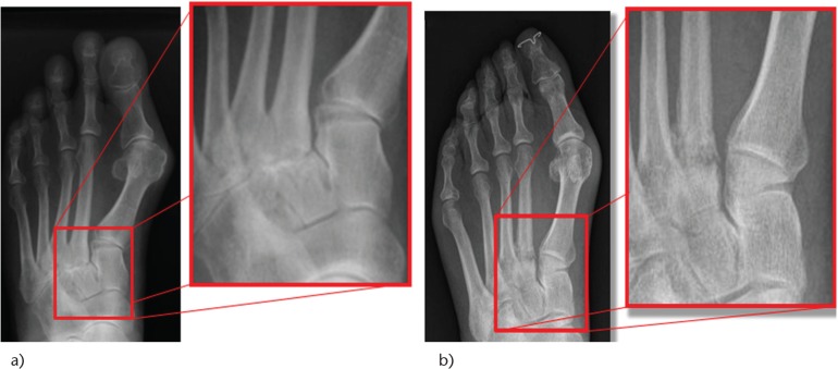
X-rayX-ray of the foot with magnified views: osteoarthritis of the second tarsometatarsal joint and fatigue fracture of the second metatarsal, indirect signs of first ray insufficiency that may be associated with claw toes.
Source : Crevoisier et al., EFORT Open Reviews, 2016, figure 1 · CC BY-NC
- Kelikian push-up test 🔑: the fundamental test for telling flexible from rigid. The examiner pushes the corresponding metatarsal head upwards and observes the passive reducibility of the toe. Reducible = flexible (conservative treatment indicated); non-reducible = rigid (surgery likely). It is the basis of the decision-making.⁷
- Drawer test / MTP Lachman (Klein 2014): assesses MTP instability from plantar plate insufficiency. The examiner stabilises the metatarsal head with one hand and moves the base of the proximal phalanx in the sagittal plane. A vertical translation > 50 % of the height of the phalanx defines a positive test. Klein 2014 showed that this physical test has a sensitivity above 95 % for a surgically confirmed plantar plate rupture.¹
- Assessing the strength of the intrinsics : the « paper grip » test (the patient tries to hold a sheet of paper under their toes while the examiner pulls it; failure marks weakness of the flexors), the toe-grip dynamometer test (validated in older people by Mickle 2009).¹⁴
| Modality | Main indication | Diagnostic performance | Level of evidence |
|---|---|---|---|
| Standard weight-bearing radiograph | An initial structural work-up: angles, metatarsal length, alignment, osteoarthritis | Excellent for bone analysis, useless for soft tissue | High (standard) |
| Dynamic ultrasound | Plantar plate, tendon sheaths, cysts, Morton's neuroma | Se/Sp ≈ 95-100 % in expert hands (Carlson 2013; Albright 2022 MA)¹⁵,¹⁶ | Moderate (operator-dependent) |
| MRI (1.5 or 3 T) | The reference for the plantar plate, marrow oedema, complex ligament injuries | Se 95 % / Sp 100 % against the surgical gold standard (Sung 2012, n=64)¹⁵ | High |
| Stress MRI (hyperextension) | Better detection of plantar plate injuries in case of doubt | Raised sensitivity in the stress position (Giuliani 2024)¹⁷ | Moderate, emerging |
🎯 Comparative performance of imaging for diagnosing a plantar plate rupture
The Albright 2022 meta-analysis (Eur J Radiol) and the Carlson 2013 and Sung 2012 data
Sources: Albright RH, Hassan M, Randich J, et al. Eur J Radiol. 2022;151:110315; Sung W, et al. J Foot Ankle Surg. 2012;51(5):570-574; Carlson RM, et al. J Foot Ankle Surg. 2013;52(6):786-791. MRI remains the gold standard; ultrasound performs comparably in expert hands but stays operator-dependent.
- Morton's neuroma : shooting or burning interdigital pain (the 3rd space), a positive Mulder sign. Confirmed by ultrasound or MRI.
- Isolated plantar plate rupture with no visible deformity: plantar MTP pain with a positive drawer test, sometimes with no gross deformity at first (to be picked up early).
- Metatarsal stress fracture : progressive pain on exertion, localised oedema, a radiograph often negative in the early phase, MRI diagnostic.
- Inflammatory arthritis : rheumatoid arthritis (MTP synovitis, radiographic erosions), gout (an acute inflammatory monoarthritis).
- Sesamoid conditions of the 1st ray.
- A complicated diabetic foot : early Charcot neuroarthropathy, a neuropathic plantar ulcer (see chapter 4).
Red flags in a consultation for a toe deformity
- A rapidly progressive deformity over a few weeks or months → suspect a neurological (CMT, cauda equina syndrome), inflammatory or neoplastic cause.
- A bilateral deformity in a young adult or an adolescent → a strong suspicion of Charcot-Marie-Tooth (refer to a neurologist).
- A plantar ulcer under a prominent metatarsal head in a person with diabetes → a neuropathic ulcer, IWGDF risk 3 → urgent referral to a diabetologist or a specialist podiatrist.
- Pain out of proportion, hot erythema, fever → suspect septic or acute crystal arthritis.
- Persistent asymmetrical oedema, a deformity with a warm foot and no trauma in a person with diabetes → suspect early Charcot neuroarthropathy.
- Associated hypoaesthesia, paraesthesia or muscle weakness → a neurological work-up.
Should toe deformities be classified, and for what benefit?
Classification is essential because it standardises communication between clinicians, guides the treatment decision and allows a prognosis. The classifications useful in physiotherapy practice:- By reducibility (a simple clinical classification, the most used): flexible stage (passively reducible) → semi-rigid → rigid.⁷
- By the joint involved : hammer (PIP), claw (MTP+PIP+DIP), mallet (DIP), crossover (medial deviation of the 2nd toe).
- The Nery 2012 classification of MTP instability from plantar plate insufficiency (stages 0 to 4 by deviation and dislocation), useful before surgery.⁹
- The Coughlin 1987 classification of the crossover toe , a historical reference still used by surgeons.¹³
Criticism and controversy
Three limits must temper the confidence placed in the assessment tools. First, the inter-examiner reliability of the clinical tests (Kelikian, drawer) varies with experience; the published studies come mainly from specialist surgical teams and the performance in everyday physiotherapy practice is probably below the 95 % reported.¹ Secondly, the classifications focus on the sagittal plane whereas the deformities are multi-planar (rotation, transverse deviation); this transverse component, whose importance is commonly accepted in practice but without data dedicated to toe deformities, stays underestimated by the usual classifications. Thirdly,dynamic ultrasound performs excellently in theory but stays highly operator-dependent : in non-expert hands its reliability drops sharply, which justifies keeping MRI as the reference when the clinical picture is in doubt.¹⁶Key points
- The history systematically explores the pain, the footwear, the course and the systemic history (diabetes, RA, neuropathies).
- The Kelikian push-up test tells flexible from rigid and guides directly the choice between conservative treatment and surgery.
- The drawer test (Klein 2014) has a sensitivity > 95 % for plantar plate rupture in expert hands; it must be part of the routine examination.
- The reference imaging is MRI (Se 95 % / Sp 100 %, the Albright 2022 meta-analysis), with dynamic ultrasound comparable in expert hands but operator-dependent.
- Major red flags: a rapidly progressive deformity, a bilateral one in a child or adolescent, ulceration in a person with diabetes, a hot arthritis.
Bibliography
- Nery C, Coughlin MJ, Baumfeld D, Raduan FC, Mann TS, Catena F. Classification of metatarsophalangeal joint plantar plate injuries: history and physical examination variables. J Surg Orthop Adv. 2014;23(4):214-223. PMID 25785472.
- Hagedorn TJ, Dufour AB, Riskowski JL, et al. Foot disorders, foot posture, and foot function: the Framingham foot study. PLoS One. 2013;8(9):e74364. PMID 24040231.
- Doty JF, Coughlin MJ. Metatarsophalangeal joint instability of the lesser toes and plantar plate deficiency. J Am Acad Orthop Surg. 2014;22(4):235-245. PMID 24668353.
- Nix S, Smith M, Vicenzino B. Prevalence of hallux valgus in the general population: a systematic review and meta-analysis. J Foot Ankle Res. 2010;3:21. PMID 20868524.
- Coughlin MJ. Crossover second toe deformity. Foot Ankle. 1987;8(1):29-39. PMID 3623360.
- Mickle KJ, Munro BJ, Lord SR, Menz HB, Steele JR. Toe weakness and deformity increase the risk of falls in older people. Clin Biomech. 2009;24(10):787-791. PMID 19751956.
- Malhotra K, Davda K, Singh D. The pathology and management of lesser toe deformities. EFORT Open Rev. 2017;1(11):409-419. PMID 28461920.
- Bus SA, Sacco ICN, Monteiro-Soares M, et al. Guidelines on the prevention of foot ulcers in persons with diabetes (IWGDF 2023 update). Diabetes Metab Res Rev. 2024;40(3):e3651. PMID 37302121.
- Morley D, Jenkinson C, Doll H, et al. The Manchester-Oxford Foot Questionnaire (MOXFQ): development and validation of a summary index score. Bone Joint Res. 2013;2(4):66-69. PMID 23673374.
- Pinsker E, Daniels TR. AOFAS position statement regarding the future of the AOFAS Clinical Rating Systems. Foot Ankle Int. 2011;32(9):841-842. PMID 22097157.
- Otter SJ, Lucas K, Springett K, et al. Foot pain in rheumatoid arthritis prevalence, risk factors and management: an epidemiological study. Clin Rheumatol. 2010;29(3):255-271. PMID 19997766.
- Nery C, Coughlin MJ, Baumfeld D, Mann TS. Lesser metatarsophalangeal joint instability: prospective evaluation and repair of plantar plate and capsular insufficiency. Foot Ankle Int. 2012;33(4):301-311. PMID 22735202.
- Gregg J, Marks P, Silberstein M, Schneider T, Kerr J. Sonographic and MRI evaluation of the plantar plate: a prospective study. Eur Radiol. 2006;16(12):2661-2669. PMID 16819605.
- Mickle KJ, Nester CJ. Morphology of the Toe Flexor Muscles in Older Adults With Toe Deformities. Arthritis Care Res (Hoboken). 2018;70(6):902-907. PMID 28834406.
- Sung W, Weil L Jr, Weil LS Sr, Rolfes RJ. Diagnosis of plantar plate injury by magnetic resonance imaging with reference to intraoperative findings. J Foot Ankle Surg. 2012;51(5):570-574. PMID 22727342.
- Carlson RM, Dux K, Stuck RM. Ultrasound imaging for diagnosis of plantar plate ruptures of the lesser metatarsophalangeal joints: a retrospective case series. J Foot Ankle Surg. 2013;52(6):786-788. PMID 23870658.
- Giuliani L, Ottonello C, Giuliani A, et al. MRI in the evaluation of plantar plate disease: diagnostic value of the "stress test". J Orthop Traumatol. 2024;25(1):65. PMID 39718683.
- Albright RH, Brooks BM, Chingre M, Klein EE, Weil LS Jr, Fleischer AE. Diagnostic accuracy of magnetic resonance imaging (MRI) versus dynamic ultrasound for plantar plate injuries: A systematic review and meta-analysis. Eur J Radiol. 2022;152:110315. PMID 35533558.
Which treatment strategies are the most effective?
Where do you start? What is the hierarchy of conservative interventions?
The first-line hierarchy of intervention, supported by the current recommendations and the few systematic reviews available, covers four complementary pillars.⁷,⁸ Pillar 1, a change of footwear (the priority intervention). 👟 Any flexible deformity calls for a footwear audit. The validated criteria are a wide and deep toe box, with a heel ≤ 2-3 cm, plus a flexible upper and an adjustable lace-up fit. The landmark RCT Torkki 2001 (JAMA, n = 209) compared, in hallux valgus, surgery, an orthosis and watchful waiting: at 1 year surgery did better than the other two arms (83 %, 46 % and 24 % of patients considering themselves improved), the orthosis bringing only transient relief at 6 months. That trial therefore does not validate conservative care as corrective treatment: it justifies offering it for the symptom, without expecting correction, and it concerns hallux valgus, not the lesser toes.⁵ The advice must be permanent : going back to the wrong shoes = a predictable recurrence. Pillar 2, foot orthoses. Orthoses play a triple role: redistributing the pressure under the metatarsal heads (a metatarsal bar, support of the transverse arch), limiting the deformity (silicone separators, thermoformed orthoplasty), improving walking comfort. High-quality evidence in the specific context of lesser toe deformities stays limited (most studies concern HV or metatarsalgia in general), but Chadchavalpanichaya 2018 (RCT n = 60) showed that a moulded silicone toe separator reduced the HVA by 3° and VAS pain by 2.2 points at 6 months (HV with associated toe deformities).¹⁰ Pillar 3, specific exercises for the intrinsic foot muscles. Set out in the next section. Pillar 4, taping and orthoplasty. Corrective taping (silicone, elastic) can relieve corns by unloading them, hold a flexible toe in position temporarily and serve as a « therapeutic bridge » for the duration of the programme. The evidence of long-term efficacy is weak but the safety is excellent.What place do intrinsic exercises hold and is there a superior approach?
Rehabilitating the intrinsic foot muscles is the cornerstone of active management and the most direct target of the pathophysiological mechanism. Three protocols have accumulated evidence of acceptable quality:- Short Foot Exercise (SFE) 💪: the reference exercise popularised by Janda and validated by Mulligan & Cook 2013 (a pilot study, n = 21). The patient tries to shorten the length of the foot actively by drawing the head of the 1st metatarsal towards the heel, without curling the toes. It preferentially activates flexor digitorum brevis (FDB) and abductor hallucis. Mulligan 2013 documented a 14 % reduction in navicular drop (a proxy for the medial arch) at 4 weeks.²⁰
- Toe Yoga / Toe Curl / Toe Grip : exercises that activate the intrinsics in isolation through several patterns (gripping a towel with the toes, curling marbles, spreading the toes). Gooding 2016 showed by T2 MRI that these exercises specifically activate the intrinsics (a raised T2 signal after effort), validating their muscular target.²¹
- Progressive strengthening programmes over 8-12 weeks : Mickle 2016 (RCT n = 85 older women) showed that a 12-week programme of progressive toe flexor strengthening significantly increased great toe flexor strength and improved functional performance.⁴¹ Taddei 2020 (RCT in runners, n = 118) showed that an 8-week foot strengthening programme significantly improved intrinsic strength and the morphology of the medial arch.¹⁹
📈 Effects of an intrinsic exercise programme over 8-12 weeks
A synthesis of the Mulligan 2013, Mickle 2016 and Taddei 2020 trials, objective and functional parameters
* Statistically significant (p < 0.05). Sources: Mulligan EP, Cook PG. Man Ther. 2013;18(5):425-430; Mickle KJ, et al. Clin Biomech. 2016;40:14-19; Taddei UT, et al. Phys Ther Sport. 2020;42:107-115. An important caveat: these studies mainly concern asymptomatic people, runners or older adults, not specifically patients with established rigid deformities.
Manual therapy, taping, technologies: how effective are they really?
Manual therapy (MTP and PIP mobilisation) : indicated above all for stiff flexible or semi-rigid deformities. Passive mobilisation into MTP plantarflexion and PIP extension can reduce stiffness and pain; but the evidence of isolated efficacy in the specific context of toe deformities is scarce (most trials concern HV or the flat foot).¹² Taping : useful in the short term for unloading corns and holding flexible toes temporarily; a measurable biomechanical effect but a negligible long-term effect on the deformity.¹⁴ Technologies (extracorporeal shockwave, laser, ultrasound): very limited or absent evidence in the specific context of toe deformities. These modalities may have a place in the associated conditions (plantar fasciitis, tendinopathy) but must not replace the first-line interventions.¹⁵When should surgery be considered and with what results?
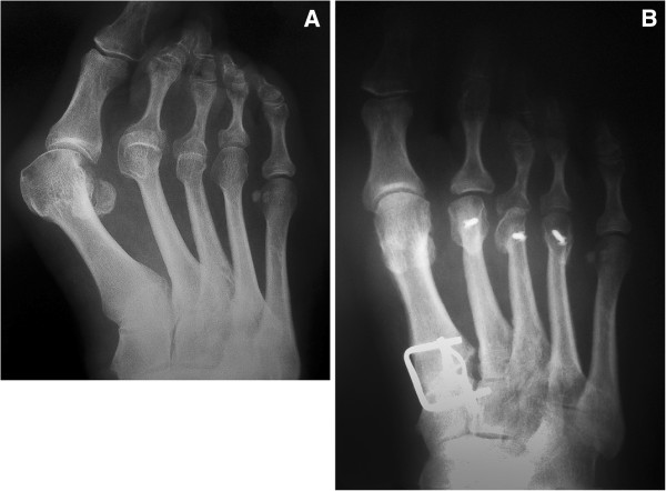
X-rayAnteroposterior X-ray of the right foot before (A) and after (B) surgery: subluxation of the first and dislocation of the second and third metatarsophalangeal joints, corrected by osteotomies of the lateral metatarsals and arthrodesis of the first ray.
Source : Popelka et al., BMC Musculoskeletal Disorders, 2012, figure 4 · CC BY
- well-conducted conservative treatment has failed over 3-6 months;
- the deformity is rigid (a negative Kelikian);
- there is MTP subluxation or dislocation with major instability;
- there is a major functional or footwear impact despite adaptation;
- there are chronic skin complications (a recurrent painful corn, ulceration).
| Procedure | Main indication | Published results | Risk / complication |
|---|---|---|---|
| PIP arthrodesis (Coughlin 2000) | A rigid hammer toe | The historical reference, high patient satisfaction | Non-union ≈ 5-15 % depending on the implant |
| PIP resection arthroplasty | An alternative for a rigid hammer toe | Less durable results than arthrodesis | A floating toe is possible |
| Weil osteotomy (Cancilleri 2003) | Metatarsalgia + deformity | « Good or excellent » ≈ 100 % in the short term (n=35) | Floating toe 28-43 % (Migues 2004)³¹ |
| Direct plantar plate repair (Flint 2017) | MTP instability from plate rupture | AOFAS 49 → 81; satisfaction 88 % (n=97 feet)³⁴ | Partial recurrence ≈ 12 % |
| FDL-FDB tendon transfer (Girdlestone-Taylor) | A flexible claw toe not amenable to conservative care | Variable; a non-destructive alternative | Adhesions, residual stiffness |
Criticism and controversies
Three areas of debate deserve to be spelled out: 1. The poverty of evidence specific to conservative care of the lesser toes. Most of the studies cited concern HV, metatarsalgia or the older foot; high-quality trials isolating flexible lesser toe deformities are scarce. The available systematic review is old and acknowledges a high risk of bias in the RCTs included. The conservative recommendations therefore rest mainly on pathophysiological extrapolation and on clinical consensus, not on robust level I evidence. 2. Custom versus prefabricated orthoses. The debate persists: while custom orthoses seem biomechanically superior, their extra cost (often 200-400 € against 30-80 €) is not systematically justified by the difference in clinical efficacy. For mild to moderate cases, a well-chosen prefabricated orthosis plus education can be enough. 3. The real capacity of intrinsic exercises to correct an established deformity. Intrinsic exercises undeniably improve strength, function and symptoms. Their capacity to correct anatomically a deformity that is already fixed, on the other hand, is very limited; their role is essentially to slow the progression, prevent worsening and improve functional tolerance.Key points
- The conservative hierarchy: suitable footwear as the absolute priority, then orthoses (a metatarsal bar), intrinsic exercises (Short Foot Exercise + Toe Yoga, 10 min a day × 8-12 weeks) and taping.
- No « winning » exercise approach: combine activation (SFE), strengthening (Toe Curl/Grip), barefoot balance and posterior chain stretching.
- Mickle 2016 and Taddei 2020 show a significant improvement in strength and function over 8-12 weeks, but few studies isolate symptomatic rigid deformities.
- Surgical indications: conservative failure at 3-6 months, a rigid stage, MTP subluxation, chronic skin complications.
- The main procedures: PIP arthrodesis (Coughlin 2000), Weil osteotomy (mind the floating toe at 28-43 %, Migues 2004), plantar plate repair (Flint 2017, AOFAS 49 → 81).
Bibliography
- Malhotra K, Davda K, Singh D. The pathology and management of lesser toe deformities. EFORT Open Rev. 2017;1(11):409-419. PMID 28461920.
- Frey-Ollivier S, Catena F, Hélix-Giordanino M, Piclet-Legré B. Treatment of Flexible Lesser Toe Deformities. Foot Ankle Clin. 2018;23(1):69-90. PMID 29362035.
- Doty JF, Coughlin MJ. Metatarsophalangeal joint instability of the lesser toes and plantar plate deficiency. J Am Acad Orthop Surg. 2014;22(4):235-245. PMID 24668353.
- Hurn SE, Vicenzino BT, Smith MD. Non-surgical treatment of hallux valgus: a current practice survey of Australian podiatrists. J Foot Ankle Res. 2016;9:16. PMID 27148407.
- Torkki M, Malmivaara A, Seitsalo S, Hoikka V, Laippala P, Paavolainen P. Surgery vs orthosis vs watchful waiting for hallux valgus: a randomized controlled trial. JAMA. 2001;285(19):2474-2480. PMID 11368700.
- Tehraninasr A, Saeedi H, Forogh B, Bahramizadeh M, Keyhani MR. Effects of insole with toe-separator and night splint on patients with painful hallux valgus: a comparative study. Prosthet Orthot Int. 2008;32(1):79-83. PMID 18330806.
- Tang SF, Chen CP, Pan JL, Chen JL, Leong CP, Chu NK. The effects of a new foot-toe orthosis in treating painful hallux valgus. Arch Phys Med Rehabil. 2002;83(12):1792-1795. PMID 12474189.
- Männikkö K, Sahlman J. The Effect of Metatarsal Padding on Pain and Functional Ability in Metatarsalgia. Scand J Surg. 2017;106(4):332-337. PMID 28737072.
- McKeon PO, Hertel J, Bramble D, Davis I. The foot core system: a new paradigm for understanding intrinsic foot muscle function. Br J Sports Med. 2015;49(5):290. PMID 24659509.
- Chadchavalpanichaya N, Prakotmongkol V, Polhan N, Rayothee P, Seng-Iad S. Effectiveness of the custom-mold room temperature vulcanizing silicone toe separator on hallux valgus: a prospective, randomized single-blinded controlled trial. Prosthet Orthot Int. 2018;42(2):163-170. PMID 28318407.
- Coughlin MJ. Operative repair of the fixed hammertoe deformity. Foot Ankle Int. 2000;21(2):94-104. PMID 10718500.
- Wei RXY, Ling SKK, Lui TH, Yung PSH. Ideal implant choice for proximal interphalangeal joint arthrodesis in hammer toe/claw toe deformity correction: a systematic review. J Orthop Surg (Hong Kong). 2020;28(1):2309499020911168. PMID 32223520.
- Cancilleri F, Marinozzi A, Martinelli N, et al. The Weil osteotomy in painful overloaded central forefoot with dislocation of the MTP joint. Foot Ankle Surg. 2003;9(4):215-218.
- Migues A, Slullitel G, Bilbao F, Carrasco M, Solari G. Floating-toe deformity as a complication of the Weil osteotomy. Foot Ankle Int. 2004;25(9):609-613. PMID 15563380.
- Flint WW, Macias DM, Jastifer JR, Doty JF, Hirose CB, Coughlin MJ. Plantar plate repair for lesser metatarsophalangeal joint instability. Foot Ankle Int. 2017;38(3):234-242. PMID 27852647.
- Mulligan EP, Cook PG. Effect of plantar intrinsic muscle training on medial longitudinal arch morphology and dynamic function. Man Ther. 2013;18(5):425-430. PMID 23632367.
- Gooding TM, Feger MA, Hart JM, Hertel J. Intrinsic foot muscle activation during specific exercises: a T2 time magnetic resonance imaging study. J Athl Train. 2016;51(8):644-650. PMID 27690528.
- Mickle KJ, Caputi P, Potter JM, Steele JR. Efficacy of a progressive resistance exercise program to increase toe flexor strength in older people. Clin Biomech (Bristol, Avon). 2016;40:14-19. PMID 27780109.
- Taddei UT, Matias AB, Duarte M, Sacco ICN. Foot Core Training to Prevent Running-Related Injuries: A Survival Analysis of a Single-Blind, Randomized Controlled Trial. Am J Sports Med. 2020;48(14):3610-3619. PMID 33156692.
Diabetic foot, rheumatoid arthritis, Charcot-Marie-Tooth: why are these subgroups vulnerable?
Diabetic foot: motor neuropathy, ulceration and IWGDF 2023 risk stratification
In the patient with diabetes, the peripheral motor neuropathy causes a progressive atrophy of the intrinsics (lumbricals, interossei), leading to the characteristic development of bilateral claw toes.³⁶,³⁷ That deformity has two major consequences:- Raised pressure under the metatarsal heads (made prominent by the MTP hyperextension) → a risk of a neuropathic plantar ulcer.
- Dorsal rubbing of the prominent PIP joints in the shoe → corns → secondary ulceration.
📋 Stratification of the risk of ulceration in the person with diabetes (IWGDF 2023)
Frequency of monitoring and intensity of management by risk category
The presence of a toe deformity in a person with diabetes and neuropathy moves the patient to risk ≥ 2, justifying quarterly monitoring, therapeutic footwear and offloading orthoses. Source: PMID 37302121.
- Every patient with diabetes over 50 or with more than 10 years of disease must undergo a screening foot examination at least annually: the 10 g Semmes-Weinstein monofilament test (neuropathy), the pedal pulses (PAD), skin inspection.
- The presence of bilateral claw toes in a person with diabetes justifies referral to a specialist podiatrist or a diabetic foot centre.
- Education about daily self-inspection of the foot (looking for corns, ulceration, fungal infection, fissures) is non-negotiable.
- Intrinsic exercises stay indicated to preserve residual function and balance, provided protective sensation is preserved and there is no active ulceration.
- Never advise a patient with diabetes to file down or cut away a corn or callus themselves (a major risk of infection).
Red flags specific to the diabetic foot
- A plantar ulcer under a metatarsal head, even painless → a relative emergency (IWGDF risk 3, immediate referral).
- Persistent asymmetrical oedema, a warm foot, a sudden deformity with no trauma → suspect early Charcot neuroarthropathy (a true emergency: immediate offloading, imaging, referral to a diabetologist).
- A corn with an erythematous halo, purulent discharge, fever → suspect a deep infection, sometimes with underlying osteitis.
- Critical ischaemia : a cold foot, rest pain, an indolent wound → severe PAD, an urgent vascular opinion.
Rheumatoid arthritis and Charcot-Marie-Tooth: the sentinel signs to recognise
The rheumatoid forefoot 🦴. Rheumatoid arthritis (RA) affects the foot in the great majority of patients at an advanced stage: the epidemiological study by Otter 2010 (n = 585 RA patients) found a forefoot pain prevalence of 63.9 %, and ankle pain of 42.7 %.³⁹ The chronic MTP synovitis progressively causes a dorsal subluxation of the phalanges, a lateral deviation of the toes, with a widening of the forefoot, and the classic picture of a severe rheumatoid forefoot. Multidisciplinary management rests on:- Rheumatological disease-modifying treatment (DMARDs, biologics, the rheumatologist's remit).
- Therapeutic footwear that is wide and deep in the toe box.
- Offloading foot orthoses with a metatarsal bar.
- Reconstructive forefoot surgery at an advanced stage, such as combined metatarsal osteotomies (Niki 2015, AOFAS RA-foot 52 → 90 at 64-108 months).³⁸
- Maintain ankle and subtalar mobility (daily triceps surae stretching).
- Strengthen the residual evertor and invertor muscles.
- Adapt the footwear and consider an ankle-foot orthosis (AFO) if there is foot drop: Phillips 2012 showed that a polypropylene AFO increases walking speed in patients with CMT.⁴³
- Refer to a specialist orthopaedic surgical consultation for severe deformities (tendon transfers, calcaneal osteotomies).
🎯 Comparison of the three vulnerable subgroups
Mechanism, prevalence of the deformity, priority specific intervention
Sources: Bus 2024 (IWGDF, PMID 37302121); Otter 2010 (PMID 19997766); Pareyson 2017 (PMID 28678038). Any toe deformity that falls outside the « ordinary HV + footwear » profile must trigger a search for a systemic cause.
Key points
- Three major subgroups converge on the toe deformity: diabetes (claw toes from motor neuropathy), RA (MTP subluxation), CMT (pes cavus + bilateral claw toes).
- In the person with diabetes, a toe deformity means ≥ IWGDF category 2 : quarterly monitoring, therapeutic footwear, offloading orthoses.
- A major red flag : a plantar ulcer under a prominent metatarsal head in a person with diabetes → immediate referral (a neuropathic ulcer, sometimes with osteitis).
- RA: forefoot pain affects 63.9 % of patients (Otter 2010), so screen systematically in the rheumatology consultation.
- CMT: bilateral claw toes in a young person call for a neurological work-up (EMG, genetics). An AFO if there is foot drop (Phillips 2012).
Bibliography
- Bus SA, Sacco ICN, Monteiro-Soares M, et al. Guidelines on the prevention of foot ulcers in persons with diabetes (IWGDF 2023 update). Diabetes Metab Res Rev. 2024;40(3):e3651. PMID 37302121.
- Schaper NC, van Netten JJ, Apelqvist J, et al. Practical guidelines on the prevention and management of diabetes-related foot disease (IWGDF 2023 update). Diabetes Metab Res Rev. 2024;40(3):e3657. PMID 37243927.
- Niki H, Hirano T, Okada H, Beppu M. Long-term outcome of joint-preserving surgery by combination metatarsal osteotomies for shortening for forefoot deformity in patients with rheumatoid arthritis. Mod Rheumatol. 2015;25(5):683-688. PMID 25608049.
- Otter SJ, Lucas K, Springett K, et al. Foot pain in rheumatoid arthritis prevalence, risk factors and management: an epidemiological study. Clin Rheumatol. 2010;29(3):255-271. PMID 19997766.
- Wicart P. Cavus foot, from neonates to adolescents. Orthop Traumatol Surg Res. 2012;98(7):813-828. PMID 23142109.
- Mickle KJ, Munro BJ, Lord SR, Menz HB, Steele JR. Toe weakness and deformity increase the risk of falls in older people. Clin Biomech. 2009;24(10):787-791. PMID 19751956.
- Mickle KJ, Nester CJ. Morphology of the Toe Flexor Muscles in Older Adults With Toe Deformities. Arthritis Care Res (Hoboken). 2018;70(6):902-907. PMID 28834406.
- Phillips MF, Robertson Z, Killen B, White B. A pilot study of a crossover trial with randomized use of ankle-foot orthoses for people with Charcot-Marie-Tooth disease. Clin Rehabil. 2012;26(8):696-704. PMID 22089961.
- Pareyson D, Saveri P, Pisciotta C. New developments in Charcot-Marie-Tooth neuropathy and related diseases. Curr Opin Neurol. 2017;30(5):471-480. PMID 28678038.
- Menz HB, Auhl M, Spink MJ. Foot problems as a risk factor for falls in community-dwelling older people: a systematic review and meta-analysis. Maturitas. 2018;118:7-14. PMID 30415759.
How do you secure a lasting recovery and prevent recurrence?
How do you make the patient an active player in their own recovery through self-management?
Lasting self-management rests on three pillars, of which patient education is the cornerstone. Pillar 1, patient education. 🧠 The patient must understand:- The biomechanics of their deformity: why their intrinsics are weak, how footwear makes it worse, why retraction of the triceps surae loads the forefoot.
- The predictable natural history without intervention (flexible → rigid).
- The permanent character of good habits: « the right shoe » is not a temporary treatment but a lasting choice.
- The warning signs that call for a further consultation: a recurrent painful corn, ulceration, a rapidly progressive deformity, night pain, a change in the skin.
- Identify a good shoe when buying (the shoehorn test: being able to move the toes freely, a deep enough box, a heel ≤ 2-3 cm for everyday wear).
- Renew their orthoses with their podiatrist (every 1-2 years depending on wear).
- Limit the at-risk shoes (high heels, narrow court shoes, tight boots) to short occasions.
- Watch their foot: a daily visual inspection (morning or evening), the monofilament test in the person with diabetes.
- Short (10-15 minutes a day); beyond that, adherence falls sharply.
- Spread across several daily sequences (« waiting exercises » in front of the television, under the desk, after the shower).
- Progressive (volume, load, complexity) with a monthly review over the first 3 months.
- Visually reinforced : an illustrated sheet, a smartphone app, monthly before-and-after photographs.
| Component | Frequency | Duration | Level of evidence |
|---|---|---|---|
| Barefoot Short Foot Exercise | 2 × 10 repetitions a day | Hold 5 sec | Moderate (Mulligan 2013) |
| Toe Curl with a towel / Toe Yoga | 1 set a day | 2-3 minutes | Moderate (Gooding 2016) |
| Triceps surae stretch (against a wall) | 2 × 30 sec, twice a day | 2 minutes | Moderate (Rao 2019) |
| Flexor digitorum longus stretch | 2 × 30 sec, once a day | 1-2 minutes | Low (consensus) |
| Barefoot balance on a stable surface | 1 session a day | 2-3 minutes | Moderate (Mickle 2016) |
| Daily skin inspection | Once a day | 1 minute | High in the person with diabetes (IWGDF 2023) |
When and how do you plan a safe return to activity and sport?
The return to physical activity after a painful period (or after surgery) must follow a logic of progression controlled by the symptoms, not a rigid calendar. Phase 1, the prerequisites before resuming :- No pain during activities of daily living (walking, prolonged standing, stairs).
- Toe flexor strength symmetrical with the healthy side (the paper-grip test or a toe dynamometer if available).
- Footwear suited to the intended activity (proper running shoes and so on).
- Complete healing and no inflammation in those operated on.
- Pain > 3/10 (visual analogue scale) persisting > 24 h after the effort → step back to the previous phase.
- The return of a corn or a skin lesion.
- Persistent oedema after effort.
Criticism and controversy
The literature on secondary prevention specific to lesser toe deformities is very thin: the current recommendations rest on extrapolation from HV, from metatarsalgia and from podiatric prevention in the person with diabetes. Trials explicitly comparing keeping up against abandoning the programme at 6-12 months are missing. The analogy with other postural conditions (chronic low back pain) nonetheless suggests that a fall in adherence at 3-6 months is the rule, not the exception, which justifies telephone reminders or scheduled follow-up consultations. Moreover, the real capacity of a patient to correct their deformity through exercise has never been shown at the anatomical level: the exercises improve strength, the function and the symptoms, but they do not « straighten » a toe that is already fixed. That nuance must be communicated honestly to the patient to avoid unrealistic expectations and a damaging disappointment.Key points
- Self-management rests on three pillars: education about the biomechanics, environmental adaptation (footwear), a short home exercise programme (10-15 min a day).
- Long-term success demands a patient able to choose their shoes and to keep up their exercises beyond the first 3 months, the critical phase of falling adherence.
- The return to activity follows 4 progressive phases (prerequisites → low impact → graded impact → specific), with symptom-based criteria for stopping.
- Heightened monitoring in the vulnerable subgroups: daily skin inspection in the person with diabetes (IWGDF 2023), rheumatology follow-up in RA, neurology follow-up in CMT.
- Communicate honestly: the exercises improve function, not the anatomy ; preventing worsening is the realistic goal.
Bibliography
- Mulligan EP, Cook PG. Effect of plantar intrinsic muscle training on medial longitudinal arch morphology and dynamic function. Man Ther. 2013;18(5):425-430. PMID 23632367.
- Mickle KJ, Caputi P, Potter JM, Steele JR. Efficacy of a progressive resistance exercise program to increase toe flexor strength in older people. Clin Biomech. 2016;40:14-19. PMID 27780109.
- Taddei UT, Matias AB, Duarte M, Sacco ICN. Foot Core Training to Prevent Running-Related Injuries: A Survival Analysis of a Single-Blind, Randomized Controlled Trial. Am J Sports Med. 2020;48(14):3610-3619. PMID 33156692.
- Gooding TM, Feger MA, Hart JM, Hertel J. Intrinsic foot muscle activation during specific exercises: a T2 time magnetic resonance imaging study. J Athl Train. 2016;51(8):644-650. PMID 27690528.
- Bus SA, Sacco ICN, Monteiro-Soares M, et al. Guidelines on the prevention of foot ulcers in persons with diabetes (IWGDF 2023 update). Diabetes Metab Res Rev. 2024;40(3):e3651. PMID 37302121.
- Menz HB, Auhl M, Spink MJ. Foot problems as a risk factor for falls in community-dwelling older people: a systematic review and meta-analysis. Maturitas. 2018;118:7-14. PMID 30415759.
- Hurn SE, Vicenzino BT, Smith MD. Non-surgical treatment of hallux valgus: a current practice survey of Australian podiatrists. J Foot Ankle Res. 2016;9:16. PMID 27148407.
- Torkki M, Malmivaara A, Seitsalo S, Hoikka V, Laippala P, Paavolainen P. Surgery vs orthosis vs watchful waiting for hallux valgus: a randomized controlled trial. JAMA. 2001;285(19):2474-2480. PMID 11368700.
- Morley D, Jenkinson C, Doll H, et al. The Manchester-Oxford Foot Questionnaire (MOXFQ): development and validation of a summary index score. Bone Joint Res. 2013;2(4):66-69. PMID 23673374.
What do the published clinical cases teach us about toe deformities?
A classic case: a flexible deformity, from conservative care to plantar plate repair
A typical presentation . A woman of 50-65 consults for progressive pain in the right forefoot present for 12-18 months, made worse by prolonged walking and by narrow shoes. Examination finds a moderate hallux valgus and a 2nd toe beginning to override (a Coughlin grade I-II crossover toe), with a painful drawer test (MTP Lachman) and a moderately increased translation (≈ 30 % of the height of the phalanx). The initial conservative trajectory. Management follows the classic hierarchy: a change of footwear (moving to a shoe with a deep toe box), foot orthoses with a metatarsal bar, corrective taping of the toe. A daily intrinsic exercise programme (Short Foot Exercise + Toe Yoga + barefoot balance) is started, completed by posterior chain stretching. This approach improves the symptoms but does not correct the transverse deviation, which progresses. Moving on to surgery. Faced with persistent MTP instability despite 6 months of well-conducted conservative care, a plantar plate repair is proposed. The reference prospective series (Flint 2017, n = 97 feet) reports the following mean results after direct plantar plate repair with or without an associated Weil osteotomy:- AOFAS forefoot score: 49 → 81 at 12 months (a clinically important difference).
- Patient satisfaction: 88 %.
- Floating toe: 27 % of the operated cases (a surgical risk to be disclosed).
📊 A typical case: the AOFAS forefoot trajectory before and after plantar plate repair
The Flint 2017 prospective cohort, n = 97 feet, minimum 12-month follow-up
Source: Flint WW, Macias DM, Jastifer JR, Doty JF, Hirose CB, Coughlin MJ. Foot Ankle Int. 2017;38(3):234-242. PMID 27852647. A caveat : the AOFAS forefoot score is criticised for its limited validity (Pinsker 2011); it remains, however, the instrument most used in the historical literature.
The diagnostic challenge: when the deformity is the presenting sign of another condition
Case 1, an isolated plantar plate rupture mimicking an early flexible deformity. A 45-year-old woman runner consults for plantar MTP2 pain of gradual onset after increasing her training volume. On inspection the 2nd toe looks slightly deviated laterally but stays flexible. The drawer test is frankly positive (translation > 50 %), confirming MTP instability. Dynamic ultrasound finds a distal rupture of the plantar plate, confirmed by MRI. The classic error would be to treat this picture as a simple « early crossover toe » to be managed conservatively; in reality, without an intervention on the plate rupture (prolonged offloading taping and then a surgical opinion), the deformity will progress inexorably to a transverse and then a vertical deviation (Doty/Coughlin 2014, Nery 2012).⁸,⁹ Case 2, a chronic metatarsalgia masking a plantar plate rupture. A 55-year-old patient with a moderate hallux valgus has a chronic metatarsalgia attributed for 18 months to « mechanical overload ». The work-up was incomplete (no drawer test, no dedicated imaging). The MRI eventually performed shows a grade 3 rupture of the MTP2 plantar plate with early subluxation. The Albright 2022 meta-analysis stresses the value of requesting imaging dedicated to the plantar plate (dynamic ultrasound or MRI) in any metatarsalgia resistant to well-conducted conservative treatment.¹⁷ Case 3, a Morton's neuroma mimicking a claw toe. Rarer but a classic trap: chronic compression of the interdigital nerve by a neuroma can cause a protective reflex contracture of the 3rd or 4th toe. The shooting pain, the positive Mulder sign and ultrasound confirmation give the diagnosis; treating the neuroma (injection or surgery) generally resolves the pseudo-deformity.Complex cases: hereditary neuropathy and the rheumatoid forefoot
Case 4, a Charcot-Marie-Tooth revealed by bilateral claw toes. A young adult of 28 consults for bilateral forefoot pain and difficulty running. Inspection reveals symmetrical bilateral claw toes with a moderate pes cavus. The absence of any history of at-risk footwear, the perfect bilaterality and the progression over several years draw attention. The neurological work-up (EMG, genetic study) confirms Charcot-Marie-Tooth type 1A (the most frequent form, a PMP22 mutation).⁴⁴ Management becomes multidisciplinary: a referring neurologist, a podiatrist (cavus orthoses), an orthopaedic surgeon for a later opinion on tendon transfers or a calcaneal osteotomy if the deformity is severe, and a physiotherapist for maintaining mobility, residual strengthening and education about an AFO if there is foot drop (Phillips 2012).⁴³ Case 5, an advanced rheumatoid forefoot. A woman of 65, with rheumatoid arthritis diagnosed 20 years ago, consults for disabling bilateral forefoot pain and major difficulty with footwear. Examination finds the classic picture: a severe bilateral HV, dorsal subluxation of MTP2-5, a fanned lateral deviation, a widened forefoot, painful submetatarsal callosities. Otter 2010 (n=585) recalls that this forefoot involvement affects 63.9 % of RA patients.³⁹ Management combines:- Optimising the rheumatological disease-modifying treatment (DMARDs, biologics).
- Custom foot orthoses with a cushioning insole and a metatarsal bar.
- Therapeutic footwear with a high toe box.
- An orthopaedic surgical opinion: the Niki 2015 series (joint-preserving combination metatarsal osteotomies) showed an AOFAS RA-foot score of 52 → 90 at 64-108 months, validating this reconstructive approach.³⁸
🔄 A decision diagram: a toe deformity in an adult patient
From screening for a systemic cause to the appropriate referral
A summary decision diagram. Any rapidly progressive deformity, any bilateral one in a young person, or any associated with a systemic background must trigger an aetiological work-up before conservative treatment is started.
Criticism and controversies
The clinical case literature on toe deformities is strongly biased. Three major limits: 1. Publication bias towards surgical successes. The published case reports and series report almost exclusively favourable trajectories; failures, serious complications and recurrences are under-reported, giving too optimistic a view of the results to expect. 2. The poverty of documented conservative trajectories. Physiotherapy case reports on flexible toe deformities are scarce; the literature on intrinsic exercise programmes concerns mainly asymptomatic cohorts (runners, older people) rather than symptomatic patients with established deformities. 3. Heterogeneity in the definition of success. Across studies, « success » may mean a symptomatic reduction, a radiographic angular correction, an improved AOFAS score or patient satisfaction; these criteria are not equivalent, and aggregating them makes comparisons between studies fragile.Key points
- The classic case : a conservative then surgical trajectory (plantar plate repair, Flint 2017: AOFAS 49 → 81; satisfaction 88 %; floating toe 27 %).
- The diagnostic challenge : an isolated plantar plate rupture can mimic an early flexible deformity; the drawer test + dedicated imaging avoid the error.
- Complex cases : bilateral claw toes in a young person → suspect CMT (EMG, genetic work-up); an advanced rheumatoid forefoot → orthoses + reconstructive surgery (Niki 2015: AOFAS RA-foot 52 → 90).
- Any atypical deformity calls for a decision pathway: bilaterality + progression + a young patient → a neurological work-up; a systemic history → multidisciplinary management.
- A critical caveat : the literature is biased towards surgical successes, under-represents conservative care and uses heterogeneous definitions of « success ».
Bibliography
- Flint WW, Macias DM, Jastifer JR, Doty JF, Hirose CB, Coughlin MJ. Plantar plate repair for lesser metatarsophalangeal joint instability. Foot Ankle Int. 2017;38(3):234-242. PMID 27852647.
- Doty JF, Coughlin MJ. Metatarsophalangeal joint instability of the lesser toes and plantar plate deficiency. J Am Acad Orthop Surg. 2014;22(4):235-245. PMID 24668353.
- Nery C, Coughlin MJ, Baumfeld D, Mann TS. Lesser metatarsophalangeal joint instability: prospective evaluation and repair of plantar plate and capsular insufficiency. Foot Ankle Int. 2012;33(4):301-311. PMID 22735202.
- Nery C, Coughlin MJ, Baumfeld D, Raduan FC, Mann TS, Catena F. Classification of metatarsophalangeal joint plantar plate injuries: history and physical examination variables. J Surg Orthop Adv. 2014;23(4):214-223. PMID 25785472.
- Albright RH, Brooks BM, Chingre M, Klein EE, Weil LS Jr, Fleischer AE. Diagnostic accuracy of magnetic resonance imaging (MRI) versus dynamic ultrasound for plantar plate injuries: A systematic review and meta-analysis. Eur J Radiol. 2022;152:110315. PMID 35533558.
- Niki H, Hirano T, Okada H, Beppu M. Long-term outcome of joint-preserving surgery by combination metatarsal osteotomies for shortening for forefoot deformity in patients with rheumatoid arthritis. Mod Rheumatol. 2015;25(5):683-688. PMID 25608049.
- Otter SJ, Lucas K, Springett K, et al. Foot pain in rheumatoid arthritis prevalence, risk factors and management: an epidemiological study. Clin Rheumatol. 2010;29(3):255-271. PMID 19997766.
- Pareyson D, Saveri P, Pisciotta C. New developments in Charcot-Marie-Tooth neuropathy and related diseases. Curr Opin Neurol. 2017;30(5):471-480. PMID 28678038.
- Phillips MF, Robertson Z, Killen B, White B. A pilot study of a crossover trial with randomized use of ankle-foot orthoses for people with Charcot-Marie-Tooth disease. Clin Rehabil. 2012;26(8):696-704. PMID 22089961.
- Pinsker E, Daniels TR. AOFAS position statement regarding the future of the AOFAS Clinical Rating Systems. Foot Ankle Int. 2011;32(9):841-842. PMID 22097157.
- Migues A, Slullitel G, Bilbao F, Carrasco M, Solari G. Floating-toe deformity as a complication of the Weil osteotomy. Foot Ankle Int. 2004;25(9):609-613. PMID 15563380.
- Malhotra K, Davda K, Singh D. The pathology and management of lesser toe deformities. EFORT Open Rev. 2017;1(11):409-419. PMID 28461920.
- Bus SA, Sacco ICN, Monteiro-Soares M, et al. Guidelines on the prevention of foot ulcers in persons with diabetes (IWGDF 2023 update). Diabetes Metab Res Rev. 2024;40(3):e3651. PMID 37302121.
How do you apply these recommendations concretely in your practice?
When, and to which other health professionals, should you refer?
The physiotherapist is often in the front line, which gives them the responsibility of referring when their own management reaches its limits. Referral is not a failure: it is a mark of quality. Criteria for referral by specialty:| Specialist | Indication for referral | Desirable timescale |
|---|---|---|
| Podiatrist | Custom orthoses; care of corns and nails; any deformity with raised pressure and a risk of ulceration | 1-4 weeks |
| Diabetologist / diabetic foot centre | A person with diabetes and a deformity ≥ IWGDF 2, a neuropathic ulcer, suspected Charcot, ischaemia | Urgent if there is an active wound (< 48 h) |
| Rheumatologist | Suspected inflammatory arthropathy (synovitis, morning stiffness, bilaterality, symmetrical MCP/MTP involvement) | 2-4 weeks |
| Neurologist | Bilateral deformities in a young adult, suspected CMT, paraesthesia, an associated motor deficit | 1-3 months |
| Orthopaedic foot and ankle surgeon | Conservative failure at 3-6 months, a rigid stage, MTP subluxation, chronic skin complications | 1-3 months |
| General practitioner | Non-urgent red flags, a systemic blood work-up needed | 1-2 weeks |
| Emergency / vascular | Critical ischaemia, a deep infection, suspected septic arthritis | Immediately (< 24 h) |
How do you measure outcomes and overcome the barriers to implementation?
Measuring outcomes objectively is essential for following a patient's trajectory, showing progress, justifying continuing (or stopping) treatment and feeding a continuous improvement process. PROMs validated for the forefoot:- MOXFQ (Manchester-Oxford Foot Questionnaire) : 16 items, 3 dimensions (pain, walking and standing, social problems). Validated by Morley 2013 (n = 671). Score 0-100, 0 = best. Much used after surgery, sensitive to change.⁹
- FFI (Foot Function Index) : 23 items, 3 subscales (pain, disability, activity limitation). Validated by Budiman-Mak 1991 and revised in 2013. Cronbach's alpha 0.73-0.95. Widely used in clinical research.⁴⁵,⁴⁶
- EQ-5D : a generic quality of life questionnaire, useful for comparing the foot's impact with other chronic conditions.
- Lack of time : the most cited barrier. The answer: build the PROMs into the pre-consultation (on paper or electronically), use standardised assessment templates.
- Lack of training in critical appraisal : it is hard to tell a robust study from a weak one. The answer: refer to validated syntheses (Cochrane, JBI), to resources such as PEDro, JOSPT and BJSM, and take part in continuing education.
- Resistance to change and habit : we tend to reproduce what we were taught. The answer: regular practice audits, peer review, interprofessional exchange.
- Lack of organisational support in community practice. The answer: quality groups, shared training, pooled resources.
📚 GRADE pyramid: the levels of evidence available for lesser toe deformities
A pragmatic ranking of the evidence by field of intervention
The pyramid illustrates the heterogeneity of the available evidence : the diabetic foot and imaging are solidly supported, while conservative care is weakly supported specifically for lesser toe deformities (extrapolation from HV and metatarsalgia).
Criticism and controversies on the recommendations
Three areas of debate persist: 1. The red flag paradox. The sensitivity of a red flag in isolation is generally low. It is their combination and their context that give them their predictive value. A corn under a metatarsal head in a 70-year-old with diabetes and neuropathy does not carry the same weight as the same corn in a young woman wearing court shoes. Clinical judgement remains central. 2. The weakness of evidence for conservative care specific to the lesser toes. As the GRADE pyramid shows, most of the conservative recommendations rest on extrapolation from HV, metatarsalgia or the older population. That extrapolation is defensible pathophysiologically but stays fragile in terms of direct evidence. 3. The tension between standardisation and personalisation. PROMs and standardised protocols improve comparability and quality, but can neglect the patient's individual goals (« being able to put my wedding shoes back on », « walking 5 km without pain »). Expert practice consists in bringing the two together: standardised tools for objective follow-up, an individualised dialogue for setting the goals.Key points
- Referral is a mark of quality, not a failure: seven typical specialists according to the context (podiatrist, diabetologist, rheumatologist, neurologist, orthopaedic surgeon, GP, emergency).
- Validated PROMs: the MOXFQ (Morley 2013) and the FFI (Budiman-Mak 1991) are to be preferred. Avoid the AOFAS forefoot score , whose validity is insufficient (Pinsker 2011).
- Barriers to evidence-based practice: time, training, habits, organisational support. The answers: building PROMs into the pre-consultation, continuing education, peer review.
- GRADE pyramid: the evidence is solid on imaging and on the diabetic foot, moderate on the exercises, weak on conservative care specific to the lesser toes.
- The clinical art consists in bringing together standardisation (PROMs, protocols) and personalisation (individual goals, life context).
Bibliography
- Morley D, Jenkinson C, Doll H, et al. The Manchester-Oxford Foot Questionnaire (MOXFQ): development and validation of a summary index score. Bone Joint Res. 2013;2(4):66-69. PMID 23673374.
- Budiman-Mak E, Conrad KJ, Roach KE. The Foot Function Index: a measure of foot pain and disability. J Clin Epidemiol. 1991;44(6):561-570. PMID 2037861.
- Budiman-Mak E, Conrad KJ, Mazza J, Stuck RM. A review of the foot function index and the foot function index – revised. J Foot Ankle Res. 2013;6(1):5. PMID 23369667.
- Pinsker E, Daniels TR. AOFAS position statement regarding the future of the AOFAS Clinical Rating Systems. Foot Ankle Int. 2011;32(9):841-842. PMID 22097157.
- Bus SA, Sacco ICN, Monteiro-Soares M, et al. Guidelines on the prevention of foot ulcers in persons with diabetes (IWGDF 2023 update). Diabetes Metab Res Rev. 2024;40(3):e3651. PMID 37302121.
- Schaper NC, van Netten JJ, Apelqvist J, et al. Practical guidelines on the prevention and management of diabetes-related foot disease (IWGDF 2023 update). Diabetes Metab Res Rev. 2024;40(3):e3657. PMID 37243927.
- Malhotra K, Davda K, Singh D. The pathology and management of lesser toe deformities. EFORT Open Rev. 2017;1(11):409-419. PMID 28461920.
- Albright RH, Brooks BM, Chingre M, Klein EE, Weil LS Jr, Fleischer AE. Diagnostic accuracy of magnetic resonance imaging (MRI) versus dynamic ultrasound for plantar plate injuries: A systematic review and meta-analysis. Eur J Radiol. 2022;152:110315. PMID 35533558.
- Hagedorn TJ, Dufour AB, Riskowski JL, et al. Foot disorders, foot posture, and foot function: the Framingham foot study. PLoS One. 2013;8(9):e74364. PMID 24040231.
- Mickle KJ, Caputi P, Potter JM, Steele JR. Efficacy of a progressive resistance exercise program to increase toe flexor strength in older people. Clin Biomech. 2016;40:14-19. PMID 27780109.
- Mulligan EP, Cook PG. Effect of plantar intrinsic muscle training on medial longitudinal arch morphology and dynamic function. Man Ther. 2013;18(5):425-430. PMID 23632367.
- Taddei UT, Matias AB, Duarte M, Sacco ICN. Foot Core Training to Prevent Running-Related Injuries: A Survival Analysis of a Single-Blind, Randomized Controlled Trial. Am J Sports Med. 2020;48(14):3610-3619. PMID 33156692.
- Flint WW, Macias DM, Jastifer JR, Doty JF, Hirose CB, Coughlin MJ. Plantar plate repair for lesser metatarsophalangeal joint instability. Foot Ankle Int. 2017;38(3):234-242. PMID 27852647.
What next after reading this?
This article is part of a collection of evidence-based clinical syntheses. A question, a comment, a correction to suggest? Contact us directly through the WhatsApp button at the bottom right of the screen.

