















In brief
Second MTP joint instability, or mechanical metatarsalgia of the second ray, is the progressive failure of the plantar plate, the main static stabiliser of the second metatarsophalangeal joint, under chronic mechanical overload. The typical pain sits under the metatarsal head, likened to a pebble in the shoe and worsened on push-off; the course runs from pre-dislocation syndrome to fixed dislocation, with the vertical drawer test as the reference examination. Management is conservative as first-line care: offloading with taping, an orthosis with a retrocapital pad and suitable footwear, completed by strengthening of the intrinsic muscles. It mainly affects women over 50 (86 % of cases).
Clinical synthesis based on the most recent meta-analyses and prospective series: Nery 2012, Albright 2022 (SR/MA imaging), Wang 2022 (SR/MA surgery, 537 patients), Kinter 2020, Coughlin / Maestro classifications.
Clinical synthesis
- Second MTP joint instability is the progressive failure of the plantar plate, the main static stabiliser of the joint, under chronic mechanical overload of the second ray.
- Mainly affects middle-aged and older women (50-70 years) and some athletes exposed to repetitive push-off (running, dance, gymnastics).
- The main anatomical risk factors are a long second metatarsal (Maestro index plus) and an associated hallux valgus, which shifts load onto the second ray.
- The condition is progressive: pre-dislocation syndrome (Yu 2002) → reducible deviation → claw toe / crossover toe → fixed MTP dislocation (Coughlin classification 0-IV).
- The typical pain lies over the metatarsal head (« a pebble in the shoe »), worsened on push-off, sometimes with local oedema.
- The vertical drawer test is the reference clinical examination: translation > 2 mm = plantar plate lesion (sensitivity 83-94 per cent, Nery 2012).
- Imaging : MRI = reference examination (Se 89 per cent / Sp 83 per cent), dynamic ultrasound = Se 95 per cent but Sp 52 per cent (Albright 2022 SR/MA).
- The main differential diagnosis is Morton's neuroma (pain in the intermetatarsal space, Mulder's sign), sometimes coexisting (reactive pseudo-neuroma).
- Management is conservative as first-line care : offloading (taping, orthosis with a retrocapital pad, suitable footwear) + intrinsic strengthening (short foot exercise).
- ESWT is an option for resistant chronic pain; corticosteroid injections carry a risk of plantar plate rupture.
- Surgery (stages III-IV or failed conservative care) rests on dorsal-approach plantar plate repair + Weil osteotomy (Wang 2022 SR/MA n=537, >80 per cent good to excellent results).
- Return to sport is guided by functional criteria (no pain, strength, push-off test) rather than by the calendar; load progression < 10 per cent / week.
- The red flags (weight loss, fixed mass, night pain, fever, history of cancer) call for prompt medical referral.
- Measure outcomes with validated PROMs (NPRS, FAAM, FFI); apply the 11 cross-cutting MSK recommendations (Lin 2020 BJSM): centre care on the patient, exercise + education, passive modalities as adjuncts.
Contents
- What are the fundamentals to know about second MTP joint instability and mechanical metatarsalgia of the second ray?
- How can second MTP joint instability be assessed and diagnosed with confidence?
- Which treatment strategies are most effective for second MTP joint instability?
- How can lasting recovery be secured and recurrence of second MTP joint instability prevented?
- What do real clinical cases teach us about second MTP joint instability?
- How do you apply these recommendations concretely in your practice?
What are the fundamentals to know about second MTP joint instability and mechanical metatarsalgia of the second ray?
How is this condition defined, who does it affect and what are the risk factors?
Instability of the second metatarsophalangeal joint (MTP2) is now regarded as one of the most frequent causes of chronic mechanical metatarsalgia in adults.12 It is a continuum of failure of the structures that stabilise the joint, foremost among them the plantar plate, a thick fibrocartilaginous structure that prevents hyperextension of the MTP joint and keeps the toe aligned.3
Metatarsalgia affects about 10 per cent of the population, with a clear female predominance and a peak after the age of 50.4 In the landmark series by Kaz and Coughlin (2007, n=169 crossover deformities), 86 per cent of the patients were women with a mean age of 59 years (33-87).5 A recent radiographic study (2024) in women undergoing hallux valgus surgery documented a MTP2 dislocation/subluxation prevalence of 34.9 per cent, significantly higher than in controls.6
Risk factors are multifactorial and combine anatomical predispositions with biomechanical constraints:
- long second metatarsal (Maestro index plus): concentrates load on the second metatarsal head during push-off. The reference measurement method remains the one described by Maestro 2003 (Foot Ankle Clin).7 Fleischer et al. (2017) showed that a second metatarsal protrusion greater than 4 mm multiplied by 2.5 the risk of plantar plate rupture (n=146, case-control study).8
- Hallux valgus: lateral deviation of the great toe reduces its role as the anteromedial pillar and shifts load onto the second ray. The prospective cohort of Coughlin and Jones (2007, JBJS Am) confirmed how often the two are associated.9
- First ray hypermobility: tarsometatarsal subluxation of the first ray increases overload of the second ray.10
- High-impact activities (running, dance, gymnastics, pivoting sports), high-heeled shoes and gastrocnemius equinus.111
Anatomical risk factors for MTP2 plantar plate rupture
Effect sizes reported in the literature (approximate odds ratios, case-control studies)
Sources: Fleischer AE et al. Foot Ankle Int. 2017;38(3):289-297 (PMID 27881742) · Coughlin MJ & Jones CP. JBJS Am. 2007;89(9):1887-1898 (PMID 17768183) · Kaz AJ & Coughlin MJ. Foot Ankle Int. 2007;28(12):1223-1237 (PMID 18173985).
What happens in the body and how does second MTP joint instability progress naturally?
The pathophysiology is a mechanical continuum: the chronic overload on the second metatarsal head first triggers a synovitis as a reaction, then microtears of the plantar plate, which progressively lengthens and loses its function as a buttress against MTP hyperextension.12 Yu et al. (2002) popularised the concept of pre-dislocation syndrome to describe this initial phase, often underdiagnosed, in which pain precedes any visible deformity.12
The Coughlin classification (first described in 1987 and later refined), taken up by Nery et al. (2012), organises the progression into 5 stages:
Coughlin / Nery classification: progressive stages of second MTP joint instability
Stacked horizontal cards (never any lettering INSIDE a triangular pyramid, good readability practice)
Coughlin MJ. Crossover second toe deformity. Foot Ankle. 1987;8(1):29-39 (PMID 3623359). Nery C, Coughlin MJ, Baumfeld D, Mann TS. Foot Ankle Int. 2012;33(4):301-311 (PMID 22735202).
The natural history, without intervention, is progressive: Nery et al. (2014, n=55 prospective) described radiographic worsening in more than 60 per cent of the patients followed for 2 years without specific treatment.13 This progression leads to stiffness, difficulty with footwear and migration of load towards the third and fourth rays (transfer metatarsalgia), with a marked functional impact.14
- Second MTP joint instability is a progressive disorder of the plantar plate, the main static stabiliser of the second metatarsophalangeal joint, under chronic mechanical overload.
- It mainly affects women over 50 (86 per cent in Kaz 2007) and some athletes exposed to repetitive push-off.
- The 2 major anatomical risk factors are index plus (long second metatarsal > 4 mm = x2.5 risk according to Fleischer 2017) and an associated hallux valgus.
- The course follows the five-stage Coughlin classification : from pre-dislocation syndrome (Yu 2002) to fixed dislocation, the optimal therapeutic window is at the reducible stages 0-2.
Chapter 1 bibliography: all references verified on PubMed / CrossRef
- Kinter CW, Hodgkins CW. Lesser Metatarsophalangeal Instability: Diagnosis and Conservative Management of a Common Cause of Metatarsalgia. Sports Health. 2020;12(4):390-394. PMID 32223694.
- Maas NM, van der Grinten M, Bramer WM, Kleinrensink GJ. Metatarsophalangeal joint stability: a systematic review on the plantar plate of the lesser toes. J Foot Ankle Res. 2016;9:32. PMID 27547243.
- Coughlin MJ. Second metatarsophalangeal joint instability in the athlete. Foot Ankle. 1993;14(6):309-319. PMID 8406245.
- Espinosa N, Maceira E, Myerson MS. Current concept review: metatarsalgia. Foot Ankle Int. 2008;29(8):871-879. PMID 18752791.
- Kaz AJ, Coughlin MJ. Crossover second toe: demographics, etiology, and radiographic assessment. Foot Ankle Int. 2007;28(12):1223-1237. PMID 18173985.
- Okuda R, Yoshimura Y, Shima H. Prevalence and radiological characteristics of the dislocation of the second metatarsophalangeal joint in patients undergoing hallux valgus surgery; a matched control study. J Orthop Sci. 2023;28(4):853-859. PMID 35803857.
- Maestro M, Besse JL, Ragusa M, Berthonnaud E. Forefoot morphotype study and planning method for forefoot osteotomy. Foot Ankle Clin. 2003;8(4):695-710. PMID 14719836.
- Fleischer AE, Klein EE, Ahmad M, Shah S, Catena F, Weil L Jr, Weil LS Sr. Association of Abnormal Metatarsal Parabola With Second Metatarsophalangeal Joint Plantar Plate Pathology. Foot Ankle Int. 2017;38(3):289-297. PMID 27881742.
- Coughlin MJ, Jones CP. Hallux valgus and first ray mobility. A prospective study. J Bone Joint Surg Am. 2007;89(9):1887-1898. PMID 17768183.
- Roukis TS, Scherer PR, Anderson CF. Position of the first ray and motion of the first metatarsophalangeal joint. J Am Podiatr Med Assoc. 1996;86(11):538-546. PMID 8961663.
- Ojofeitimi S, Bronner S, Becica L. Conservative Management of Second Metatarsophalangeal Joint Instability in a Professional Dancer: A Case Report. J Orthop Sports Phys Ther. 2016;46(2):114-123. PMID 26755404.
- Yu GV, Judge MS, Hudson JR, Seidelmann FE. Predislocation syndrome. Progressive subluxation/dislocation of the lesser metatarsophalangeal joint. J Am Podiatr Med Assoc. 2002;92(4):182-199. PMID 11961086.
- Nery C, Coughlin MJ, Baumfeld D, Kiss J, Mann TS. Prospective evaluation of protocol for surgical treatment of lesser MTP joint plantar plate tears. Foot Ankle Int. 2014;35(9):876-885. PMID 24958766.
- Coughlin MJ. Crossover second toe deformity. Foot Ankle. 1987;8(1):29-39. PMID 3623359.
- Nery C, Coughlin MJ, Baumfeld D, Mann TS. Lesser metatarsophalangeal joint instability: prospective evaluation and repair of plantar plate and capsular insufficiency. Foot Ankle Int. 2012;33(4):301-311. PMID 22735202.
How can second MTP joint instability be assessed and diagnosed with confidence?
Which questions should you ask to understand the patient and their history?
The history steers the diagnostic hypothesis before any physical examination. The cardinal pain of second MTP joint instability is typically described as a sensation of « walking on a pebble », « a fold in the sock » or « a marble under the foot », localised precisely under the second metatarsal head.12 This pain is worse on push-off (the terminal stance phase of gait, high heels, running, jumping) and relieved by rest.3 Local oedema that does not settle with rest points to an associated synovitis.4
The following should be clarified in every case:
- The mode of onset : insidious (chronic, the typical degenerative instability) or sudden (acute trauma, plantar plate rupture after hyperextension).
- The appearance of a deviation (medial then dorsal, crossover toe): a marker of worsening and of progression towards the higher Coughlin stages.5
- The podiatric history : hallux valgus, forefoot surgery, previous corticosteroid injections (iatrogenic risk of rupture).1
- The footwear habits and sporting activity (running, dance, pivoting sports).
- The red flags : dull night pain, weight loss, fever, palpable mass, history of cancer, all of which call for a medical work-up.6
Which clinical tests should be performed and which other conditions must be ruled out?
Clinical examination is the cornerstone of diagnosis. After inspection (deviation, oedema, plantar hyperkeratosis) and targeted palpation (exquisite tenderness under the second metatarsal head, just distal to the phalanx-metatarsal junction), the clinician performs the specific manoeuvres:
- Vertical drawer test (Lachman test of the foot): the reference manoeuvre. The clinician stabilises the metatarsal head with one hand and translates the base of the proximal phalanx vertically with the other. A displacement > 2 mm or > 50 per cent of the joint height compared with the unaffected side defines a positive test. Reported sensitivity is 83-94 per cent in the literature for detecting plantar plate ruptures (Nery 2012, Nery 2017).47
- Paper pull-out test: the patient holds a sheet of paper under the toe with active plantarflexion. Weakness points to the intrinsic muscle deficit that is often associated.8
- Mulder's test: transverse compression of the metatarsal heads: positive (click + radiating pain) in Morton's neuroma, classically negative in isolated second MTP joint instability.9
- Palpation of the intermetatarsal spaces : pain between the heads = Morton; pain under the head = MTP2.
Diagnostic performance of the clinical tests and of imaging
Reported sensitivity and specificity (Nery 2012 prospective, Albright 2022 SR/MA n=11 studies)
Sources: Nery C et al. Foot Ankle Int. 2012;33(4):301-311 (PMID 22735202) · Albright RH et al. Eur J Radiol. 2022;152:110308: SR/MA of 11 studies, 227 plates on MRI and 238 plates on US, compared against the operative reference standard.
The differential diagnosis is crucial, because the pitfalls are many. The conditions to rule out include:
- Morton's neuroma: pain over the intermetatarsal space (most often the third), Mulder's sign typically positive. Caution: a reactive pseudo-neuroma is possible in chronic second MTP joint instability with pericapsular inflammation.10
- Stress fracture of the second metatarsal: bony pain, tenderness on palpation of the metatarsal shaft, a context of rapidly increased load. MRI or bone scintigraphy to confirm.11
- Freiberg's disease (Freiberg infraction): avascular necrosis of the second metatarsal head (5:1 female, typically 11-17 years). Plain radiograph: flattening of the head, cystic lesions. MRI: low T1 signal / high T2 signal.12
- Inflammatory synovitis (rheumatoid arthritis, spondyloarthritis): involvement is readily polyarticular, with inflammatory blood markers (CRP, RF, ACPA).
- Sesamoiditis of the second ray or MTP osteoarthritis.
| Condition | Typical location | Key test | Imaging of choice |
|---|---|---|---|
| Second MTP joint instability | Under the second metatarsal head | Vertical drawer + | MRI or dynamic US |
| Morton's neuroma | Intermetatarsal space (third+++) | Mulder + / radiating pain | US (Se 95%) |
| Freiberg infraction | Second metatarsal head, adolescent girl | Head palpation + radiograph | Radiograph + MRI |
| Stress fracture | Metatarsal shaft 2/3 | Palpation of the bone shaft | MRI / scintigraphy |
| Inflammatory synovitis | Polyarticular | Blood tests | US / MRI |
Should patients be classified, and what are the benefits?
Yes: classification makes severity objective, standardises communication between clinicians and, above all, helps to guide the treatment decision (conservative at the reducible stages 0-2, surgical at the fixed stages 3-4).413 The five-stage Coughlin/Nery classification (see chapter 1) is today the reference standard. An arthroscopic classification from Nery 2015 (grades 0-IV) complements it intraoperatively to quantify the extent of the tear (longitudinal, transverse, medial, complete with retraction).14
When should MRI or ultrasound be ordered to confirm the diagnosis?
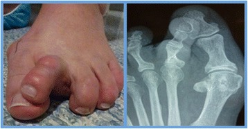
X-rayClinical photograph and anteroposterior X-ray of the foot: complex crossover deformity of the second toe with subluxation of the metatarsophalangeal joint, visible both clinically and radiographically.
Source : Gilheany et al., Journal of Foot and Ankle Research, 2015, figure 2 · CC BY
Imaging is not routine: a positive vertical drawer test is usually enough to make the diagnosis and to start conservative treatment. It becomes essential:
- in the event of clinical discordance or of an atypical presentation ;
- when the differential diagnosis is uncertain (Morton, fracture, Freiberg);
- in the pre-operative setting to quantify the extent of the lesion and to plan the surgical approach.
The Albright et al. SR/MA (2022, Eur J Radiol) pooling 11 studies (227 plantar plates on MRI, 238 on ultrasound, operative reference standard) establishes that:
- MRI : pooled sensitivity 89 per cent (95% CI 0.84-0.93), specificity 83 per cent (95% CI 0.64-0.94). The reference examination.
- Dynamic ultrasound : sensitivity 95 per cent (95% CI 0.91-0.98) but specificity of only 52 per cent (95% CI 0.37-0.68). Useful as a screening test (high NPV) but to be read with caution as a confirmatory test.15
A negative ultrasound in a patient with an equivocal clinical test is reassuring; a positive ultrasound needs MRI confirmation if surgery is being considered.
Forefoot-specific red flags not to be missed
- Dull, non-mechanical night pain: suspected malignancy (osteoid osteoma, metastasis, soft-tissue tumour).
- Palpable mass, firm, growing: suspected tumour.
- Redness, fever, pain on bony pressure: osteomyelitis (particularly in people with diabetes).
- History of cancer + focal bone pain: metastasis.
- Unexplained weight loss + persistent foot pain: oncological work-up.
- Diabetic foot with an ulcer + metatarsal pain: risk of osteomyelitis / Charcot foot.
- Polyarthritis: morning stiffness > 60 minutes, symmetrical involvement, MCP joints affected: inflammatory rheumatic disease.
Any red flag => prompt medical referral (general practitioner, rheumatologist, emergency care depending on the context) before physiotherapy management.
- The history must identify pain under the second metatarsal head of the « pebble in the shoe » type, worse on push-off, and look for progressive deviation of the toe.
- The vertical drawer test is the reference clinical test (sensitivity 83-94 per cent, Nery 2012). A translation > 2 mm defines a positive result.
- The main differential diagnosis is Morton's neuroma (Mulder's sign, pain between the heads): the two conditions can coexist.
- Imaging : MRI as the reference (89/83), ultrasound as a screening test (95/52), Albright 2022 SR/MA. No routine imaging when the clinical picture is typical.
- The Coughlin classification in five stages guides the decision: conservative 0-2, surgical 3-4, and always look for bony or neoplastic red flags.
Chapter 2 bibliography: all references verified on PubMed / CrossRef
- Kinter CW, Hodgkins CW. Lesser Metatarsophalangeal Instability: Diagnosis and Conservative Management of a Common Cause of Metatarsalgia. Sports Health. 2020;12(4):390-394. PMID 32223694.
- Thompson FM, Hamilton WG. Problems of the second metatarsophalangeal joint. Orthopedics. 1987;10(1):83-89. PMID 3809019.
- Coughlin MJ. Second metatarsophalangeal joint instability in the athlete. Foot Ankle. 1993;14(6):309-319. PMID 8406245.
- Nery C, Coughlin MJ, Baumfeld D, Mann TS. Lesser metatarsophalangeal joint instability: prospective evaluation and repair of plantar plate and capsular insufficiency. Foot Ankle Int. 2012;33(4):301-311. PMID 22735202.
- Coughlin MJ. Crossover second toe deformity. Foot Ankle. 1987;8(1):29-39. PMID 3623359.
- Finucane LM, Downie A, Mercer C, et al. International Framework for Red Flags for Potential Serious Spinal Pathologies. J Orthop Sports Phys Ther. 2020;50(7):350-372. PMID 32438853.
- Nery C, Coughlin MJ, Baumfeld D, Lemos AVKC, Schiefer S, Shibuya N. How to classify plantar plate injuries: parameters from history and physical examination. Rev Bras Ortop. 2015;50(6):720-728. PMC4868080.
- McKeon PO, Hertel J, Bramble D, Davis I. The foot core system: a new paradigm for understanding intrinsic foot muscle function. Br J Sports Med. 2015;49(5):290. PMID 24659509.
- Mulder JD. The causative mechanism in Morton s metatarsalgia. J Bone Joint Surg Br. 1951;33-B(1):94-95. PMID 14814166.
- Bencardino J, Rosenberg ZS, Beltran J, Liu X, Marty-Delfaut E. Morton's neuroma: is it always symptomatic?. AJR Am J Roentgenol. 2000;175(3):649-653. PMID 10954445.
- Fetzer GB, Wright RW. Metatarsal shaft fractures and fractures of the proximal fifth metatarsal. Clin Sports Med. 2006;25(1):139-50, x. PMID 16324980.
- Carmont MR, Rees RJ, Blundell CM. Current concepts review: Freiberg s disease. Foot Ankle Int. 2009;30(2):167-176. PMID 19254514.
- Kaz AJ, Coughlin MJ. Crossover second toe: demographics, etiology, and radiographic assessment. Foot Ankle Int. 2007;28(12):1223-1237. PMID 18173985.
- Nery C, Coughlin MJ, Baumfeld D, Raduan F, Mann TS, Catena F. Prospective evaluation of protocol for surgical treatment of lesser MTP joint plantar plate tears. Foot Ankle Int. 2014;35(9):876-885. PMID 24958766.
- Albright RH, Brooks BM, Chingre M, Klein EE, Weil LS Jr, Fleischer AE. Diagnostic accuracy of magnetic resonance imaging (MRI) versus dynamic ultrasound for plantar plate injuries: A systematic review and meta-analysis. Eur J Radiol. 2022;152:110308. doi:10.1016/j.ejrad.2022.110308.
Which treatment strategies are most effective for second MTP joint instability?
Where to start? What is the hierarchy of conservative interventions?
Management begins almost always with a conservative approach centred on mechanically offloading the second MTP joint and controlling local inflammation.1 The review by Kinter and Hodgkins (2020, Sports Health) summarises the validated modalities:2
- Immediate mechanical offloading: the taping of the second toe in slight plantarflexion limits MTP hyperextension, the main aggravating mechanism. Rapid symptomatic effect (Adams 2016: continuous use in a dancer, with a successful outcome).3
- Footwear modification: a shoe with a wide and deep toe box, a rigid or rocker sole (rocker sole) to reduce MTP flexion at push-off. Avoid high heels.2
- Foot orthosis with a retrocapital pad: a metatarsal pad placed behind the second metatarsal head to offload it. This is the standard of care as first-line treatment.24
- Management of sporting load: temporary reduction of impact activities, offloaded alternatives (swimming, cycling); gradual resumption with the « < 10 per cent / week » rule (Nielsen 2014 JOSPT).5
- Patient education: the nature of the condition, the role of mechanical overload, the importance of adherence.
Decision algorithm: second MTP joint instability / metatarsalgia of the second ray
Clinical pathway from suspicion to the conservative / surgical decision
Summary decision flow. Wang G et al. J Foot Ankle Surg. 2022;61(5):1108-1116: SR/MA of 12 studies, 537 patients, with the dorsal approach + Weil osteotomy predominating at advanced stages.
What is the role of intrinsic foot exercise and which approach is superior?
Therapeutic exercise is the active pillar of management in the medium and long term. The main objective is strengthening of the intrinsic foot muscles (abductor hallucis, lumbricals, flexor digitorum brevis) that make up the « foot core system » (McKeon 2015, BJSM).6
The short foot exercise (SFE) is the best-documented intervention: the patient learns to actively shorten the foot by drawing the head of the first metatarsal towards the heel, without curling the toes. Jung et al. (2011, Phys Ther Sport) showed on EMG that the SFE activates abductor hallucis significantly more than a simple toe curl (p<0.001), and that the effect is greater in single-leg stance.7 Mulligan and Cook (2013, Man Ther) confirmed in a pilot trial that 4 weeks of SFE significantly increase arch height in subjects with marked collapse.8
The meta-analysis by Wei et al. (2022, PLOS One) on 16 studies confirms that intrinsic foot muscle training improves strength and dynamic balance and reduces navicular drop, but the specific effect on pain remains modest, which argues for combining it with an orthosis and taping.9
Beyond the foot, adding posterior chain work (triceps surae, gastrocnemius stretching, ankle/hip balance) and control of pronation is useful to reduce the push-off forces focused on the second ray.2
Manual therapies, ESWT, injections: how effective are they really?
Several adjuvant therapies are available; their level of evidence varies widely:
- Manual therapy (intermetatarsal, tarsometatarsal and ankle mobilisations): short-term pain relief reported in case series, low level of evidence (no good-quality RCT specific to the second MTP joint).
- ESWT (extracorporeal shockwave therapy) : a validated option for resistant chronic metatarsalgias (> 3 months). For the second MTP joint specifically, the data remain limited but consistent with the effectiveness seen in other plantar tendinopathies / enthesopathies.
- Corticosteroid injections : their use is controversial. They relieve inflammation but carry a far from negligible risk of rupture of the plantar plate that is already weakened, and of fat pad atrophy, which can worsen the instability in the medium term.210 Recent recommendations (Kinter 2020) advise against their routine use and restrict them to very selected situations, under supervision.
- Prolotherapy : used in the Adams 2016 case (a dancer, successful outcome) but with a low level of evidence.3
| Modality | Level of evidence (GRADE) | Main effect | Caveats |
|---|---|---|---|
| Retrocapital orthosis | High | Mechanical offloading: standard of care | Custom-made no better than well-fitted prefabricated devices (Chapman 2018 SR) |
| Protective taping | Moderate | Short-term pain relief + protection | Effect limited over time if used alone |
| Short foot exercise | Moderate | Intrinsic strengthening, balance | Modest effect on pain when used alone: combine it |
| Education + load modification | High | Cornerstone (Lin 2020 MSK recommendations) | Requires the patient to buy in |
| ESWT | Moderate | Pain relief in chronic cases (extrapolated) | Limited data specific to the second MTP joint |
| Manual therapy | Low | Short-term pain relief | Case series only, no RCT |
| Corticosteroid injection | Low (unfavourable) | Local anti-inflammatory effect | Risk of plate rupture + atrophy |
| Dorsal surgical repair + Weil | Moderate | > 80% good-excellent (Wang 2022 n=537) | Residual stiffness ~20%: evidence level: case series |
When should surgery be considered and what results can be expected?
Surgery is indicated at the fixed stages 3-4 or after failure of 3-6 months of well-conducted conservative treatment. The SR/MA by Wang et al. (2022, J Foot Ankle Surg) covering 12 studies that pooled 537 plantar plate tears treated surgically shows:11
- There is a significant improvement in the pain VAS and the AOFAS score at 2 years.
- The dorsal approach (Watson 2014) with direct repair + Weil osteotomy added is the commonest and best-studied combination.12
- More than 80 per cent good to excellent results, but a residual stiffness of the MTP joint in about 20 per cent of those operated on: the most frequently reported complication.
- There is a recurrence rate of about 7.6 per cent (Prissel 2017, plantar approach).13
The direct plantar approach (Prissel 2017) offers excellent visualisation but exposes the patient to plantar scar complications (up to 10 per cent, Sharpe 2025).14 In the event of an associated hallux valgus, correcting it at the same time is essential on pain of mechanical failure of the MTP2 repair.15
- Management is staged : 1) offloading (taping, retrocapital orthosis, footwear); 2) intrinsic strengthening (short foot exercise); 3) load modification; 4) ESWT if the problem is chronic; 5) surgery if it fails or the stage is advanced.
- The foot orthosis with a retrocapital pad is the standard of care (GRADE high, Chapman 2018 SR).
- The corticosteroid injections must be used with caution (iatrogenic risk of plantar plate rupture and fat pad atrophy).
- Surgery (dorsal repair + Weil) gives > 80 per cent good results (Wang 2022 SR/MA n=537), but ~20 per cent residual stiffness.
- The simultaneous correction of the associated anatomical factors (long second metatarsal, hallux valgus) determines lasting success.
Chapter 3 bibliography: all references verified on PubMed / CrossRef
- Maas NM, van der Grinten M, Bramer WM, Kleinrensink GJ. Metatarsophalangeal joint stability: a systematic review on the plantar plate of the lesser toes. J Foot Ankle Res. 2016;9:32. PMID 27547243.
- Kinter CW, Hodgkins CW. Lesser Metatarsophalangeal Instability: Diagnosis and Conservative Management of a Common Cause of Metatarsalgia. Sports Health. 2020;12(4):390-394. PMID 32223694.
- Ojofeitimi S, Bronner S, Becica L. Conservative Management of Second Metatarsophalangeal Joint Instability in a Professional Dancer: A Case Report. J Orthop Sports Phys Ther. 2016;46(2):114-123. PMID 26755404.
- Espinosa N, Maceira E, Myerson MS. Current concept review: metatarsalgia. Foot Ankle Int. 2008;29(8):871-879. PMID 18752791.
- Nielsen RO, Parner ET, Nohr EA, Sorensen H, Lind M, Rasmussen S. Excessive progression in weekly running distance and risk of running-related injuries: an association which varies according to type of injury. J Orthop Sports Phys Ther. 2014;44(10):739-747. PMID 25155475.
- McKeon PO, Hertel J, Bramble D, Davis I. The foot core system: a new paradigm for understanding intrinsic foot muscle function. Br J Sports Med. 2015;49(5):290. PMID 24659509.
- Jung DY, Kim MH, Koh EK, Kwon OY, Cynn HS, Lee WH. A comparison in the muscle activity of the abductor hallucis and the medial longitudinal arch angle during toe curl and short foot exercises. Phys Ther Sport. 2011;12(1):30-35. PMID 21256447.
- Mulligan EP, Cook PG. Effect of plantar intrinsic muscle training on medial longitudinal arch morphology and dynamic function. Man Ther. 2013;18(5):425-430. PMID 23632367.
- Wei Z, Zeng Z, Liu M, Wang L. Effect of intrinsic foot muscles training on foot function and dynamic postural balance: A systematic review and meta-analysis. PLoS One. 2022;17(4):e0266525. PMID 35442981.
- Baker JR, Albright R, Jameson R, et al. Treatment of Lesser Metatarsophalangeal Joint Instability With Plantar Plate Repair: A Systematic Review and Meta-Analysis. J Foot Ankle Surg. 2022;61(5):1114-1118. PMID 35283034.
- Watson TS, Reid DY, Frerichs TL. Dorsal Approach for Plantar Plate Repair With Weil Osteotomy: Operative Technique. Foot Ankle Int. 2014;35(7):730-739. PMID 24850163.
- Sharpe BD, Ebaugh MP, Philbin TM, Prissel MA, Hyer CF, Berlet GC, Goss DA. Direct Plantar Approach to Plantar Plate Repair and Associated Wound Complications. Foot Ankle Spec. 2025;18(2):134-140. doi:10.1177/19386400221118500.
- Coughlin MJ, Jones CP. Hallux valgus and first ray mobility. A prospective study. J Bone Joint Surg Am. 2007;89(9):1887-1898. PMID 17768183.
How can lasting recovery be secured and recurrence of second MTP joint instability prevented?
How can the patient be made an active partner in their own recovery through self-management?
Self-management (self-management) is the cornerstone of lasting results. Therapeutic education gives the patient the knowledge and skills to manage the condition day to day: understanding the role of overload on the plantar plate, identifying aggravating factors (unsuitable shoes, spikes in activity), and knowing how to react to early symptoms.1
The key components of self-management are:
- Intrinsic strengthening at home: the patient continues the short foot exercise and toe yoga for 5-10 minutes / day. The Wei 2022 meta-analysis confirms the effects on posture and balance, prerequisites for a good distribution of pressure.2
- Self-monitoring of pain: the patient learns to modulate activity while staying below an acceptable threshold (typically ≤ 3/10 on the NPRS) during and after exercise, with no worsening on waking the next day. This approach allows safe graded exposure.3
- Sensible use of adjuncts: the orthosis and taping are tools of transition, not of dependence. The patient should learn to apply a simple tape job themselves during a flare.
- Footwear modification day to day: lasting avoidance of pointed shoes / high heels for women at risk.4
Lin et al. (2020, BJSM) identified 11 consistent recommendations drawn from the most robust MSK guidelines: centre care on the patient, screen for red flags, assess psychosocial factors, use imaging selectively, carry out a physical examination, measure progress, provide education and information, prescribe exercise, use passive modalities as adjuncts only, avoid surgery when it is not needed, and encourage people to stay active.5
When and how should a safe return to sport and activity be planned?
Return to sport must be guided by functional criteria and not by a rigid calendar. The decision-rules model validated by Grindem et al. (2016, BJSM) on the Delaware-Oslo ACL cohort (an 84 per cent reduction in re-injury risk through adherence to the functional criteria) provides a solid analogy: wait for symmetry of strength and a minimum delay before returning to pivoting activities.6
For the second MTP joint, a structured progression in 3 phases is recommended:
Phases of return to activity: functional criteria for the second MTP joint
Mechanical load progression guided by objective markers (by analogy with the Grindem 2016 ACL decision rules)
Decision rules adapted from Grindem H et al. Br J Sports Med. 2016;50(13):804-808 (PMID 27162233): 84% reduction in reinjury through adherence to the functional criteria. The 10% rule: Nielsen RO et al. J Orthop Sports Phys Ther. 2014;44(10):739-747 (PMID 25155475).
The psychological dimension is decisive. The kinesiophobia, the fear of recurrence and the readiness (psychological preparedness) are major predictors of a successful return to activity. The work of Ardern et al. (2014, BJSM) on the ACL-RSI score documented this dimension in the ACL setting, and it transfers to any return after a foot condition.7
The shared decision-making (collaborative decision-making) between physiotherapist and patient must remain central: the patient's personal goals, tolerance of the risk of recurrence, and work and leisure constraints.8
- Preventing recurrence depends on making the patient autonomous through therapeutic education (Lin 2020: 11 cross-cutting MSK recommendations).
- The continued intrinsic strengthening (daily short foot exercise) maintains the dynamic stability of the forefoot.
- Return to sport is guided by functional criteria (pain, symmetrical strength, functional tests passed) and not by a rigid calendar.
- The rule of < 10 per cent increase in load per week (Nielsen 2014) is a simple and effective tool for avoiding overload.
- The psychological readiness (confidence, kinesiophobia) is a key predictor of success: adopt a biopsychosocial approach.
Chapter 4 bibliography: all references verified on PubMed / CrossRef
- Lorig KR, Holman H. Self-management education: history, definition, outcomes, and mechanisms. Ann Behav Med. 2003;26(1):1-7. PMID 12867348.
- Wei Z, Zeng Z, Liu M, Wang L. Effect of intrinsic foot muscles training on foot function and dynamic postural balance: A systematic review and meta-analysis. PLoS One. 2022;17(4):e0266525. PMID 35442981.
- Gabbett TJ. The training-injury prevention paradox: should athletes be training smarter and harder? Br J Sports Med. 2016;50(5):273-280. PMID 26758673.
- Kinter CW, Hodgkins CW. Lesser Metatarsophalangeal Instability: Diagnosis and Conservative Management. Sports Health. 2020;12(4):390-394. PMID 32223694.
- Lin I, Wiles L, Waller R, et al. What does best practice care for musculoskeletal pain look like? Eleven consistent recommendations from high-quality clinical practice guidelines: systematic review. Br J Sports Med. 2020;54(2):79-86. PMID 30826805.
- Grindem H, Snyder-Mackler L, Moksnes H, Engebretsen L, Risberg MA. Simple decision rules can reduce reinjury risk by 84% after ACL reconstruction: the Delaware-Oslo ACL cohort study. Br J Sports Med. 2016;50(13):804-808. PMID 27162233.
- Ardern CL, Österberg A, Tagesson S, Gauffin H, Webster KE, Kvist J. The impact of psychological readiness to return to sport and recreational activities after anterior cruciate ligament reconstruction. Br J Sports Med. 2014;48(22):1613-1619. PMID 25293342.
- Foster NE, Anema JR, Cherkin D, et al. Prevention and treatment of low back pain: evidence, challenges, and promising directions. Lancet. 2018;391(10137):2368-2383. PMID 29573872.
- Nielsen RO, Parner ET, Nohr EA, et al. Excessive progression in weekly running distance and risk of running-related injuries: an association which varies according to type of injury. J Orthop Sports Phys Ther. 2014;44(10):739-747. PMID 25155475.
What do real clinical cases teach us about second MTP joint instability?
Conservative case: resolution without surgery in the dancer described by Adams 2016
The case report by Adams, Pozzi and Wallace (2016, JOSPT, PMID 26755404) describes a 33-year-old professional dancer with insidious pain over the second and third MTP joints, present for several months.1 Clinical examination showed tenderness on palpation of the metatarsal heads and a positive vertical drawer test, and imaging (MRI) confirmed:
- MTP1 osteoarthritis;
- Calcification + MTP2 capsulitis;
- Rupture of the MTP2 plantar plate (Coughlin stage 2).
The multimodal conservative treatment strategy comprised:
- Taping for protection, holding the second toe in plantarflexion, applied for all physical activity;
- Padding (a retrocapital metatarsal pad) built into the dance shoe;
- Physiotherapy: intrinsic strengthening (short foot, toe yoga), posterior chain work, motor control of the foot, graded exposure to load;
- Activity modification: temporary reduction of jumping, replaced by floor work;
- Courses of prolotherapy, as an add-on (4 injections);
- Follow-up over 37 physiotherapy sessions across 16 weeks.
At discharge, the patient had resumed her professional activity in full (complete choreography), continuing the taping and the padding for performances. This case is emblematic of the conservative therapeutic window of the early stages and of the need for a multimodal approach combining offloading, active strengthening and activity modification.1
Surgical case: the Nery 2012 prospective series
The founding prospective series of Nery, Coughlin, Baumfeld and Mann (2012, Foot Ankle Int, PMID 22735202) brings together 22 MTP joints with reducible instability (Coughlin stages 1-3) that underwent direct plantar plate repair through a dorsal approach with or without an associated Weil osteotomy.2
Main results:
- Significant improvement in the AOFAS and pain VAS scores ;
- Return to daily activities at about 6-8 weeks;
- High patient satisfaction score at 12 months;
- Most frequently reported complication: residual stiffness of the MTP joint in about 20 per cent of those operated on;
- No recurrence at 12 months in this series.
This series established the dorsal approach as the reference technique, later refined by Watson (2014) and confirmed by the Wang 2022 SR/MA (537 patients).34
The diagnostic challenge: when second MTP joint instability mimics Morton's neuroma
The confusion between second MTP joint instability and Morton's neuroma is the commonest diagnostic error, and its consequences can be serious: the repeated corticosteroid injections given for a presumed « neuroma » on an already weakened plantar plate precipitate complete rupture.5
The typical case described in the literature: a middle-aged woman with pain under the forefoot, given corticosteroid injections for a supposed neuroma without lasting improvement. Reassessment reveals:
- Pain under the head of the second metatarsal (not in the 2-3 space);
- Positive vertical drawer test (translation > 2 mm);
- Mulder's sign negative or equivocal;
- Dynamic ultrasound demonstrates the dorsal subluxation of the phalanx + a tear of the plate;
- MRI confirms the rupture.6
The trap is all the more common because the two conditions can coexist : chronic instability of the second MTP joint generates a reactive pseudo-neuroma (pericapsular thickening) that can be mistaken for a true intermetatarsal neuroma on imaging.5
Complex case study: hallux valgus + plantar plate tear + combined surgery
Complex cases illustrate the multifactorial aetiology of second MTP joint instability and the need for a comprehensive surgical approach. Vakhshori et al. (2022, Foot Ankle Surg) documented that 34.9 per cent of the women operated on for hallux valgus had a concomitant MTP2 subluxation, against 0 per cent in the matched controls: a highly significant difference.7
In these situations, the simultaneous correction of the hallux valgus (scarf, chevron or Lapidus osteotomy depending on the context) AND of the MTP2 plantar plate (dorsal repair + Weil) is essential on pain of mechanical failure. Neglecting the hallux valgus exposes the patient to recurrence of the second MTP joint instability within 12-18 months, because load remains badly distributed.8
The Adams 2016 case already cited illustrates another form of complexity: multi-joint involvement (MTP1 osteoarthritis + second MTP joint instability + capsulitis) in a dancer, where surgery would have compromised her career and where the multimodal conservative approach allowed a return to performance.1
Critique and controversies: the place of the clinical case
GRADE evidence pyramid: second MTP joint instability / metatarsalgia of the second ray
Format of stacked horizontal cards (never any lettering INSIDE a triangular pyramid, good readability practice)
GRADE classification (Grading of Recommendations Assessment, Development and Evaluation) applied to the key sources cited in this article.
Case reports have well-identified limitations:
- Publication bias: successful cases are over-represented compared with failures;
- Level V evidence (CEBM), no causality and no generalisation are possible;
- Heterogeneity of reporting, no universal standardisation of outcomes;
- Over-representation of surgery: a relative shortage of detailed conservative cases with confirmatory imaging (the Adams 2016 case stands out as a remarkable exception).
Clinical cases are an excellent teaching tool and a good way to expose pitfalls, but major treatment decisions must rest on SR/MAs (Maas 2016, Albright 2022, Wang 2022, Wei 2022) and large prospective series.
- The Adams 2016 case (JOSPT) demonstrated the feasibility of a complete conservative resolution (37 sessions, 16 weeks, return to professional dance) in a patient with a documented plantar plate rupture.
- The surgical series (Nery 2012, Wang 2022 SR/MA n=537) confirm > 80 per cent good results after dorsal repair + Weil, with ~20 per cent residual stiffness.
- The main diagnostic trap is confusion with Morton's neuroma: the two can coexist (a reactive pseudo-neuroma on chronic instability).
- Complex cases are a reminder that 34.9 per cent of surgical hallux valgus cases have a concomitant MTP2 subluxation (Vakhshori 2022) that must be corrected at the same time.
- Clinical cases are signals and teaching tools, not proof: major decisions rest on SR/MAs and prospective series.
Chapter 5 bibliography: all references verified on PubMed / CrossRef
- Ojofeitimi S, Bronner S, Becica L. Conservative Management of Second Metatarsophalangeal Joint Instability in a Professional Dancer: A Case Report. J Orthop Sports Phys Ther. 2016;46(2):114-123. PMID 26755404.
- Nery C, Coughlin MJ, Baumfeld D, Mann TS. Lesser metatarsophalangeal joint instability: prospective evaluation and repair of plantar plate and capsular insufficiency. Foot Ankle Int. 2012;33(4):301-311. PMID 22735202.
- Watson TS, Reid DY, Frerichs TL. Dorsal Approach for Plantar Plate Repair With Weil Osteotomy: Operative Technique. Foot Ankle Int. 2014;35(7):730-739. PMID 24850163.
- Baker JR, Albright R, Jameson R, et al. Treatment of Lesser Metatarsophalangeal Joint Instability With Plantar Plate Repair: A Systematic Review and Meta-Analysis. J Foot Ankle Surg. 2022;61(5):1114-1118. PMID 35283034.
- Kinter CW, Hodgkins CW. Lesser Metatarsophalangeal Instability: Diagnosis and Conservative Management. Sports Health. 2020;12(4):390-394. PMID 32223694.
- Albright RH, Brooks BM, Chingre M, Klein EE, Weil LS Jr, Fleischer AE. Diagnostic accuracy of magnetic resonance imaging versus dynamic ultrasound for plantar plate injuries: A systematic review and meta-analysis. Eur J Radiol. 2022;152:110308. doi:10.1016/j.ejrad.2022.110308.
- Okuda R, Yoshimura Y, Shima H. Prevalence and radiological characteristics of the dislocation of the second metatarsophalangeal joint in patients undergoing hallux valgus surgery; a matched control study. J Orthop Sci. 2023;28(4):853-859. PMID 35803857.
- Coughlin MJ, Jones CP. Hallux valgus and first ray mobility. A prospective study. J Bone Joint Surg Am. 2007;89(9):1887-1898. PMID 17768183.
- Fleischer AE, Klein EE, Ahmad M, Shah S, Catena F, Weil L Jr, Weil LS Sr. Association of Abnormal Metatarsal Parabola With Second Metatarsophalangeal Joint Plantar Plate Pathology. Foot Ankle Int. 2017;38(3):289-297. PMID 27881742.
- Maas NM, van der Grinten M, Bramer WM, Kleinrensink GJ. Metatarsophalangeal joint stability: a systematic review on the plantar plate of the lesser toes. J Foot Ankle Res. 2016;9:32. PMID 27547243.
How do you apply these recommendations concretely in your practice?
When and to which other healthcare professionals should you refer?
One of the key skills of the physiotherapist working in direct access is the identification of situations that fall outside their scope of practice. This triage rests on two registers: red flags (suspected serious pathology) and yellow flags (psychosocial factors that predict chronicity).
The international framework of Finucane et al. (2020, JOSPT) stresses that red flags taken in isolation have a low positive predictive value: it is their clustering (a constellation) that should raise the alarm and prompt a rapid referral.1 For the forefoot, the signs to take in are: dull night pain, a firm immobile mass, redness-fever-swelling (suspected infection / Charcot foot in diabetes), a history of cancer, unexplained weight loss, and polyarthritis with prolonged morning stiffness.
Referral should therefore be considered in these scenarios:
- To a general practitioner, podiatrist, rheumatologist or orthopaedic surgeon : red flags present, no progress after 3-6 months of well-conducted conservative treatment, severe deformity (Coughlin stages 3-4), discussion of interventional alternatives (ESWT, surgery).2
- To an orthotist / specialised podiatrist : design and fitting of a custom foot orthosis with a good retrocapital pad (Chapman 2018 confirms similar effectiveness between custom and well-fitted prefabricated devices, but the fitting calls for expertise).3
- To a psychologist / CBT approach : predominant yellow flags (marked kinesiophobia, catastrophising, interfering depressive symptoms) that hold recovery back.4
- To a pain specialist / multidisciplinary team : complex chronic pain, suspected central sensitisation, multiple comorbidity.5
The interprofessional collaboration is a quality-of-care factor recognised by the guidelines (Lin 2020): clear referral protocols, shared reports, shared decision-making with the patient.6
How do you measure outcomes and overcome barriers to implementation?
Measuring outcomes objectively is essential to justify continuing treatment, adjust management and document progress. The validated PROMs for metatarsalgia are:
- NPRS (Numeric Pain Rating Scale, 0-10): pain intensity, MCID about 2 points;
- FAAM (Foot and Ankle Ability Measure): 29 items + 8 sport items, validated for foot/ankle function;
- FFI (Foot Function Index): pain + disability + activity limitation;
- AOFAS (American Orthopaedic Foot & Ankle Society score): much used in surgery but criticised for its limited psychometric properties.
Systematic use of these tools allows tracking over time and shared decision-making. Their implementation in daily practice runs into several classic barriers, however:
- Barriers on the therapist's side: lack of time, little familiarity with the tools, residual biomedical beliefs, the inertia of habit;
- Barriers on the patient's side: expectations geared towards passive techniques, low health literacy, socio-economic constraints;
- Systemic barriers: reimbursement of passive modalities rather than education/exercise, lack of organisational support, imperfect interprofessional communication.6
The 11 cross-cutting recommendations identified by Lin et al. (2020) for MSK conditions apply without reservation to the second MTP joint: (1) patient-centred care, (2) red flag screening, (3) yellow flag assessment, (4) selective imaging, (5) full physical examination, (6) monitoring of progress, (7) education and information, (8) exercise prescription, (9) passive modalities as adjuncts only, (10) surgery as a last resort, (11) staying active encouraged.6
Critique and controversy: beyond the simple application of the recommendations
Several nuances need to be taken on board:
- The « tyranny of red flags » is an emerging criticism: an excessive, anxiety-provoking search can paradoxically increase the use of unnecessary imaging, generate iatrogenic anxiety and reinforce negative beliefs about the « fragility » of the body. The skill lies in the ability to reassure as much as to raise the alarm.1
- The theory/practice gap in the biopsychosocial model remains wide: many therapists say they have adopted it but in practice stay focused on the biomechanical side. Addressing yellow flags calls for advanced communication skills.5
- The reduction of the patient to a score through PROMs: better numbers do not amount to the patient's own satisfaction: PROMs are a starting point for the conversation, not an end in themselves.
- The rigid application of the recommendations must always leave room for individualisation: goals, sporting and occupational context, patient preferences.
- Identifying red flags (night pain, mass, fever, weight loss, history of cancer) calls for prompt medical referral, but their predictive value in isolation is low (Finucane 2020).
- The yellow flags (kinesiophobia, catastrophising) are major predictors of chronicity and justify a biopsychosocial approach or collaboration with a psychologist / CBT.
- The interprofessional collaboration (podiatrist, sports physician, surgeon, psychologist) is a quality-of-care factor.
- The PROMs (NPRS, FAAM, FFI) are essential for measuring progress, but they do not replace the patient-centred conversation.
- The 11 MSK recommendations (Lin 2020 BJSM) apply to the second MTP joint: centre care on the patient, screen for flags, exercise + education, selective imaging, passive modalities as adjuncts only.
Chapter 6 bibliography: all references verified on PubMed / CrossRef
- Finucane LM, Downie A, Mercer C, et al. International Framework for Red Flags for Potential Serious Spinal Pathologies. J Orthop Sports Phys Ther. 2020;50(7):350-372. PMID 32438853.
- Kinter CW, Hodgkins CW. Lesser Metatarsophalangeal Instability: Diagnosis and Conservative Management. Sports Health. 2020;12(4):390-394. PMID 32223694.
- Espinosa N, Maceira E, Myerson MS. Current concept review: metatarsalgia. Foot Ankle Int. 2008;29(8):871-879. PMID 18752791.
- Foster NE, Anema JR, Cherkin D, et al. Prevention and treatment of low back pain: evidence, challenges, and promising directions. Lancet. 2018;391(10137):2368-2383. PMID 29573872.
- O Sullivan PB, Caneiro JP, O Keeffe M, et al. Cognitive Functional Therapy: An Integrated Behavioral Approach for the Targeted Management of Disabling Low Back Pain. Phys Ther. 2018;98(5):408-423. PMID 29669082.
- Lin I, Wiles L, Waller R, et al. What does best practice care for musculoskeletal pain look like? Eleven consistent recommendations from high-quality clinical practice guidelines: systematic review. Br J Sports Med. 2020;54(2):79-86. PMID 30826805.
- Ojofeitimi S, Bronner S, Becica L. Conservative Management of Second Metatarsophalangeal Joint Instability in a Professional Dancer: A Case Report. J Orthop Sports Phys Ther. 2016;46(2):114-123. PMID 26755404.
- Martin RL, Irrgang JJ, Burdett RG, Conti SF, Van Swearingen JM. Evidence of validity for the Foot and Ankle Ability Measure (FAAM). Foot Ankle Int. 2005;26(11):968-983. PMID 16309613.
- Budiman-Mak E, Conrad KJ, Roach KE. The Foot Function Index: a measure of foot pain and disability. J Clin Epidemiol. 1991;44(6):561-570. PMID 2037861.
- Hawker GA, Mian S, Kendzerska T, French M. Measures of adult pain: Visual Analog Scale for Pain (VAS Pain), Numeric Rating Scale for Pain (NRS Pain), McGill Pain Questionnaire (MPQ), Short-Form McGill Pain Questionnaire (SF-MPQ), Chronic Pain Grade Scale (CPGS), Short Form-36 Bodily Pain Scale (SF-36 BPS), and Measure of Intermittent and Constant Osteoarthritis Pain (ICOAP). Arthritis Care Res. 2011;63 Suppl 11:S240-252. PMID 22588748.
And after this read?
This article is part of a collection of evidence-based clinical syntheses. A question, a comment, a correction to suggest? Contact us directly through the WhatsApp button at the bottom right of the screen.

