














On-siteDPCFIFPL

Upper limb
Advance your hand and wrist rehabilitation: assess and empower your patients
Tessadit Aissaoui
Go further
Mallet finger: the Doyle classification, custom against prefabricated splint (0 % failure against 23.8 %), how long to immobilise, and rehabilitation.
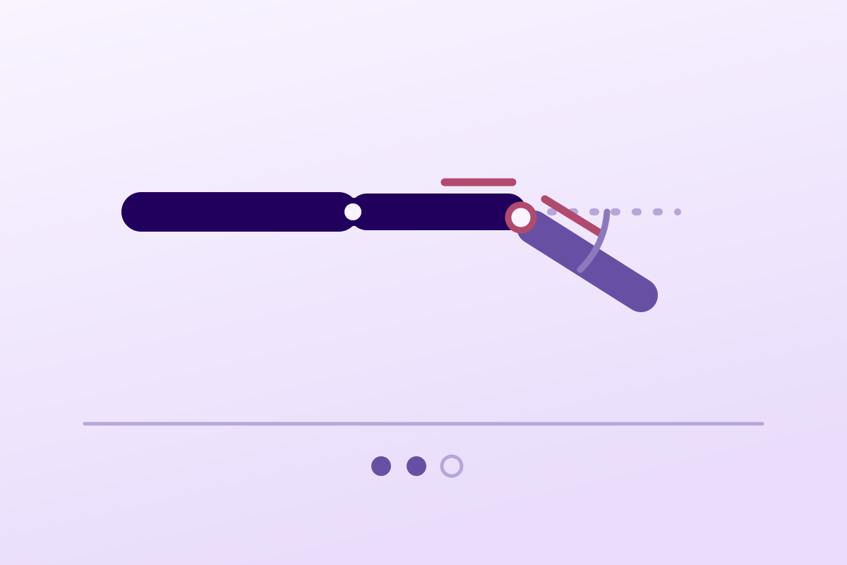
The extension lag does not regress spontaneously: continuous extension immobilisation, for six to eight weeks, is the first-line treatment.
23.8%of treatment failures with a prefabricated splint
What follows covers each of these points in detail, with sources. It is there if you need it.
This topic is taught in a course: Advance your hand and wrist rehabilitation: assess and empower your patients, on-site with Tessadit Aissaoui.
1 / 11
10 article chapters · 36 min in total
Understanding it◔ 2 min
The signature of mallet finger: flexed actively, but extensible passively, in two seconds.
Who and how◔ 3 min
The dominant hand is affected in about 74 % of cases, which weighs on the occupational impact.
Classifying it◔ 3 min
A palmar subluxation of the phalanx tips the balance towards surgery, whatever the fragment size.
Choosing a splint◔ 5 min
The splint does not change the final deficit, it changes the odds of completing treatment.
Duration of splinting◔ 3 min
The reference protocol: six to eight weeks of continuous extension of the DIP joint.
Surgery◔ 3 min
Surgery and non-surgical treatment give clinically equivalent results.
Work and sport◔ 3 min
Surgery speeds up nothing: it lengthens return to work by 9 days, in the only trial that measured it.
Rehab◔ 5 min
After splint removal, a loss of extension appears, partly regained through active exercise.
Case reports◔ 3 min
Published cases never describe an ordinary mallet finger, which is what makes them instructive.
In practice◔ 6 min
For this condition, the first consultation is worth more than all the following ones.
Train on this
Upper limb
Tessadit Aissaoui
Next comes practice: the course that teaches this topic, with Tessadit Aissaoui.
Our courses on this topic may be covered by DPC or FIFPL, the French continuing-education funding schemes, depending on your profession and situation.
A fingertip that will no longer straighten after an unremarkable knock: tucking in a sheet, catching a ball badly, pulling up a sock. The injury is simple, the treatment looks trivial, and that is exactly where the result is decided: between a splint that holds for eight weeks and a splint that gives up, the gap measured in a randomised trial is 23.8 points of treatment failure.
Summary written from primary sources verified one by one on PubMed: identifier resolved, journal, year and author list checked, abstract read before citation. Full bibliography at the end of the article.
Mallet finger in three figures
What three independent pieces of work establish, and which moves the question from “should we operate?” to “which splint, and worn for how long?”
Sources: Lin JS, Samora JB. J Hand Surg Am 2018;43(2):146-163.e2 (PMID 29174096); O'Brien LJ, Bailey MJ. Arch Phys Med Rehabil 2011;92(2):191-198 (PMID 21272714); Gruber JS, Bot AG, Ring D. Hand (N Y) 2014;9(2):145-150 (PMID 24839414).
What to take away before reading the rest
A lesion of a few millimetres, an extensor mechanism that loses its terminal pulley, and a joint that tips over. Understanding the mechanics avoids the two most frequent errors: underestimating the length of treatment, and overestimating the value of surgery.
Mallet finger denotes a deformity of the fingertip in which the distal interphalangeal joint stays flexed and can no longer be actively extended. It results from a traumatic interruption of the terminal extensor mechanism, either at its insertion on the distal phalanx or slightly proximal to it at the joint2.
The sign that identifies it is also the one that distinguishes it from stiffness: the joint can be extended passively, but that extension is not maintained as soon as you let go2. It is the test to do routinely, and it takes two seconds.
Three consequences follow, and they govern everything else:
The splint does not “hold” the finger: it holds the two ends of the tendon close enough to each other, for long enough, for them to meet. A single flexion is enough to undo weeks of work.
The injury can be purely tendinous, the tendon pulls off its insertion, or bony, the tendon taking with it a fragment of the dorsal cortex of the distal phalanx. Both forms produce the same deformity, but they are not treated in quite the same way when the fragment is large or the joint subluxes: that is the subject of the chapter on classification.
Key point
A sportsperson's injury, so they say. That is true of only some cases, and the most frequent mechanism in community practice is so unremarkable that it surprises the patient themselves.
The injury is mostly seen in young men, in the third and fourth decades ; after the fifth decade, the difference between the sexes disappears2. The reference NCBI chapter adds a nuance about frequency: the picture is seen in young to middle-aged men, but also in older women1.
The dominant hand is involved in about 74 % of cases2, which is not neutral for the impact on work: eight weeks of splinting on the dominant index finger of a tradesperson is not managed like the non-dominant little finger of an office worker.
As for which fingers are affected, the order of frequency is consistent from one source to another: the middle finger first, then the ring finger, the little finger and the index ; involvement of the thumb is rare2. The NCBI chapter for its part mentions the little, ring and middle fingers of the dominant hand as the most frequently involved1: the exact order varies, but it is indeed the last three rays that dominate.
The mechanisms classically described number three2 :
A case published in the Journal of Orthopaedic Case Reports also recalls this injury's place in sports trauma: according to its authors it accounts for 2 % of sports emergencies and is the most frequent closed tendon injury in contact sports and in the workplace15.
The unremarkable nature of the triggering movement is a clinical datum in its own right: it explains late presentations, and it also explains that the injury occurs outside any sporting context. A case published in Anaesthesia Reports thus describes a mallet finger sustained by an anaesthetist during tracheal intubation, treated without surgery, with a prolonged absence from clinical work before full recovery17.
Four practical markers
Data from the reference review and the NCBI chapter
Sources: Khera B, Chang C, Bhat W. Acta Biomed 2021;92(5):e2021246 (PMID 34738569); Benabdallah O, Benabdallah R. J Orthop Case Rep 2022;12(11):60-64 (PMID 37013228).
A mallet finger with no injury is not a mallet finger
It is a sign, and it requires the cause to be looked for. Two published cases demonstrate it. A 35-year-old woman developed the deformity gradually, over more than twenty days, with no injury at all, with palpable nodules at the distal interphalangeal joint: it was a pigmented villonodular synovitis, a condition never previously reported with this presentation15. A 4-year-old child presented with a painless deformity that had worsened progressively over a year, with a palpable bony mass proximal to the joint: an osteochondroma of the middle phalanx was pushing the terminal tendon aside, and the initial radiograph was not conclusive16. Gradual onset, no injury, a palpable mass, a paediatric age : four reasons to refer rather than splint.
Key point
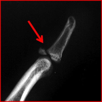
X-rayLateral finger radiograph: mallet finger with a small dorsal bony avulsion fragment at the base of the distal phalanx, red arrow on the lesion.
Source : Avery et al., Journal of Orthopaedic Surgery and Research, 2016, figure 14 · CC BY
One classification is used universally. It comes down to four types, it is read on a lateral radiograph, and it is what separates the fingers that are splinted from those that are operated on.
| Type | Injury | Usual direction |
|---|---|---|
| I | Closed injury, with or without a small dorsal avulsion fracture | Splinting: this is the very great majority of cases2 |
| II | Open injury from a clean laceration | Surgical management of the wound and the tendon |
| III | Open injury from deep abrasion, with loss of skin and tendon substance | Surgery, with coverage and reconstruction |
| IV-A | Transphyseal fracture (child and adolescent) | Depending on displacement; surgical opinion |
| IV-B | Fragment involving 20 - 50 % of the articular surface | Case-by-case decision, splinting possible |
| IV-C | Fragment involving more than 50 % of the articular surface | Risk of palmar subluxation: surgical opinion |
The operative indications retained by that same chapter are explicit: subluxation of the distal interphalangeal joint, a fragment involving more than a third of the articular surface, a joint gap of more than 2 mm, rupture or complete division of the extensor tendon, and complex injuries1.
Doyle's four types, at a glance
What the lateral radiograph shows, and what it implies
Source: Mallet Finger Injuries, StatPearls, NCBI Bookshelf NBK459373 (PMID 29083648). The original classification is Doyle's; a modification was proposed by Sivakumar et al. to reduce inter-observer error (PMID 36509592).
A strict lateral view of the finger, centred on the distal interphalangeal joint, is enough. Three things are looked for on it, in this order:
One useful detail so as not to over-treat: in a retrospective comparison of early and delayed splinting, the presence or absence of a dorsal rim fracture involving less than a third of the articular surface did not change the final result9. A small bony flake therefore does not change what is done.
Key point
It is the only point in this condition where several randomised trials answer each other. And their message is not the expected one: the orthosis does not change the final extensor lag, it changes the probability that the treatment is seen through.
The Cochrane review of interventions for mallet finger included four trials totalling 278 participants and 283 fingers. Its verdict is clear and should be quoted as it stands: the evidence from randomised trials was insufficient to establish the relative effectiveness of the different orthoses, custom-made or prefabricated, and insufficient too to determine when surgery is indicated4.
The authors nevertheless draw two practical lessons from it, and those are the ones that have aged best: an orthosis worn continuously for weeks must be robust enough for daily use, and the patient's adherence to the wearing instructions is central4. One of the included trials reported a splint that fell apart (degraded rubber coating, rusted metal wires) and which participants unsurprisingly liked less.
A single-blind multicentre randomised controlled trial compared three orthoses in 64 patients with an acute type 1a or 1b mallet finger: a prefabricated Stack splint (control), a padded dorsal aluminium splint, and a custom-made thermoplastic orthosis. All were worn continuously for eight weeks, followed by gradual weaning over four weeks with exercises7.
The primary result is a non-result: no difference in extensor lag between the three groups, at either twelve or twenty weeks. The secondary result, on the other hand, is spectacular: treatment failure affected 23.8 % of patients in the Stack group and 23.8 % in the dorsal aluminium group, against none in the custom-made thermoplastic group (p = 0.04)7. The authors also note a moderate negative correlation between adherence and extensor lag: the less the patient wears it, the greater the lag.
Three orthoses, the same final lag, three different failure rates
Multicentre randomised controlled trial, 64 patients, acute type 1a-1b mallet finger, 8 weeks of continuous wear
Source: O'Brien LJ, Bailey MJ. Single blind, prospective, randomized controlled trial comparing dorsal aluminum and custom thermoplastic splints to stack splint for acute mallet finger. Arch Phys Med Rehabil 2011;92(2):191-198 (PMID 21272714).
A systematic review with meta-analysis pooled seven controlled trials totalling 491 participants to compare types of orthosis on three outcomes: complications, extensor lag and success rate by Abouna and Brown's criteria6.
The result is consistent with the previous one, and this time it concerns tolerance:
Skin complications: prefabricated against custom-made orthosis
Meta-analysis of 7 controlled trials, 491 participants: relative risk and 95 % confidence interval
Source: Witherow EJ, Peiris CL. Custom-made finger orthoses have fewer skin complications than prefabricated finger orthoses in the management of mallet injury: a systematic review and meta-analysis. Arch Phys Med Rehabil 2015;96(10):1913-1923.e1 (PMID 26163944).
Two trials have studied it, with converging but nuanced results.
The first compared, in 57 subjects, immobilisation with a thermoformable cast (Quickcast) against a removable lever-type thermoplastic orthosis. The removable group had an extensor lag 5° greater on average at twelve weeks (p = 0.05), and two factors proved unfavourable: marked oedema and older age14.
The second, in 58 patients, compared the same Quickcast against a custom-made thermoplastic orthosis. This time no difference in final lag or in recourse to surgery, but fewer skin complications (33 % against 64 %) and less pain in the cast group13.
Reading these studies together is clear enough to guide practice: what matters is that the finger never flexes. A removable orthosis that is well tolerated and actually worn is worth a cast; a removable orthosis taken off for washing loses its advantage.
The instruction to give, word for word
“The splint stays on day and night, without exception, for eight weeks. For washing, the finger stays straight: you only remove the splint while holding the last phalanx in extension with the other hand, you clean it, you dry it, and you put it back. If the finger bends even once, the count starts again from zero.” That last sentence is not a teaching device: the reference chapter states it as such1, and O'Brien's trial shows that adherence correlates directly with the result7.
Key point
Two questions the patient always asks, and to which the literature gives unusually clear answers for this condition.
The reference protocol is stable from one source to another: continuous immobilisation of the distal interphalangeal joint in full extension for six to eight weeks, followed by night-time wear for a further two to six weeks1. The overview review keeps six to eight weeks of permanent wear then gradual weaning over the following two weeks2, and O'Brien's trial applied eight weeks of continuous wear followed by graded weaning over four weeks with exercises7.
One point of functional anatomy is worth explaining to the patient: only the distal interphalangeal joint should be immobilised. The proximal interphalangeal joint stays free and must be mobilised from day one, that is what prevents the flexion stiffness that is most disabling in the long term.
The timetable for splinting
Protocol from the reference chapter and O'Brien's randomised trial
Sources: Mallet Finger Injuries, StatPearls, NCBI Bookshelf NBK459373 (PMID 29083648); Khera B et al. Acta Biomed 2021 (PMID 34738569); O'Brien LJ, Bailey MJ. Arch Phys Med Rehabil 2011 (PMID 21272714).
The question was put to a randomised trial. Fifty-one patients who had completed six to eight weeks of continuous immobilisation were allocated between a month of additional night splinting and none. No difference was found in final extensor lag, in disability, or in satisfaction8.
That same trial gives two figures to have in mind before promising the patient a result: the mean final extensor lag was 14°, and 34 % of patients (14 of the 41 assessed in person) kept a lag of 20° or more. The authors put it plainly: residual lags of 20° and more are common. The initial lag and the initial flexion did in fact predict the final lag, explaining 28 % of its variability8.
A well-treated mallet finger leaves on average fourteen degrees of lag. Saying so at the first consultation stops the patient experiencing their normal result as a failure.
This is the most frequent situation in the clinic, since the triggering movement is unremarkable and the pain is modest. The answer is reassuring and it is not new.
A comparison of two groups of 40 patients (treatment started within two weeks of injury on one side, more than four weeks after on the other), measured the success of splinting, defined as active extension with no more than 10° of residual lag. Success in 17 patients out of 21 in the early group, and in 15 out of 19 in the delayed group. Neither the presence of a dorsal rim fracture involving less than a third of the articular surface, nor the type of splint used, changed the final result. The authors conclude that splinting was as effective in the late-treated population as in the early-treated one9.
What that changes in the clinic
A patient who presents six weeks after their injury has not “missed their chance”. They should be offered the full splinting treatment, with the same requirement of continuous wear, and without being led to believe that the delay means surgery. The data date from 1994 and have never been contradicted since; they do not remove the need for a surgical opinion when the injury falls outside type I, but they avoid giving up on principle.
Key point
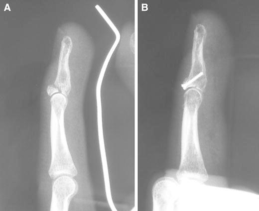
X-rayLateral finger radiographs before (a) and after (b) treatment: dorsal avulsion fracture at the base of the distal phalanx, anatomical reduction achieved and fixed with a screw.
Source : Shimura et al., Journal of orthopaedic science : official journal of the Japanese Orthopaedic Association, 2014, figure 3 · CC BY
The question looks decisive; it matters far less than the choice of orthosis, and the trials that have put it directly all give the same answer.
A systematic review gathered 44 studies reporting clinical results: 22 on surgical treatments, 17 on non-surgical ones. The two figures to remember are remarkably close5 :
Five studies compared the two strategies directly, with divergent results and recommendations. The authors' conclusion is explicit: both treatments give excellent clinical results, the available evidence does not allow us to determine when surgery is indicated, and these treatments appear equivalent and should be individualised5. It is the same conclusion the Cochrane review had drawn fourteen years earlier4.
On tendinous mallet finger. One trial allocated 48 patients between a thermoplastic orthosis and Kirschner wire fixation. At sixteen weeks, no significant difference in final extension or in extensor lag. Both groups showed the same pattern of early loss of extension after the immobilisation was removed, partly recovered through regular active exercise11.
On bony mallet finger with a fragment greater than a third, without initial subluxation. A level I randomised trial compared splinting with extension-block pinning in 32 patients randomised, 28 analysed. At six months, no difference in active extensor lag or in function and pain scores. Better still: flexion, active range and pulp-to-palm distance were better in the splinting group. But three patients in that group developed a secondary subluxation, which leads the authors to a nuanced conclusion: splinting is safe and effective for restoring mobility, it does not sufficiently prevent secondary subluxation, and radiographic monitoring during immobilisation appears necessary10.
On the comparison with an anchor technique. A level Ib randomised trial compared an anchor technique with conservative treatment in 29 patients. An extensor lag of 8.1° after surgery against 6.1° with conservative treatment, return to work at 63.2 days against 53.7, final distal interphalangeal flexion of 54.5° against 58.3°. And one result that is not trivial: radiographic joint degeneration was seen in four patients in the surgical group and in none in the conservative group at one year12.
Surgery against splinting: what the comparative data show
Systematic review of 44 studies, completed by three randomised trials
Sources: Lin JS, Samora JB. J Hand Surg Am 2018;43(2):146-163.e2 (PMID 29174096); Batıbay SG, Akgül T, Bayram S, Ayık Ö, Durmaz H. J Hand Ther 2018;31(4):429-436 (PMID 28966061).
Key point
The cost of this injury is almost never medical: it is an occupational and sporting cost, and it falls on the dominant hand three times out of four.
Two figures set out the problem. The dominant hand is involved in about 74 % of cases2, and treatment requires six to eight weeks of continuous immobilisation1. Applied to a tradesperson, a cook, a healthcare worker, a musician or a hairdresser, that is not settled with a prescription: it is planned for.
The sporting side is documented too. Mallet finger is said to account for 2 % of sports emergencies and to be the most frequent closed tendon injury both in contact sports and in the workplace15. The classic mechanism, a ball caught on the pulp with the finger extended, mostly concerns ball sports where the catch is made bare-handed.
Only one quantified figure for return exists in the randomised literature, and it comes from a level Ib trial comparing an anchor technique with conservative treatment in 29 patients: return to work at 63.2 days on average after surgery, against 53.7 days after conservative treatment12. Nine days apart, in favour of non-surgical treatment: one more argument, if one were needed, for not operating on a closed injury by default.
For return to sport, one has to be frank: no study has evaluated the terms or the timing of return to sport in this injury. What is done in practice is reasoning (a gradual return once active extension is achieved and stable, protection during activities exposing the finger to a direct blow) and it must be presented as such to the patient and the coach.
The case of the anaesthetist published in Anaesthesia Reports illustrates the order of magnitude of the impact when the hand is the tool of the trade: the injury, managed without surgery, required a significant absence from clinical work before a good recovery and a return to work17. The authors devote part of their discussion to contributing factors and prevention strategies, which, for an injury reputed to be benign, says enough about its real cost.
| Context | What the literature says | Adaptation to discuss |
|---|---|---|
| Heavy manual work | Return to work at 53.7 days on average after conservative treatment12 | A protective glove over the orthosis, adjusted or changed duties for the length of treatment |
| Caring professions and fine movements | One published case of prolonged absence from clinical work after an injury sustained in theatre17 | The thinnest possible orthosis, technical movements tried out before returning |
| Ball sports | The classic mechanism of a direct blow to the pulp; 2 % of sports emergencies15 | Stop catching situations during immobilisation; no data on timing of return |
| Keyboard work | No specific data | Compatible with wearing the orthosis in most cases; check there is no forced flexion when typing |
| Older patient, marked oedema | Marked oedema and older age associated with a greater final extensor lag14 | Close monitoring of the orthosis and of swelling, non-removable immobilisation to be considered |
Three messages, and it is better to give them together:
Key point
This is the part of treatment that falls to the physiotherapist, and paradoxically the least studied. A recent trial nevertheless brings something we did not have, and it concerns exactly that moment.
A level II randomised trial compared, in 48 patients with a tendinous mallet finger, a thermoplastic orthosis against Kirschner wire fixation. Beyond its primary result, no difference at sixteen weeks, it documents a clinical observation every practitioner knows and nobody had quantified: a loss of extension occurs in the days following removal of the immobilisation, whatever it was, and it is partly recovered through routine active exercise11.
The authors conclude that sustained active exercise appears to favour recovery of extension at the distal interphalangeal joint11. The wording is cautious, it is a secondary result of a trial that was not testing this question, but it finally gives some support to what practice has always done.
The trial brings a second element, useful for prediction: the final extension of the joint was correlated with the extension obtained under immobilisation (r = 0.60) and with the maximum extension of the contralateral finger (rho = 0.54)11. In other words, you have to look at the healthy finger opposite before setting a goal: a patient whose contralateral distal interphalangeal joint does not go beyond 0° will not do better on the injured side.
During immobilisation (weeks 0 to 8). The physiotherapist's role is not nil, it is preventive:
At weaning (weeks 8 to 12). The trial protocols provide for gradual removal over two to four weeks with exercises27. In practice:
What not to do. Adding a night orthosis after a correctly conducted full immobilisation adds nothing: the randomised trial that tested it found no difference in lag, disability or satisfaction8. And forcing flexion of the distal interphalangeal joint in the first weeks of weaning has no basis at all, whereas the risk of recreating tendon lengthening is mechanically real.
An editorial appraisal applying GRADE principles to the question of treating traumatic mallet finger. No formal GRADE assessment and no learned society guideline exists for this condition: the Cochrane review explicitly stopped at a finding of insufficient evidence4.
A custom-made thermoplastic orthosis rather than a prefabricated one. Multicentre randomised trial: 23.8 % treatment failure against 0 %7. Meta-analysis of 7 trials: risk of skin complication multiplied by 3.17 with prefabricated, by 6.72 against custom-made thermoplastic6. Downgraded from “high” because the final extensor lag does not in fact differ.
Do NOT add a night orthosis after full immobilisation. Randomised trial in 51 patients: no difference in lag, disability or satisfaction8. Direct evidence of an absence of benefit, on a relevant outcome.
Splinting rather than surgery as first line for closed injuries without subluxation. Systematic review of 44 studies: lag 5.7° against 7.6°, complications 14.5 % against 12.8 %5 ; three randomised trials with no difference101112, one of them with more joint degeneration on the surgical side12.
Continuous immobilisation in extension for 6 to 8 weeks. No trial has compared immobilisation with doing nothing, that would hardly be ethical. It is the basis of every trial protocol711 and the overview review notes that the lag does not regress spontaneously2.
Regular active exercise after removal of the immobilisation. A secondary result of a randomised trial: the early loss of extension after removal was partly recovered through routine active exercise, and sustained exercise appears to favour recovery11. That was not the question tested.
Oedema control during immobilisation. In a randomised trial, marked oedema and older age had a negative influence on the final extensor lag14. An observed association, not the effect of a tested intervention.
Mobilisation of the proximal interphalangeal joint from day one. No study has evaluated it in this indication. Consistent with the mechanics and without risk to the lesion, which does not involve that joint.
Gradual weaning over 2 to 4 weeks. A protocol applied by the trials7, never compared with abrupt removal. Reasoned practice, not demonstrated.
Passive mobilisation or massage of the distal interphalangeal joint during immobilisation. Contrary to the very principle of the treatment: any flexion restarts the count1. An unfavourable benefit-risk balance by construction.
Electrophysical agents (ultrasound, laser, currents) for mallet finger: no study identified in this indication.
What should prompt a return to the surgical opinion
Key point
Three published cases, with their identifiers. None describes an ordinary mallet finger: those are precisely the ones that teach something.
Case 1: A mallet finger that develops over three weeks, with no injury
Benabdallah O, Benabdallah R. Journal of Orthopaedic Case Reports 2022;12(11):60-6415: free full text (PMC10066668).
A 35-year-old woman presents with a mallet deformity of the right index finger. On questioning, she reports no injury : the deformity built up gradually over more than twenty days, preceded by moderate pain and burning sensations at the level of the third phalanx. On palpation, nodules are felt at the distal interphalangeal joint and on the dorsal aspect of the second phalanx. The radiograph shows the classic deformity, with no associated bony lesion.
A diagnosis of pigmented villonodular synovitis was suspected intraoperatively on finding haemosiderin in the tendon sheath and in the distal joint. Treatment combined excision of the mass, tenosynovectomy and tendon reinsertion.
What this case teaches. The authors stress that mallet finger occurs “always after a traumatic aetiology” and that this cause had never been reported. For practice, the rule is simple and requires no expertise: gradual onset plus no injury plus palpable nodules equals investigation, not splinting.
Case 2: A 4-year-old child, a painless deformity, an inconclusive radiograph
Aprilya D, Satria O, Vianney MM. International Journal of Surgery Case Reports 2025;129:11110516: free full text (PMC11932670).
The parents consult about a “bent” left little finger in a 4-year-old child. The deformity, painless, had progressed over a year, with no identifiable injury. Examination finds a palpable bony mass proximal to the angled distal interphalangeal joint. The radiograph was not conclusive and suggested an old fracture of the middle phalanx.
Intraoperatively, a sessile exostosis of the epimetaphysis of the middle phalanx was pushing the terminal extensor tendon towards the ulnar border. Biopsy confirmed an osteochondroma. Reconstruction of the terminal tendon and of the skin followed the resection; at two years of follow-up, there was no infection, no recurrence of the deformity and no tumour recurrence.
What this case teaches. Two flags were present together, the age and the absence of injury, and a third element could have been falsely reassuring: a radiograph interpreted as the sequel of a fracture. In a child, a progressive, painless mallet deformity is not a traumatic mallet finger, and an inconclusive radiograph is not a normal radiograph.
Case 3: An accident at work, non-surgical management, a long absence
de Vena Franks PL, Lightfoot NJ. Anaesthesia Reports 2020;8(2):127-13017: free full text (PMC7656317).
An anaesthetist injures themselves during induction, at tracheal intubation. The authors note that injuries to healthcare workers are frequent but rarely published, and that this mechanism had never been described. The injury was treated without surgery ; after a significant absence from clinical work, the doctor recovered well and returned to work. The article discusses the contributing factors and prevention strategies.
What this case teaches. It illustrates the most frequent and most unremarkable mechanism, forced flexion against resistance during an ordinary movement, and it recalls the real cost of this injury, which is not medical but occupational. Eight weeks of splinting on a working finger is something to plan with the patient at the first consultation: workplace adjustment, gloves, adapted equipment, and informing the employer.
Key point
A frequent injury, a long treatment, and a result that depends almost entirely on what the patient does between sessions: this is a condition in which the first consultation is worth more than all the others.
| What you measure | Why | How often |
|---|---|---|
| Active extensor lag in degrees | The primary outcome of every trial cited; comparable with the literature78 | At the outset, then at every assessment after weaning |
| Skin condition under the orthosis | The most frequent complication, up to three times more likely with a prefabricated model6 | At every session |
| Reported adherence | Correlated with the final lag in the multicentre trial7 ; it is the main lever | At every session |
| Range of the proximal interphalangeal joint | Its stiffness is the most troublesome functional sequela, and it is avoidable | At every session |
| Oedema | An unfavourable factor measured on the final lag14 | Weekly at first |
| Extension of the contralateral distal interphalangeal joint | Correlated with the final extension on the injured side11 : it sets a realistic goal | Once, at the outset |
| Follow-up radiograph for large bony mallet fingers | Three secondary subluxations under splinting in the level I trial10 | As prescribed, during immobilisation |
Not always, and it is better to know that from the start. In the randomised trial that is most informative on this point, the mean final extensor lag was 14°, and 34 % of patients kept a lag of 20° or more8. The systematic review gives more favourable means: 7.6° after splinting5, but a moderate residual lag is the rule rather than the exception, and it is most often well tolerated.
The count starts again from zero. That is not a teaching device: the reference chapter states that any flexion of the joint during the splinting process means starting again1. The healing tendon is brought back into a lengthened position, and the work of the past weeks is lost.
Yes, provided you hold the last phalanx in extension with the other hand throughout: you slide the splint off, you clean, you dry, you put it back, never letting the finger drop. If the patient is not sure they can do that, non-removable immobilisation is an alternative: in a randomised trial it gave 5° less lag than a removable orthosis at twelve weeks14.
No, for closed injuries without subluxation. The systematic review of 44 studies finds 5.7° of lag after surgery against 7.6° after splinting, with comparable complication rates (14.5 % against 12.8 %), and concludes that both treatments are equivalent and should be individualised5. A randomised trial even observed radiographic joint degeneration in four operated patients against none of the conservatively treated ones at one year12.
No. A comparison of two groups of 40 patients, treatment started within two weeks against more than four weeks after the injury, found success in 17 patients out of 21 in the early group and 15 out of 19 in the delayed group9. The full splinting treatment remains indicated, with the same requirement about wear.
The question is worth turning round: the off-the-shelf prefabricated splint, of the Stack type, showed 23.8 % treatment failure against 0 % for a custom-made thermoplastic orthosis in a multicentre randomised trial7, and a risk of skin complication multiplied by nearly seven in meta-analysis6. A custom-made orthosis, made by a trained physiotherapist or occupational therapist, is not a luxury: it is what makes the difference between a treatment seen through and a treatment abandoned.
Not during the eight weeks of continuous wear, except for activities that pose no risk to the finger and with the orthosis in place. No study has evaluated the terms of return to sport in this injury; the practical marker is that of weaning: a gradual return once active extension is achieved and stable, with protection during activities carrying a risk of a direct blow.
No, and it warrants an opinion. It is the swan-neck deformity : the loss of distal pull transfers force to the central slip and tips the proximal interphalangeal joint into hyperextension. Its correction is surgical2, and it is picked up by monitoring that joint at every session.
On the same subject
Every reference was verified individually in PubMed: identifier resolved, journal, year, volume and author list checked, abstract read to make sure the source does establish what is attributed to it.
Method and limitations
Sources searched in PubMed through the NCBI E-utilities services, crossing mallet finger with classification, orthoses, length of immobilisation, surgery, rehabilitation and case reports. Every identifier was resolved and every abstract read before citation. A strength of this file, rare in the field: six randomised trials and a Cochrane review: mallet finger is by far the best studied of the three subjects in this series. Main limitation : those trials compare orthoses with each other or splinting with surgery, never treatment with doing nothing, and none has evaluated a rehabilitation protocol as such. The levels of evidence in the table of modalities are an editorial appraisal applying GRADE principles, and not a published GRADE assessment: the Cochrane review explicitly stopped at a finding of insufficient evidence. Article written on 14 August 2026.
Behind this article
An author who explains, a reviewer who checks.
How we write and check our content

Physiotherapist · co-founder of Physio Learning
Marked for life by his first four-hour lecture without a single image, he took a master’s in instructional design so that it would never happen to anyone again. He hunts down publication bias and unreadable slides with the same intransigence.

Head of scientific content
Physiotherapist specialising in neuro-musculoskeletal practice and holder of a master’s in public health. He checks the methodological rigour of every article: primary sources, levels of evidence, no exceptions.
