
















On-siteDPCFIFPL

Upper limb
Hand rehabilitation and splinting: refining and deepening your practice
Jean Lichtle
Go further
Heberden's nodes (distal joint) and Bouchard's nodes (proximal joint): finger osteoarthritis, why some hurt, ruling out rheumatoid arthritis, rehab.

What the patient feels is bone, an osteophyte, never inflammation eating away.
7.6%of the world population affected by osteoarthritis, all locations combined, in 2020
What follows covers each of these points in detail, with sources. It is there if you need it.
This topic is taught in a course: Hand rehabilitation and splinting: refining and deepening your practice, on-site with Jean Lichtle.
1 / 13
12 article chapters · 58 min in total
Anatomy◔ 4 min
A nodule is bone: a marginal osteophyte, palpable because the skin there is thin.
Epidemiology◔ 6 min
Three figures circulate for hand osteoarthritis prevalence, and they differ by a factor of five.
Dissociate pain◔ 4 min
Contrary to the image of mechanical wear, deformity and pain do not go together.
Diagnose◔ 6 min
No single test suffices, but the combination of tests clearly raises the probability of the diagnosis.
Rule out RA◔ 6 min
Missing a rheumatoid arthritis loses months in a disease whose prognosis depends on delay.
Prognosis◔ 4 min
What is visible on imaging and what hurts do not progress together.
Rehabilitation◔ 10 min
Citing the studies rather than a homemade protocol is the only way to announce a realistic benefit.
Splints and aids◔ 5 min
Splints, assistive devices, joint economy: reduce load, not reinforce structure.
Surgery◔ 2 min
Surgery is considered for disabling pain despite conservative treatment, never on the X-ray.
Case reports◔ 3 min
Three cases each illustrate a pitfall: doubt resolved by ultrasound, or what the tissue reveals.
Talking to the patient◔ 3 min
What the patient will know about their fingers, they will know from the physiotherapist, in a few minutes.
Questions◔ 5 min
Nodules are newly formed bone; their link with radiographic osteoarthritis is firmly established.
Train on this
Upper limb
Jean Lichtle
Next comes practice: the course that teaches this topic, with Jean Lichtle.
Our courses on this topic may be covered by DPC or FIFPL, the French continuing-education funding schemes, depending on your profession and situation.
Fingers that get bigger at the last phalanges, that deviate slightly, and a patient who came in for her shoulder and ends up laying them on the table, asking: "and these, doctor, are they going to keep going?". Interphalangeal finger osteoarthritis is probably the condition the physiotherapist sees most often without ever having treated it. This article covers the long fingers, that is, the distal and proximal interphalangeal joints. For osteoarthritis of the thumb base, trapeziometacarpal osteoarthritis, which is another location with different management, see our dedicated article.
Synthesis written from primary sources verified one by one on PubMed. Every figure carries its reference at the point where it is written; the full bibliography, with PMID and DOI identifiers, appears at the end of the article.
Finger osteoarthritis in three figures
A very common disease, a diagnosis that never rests on a single sign, and a treatment whose measured effect is modest
Sources: Zhang Y et al. Am J Epidemiol 2002;156(11):1021-1027 (PMID 12446258); Zhang W et al. Ann Rheum Dis 2009;68(1):8-17 (PMID 18250111); Østerås N et al. Cochrane Database Syst Rev 2017;1:CD010388 (PMID 28141914).
What to take away before reading the rest
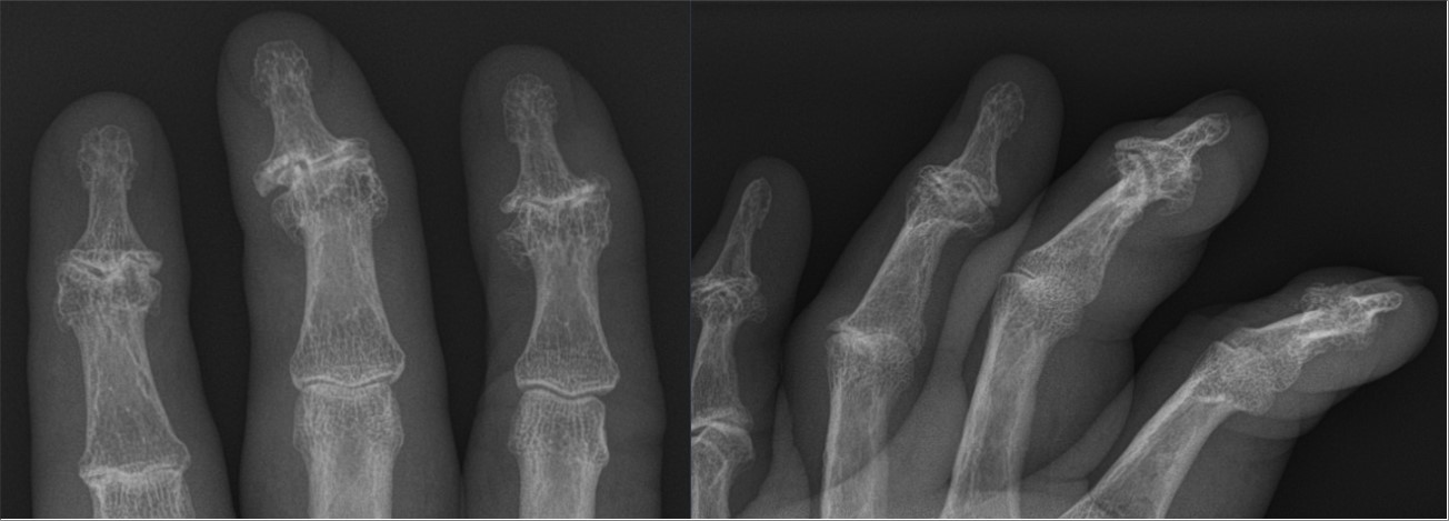
X-rayRadiograph of the fingers, frontal and oblique views: large marginal osteophytes at the distal interphalangeal joints, a deformity typical of Heberden's nodes.
Source : Hellerhoff, Wikimedia Commons · CC BY-SA
Two proper names from the eighteenth and nineteenth centuries today designate two levels of the same finger. Knowing which is which is not a nicety of vocabulary: it is what allows you to tell the patient, in three seconds, whether what they are showing matches what you think.
The long finger has three joints in series. From the palm towards the nail: the metacarpophalangeal joint (MCP), between the head of the metacarpal and the first phalanx; the proximal interphalangeal joint (PIP), between the first and second phalanges; the distal interphalangeal joint (DIP), between the second and third phalanges, just under the nail. The thumb, which has only two phalanges, has only one interphalangeal joint, and it also has a joint the other fingers do not: the trapeziometacarpal joint, at the base of the thenar eminence.
Finger osteoarthritis has a topography of its own, and that is almost a diagnostic sign in itself:
Where the nodes sit, and where they do not
The topography of finger osteoarthritis is so stereotyped that it constitutes a diagnostic argument in itself
Location frequencies from Dreiser RL, Maheu E, Guillou GB. Osteoarthritis Cartilage 2000;8 Suppl A:S25-28 (PMID 11156490), a series of 261 patients with symptomatic hand osteoarthritis. Rarity of metacarpophalangeal involvement: Haugen IK et al. Ann Rheum Dis 2011;70(9):1581-1586 (PMID 21622766). Diagram with no metric anatomical value.
The question the patient asks is almost never "what have I got?" but "what is this lump?". The answer fits in one word: it is bone. Nodes are osteophytes, marginal bony outgrowths that osteoarthritis builds around the rim of the joint, and which become palpable because the overlying tissues there are very thin.
This identification is not a clinical hunch: it has been measured. In the GOAL study, 1,939 subjects carrying at least one Heberden's or Bouchard's node were examined and then X-rayed. The association between the presence of a node and the underlying radiographic osteoarthritis ranged from an odds ratio of 2.26–21.23 depending on the joint, with a dose-effect relationship between the clinical severity of the node and the extent of the radiographic lesions11. Two secondary results from this same study are worth knowing, because they answer two questions patients ask: nodes are more frequent on the dominant hand, and more frequent in women11.
Saying "this is bone, not inflammation" defuses in one sentence the most widespread fear: that of having "the disease that eats you away", that is, rheumatoid arthritis.
One detail is worth noting, because it often appears next to a Heberden's node without the link being made: the mucous cyst. This is a soft, translucent swelling on the back of the finger between the DIP joint and the nail base. Its 2024 reference review recalls that it is associated with osteoarthritis of that joint, the osteophyte probably being the main causal factor, and that nail deformity may be the first visible abnormality, even before the cyst is obvious32. A patient who consults for a nail that is becoming grooved or ridged therefore sometimes actually has finger osteoarthritis.
The glossary to give the patient
Heberden's node = hard lump at the last joint of the finger, under the nail. Bouchard's node = the same thing at the middle joint. Digital osteoarthritis, finger osteoarthritis and interphalangeal osteoarthritis all designate the same disease. Thumb-base osteoarthritis = osteoarthritis of the base of the thumb, a neighbouring but distinct location. If the radiology report speaks of "osteoarthritis of the DIP and PIP joints", it is describing exactly these nodes.
Key points
It is one of the most common joint diseases of the second half of life, and the gap between what radiography sees and what the patient complains of is wider here than anywhere else.
Osteoarthritis, all locations combined, affected 595 million people in 2020, that is 7.6 % of the world population, according to the Global Burden of Disease 2021 analysis. The projections from that same analysis announce a rise of 48.6 % in the number of cases of hand osteoarthritis by 2050, under the effect of ageing and population growth alone9.
Three figures circulate for "the" prevalence of hand osteoarthritis, and they differ by a factor of five. This is not a contradiction, it is a question of definition. The Framingham cohort X-rayed both hands of its participants at inclusion and then nine years later, and distinguished three entities6 :
The most useful lesson of this table is not in the absolute values, it is in the way the ratio between the sexes changes according to the definition adopted. The female predominance, so often quoted as self-evident, is in reality modest on radiography (44.2 against 37.7) and clear-cut as soon as one considers the forms that hurt or that erode (a ratio close to 2 to 1, and to 3 to 1)6. In other words, men have almost as much finger osteoarthritis as women; it is the symptomatic forms that are female.
Hand osteoarthritis: what the definition changes
Age-standardised prevalences in the Framingham cohort, depending on whether radiography alone, symptoms or erosions are used
Source: Haugen IK, Englund M, Aliabadi P et al. Prevalence, incidence and progression of hand osteoarthritis in the general population: the Framingham Osteoarthritis Study. Ann Rheum Dis 2011;70(9):1581-1586 (PMID 21622766). Prevalences standardised to the 2000 United States population, 40 to 84 years.
A second cohort, that of Rotterdam, measured the same thing in 3,430 subjects aged 55 and over, with consistent results: 56 % radiographic osteoarthritis and 11 % symptomatic osteoarthritis. The erosive form there represented 2.8 % of the general population but 10.2 % of symptomatic subjects, and its presence was associated with an adjusted odds ratio of 3.6 for pain and 2.4 for disability8. An erosion is therefore not a radiological detail: it is the marker of a form that causes more suffering.
In the oldest subjects, the frequency of symptomatic forms becomes considerable. In the Framingham survey conducted in 1,041 people aged 71 to 100, 26.2 % of women and 13.4 % of men had symptomatic hand osteoarthritis. These subjects had a maximum grip strength reduced by 10 %, reported more difficulty writing or handling small objects (odds ratio 3.4), and more difficulty, both self-reported and observed, in carrying a 4.5 kg parcel (odds ratios 1.7 and 1.6)7.
France has a dedicated prospective hospital cohort for this condition, DIGICOD, set up in Paris and published in 2021. It included 426 patients followed for symptomatic hand osteoarthritis, and its baseline table is the identikit picture of the patient the physiotherapist meets in consultation: 84 % women, 66.7 years mean age, a mean disease duration of 12.6 years, activity pain of 44.4 mm out of 100, a mean FIHOA functional index of 19.9 out of 100, metabolic syndrome in 36.5 % of them, and an erosive form in 45.8 %10.
This last figure is far higher than that of the general population, and that is expected: DIGICOD is a hospital cohort, therefore enriched in severe forms. This must be remembered before quoting "46 % erosive forms" as an epidemiological datum: it describes a rheumatology caseload, not the patients of a community practice.
The typical French patient of a hospital cohort
Baseline characteristics of the 426 DIGICOD patients, the French prospective hand osteoarthritis cohort
Source: Sellam J, Maheu E, Crema MD et al. The DIGICOD cohort. Joint Bone Spine 2021;88(4):105171 (PMID 33689840). Erosive form defined by at least one joint in the erosive or remodelling phase according to the Verbruggen-Veys score.
The "why me" question comes up at every consultation. The honest answer is that associated factors are better known than causal ones, and that a 2021 systematic review devoted specifically to the prognostic factors of interphalangeal osteoarthritis says so bluntly. Out of 18 studies and 49 factors tested, only eight proved prognostic, and all with a limited or moderate level of evidence: advanced age in women and female sex (moderate evidence); family history of Heberden's nodes, Kashin-Beck disease, advanced age in men, being a dentist in men, finger fracture and parity (limited evidence)17.
The most important finding of this review is negative, and it deserves to be quoted as it stands: no prognostic factor for a symptomatic form has been identified17. In other words, we cannot say today which patient with nodes will have pain, and which will never have any trouble.
Heredity, for its part, is solid, and its shape is instructive. A study of 992 female twins modelled radiographic osteoarthritis at five sites: DIP, PIP, trapeziometacarpal, hip and knee. All were heritable, and the genetic influences were strongly correlated between the joints of the hand. But there was no evidence for shared genetic pathways between the hand, the hip and the knee19. A patient whose mother had "deformed fingers" therefore has an increased risk for their own fingers, and that risk says nothing about their knees.
In the field, the question of a link with manual work is raised at every consultation. The available data remain more solid for the thumb base than for the long fingers: it is for the trapeziometacarpal joint that a high occupational load has been identified as a clinically relevant risk factor33. For the interphalangeal joints, the 2021 systematic review retains only one occupation, that of dentist in men, and with a limited level of evidence17. Answering "your work made your finger osteoarthritis" therefore goes well beyond what the data allow.
Key points
This is the question the patient asks without putting it into words, and the one whose answer most changes how they live with their hands. Over the past fifteen years, it has received a measured answer rather than a theoretical one.
The image of a "wear and tear" osteoarthritis, purely mechanical, predicts that deformity and pain should go together. They do not. The clearest demonstration comes from the Bruneck population study, which examined and scanned 293 subjects aged 65 and over, split into three groups: those who met the ACR criteria for hand osteoarthritis, those who had finger joints that were enlarged but painless, and those who had no clinical abnormality at all12.
The result fits into two lines. Osteophytes were present in almost everyone, whatever the group: 100 % of symptomatic subjects, 99.4 % of subjects with painless enlarged fingers, and even 93.9 % of subjects with no clinical abnormality at all. By contrast, synovitis on grey-scale clearly separated the groups: 94 % in the symptomatic subjects against 67 % in those with painless fingers, and the Doppler signal 33 % against 13 %12.
Painful or painless enlarged fingers: what really separates them
The osteophyte is everywhere; it is inflammation that distinguishes the finger that hurts from the finger that merely grows
Source: Gasperi N, Schreiber N, Bosch P et al. Ultrasound-detected inflammation is more common in clinically manifest hand osteoarthritis than in painless bony enlarged finger joints: subanalysis of the population-based Bruneck study. Ther Adv Musculoskelet Dis 2022;14:1759720X221096382 (PMID 35586515). In subjects with no clinical abnormality at all, osteophytes were still present in 93.9 % of cases and synovitis in 39.4 %.
This result is not isolated. In a series of 55 patients scanned joint by joint, synovitis, effusion, synovial thickening and the Doppler signal were all associated with pain on palpation, with odds ratios of 4.1–4.9 and a dose-dependent relationship13. In the Norwegian Nor-Hand cohort, 290 patients, grade 2 or 3 synovitis at the interphalangeal joints multiplied by 3.17 the probability of pain on palpation in the same joint14.
An important nuance emerges from this last study, and it deserves to be remembered by the rehabilitation practitioner: at the interphalangeal joints, inflammation was linked to local pain, but not to overall hand pain, nor to function, nor to grip strength. Those links existed only for the trapeziometacarpal joint14. That is a fundamental difference between the two locations: the osteoarthritic thumb affects the whole hand, the osteoarthritic interphalangeal joint hurts where it is.
Finally, the meta-analysis of 32 ultrasound and MRI studies, covering nearly 2,000 participants, confirms the association between inflammatory signs and joint tenderness (odds ratios of 2.2 to 2.6) as well as with radiographic progression (odds ratios of 4.3 to 6.5), while concluding that the relationship is inconsistent with self-reported pain15. That is an honest nuance: inflammation explains part of the pain, not all of it.
Deformity is bone, and it does not go away. Pain is largely inflammation, and inflammation comes and goes. These are two distinct phenomena, and the patient has a right to know it.
It makes it possible to say three true things, all of which are reassuring without being false:
Key points
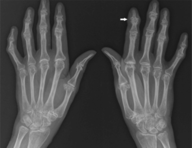
X-rayRadiograph of both hands: joint space narrowing in most metacarpophalangeal, proximal and distal interphalangeal joints, with osteophytes, arrow on an affected joint.
Source : de Wergifosse et al., The open rheumatology journal, 2014, figure 3 · CC BY-NC
No single test suffices, and that is the official conclusion of a European task force, not editorial caution. The diagnosis is built by accumulation, and it is built very well without imaging.
The EULAR recommendations devoted to the diagnosis of hand osteoarthritis, published in 2009, measured the sensitivity, specificity and likelihood ratio of each sign taken in isolation. Their conclusion is clear: no single test allows hand osteoarthritis to be defined, all having a likelihood ratio below 10, but combining the tests considerably increases the probability of the diagnosis3.
The figure they give as an example is the most telling in the whole diagnostic literature on this subject, and it deserves to be known by heart: the probability that a subject has hand osteoarthritis is 20 % when Heberden's nodes are present on their own ; it rises to 88 % when age over 40, a family history of nodes and radiographic joint space narrowing at any finger joint are added3.
Diagnostic approach to painful, swollen fingers
The tree does not seek to confirm osteoarthritis: it seeks first to rule out what must not be missed
Built from: Zhang W et al. Ann Rheum Dis 2009;68(1):8-17 (PMID 18250111) for the composite approach; Altman R et al. Arthritis Rheum 1990;33(11):1601-1610 (PMID 2242058) for the metacarpophalangeal criterion; Aletaha D et al. Arthritis Rheum 2010;62(9):2569-2581 (PMID 20872595) for the rheumatoid arthritis criteria. The 30-minute morning stiffness threshold is a clinical sorting landmark, not a criterion validated on its own; it is consistent with the mean duration of 20 minutes measured in symptomatic finger osteoarthritis (PMID 11156490).
The classification criteria of the American College of Rheumatology, published in 1990, were built by comparing osteoarthritic patients with patients who had hand symptoms of another origin, including rheumatoid arthritis and spondyloarthropathies. This methodological detail explains why they constitute, in practice, an excellent tool for differentiation4.
| Criterion | Content | What it means clinically |
|---|---|---|
| Prerequisite | Pain, discomfort or stiffness of the hand | The criterion applies only to a symptomatic hand: it does not classify radiographs |
| 1 | Swelling of hard tissue in at least 2 of the 10 selected joints | An osteophyte feels hard and fixed on palpation, unlike a synovitis, which rolls under the finger |
| 2 | Fewer than 3 swollen metacarpophalangeal joints | The major differential criterion. Three or more swollen MCP joints take the case out of the osteoarthritic frame |
| 3 | Hard swelling of at least 2 distal interphalangeal joints | The topographical signature of finger osteoarthritis: Heberden's nodes |
| 4 | Deformity of at least 1 of the 10 selected joints (required only if fewer than 2 DIP joints are affected) | A fallback route for forms with proximal or trapeziometacarpal predominance |
Three of these four criteria are enough to classify, with a sensitivity of 94 % and a specificity of 87 %. The classification-tree version, which performs better, reaches 92 % sensitivity and 98 % specificity. The authors also conclude, and this is a sentence worth keeping, that radiography contributes less than the clinical examination to classifying symptomatic hand osteoarthritis4.
Inspection first, comparative, hands laid flat. Note the swellings and at which level they sit, the lateral deviations (an osteoarthritic DIP joint frequently deviates radially or ulnarly), the extension lag of the last phalanx, the state of the nails, and any dorsal mucous cyst.
Palpation next, joint by joint. The question to settle is the nature of the swelling: hard and fixed, it signals an osteophyte, therefore osteoarthritis; elastic, warm, giving a sense of rebound, it signals synovitis. It is the manoeuvre that discriminates best at the bedside, and it costs nothing.
Mobility, actively then passively. The extension lag of the DIP joint is frequent and often more troublesome than the flexion lag, because it hampers laying the hand flat. Pulp-to-palm distance is also measured, being simple and reproducible.
Strength, finally. Grip strength on a dynamometer and thumb-index pinch. It is the parameter that best documents the impact, and the Framingham cohort had measured a 10 % loss of maximum grip strength in subjects with symptomatic hand osteoarthritis7.
To quantify the disability, the reference tool in French is the Dreiser functional index (FIHOA), a 10-item questionnaire administered by the examiner. Its sensitivity to change over six months was measured in a randomised trial, with a mean standardised response of 0.58, lower than that of the simple visual analogue pain scale (0.87)36. In other words, to follow a patient's progress, a well-kept VAS remains at least as informative as a functional score.
Not to make the diagnosis. The clinical picture is sufficiently characteristic, and the ACR criteria showed precisely that imaging contributed less than the examination4. Radiography retains three useful indications: documenting the presence of erosions, which identifies a more painful and more progressive form818 ; resolving a doubt in an atypical presentation; and preparing a surgical opinion.
Ultrasound, for its part, has a clear place when doubt about inflammatory rheumatic disease persists. It allows synovitis and erosions to be confirmed or ruled out, as in the case report below where it was precisely ultrasound that corrected the diagnosis34.
Red flags on examination of a painful hand
Key points
This is the only place in this article where an error is costly. Finger osteoarthritis taken for rheumatoid arthritis leads to needless investigations and worry; rheumatoid arthritis taken for osteoarthritis loses months of treatment in a disease whose prognosis depends on how quickly care begins.
The two diseases present in the same way in the patient's eyes: fingers that swell, that hurt, and that become deformed. Yet they have almost nothing in common when looked at closely. The confusion arises because everyday language calls both wear and tear and an autoimmune disease "rheumatism".
The site publishes a full synthesis on rheumatoid arthritis ; this chapter deals only with sorting, that is, with the practical question: does what I have in front of me warrant a prompt rheumatology opinion?
Two diseases, two signatures
What to look at, in order, to decide between finger osteoarthritis and rheumatoid arthritis
Built from: Altman R et al. Arthritis Rheum 1990;33(11):1601-1610 (PMID 2242058); Aletaha D et al. Arthritis Rheum 2010;62(9):2569-2581 (PMID 20872595); Zhang W et al. Ann Rheum Dis 2009;68(1):8-17 (PMID 18250111). Mean duration of morning stiffness in symptomatic finger osteoarthritis: 20 ± 27.6 minutes, measured in 261 patients (PMID 11156490). Proportion of normal CRP: DIGICOD cohort, 90 % (PMID 33689840).
First axis, topography. It is the most discriminating, and it is free. Osteoarthritis lodges in the interphalangeal joints and leaves the metacarpophalangeal joints alone; the ACR criteria formalised this by requiring fewer than three swollen MCP joints to classify osteoarthritis4, and the Framingham cohort confirmed that incident osteoarthritis of the MCP joints and the wrist was rare6. Rheumatoid arthritis does the opposite: it preferentially occupies the MCP joints and the wrists, and the DIP joint is classically spared. In the 2010 ACR/EULAR criteria, the domain that carries the most weight is precisely the number and site of the affected joints, scored from 0 to 5 out of a total score of 105.
Second axis, the consistency of the swelling. Hard, fixed, bony, it is an osteophyte. Soft, elastic, warm, with a sense of rebound, it is a synovitis. This manoeuvre costs only a few seconds per joint and it is more informative than many investigations.
Third axis, the timing and duration of stiffness. This is the point most often used badly, because people ask about the existence of morning stiffness instead of asking about its duration. Yet finger osteoarthritis produces it too: in a series of 261 patients with symptomatic hand osteoarthritis, the mean duration of morning stiffness was 20 minutes (standard deviation 27.6)36. The right question is therefore not "are you stiff in the morning?", to which everyone answers yes, but "how long does it take you, in the morning, to get your hands back?". A patient who answers "an hour and a half" is not in the osteoarthritis picture.
Fourth axis, the course through the day and over time. Osteoarthritic pain is mechanical: it appears with use and eases with rest, with flares lasting a few weeks. Inflammatory pain wakes the patient at night, in the second half of the night, and improves with limbering up. The duration of symptoms is in fact one of the four scored domains of the ACR/EULAR criteria for rheumatoid arthritis5.
Red flags: when to refer without delay
The practical rule. The physiotherapist does not have to settle this diagnosis; they have to spot that the picture falls outside the frame and to inform the general practitioner. In rheumatoid arthritis, the very aim of the 2010 criteria was to identify earlier the patients at risk of persistent, erosive disease, so that treatment can be started sooner5. Delay is the variable that can be acted upon.
| Diagnosis | What puts you on the trail | What should raise the alarm |
|---|---|---|
| Finger osteoarthritis | DIP and PIP joints, hard swelling, short morning stiffness, family history, after the age of 50 | Nothing, if the picture is complete |
| Erosive finger osteoarthritis | Same topography, but more inflammatory flares, more marked pain and disability | Adjusted odds ratios of 3.6 for pain and 2.4 for disability8, and it predicts radiological progression18 |
| Rheumatoid arthritis | MCP joints and wrists, soft and symmetrical swelling, long morning stiffness, inflammatory syndrome | Refer without delay: the prognosis depends on how early treatment starts5 |
| Psoriatic arthritis | DIP involvement possible, skin or nail psoriasis, dactylitis, enthesopathy, axial involvement | This is the trap of this chapter: it shares the topography of osteoarthritis. Two published cases in fact combine erosive osteoarthritis and psoriatic arthritis in the same patient35 |
| Chondrocalcinosis and gout | Sudden flare, one or two joints, tight, red skin, resolution within a few days | The diagnosis rests on the crystals in the joint fluid, and it can coexist with osteoarthritis35 |
| Mucous cyst | Soft, translucent swelling on the back of the finger, between the DIP joint and the nail, with nail deformity | This is not a differential diagnosis but a complication: it reflects underlying DIP osteoarthritis32 |
| Systemic causes | Nodular-looking periarticular swellings with no radiographic osteoarthritis | A published case describes periarticular cholesterol deposits mimicking Bouchard's and Heberden's nodes in familial hypercholesterolaemia38 |
Key points
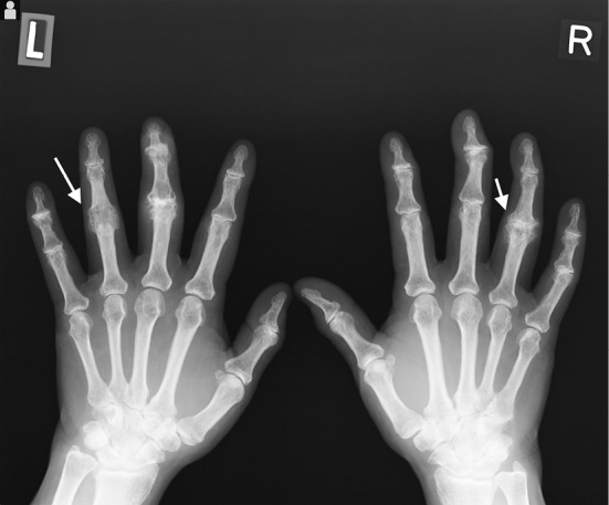
X-rayRadiograph of both hands: bony ankylosis of one proximal interphalangeal joint on each side, arrows, a deformity characteristic of erosive osteoarthritis.
Source : Takizawa et al., Internal Medicine, 2017 · CC BY-NC-ND
This is the question that causes the most anxiety, and the one on which good-quality longitudinal data are available. They say something counter-intuitive: what is visible and what hurts do not progress together.
A Dutch cohort followed 289 patients for six years, with standardised clinical and radiographic assessment at inclusion and at the end. The results are worth knowing, because they make it possible to answer the patient honestly16 :
This dissociation was found again in an independent Belgian cohort, LIHOC, which followed 203 patients for two years: all the radiographic scores worsened significantly even in the absence of clinical worsening. In that same cohort, the predictive factors differed according to the outcome considered: the initial pain level predicted clinical progression, whereas the presence of erosive or swollen joints predicted radiological18.
A radiograph that worsens does not mean the patient will fare worse, and a patient who is improving does not have a radiograph that improves. These are two parallel stories.
The radiological literature has long described interphalangeal joints as evolving through predictable anatomical phases, rather than in a linear fashion. The Verbruggen and Veys scoring system, still used today, including in the French DIGICOD cohort10, describes their succession: a normal phase, then the appearance of the classic osteoarthritic lesions, then loss of the joint space, then in some cases a frank erosive phase with destruction of the subchondral bone plate, and finally, after these erosive episodes, remodelling processes20.
It is this last phase, the remodelling one, that gives its basis to the everyday clinical observation: a joint that has finished transforming is often less painful than while it was transforming. Histological analysis of two proximal interphalangeal joints surgically replaced for erosive osteoarthritis points the same way: it shows active osteoclastic subchondral bone resorption, but also peripheral fibrocartilaginous resurfacing with remodelling of the exposed bone, and a synovium that is only moderately hypertrophic37.
An intellectual caution is called for here. That the pain of a given joint decreases once the remodelling phase is reached is consistent with the anatomical phases described, with the clinical-radiological dissociation measured over six years, and with the very existence of a large population of enlarged but painless fingers. It is not, however, a trial result: no study has directly measured the course of pain joint by joint through these phases. One can therefore tell the patient that established deformity is not synonymous with increasing pain, without promising that their pain will stop on a fixed date.
The anatomical phases of an osteoarthritic interphalangeal joint
The joint does not deteriorate linearly: it goes through phases, including a remodelling phase that follows the erosive episodes
Anatomical phases after Verbruggen G, Goemaere S, Veys EM. Clin Rheumatol 2002;21(3):231-243 (PMID 12111630); the letters N, S, J, E and R follow the nomenclature of the anatomical phase scoring system. Six-year progression data: Bijsterbosch J et al. Ann Rheum Dis 2011;70(1):68-73 (PMID 20736393).
Honesty requires recalling the negative result of the systematic review devoted to the prognostic factors of interphalangeal osteoarthritis: out of 49 factors tested in 18 studies, none predicted the occurrence of a symptomatic form17. The clinician who promises a patient a precise trajectory is making it up.
What can be said, on the other hand, rests on measurements:
Key points
There are international recommendations on hand osteoarthritis, two meta-analyses on exercise and several randomised trials. Citing them rather than proposing a home-made protocol is the only way to announce to the patient a benefit that matches what they will actually get.
Two texts are authoritative. The EULAR recommendations, updated in 2018, built by a task force of 19 clinicians, health professionals and patients from ten European countries: five general principles and ten recommendations, of which the first three cover the non-pharmacological (education, assistive devices, exercises, splints)1. And the 2019 ACR and Arthritis Foundation recommendations, which explicitly apply the GRADE methodology and distinguish strong from conditional recommendations2.
That the order of these recommendations places the non-pharmacological first is not a courtesy: it is the translation of the fact that no disease-modifying treatment exists in this condition. Conventional and biological DMARDs are moreover explicitly discouraged by EULAR1, and the COLOR trial, conducted against placebo in 100 patients, showed that colchicine did not relieve finger pain (a difference of 0.4 mm out of 100, confidence interval of -7.6 – 6.7) while causing more adverse effects (72 % against 44 %)30.
This is the point on which one must be most precise, because it is the one where the temptation to embellish is strongest.
The Cochrane review of 2017 pooled seven trials. In the five trials and 381 participants that reported pain, exercise reduced it with a standardised mean difference of -0.27 (95 % confidence interval: -0.47 to -0.07). Translated into language a patient can understand, that represents an absolute reduction of 5 % on a scale of 0 to 10, that is 0.5 points compared with the control group, with a number needed to treat of 9. Function improved by -0.28 (-0.58 to 0.02), an interval which crosses zero, and joint stiffness by -0.36 (-0.58 to -0.15). The overall quality of the evidence was judged low, because of the absence of blinding on self-reported outcomes and the imprecision of the estimates. Finally, and this is the result most often passed over in silence: the positive effect on pain, function and stiffness was not maintained at medium and long-term follow-up21.
The JOSPT meta-analysis of 2024, more recent and larger (14 trials, 1,341 participants), finds the same pattern of results with slightly larger effect sizes in the short term22 :
What hand exercises achieve, with their uncertainty
Standardised mean differences and 95 % confidence intervals, expressed in favour of exercise
Sources: Østerås N, Kjeken I, Smedslund G et al. Exercise for hand osteoarthritis. Cochrane Database Syst Rev 2017;1:CD010388 (PMID 28141914); Huang L, Zhang ZY, Gao M et al. The Effectiveness of Exercise-Based Rehabilitation in People With Hand Osteoarthritis. J Orthop Sports Phys Ther 2024;54(7):457-467 (PMID 38506711). The signs of the standardised mean differences have been presented as absolute values, oriented in favour of exercise, to allow comparison between outcomes running in opposite directions.
The most recent systematic review, published in 2025 and based on 65 new randomised studies since 2017, does not change this picture but refines it: it retains low-certainty evidence in favour of a small effect of hand exercises on long-term pain, and stresses, which is of direct interest to the rehabilitation practitioner, that there is a lack of interventions that effectively improve grip strength23.
The most instructive trial in this whole literature is a British 2x2 factorial trial, which randomised 257 patients aged 50 and over into four groups: information leaflet alone, joint protection, hand exercises, or both. The interventions were delivered by nine occupational therapists, in four group sessions24.
At six months, on the primary outcome (the OARSI/OMERACT responder rate):
A secondary benefit of joint protection lasted a long time: the improvement in self-efficacy in the face of pain was significant at 3 months, 6 months and still at 12 months24. No adverse effects were reported.
In the only trial that compared them head to head, teaching the patient to use their hands differently did better than making them exercise their hands.
This result does not say that exercises should be abandoned: it says that the consultation time devoted to teaching new ways of moving is not time wasted, and that it is probably better invested than extra time spent adding exercises. That is consistent with the place the EULAR recommendations give to education and assistive devices, even before exercises, in their order of presentation1.
| Option | What was measured | Certainty | What to do with it |
|---|---|---|---|
| Education and self-management | The strong ACR recommendation for self-efficacy and self-management programmes, in all osteoarthritis locations2 ; general EULAR principle1 | Strong recommendation | Routine, from the first session onwards |
| Joint protection | 33 % responders against 21 % at 6 months, p = 0.03; self-efficacy gain maintained at 12 months24 | One good-quality randomised trial | At least as much of a priority as exercises |
| Assistive devices | The moderate long-term effect on function, certainty moderate23 ; +1.8 points of COPM performance, effect size 0.925 | Moderate | The best benefit-to-effort ratio in the whole table |
| Exercises: stiffness | SMD -0.33 (-0.51; -0.16), certainty moderate22 | Moderate | This is the best-supported indication for exercises |
| Exercises: pain | SMD -0.27, that is 0.5 points out of 1021 ; -0.65 in the short term22 | Low | To be offered while stating the real magnitude |
| Exercises: grip strength | SMD +0.21 (0.03; 0.38)22 ; the 2025 review notes the absence of an effective intervention on this outcome23 | Low | Do not make it the main objective |
| Exercises beyond 6 months | No additional effect on pain, function and stiffness22 ; benefit not maintained21 | Low | Do not promise a lasting benefit |
| Night splint for the DIP joint | Mean pain reduced at 3 months (p = 0.002), extension lag improved (p = 0.016), in 26 subjects26 | Very low: non-randomised controlled trial, small sample | The most specific intervention for the long fingers, to be offered on one targeted joint |
| Finger splints (other than the thumb) | The conditional ACR recommendation for splints on hand joints other than the trapeziometacarpal joint2 ; EULAR recommendation1 | Conditional | Worth trying, while assessing tolerance |
| Thermal modalities, paraffin | The conditional ACR recommendation2 ; randomised trial of 56 patients: pain at rest and on activity improved up to 12 weeks, and strength maintained while the control group lost some27 | Conditional | Useful in preparation for a session or during a flare |
| Combined functional consultation | A single interdisciplinary session: grip strength up in the treated group, down in the usual-care group (p = 0.001), 151 patients28 | One randomised trial | A realistic model for a one-off consultation |
| Topical NSAIDs | The first-line drug treatment according to EULAR, preferred to systemic forms1 ; certainty high for a small short-term effect on function23 | High for a small effect | Worth reminding the patient and the prescriber |
| Intra-articular corticosteroids | Generally not recommended in hand osteoarthritis, but to be considered for a painful interphalangeal osteoarthritis1 | An explicitly provided exception | An option for a resistant localised flare |
| Colchicine | A difference of 0.4 mm out of 100 against placebo, and more adverse effects30 | A negative randomised trial | Do not offer |
| Conventional and biological DMARDs | Discouraged by EULAR in hand osteoarthritis1 | A negative recommendation | Do not offer |
Moderate certainty
Assistive devices on long-term function, and exercises on stiffness. These are the only two statements in this article that can be made without heavy reservations.
Low certainty
Exercises on pain, function and grip strength in the short term. The effect exists, it is small, and it does not survive six months. Saying so to the patient does not weaken the management, it prevents disappointment.
Very low certainty
Night splint for a distal interphalangeal joint. A single trial, controlled but not randomised, in 26 subjects. It is nevertheless the intervention most specifically studied on the long fingers, and it carries no risk.
Negative data
Colchicine, and disease-modifying antirheumatic drugs. The first has been tested against placebo and showed nothing; the second are explicitly discouraged by EULAR.
The protocols of the trials included in the Cochrane review varied a great deal, but all aimed to improve muscle strength and joint stability or function, with frequencies ranging from two or three times a week to three or four times a day. Self-reported adherence to the prescribed frequency lay between 78 % and 94 %21. This high adherence is an encouraging finding: patients do the exercises when they are explained to them.
Adverse effects deserve a sentence. Three trials reported them: they were few and not serious, consisting of increased inflammation in one finger joint and hand pain. The relative risk of an adverse event was 4.55, but with a confidence interval running from 0.53 to 39.31, that is, an estimate too imprecise to draw a conclusion21. Practical translation: these hands can be exercised, while watching for flares.
| Objective | Content | Dosing landmarks | Points to watch |
|---|---|---|---|
| Mobility | Analytical flexion-extension of each DIP and PIP joint, full fist, hook fist, thumb-to-finger opposition, pulp-to-palm distance | 1 to 2 times a day, 10 repetitions per movement, without forcing at end of range | This is the indication with the best certainty (stiffness). Warn that range will not return to normal |
| Strength and grip | Squeezing a foam ball, graded-resistance putty, thumb-index and thumb-middle pinch, extension against a finger elastic | 2 to 3 times a week, low to moderate resistance, stop if pain persists for more than 24 hours | The expected gain is small (SMD 0.21); do not make it the central objective |
| Function | Real everyday actions: doing up buttons, opening a jar with the adapted technique, turning a key, holding a wide-barrelled pen | Built into activities, with no dedicated session | This is where joint protection takes over from exercises |
| Preparation | Paraffin bath or local heat before the session or during a flare | A conditional recommendation; the protocol tested was 5 sessions a week for 3 weeks27 | A comfort modality, not a disease-modifying treatment |
One trial is worth knowing because it comes close to occupational health conditions: 29 car industry workers with hand osteoarthritis were randomised between a finger exercise programme combined with paraffin baths and paraffin baths alone. The combined group did significantly better on grip strength (p = 0.015), pain and function29. The sample is small, but the conclusion is consistent with the rest: exercise adds something to the passive modality.
Key points
This is the side of management with the most solid data, and paradoxically the one to which the least time is devoted. Three tools, three different levels of evidence, and one common logic: reduce the load rather than reinforce the structure.
This is the only splinting intervention studied specifically on the long fingers, and it is worth knowing, because most of the hand splinting literature concerns the thumb.
The trial, prospective, with the radiologist blinded, internally controlled but not randomised, included 26 subjects with painful, deforming DIP osteoarthritis. Each subject designated one intervention joint and one control joint. A custom-made gutter splint was worn every night for three months on the intervention joint only, with clinical and radiological assessment at inclusion, at 3 and at 6 months26.
The results: mean pain and maximum pain in the splinted joint were significantly lower at 3 months than at baseline (p = 0.002 and p = 0.02); the difference between the splinted joint and the control joint reached significance at 6 months (p = 0.049), that is, three months after the splint was stopped; and the extension lag improved significantly in the splinted joints, including compared with the contralateral counterpart joint (p = 0.016). The authors conclude that this is a safe and simple modality, with no particular non-adherence, no increase in stiffness and no joint restriction26.
The limitations must be stated : 26 subjects, no randomisation, and a primary outcome compared with baseline rather than with the control. That is very low certainty in the GRADE sense. But the intervention is risk-free, inexpensive, and it answers the patient's twofold complaint: the pain and the finger that no longer lies flat.
In practice
Make a thermoplastic dorsal or palmar gutter splint immobilising only the most painful DIP joint, in a comfortable extension position, leaving the PIP and MCP joints free. Wear it at night only. Reassessment at six weeks on pain and on the extension lag. One joint at a time: that is how the trial was conducted, and it lets the patient judge the usefulness for themselves by comparing their two hands.
This is the modality with the highest certainty in the whole of non-pharmacological management. The 2025 systematic review concludes that there is moderate-certainty evidence that assistive devices have a moderate long-term effect on function23.
The trial supporting it is clear-cut. Seventy patients with hand osteoarthritis were randomised between information alone and information plus the provision of assistive devices and splints, with assessment at three months using the Canadian Occupational Performance Measure. The equipped group gained 1.8 points out of 10 in performance (confidence interval 1.1 to 2.6) and 1.7 points in satisfaction, which corresponds to an effect size of 0.9, that is, a large effect25. Compare that with the 0.27 of exercises on pain.
The aids to offer are ordinary and available in a pharmacy or a supermarket: a jar opener fixed to the worktop, enlarged grips for cutlery and pens, spring-loaded scissors, an ergonomic knife with a perpendicular blade, a lever key turner, a chopping board with spikes, lever taps, a zip puller.
This is the intervention which, in the only trial that compared it with exercises, did better than they did24. Its principle is to learn to perform the same actions while placing less load on the small joints.
The six principles of hand joint economy
Classic occupational therapy practice content, to be adapted to the patient's real activities
Content from everyday occupational therapy practice, presented here as a teaching framework and not as a trial protocol. The effectiveness of joint protection as an intervention was demonstrated in a randomised factorial trial (Dziedzic K et al. Ann Rheum Dis 2015;74(1):108-118, PMID 24107979), whose publication does not detail the content session by session. The two-hour landmark is a usual teaching rule, not one derived from a trial.
One field remark, to close this chapter. The trials that tested these interventions had them delivered by occupational therapists, in group sessions for the British trial24, and by health professionals from several disciplines trained in rheumatology for the Austrian trial28. The latter in fact showed that a single individual session, followed by a telephone call at one month, was enough to increase grip strength in the treated group while it decreased in the group receiving usual care (p = 0.001, 151 patients)28. That is a format that can be transposed to private practice.
Key points
Surgery of the osteoarthritic long fingers is rare, late, and effective on pain at the price of an accepted stiffness. Knowing what it offers makes it possible to talk about it without brandishing it as a threat or presenting it as a simple solution.
The EULAR recommendations devote their ninth recommendation to surgery1. The principle is constant throughout osteoarthritis: it is considered when pain remains disabling despite well-conducted conservative management, and not on the radiographic appearance.
This is the reference operation at that level. The joint is fixed in a functional position, generally with a headless compression screw. The reasoning is simple: the DIP joint has a small useful range, and losing it is better tolerated than its pain.
The reference series covers 102 fingers in 59 patients, with a mean follow-up of 26 months. The results are good: 89 of the 102 cases fully satisfied on the patients' side, and 9 satisfied; only four complications (two prominent implants, one type 1 complex regional pain syndrome, one symptomatic bony callus), all revised surgically; no non-union, no malunion, no nail dystrophy, no infection, and union of every arthrodesis31.
At the proximal level, loss of mobility is much less well tolerated, because the PIP joint provides most of the curling of the finger. Prosthetic replacement therefore has its place there. Histological analysis of two PIP joints replaced with an uncemented ceramic prosthesis, in a context of erosive osteoarthritis, in fact provided interesting information on the mechanism of damage: complete erosion of the articular cartilage with peripheral fibrocartilaginous resurfacing, sclerosis and remodelling of the exposed bone, and above all osteoclastic activity with resorption lacunae in the subchondral bone, the synovium being only moderately hypertrophic37. The authors draw from this the hypothesis of cartilage resorption starting from the subchondral bone, and not from the joint surface.
This is a frequent reason for surgical referral and one often misunderstood by the patient, who thinks a "lump" is being removed when the procedure is actually on the joint. The 2024 reference review is explicit: the most effective treatment to prevent recurrence is surgical, combining joint debridement and excision of the cyst32. Removing the cyst without treating the osteophyte is treating the consequence.
What justifies a referral
Key points
Three published observations, chosen because each illustrates a different trap: the diagnostic doubt resolved by ultrasound, the coexistence of several diseases in the same hand, and what is found when the tissue itself is examined.
An osteoarthritis that looked like rheumatoid arthritis
Lewis KL, Battaglia PJ. J Chiropr Med 2019;18(1):56-60 (PMID 31193241)
The presentation. A woman aged 48 years consults for bilateral hand pain of one year's duration, with stiffness, swelling and redness of the proximal and distal interphalangeal joints.
The trap. Redness, swelling, bilateral involvement and a relatively young age: the picture first suggests rheumatoid arthritis. The blood tests and plain radiographs came back equivocal, that is, neither confirming nor excluding.
What settled it. Diagnostic ultrasound, which showed neither synovitis nor erosion in the joints of the hand. The diagnosis retained was bilateral symptomatic hand osteoarthritis.
What followed. Conservative treatment combining mobilisations, instrument-assisted soft tissue therapy and laser. At one month, the patient had regained a full and painless range, her strength, and full working activity.
What the case teaches. First, that finger osteoarthritis can present signs that look inflammatory, which is consistent with the ultrasound data cited above: synovitis is frequent in it1213. Next, that in case of doubt, the useful imaging is not radiography but ultrasound. Finally, and this is what the authors stress, that what is at stake in this sorting is the referral : rheumatology if it is rheumatoid arthritis, conservative management if it is osteoarthritis.
When three diseases share the same hand
Hoxha A, Ruffatti A, Alberioli E et al. Clin Rheumatol 2016;35(7):1885-1889 (PMID 25833145)
The presentation. Two women aged 71 and 85, both followed for long-standing osteoarthritis and carrying discreet psoriasis, consult for a painful, swollen joint: the first interphalangeal joint for one, the wrist for the other.
What was done. A joint aspiration, in both cases. The fluid was inflammatory and contained calcium pyrophosphate crystals. Radiographs of the hands, feet and knees simultaneously showed the characteristic features of erosive osteoarthritis, of psoriatic arthritis and of chondrocalcinosis. HLA typing found predisposing alleles in both patients.
What the case teaches. That the reasoning "it is osteoarthritis, therefore it is nothing else" is wrong. Known finger osteoarthritis does not immunise against inflammatory rheumatic disease or against crystal arthropathy, and an osteoarthritic patient one of whose joints suddenly becomes inflamed deserves an examination, not a shrug. It is also a reminder that psoriasis, even discreet, is to be looked for on examination: skin folds, scalp, nails.
What is seen when the tissue is examined
Favero M, Perino G, Valente ML et al. Skeletal Radiol 2017;46(3):385-391 (PMID 28054155)
The context. Two patients with erosive finger osteoarthritis underwent replacement of a proximal interphalangeal joint with an uncemented ceramic prosthesis. The surgical specimens were analysed histologically, with radiological correlation.
What was found. Complete erosion of the articular cartilage, with in places a peripheral fibrocartilaginous resurfacing ; sclerosis and remodelling of the exposed bone; an osteoclastic activity with resorption lacunae in the subchondral bone and around degenerative fibromyxoid pseudocysts; coarse trabeculation of the cancellous bone; and marginal osteophytes. The synovial membrane, for its part, was only moderately hypertrophic, with a sparsely cellular stroma.
What the case teaches. The authors propose that, in erosive osteoarthritis, cartilage resorption comes from the subchondral bone by an osteoclastic route, and not from the joint surface. Two practical consequences: synovitis is not the sole driver of this form, and the remodelling phase described by radiologists has a tissue reality, which gives a basis to what is observed clinically, namely joints that end up stabilising, deformed.
What these three cases have in common
In all three, the initial diagnosis or the initial picture of the disease was inadequate, and it was a targeted additional investigation that changed management: an ultrasound scan, an aspiration, a histological analysis. None was corrected by one more plain radiograph. The message that transposes to practice is simple: faced with a picture that does not fit, what is needed is not one more image, it is a different investigation, and it is requested by a letter to the general practitioner.
The patient you are seeing for their shoulder or their knee may never consult for their hands. What they know about it, they will know from you. Here is enough to hold that conversation in a few minutes, without promising anything false.
"What you can feel is bone, not inflammation eating away at you." It is true (nodes are osteophytes11), it is reassuring, and it answers the real fear, which is that of rheumatoid arthritis.
"Deformity and pain are not the same thing." Many people have enlarged fingers with no pain at all: in the Bruneck study, that group really did exist, and 99.4 % of its members had osteophytes on ultrasound12. A finger that grows does not herald a finger that will hurt more and more.
"Over six years, one person in two does not get worse." That is what the six-year cohort measured: about half the patients report a deterioration, so the other half do not, and the course of the radiographs does not predict that of the symptoms16.
"It is not because you used your hands too much." Guilt is frequent, and it is unfounded for the long fingers: the systematic review of prognostic factors in interphalangeal osteoarthritis retains only one occupation, with a limited level of evidence17. Occupational load is documented for the thumb base33, not for the interphalangeal joints.
"We cannot make the nodes disappear, but we can change what you manage to do." This is the honest statement of the scope: no disease-modifying treatment1, but one modality with a large effect on function, assistive devices2325.
| Step | What is done | Why |
|---|---|---|
| 1. Sort | Which joints, hard or soft, duration of morning stiffness, systemic signs, psoriasis | This is the only step where an error is costly45 |
| 2. Name | Say the name, say that the node is bone, say that deformity and pain are dissociated | That is what changes the experience, and it costs only two minutes12 |
| 3. Measure | Pain VAS, grip and pinch strength, pulp-to-palm distance, Dreiser index if a score is wanted | The VAS was more sensitive to change than the functional score36 |
| 4. Equip | Two or three assistive devices chosen for the activities the patient mentions themselves | Best certainty and largest effect size in the whole table2325 |
| 5. Protect | The principles of joint economy, applied to three concrete actions in their day | Did better than exercises in the trial that compared them24 |
| 6. Mobilise | A short self-exercise programme, stating the real magnitude of the benefit | Best certainty on stiffness22, high adherence in the trials21 |
| 7. Splint if needed | A night splint on the most painful DIP joint, one at a time | The intervention best studied specifically on the long fingers26 |
| 8. Reassess | At six weeks, on pain and function, never on a radiograph | Radiological progression and clinical course are independent1618 |
The patient is not asking for their old fingers back. They are asking to be told what lies ahead, and to be shown how to go on opening their jars.
Key points
No. They are osteophytes, that is, new bone formed around the rim of the joint, and their association with the underlying radiographic osteoarthritis is firmly established, with a dose-effect relationship between their clinical severity and the extent of the lesions11. No medical treatment makes them regress. The inflammatory swelling that accompanies them during flares, on the other hand, can subside, and that is often what the patient sees varying.
Not necessarily, and it cannot be predicted. In the Framingham cohort, the development of erosive disease occurred mainly in people who already had non-erosive osteoarthritis at inclusion, rather than in those who had none6. The joints most often affected are the distal interphalangeal joints (67.6 % of patients in a symptomatic series), ahead of the thumb base (62 %) and the proximal interphalangeal joints (47.5 %)36.
Yes, and the genetic component is specific to the hand. A study of 992 female twins showed that genetic influences were strongly correlated between the joints of the hand, but that there was no evidence for shared genetic pathways between the hand, the hip and the knee19. A family history of Heberden's nodes is in fact one of the identified prognostic factors, with a limited level of evidence17, and it was found in 48 % of patients in a French series36. Having "your mother's hands" therefore says nothing about your future knees.
No, they are two different diseases. Finger osteoarthritis affects the interphalangeal joints while sparing the metacarpophalangeal joints, swells hard, comes with about twenty minutes of morning stiffness36 and with normal blood tests in nine cases out of ten10. Rheumatoid arthritis does the opposite. Any doubt calls for a prompt specialist opinion, because its prognosis depends on how early treatment starts5. The topic is covered in our article devoted to rheumatoid arthritis.
Not to make the diagnosis. The authors of the ACR criteria concluded that radiography contributed less than the clinical examination to classifying symptomatic hand osteoarthritis4. It retains its value for looking for erosions, which identify a more painful form8, for resolving a doubt, or before a surgical opinion. Where there is doubt about inflammatory rheumatic disease, ultrasound settles the question, not one more X-ray.
No. Hand exercises are safe: in the Cochrane review, the few adverse effects reported were limited to increased inflammation in one joint and hand pain, with no serious events21. The aim is not to use your hands less, but to use them differently, which is precisely what joint economy and assistive devices are about2425.
Yes, but modestly. The Cochrane review puts the gain in pain at about 0.5 points on a 10-point scale compared with no exercise, on low-quality evidence, and this effect was not maintained in the medium and long term21. The 2024 meta-analysis finds somewhat larger short-term effects, the only moderate certainty being for stiffness22. Stating this order of magnitude avoids a disappointment which, in turn, is what really makes people stop the programme.
On the long fingers, the available trial concerns a gutter splint worn at night on a single distal interphalangeal joint for three months: pain decreased and the extension lag improved, with no added stiffness or non-adherence26. The study covered 26 subjects and was not randomised, so certainty is very low, but the intervention is simple and carries no risk. For the thumb, splinting is the subject of a strong ACR recommendation2, which is not the case for the other joints of the hand, where the recommendation is conditional.
The sources cited in this article do not address this question, and it would not be honest to assert an answer. What can be said is that local heat has been tested: a randomised trial of 56 patients showed that a paraffin bath programme improved pain at rest and during daily activities, with an effect lasting twelve weeks27, and the ACR issues a conditional recommendation in favour of thermal modalities2. If a patient finds relief from heat, there is nothing against using it.
Only one compound appears in the recommendations: chondroitin sulphate, which EULAR mentions for symptomatic relief1 and which the ACR retains as a conditional recommendation for the hand2. Conversely, colchicine has been tested against placebo in 100 patients and produced no benefit on pain, with more adverse effects30. None of these options alters the course of the disease.
Not genetically. The twin study cited above found no evidence for shared genetic factors between hand, hip and knee, and concludes that there are site-specific aetiological differences19. This does not mean that the same patient cannot have both, but that one does not predict the other through heredity.
The sources cited here do not give an average flare duration. What is documented is that the mean number of painful flares over the preceding twelve months was 4.4 in a series of 261 symptomatic patients, with a mean of 3.7 painful joints36. The intermittent pattern is therefore the rule, which is consistent with the largely inflammatory origin of the pain1314.
To go further on the site
Method and limitations
The sources were searched in PubMed via the NCBI E-utilities services, crossing hand osteoarthritis and interphalangeal osteoarthritis with epidemiology, diagnosis, differential diagnosis, imaging, exercise, splints, assistive devices, joint protection and surgery. Every identifier was resolved and every abstract read before citation ; two older case reports, spotted during the search and relevant in substance, were set aside for lack of an accessible abstract allowing verification of what they establish, and the thirty-eighth reference is flagged as having been verified only on its title.
Three limitations should be known to the reader. First, a large part of the hand rehabilitation literature concerns hand osteoarthritis as a whole, trapeziometacarpal joint included: the effect sizes of the meta-analyses cited are therefore not specific to the interphalangeal joints, and the only trial genuinely specific to the long fingers is the night splint one, in 26 subjects and without randomisation. Next, the certainty ratings reproduce the GRADE appraisals published by the sources cited where they exist; the ranking in the table of modalities, for its part, is an editorial appraisal applying GRADE principles, and not a published GRADE assessment. Finally, the idea that a joint becomes less painful once its transformation is complete is consistent with the anatomical phases described, with the clinical-radiological dissociation measured and with the existence of a population of painless enlarged fingers, but no study has directly measured this trajectory joint by joint : it is presented here as an inference, never as a result.
Article written on 15 August 2026.
Behind this article
An author who explains, a reviewer who checks.
How we write and check our content

Physiotherapist · co-founder of Physio Learning
Marked for life by his first four-hour lecture without a single image, he took a master’s in instructional design so that it would never happen to anyone again. He hunts down publication bias and unreadable slides with the same intransigence.

Head of scientific content
Physiotherapist specialising in neuro-musculoskeletal practice and holder of a master’s in public health. He checks the methodological rigour of every article: primary sources, levels of evidence, no exceptions.
