















In brief
Piriformis syndrome is a non-discogenic compression of the sciatic nerve by the piriformis muscle, now folded into the broader concept of deep gluteal syndrome (DGS). It presents as deep buttock pain radiating down the back of the thigh, most often stopping above the knee and aggravated by prolonged sitting; it affects women aged 30 to 50 preferentially. Diagnosis is clinical and by exclusion. First-line treatment is conservative and multimodal (6 to 12 weeks): education, piriformis stretching, strengthening of the hip abductors and extensors, and neural mobilisation.
Clinical synthesis based on the most recent systematic reviews and prospective data: Martin KSSTA 2014, Hopayian SR 2018, Kay Arthroscopy 2017, Poutoglidou Cureus 2020, Külcü TJPMR 2024.
Clinical summary
- Piriformis syndrome is a non-discogenic compression of the sciatic nerve by the piriformis muscle. It belongs to the broader concept of deep gluteal syndrome (DGS), which covers any subgluteal compression of the nerve (Martin J Hip Preserv Surg 2015, Hernando Skeletal Radiol 2015).
- It preferentially affects adult women aged 30-50. The commonly quoted F/M ratio (6:1) rests on old clinical series; the true prevalence in the population remains poorly estimated.
- The anatomical variants of the sciatic nerve are the main mechanical predisposition: 16.9 % pooled variants across 6062 cadavers (Smoll 2010), confirmed at 16-22 % by the Poutoglidou 2020 meta-analysis.
- Diagnosis is clinical, by exclusion : no single test is definitive. The combination of the active piriformis test + the seated piriformis stretch reaches sens. 91 % / spec. 80 % (Martin KSSTA 2014, n=52, endoscopic neurolysis).
- The mandatory differential diagnoses: lumbar radiculopathy, sacroiliac involvement, femoroacetabular impingement, proximal hamstring tendinopathy, gluteal vascular anomalies.
- The classification into primary (intrinsic) and secondary (trauma, repeated microtrauma, sporting hypertrophy) steers the treatment strategy.
- First-line treatment is conservative and multimodal (6-12 weeks) : education, piriformis stretching, strengthening of the hip abductors and extensors, neural mobilisation (Tonley JOSPT 2010, Probst PM R 2019).
- Radial ESWT is equivalent to corticosteroid injection over 12 weeks (Ahadi J Bodyw Mov Ther 2023, n=35, RCT).
- Piriformis dry needling of trigger points is an adjunctive option (moderate evidence, methodological heterogeneity).
- Making the patient autonomous through self-stretching, home strengthening and ergonomics (sitting < 30 min, a cushion) is crucial in preventing recurrence.
- The return to sport is guided by functional criteria and not by a calendar: complete freedom from pain, symmetrical range, abductor strength ≥ 90 % of the healthy side (Ardern BJSM 2016).
- The endoscopic neurolysis of the sciatic nerve with release of the piriformis is indicated for refractory cases > 6 months or for symptomatic anatomical variants (Kay Arthroscopy 2017 SR, Park BMC Musculoskelet 2016).
- The conceptual shift towards deep gluteal syndrome (DGS) obliges the clinician to assess beyond the piriformis alone: the gemelli, obturator internus, quadratus femoris, the fibrovascular bands.
- The red flags (IFOMPT, Finucane JOSPT 2020) mandate medical referral: progressive motor deficit, suspected cauda equina, a mass, a vascular anomaly, fever.
- Measuring outcomes with PROMs (the Lower Extremity Functional Scale, pain VAS, the Modified Harris Hip Score) is essential to make progress objective and to demonstrate the value of care.
Contents
- What are the fundamentals to know about piriformis syndrome and DGS?
- How do you assess and diagnose piriformis syndrome with confidence?
- What should you know about the anatomical variants of the sciatic nerve (Beaton-Anson)?
- Which treatment strategies are the most effective for piriformis syndrome?
- How do you secure lasting recovery and prevent recurrence of piriformis syndrome?
- What do real clinical cases teach us about piriformis syndrome?
- How do you apply these recommendations concretely in your practice?
What are the fundamentals to know about piriformis syndrome and DGS?
Piriformis syndrome historically denotes a non-discogenic compression of the sciatic nerve by the piriformis muscle, presenting as buttock pain radiating along the sciatic path and mimicking a “true” radicular sciatica.¹ The recent international literature now places this entity within a broader nosological frame: the deep gluteal syndrome (Deep Gluteal Syndrome, or DGS), which encompasses every subgluteal compression of the sciatic nerve, the piriformis being only the most frequent cause among the gemelli, the obturator internus, the quadratus femoris and the fibrovascular bands.²,³
How is this condition defined, who does it affect and what are the risk factors?
Piriformis syndrome and DGS share a common definition: an extrapelvic entrapment of the sciatic nerve in the subgluteal space, distinct from any discal or radicular origin.⁴ The typical presentation combines deep buttock pain, posterior thigh radiation (most often stopping above the knee) and exacerbation by sitting.⁵
The epidemiology remains debated because no rigorous population study quantifies the true prevalence. The historical range of 5 to 36 % of sciatica⁶ comes from retrospective clinical studies with no homogeneous diagnostic criteria. Caution is needed: these figures cannot be generalised to the general population.
Female sex is over-represented in almost every clinical series, with a reported F/M ratio of around 6:1 in some older cohorts.⁷ This difference may reflect anatomical (pelvic width, Q angle), hormonal and behavioural factors (high heels, asymmetrical postures in prolonged sitting). This ratio comes from clinical populations (women consult more), however, not from population studies, which limits its external validity.⁸
The age band usually involved is 30-50 years, the period when prolonged seated work, chronic sporting practice and any age-related musculotendinous changes combine to create fertile ground.¹,⁹
The risk factors are classically split into two groups:
- Primary causes (≈ 15 %): intrinsic disease of the muscle (spasm, fibrosis, myositis, isolated hypertrophy).¹⁰
- Secondary causes (≈ 85 %) :
- Macrotrauma : a fall onto the buttock, a road traffic accident, a direct contusion; it can generate a local haematoma and perineural scar fibrosis.
- Repeated microtrauma 🏃: running on hard ground, cycling, prolonged walking; mechanical overload of the piriformis and the deep external rotators.
- Prolonged sitting 💺: on a hard surface, in the car, at the desk; “ wallet neuritis ” (a wallet in the back pocket) is its caricature.⁵,⁶
- Piriformis hypertrophy in the athlete: sprinters, dancers, cyclists; overload of the external rotators against a short muscle excursion.
- After hip surgery : scarring, haematoma, biomechanical change after total replacement or arthroscopy.²
What happens in the body, and how does piriformis syndrome evolve naturally?
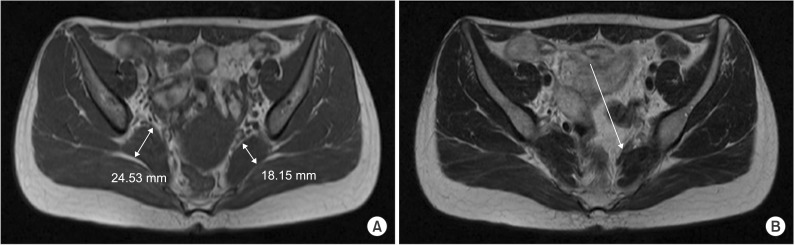
MRIAxial pelvic MRI: asymmetric enlargement of the right piriformis muscle (A) and a dark signal band anterior to the left piriformis (B), in a patient with bilateral piriformis syndrome.
Source : Moon et al., Annals of Rehabilitation Medicine, 2015, figure 2 · CC BY-NC
The piriformis is a small deep external rotator of the hip in extension, which becomes an abductor when the hip is flexed to 90°. It runs from the sacrum (S2-S4) to the greater trochanter and passes through the greater sciatic foramen, where it is topographically intertwined with the sciatic nerve.¹
The mechanical conflict arises when the piriformis increases in volume (hypertrophy, spasm, post-traumatic oedema) or loses its suppleness (fibrosis), compressing the sciatic nerve directly against the bony structures of the pelvis. This compression triggers a pathological cascade:
- Local inflammation (mechanical neuritis)
- Intraneural ischaemia through compression of the vasa nervorum
- In chronicity: damage to the myelin sheath (segmental demyelination) and disturbed nerve conduction
It is this pathophysiology that generates the typical symptoms: radiating pain, paraesthesia, a burning or numb sensation.⁵,¹⁰
The natural history is variable and poorly documented by good prospective studies. Most patients see their symptoms improve significantly with well-conducted conservative management (education, stretching, activity modification, strengthening).⁵,⁶ Untreated, a subgroup moves towards chronicity with persistent nerve compression, constant pain, and even muscle atrophy of the lower limb in prolonged forms.¹
- Piriformis syndrome is a non-discogenic compression of the sciatic nerve, folded today into the broader concept of deep gluteal syndrome (DGS).
- The true prevalence remains imprecise (5-36 % of sciatica according to the clinical series); the F/M ratio of ~6:1 comes from clinical cohorts, not population ones.
- The anatomical variants of the sciatic nerve are the main mechanical predisposition (16.9 % of cadavers, Smoll 2010).
- The secondary causes (> 85 %) are mechanical: direct trauma, repeated microtrauma, prolonged sitting, sporting hypertrophy.
- The pathophysiology combines inflammation, intraneural ischaemia and, in chronicity, segmental demyelination.
- The prognosis is good with early, well-matched conservative management.
Bibliography, chapter 1
- Hicks BL, Lam JC, Varacallo MA. Piriformis Syndrome. In: StatPearls. Treasure Island (FL): StatPearls Publishing; 2023. PMID: 28846222.
- Carro LP, Hernando MF, Cerezal L, Navarro IS, Fernandez AA, Castillo AO. Deep gluteal space problems: piriformis syndrome, ischiofemoral impingement and sciatic nerve release. Muscles Ligaments Tendons J. 2016;6(3):384-396. PMID: 28066745.
- Martin HD, Reddy M, Gómez-Hoyos J. Deep gluteal syndrome. J Hip Preserv Surg. 2015;2(2):99-107. doi:10.1093/jhps/hnv029.
- Geler Külcü D. Deep Gluteal syndrome: An underestimated cause of posterior hip pain. Turk J Phys Med Rehabil. 2024;70(1):4-16. PMID 38549816.
- Probst D, Stout A, Hunt D. Piriformis Syndrome: A Narrative Review of the Anatomy, Diagnosis, and Treatment. PM R. 2019;11(Suppl 1):S54-S63. PMID: 31102324.
- Siddiq MAB. Piriformis Syndrome and Wallet Neuritis: Are They the Same? Cureus. 2018;10(5):e2606. doi:10.7759/cureus.2606.
- Boyajian-O'Neill LA, McClain RL, Coleman MK, Thomas PP. Diagnosis and management of piriformis syndrome: an osteopathic approach. J Am Osteopath Assoc. 2008;108(11):657-664. PMID: 19011229.
- Hopayian K, Danielyan A. Four symptoms define the piriformis syndrome: an updated systematic review of its clinical features. Eur J Orthop Surg Traumatol. 2018;28(2):155-164. PMID: 28836092.
- Cass SP. Piriformis syndrome: a cause of nondiscogenic sciatica. Curr Sports Med Rep. 2015;14(1):41-44. PMID: 25574881.
- Vij N, Kiernan H, Bisht R, Singleton I, Cornett EM, Kaye AD, et al. Surgical and Non-surgical Treatment Options for Piriformis Syndrome: A Literature Review. Anesth Pain Med. 2021;11(1):e112825. PMID: 34221947.
How do you assess and diagnose piriformis syndrome with confidence?
Diagnosing piriformis syndrome, and more broadly deep gluteal syndrome (DGS), remains a challenge because neither a pathognomonic sign nor a gold standard exists. The approach is a rigorous one: first rule out the more frequent and better-defined causes of buttock pain, then gather a converging body of clinical evidence pointing to a subgluteal compression.¹,²
Which questions should you ask to understand the patient and their history?
The history is the foundation of the diagnosis. Several features carry strong orienting value :
- Site and radiation : deep buttock pain, often at the level of the sciatic notch, radiating down the back of the thigh but stopping most often above the knee. Radiation as far as the foot points rather to an L5/S1 lumbar radiculopathy.³
- Prolonged sitting as an aggravator 💺: particularly on a hard surface, while driving, or with a wallet in the back pocket (the “wallet sign”).¹,⁴
- Aggravation by activity : running, climbing stairs, moving from sitting to standing.²
- A history of direct trauma (a fall, a buttock contusion), of overuse (running, cycling) or of hip surgery.²,⁵
- Neuropathic character of the pain: burning, tingling, numbness, with no true motor deficit and no lost reflexes (which would point to a radiculopathy).³
The systematic review of Hopayian & Danielyan 2018 identified four key symptoms that together make a strong diagnostic cluster: buttock pain, pain aggravated by sitting, external tenderness over the greater sciatic notch, and pain on any manoeuvre that increases piriformis tension.⁶
Which clinical tests, and which other conditions should you rule out?
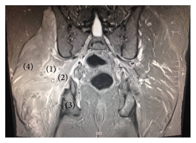
MRICoronal T2 pelvic MRI: signal abnormality of the right pelvitrochanteric muscles, with the piriformis labelled 1, obturator internus 2, ischium 3 and gluteus maximus 4.
Source : McCabe et al., Case Reports in Orthopedics, 2016, figure 1 · CC BY
The physical examination combines provocation tests specific to the piriformis with a systematic differential diagnosis. The reference work of Martin et al. KSSTA 2014 (52 patients with sciatic entrapment confirmed at endoscopic neurolysis) quantified for the first time the diagnostic performance of two tests:
The main tests to know, and how to perform them:
- Active Piriformis Test (Martin 2014): side-lying with the healthy side down, the patient holds an active abduction and external rotation against the clinician's resistance. Positive if the buttock pain is reproduced.⁷
- Seated Piriformis Stretch Test : seated, the ankle of the symptomatic side resting on the opposite knee, trunk flexed forwards. Positive if the pain is reproduced.⁷
- FAIR test (Flexion-Adduction-Internal Rotation): side-lying, hip flexed to 60°, the clinician applies passive adduction and internal rotation. Positive if the pain is reproduced.⁸
- Pace sign : pain and weakness on resisted abduction and external rotation in sitting.²
- Freiberg sign : pain on forced passive internal rotation with the hip extended.²
- Deep palpation of the piriformis region (the sciatic notch): it can reproduce the radiation.¹
The differential diagnosis is the most important step. You must systematically rule out:
- L4-S1 lumbar radiculopathy : a full neurological examination (strength, reflexes, dermatomes), the straight leg raise / slump test, spinal imaging where there is doubt.⁹
- Sacroiliac disease : the Laslett cluster (distraction, compression, Gaenslen, sacral thrust, thigh thrust); at least 3 positive tests out of 5 points strongly.¹⁰
- Femoroacetabular impingement and intra-articular hip disease: the FADIR test, the FABER test, joint range.¹¹
- Proximal hamstring tendinopathy : pain on palpation of the ischial tuberosity, pain on resisted lengthening.¹²
- Vascular disease , rare (aneurysm, malformation), where the pain is pulsatile or resistant to well-conducted treatment.
Imaging is not required to diagnose piriformis syndrome, but it is useful in order to rule out other conditions. Lumbar MRI and pelvic MRI are the reference investigations. Magnetic resonance neurography (MRN) can show piriformis hypertrophy, an abnormal sciatic nerve signal or an anatomical variant, but it remains a second-line investigation.¹²
Should patients with piriformis syndrome be classified, and what are the benefits?
Yes: the primary/secondary classification is clinically useful in steering the treatment strategy:
- Primary syndrome (≈ 15 %): intrinsic disease (spasm, fibrosis, myositis, isolated hypertrophy, a symptomatic anatomical variant). Initial treatment remains conservative, but failure should bring surgery into the discussion sooner.¹
- Secondary syndrome (≈ 85 %): an identifiable cause (trauma, overuse, after surgery). Treating the cause + multimodal physiotherapy is very often enough.⁵
A complementary anatomical classification uses the Beaton-Anson types (the relationship between the sciatic nerve and the piriformis), detailed in the next chapter.
Critique and controversy: a catch-all diagnosis?
The validity of piriformis syndrome as a distinct diagnostic entity is the subject of serious academic debate. Many experts stress that it is over-diagnosed, applied whenever sciatica without a discal cause is found.² The poor sensitivity and specificity of the isolated clinical tests, the absence of a non-invasive gold standard and the conceptual shift towards DGS all support that criticism. The most robust strategy therefore remains:
- Rule out the more frequent differential diagnoses rigorously.
- Combine ≥ 2 positive provocation tests.
- Document the absence of any neurological deficit.
- Reserve imaging for atypical or refractory cases.
- Progressive motor deficit of the lower limb (foot drop, weak extension or flexion)
- Suspected cauda equina syndrome : bladder or bowel disturbance, saddle anaesthesia
- A history of cancer + pain that persists at rest
- Fever, night sweats, unexplained weight loss
- Pulsatile pain or a palpable mass (suspected aneurysm, tumour)
- Rapid deterioration or a major acute post-traumatic picture
- The diagnosis of piriformis syndrome is a clinical diagnosis of exclusion.
- The history looks for: buttock pain, aggravation on sitting, radiation stopping above the knee, the wallet sign.
- The combination of the Active Piriformis Test + the Seated Piriformis Stretch reaches sens. 91 % / spec. 80 % (Martin KSSTA 2014, n=52).
- Mandatory differential diagnoses: lumbar radiculopathy, sacroiliac, FAI, proximal hamstring tendinopathy.
- Imaging serves mainly to rule out the other conditions; MRN is a second-line option.
- The primary vs secondary classification steers the treatment strategy and how quickly surgery is considered where it fails.
Bibliography, chapter 2
- Hicks BL, Lam JC, Varacallo MA. Piriformis Syndrome. StatPearls. 2023. PMID: 28846222.
- Cass SP. Piriformis syndrome: a cause of nondiscogenic sciatica. Curr Sports Med Rep. 2015;14(1):41-44. PMID: 25574881.
- Probst D, Stout A, Hunt D. Piriformis Syndrome: A Narrative Review of the Anatomy, Diagnosis, and Treatment. PM R. 2019;11(Suppl 1):S54-S63. PMID: 31102324.
- Siddiq MAB. Piriformis Syndrome and Wallet Neuritis: Are They the Same? Cureus. 2018;10(5):e2606. doi:10.7759/cureus.2606.
- Carro LP, Hernando MF, Cerezal L, et al. Deep gluteal space problems. Muscles Ligaments Tendons J. 2016;6(3):384-396. PMID: 28066745.
- Hopayian K, Danielyan A. Four symptoms define the piriformis syndrome: an updated systematic review of its clinical features. Eur J Orthop Surg Traumatol. 2018;28(2):155-164. PMID: 28836092.
- Martin HD, Kivlan BR, Palmer IJ, Martin RL. Diagnostic accuracy of clinical tests for sciatic nerve entrapment in the gluteal region. Knee Surg Sports Traumatol Arthrosc. 2014;22(4):882-8. PMID 24217716.
- Boyajian-O'Neill LA, McClain RL, Coleman MK, Thomas PP. Diagnosis and management of piriformis syndrome: an osteopathic approach. J Am Osteopath Assoc. 2008;108(11):657-664. PMID: 19011229.
- Fishman LM, Dombi GW, Michaelsen C, et al. Piriformis syndrome: diagnosis, treatment, and outcome--a 10-year study. Arch Phys Med Rehabil. 2002;83(3):295-301. PMID 11887107.
- Laslett M. Evidence-based diagnosis and treatment of the painful sacroiliac joint. J Man Manip Ther. 2008;16(3):142-152. PMID: 19119403.
- Geler Külcü D. Deep Gluteal syndrome: An underestimated cause of posterior hip pain. Turk J Phys Med Rehabil. 2024;70(1):4-16. PMID 38549816.
- Michel F, Decavel P, Toussirot E, Tatu L, Aleton E, Monnier G, Garbuio P, Parratte B. The piriformis muscle syndrome: an exploration of anatomical context, pathophysiological hypotheses and diagnostic criteria. Ann Phys Rehabil Med. 2013;56(4):300-311. PMID: 23684469.
What should you know about the anatomical variants of the sciatic nerve (Beaton-Anson)?
The anatomical variants of the sciatic nerve in its relationship with the piriformis muscle are the main mechanical predisposition to piriformis syndrome and to DGS. Knowing them is essential in order to interpret an MRI, plan an injection and, above all, understand why some patients are refractory to conservative treatment.¹,²
How are the variants classified, and how common are they?
The historical classification of Beaton & Anson (1937) distinguishes six types according to the division of the sciatic nerve into tibial and fibular branches and their path relative to the piriformis:
The cadaveric meta-analysis of Smoll 2010 remains the reference: across 6062 pooled cadavers from 18 studies, the prevalence of a variant (other than type A) is 16.9 %, with a wide range between studies (1.5 % to 35.8 %) reflecting methodological heterogeneity (dissection, definition, ethnicity).¹
The more recent meta-analysis of Poutoglidou 2020 confirmed those values by including MRI and operative series (>10 % of subjects carry a variant; up to 22 % in East Asians).² Type B (the fibular branch through the piriformis) remains the most frequent symptomatic variant.
What are the clinical and surgical implications?
Not every variant is symptomatic. Most carriers of a type B/C/D variant remain asymptomatic all their lives, which makes it a factor of mechanical predisposition and not a direct cause. Symptoms appear when an additional factor (microtrauma, sporting hypertrophy, mechanical overload) triggers the conflict.²,³
Three major clinical implications:
- Suspicion in a refractory case : a patient not responding to well-conducted conservative treatment for more than 6 months should raise the possibility of an anatomical variant. Magnetic resonance neurography (MRN) is the second-line investigation for showing it.⁴
- A targeted surgical strategy : knowing the variant before surgery allows the approach (endoscopic vs open) and the extent of the release (isolated piriformis release vs partial resection) to be adapted.⁵
- Caution during injections : a type D variant (the whole nerve through the piriformis) carries an increased risk of direct neurotoxicity if the needle crosses the nerve.
The endoscopic neurolysis of the sciatic nerve with release of the piriformis is the reference procedure for refractory symptomatic variants. The systematic review of Kay et al. (Arthroscopy 2017) analysed all the published surgical series in DGS: significant improvement in pain score and function, with few complications. This minimally invasive approach has largely supplanted the historical open surgery.⁵
- The Beaton-Anson classification distinguishes 6 types of sciatic nerve / piriformis relationship.
- The type A configuration (the normal one, nerve below the piriformis) accounts for ~83 % of the population.
- The pooled variants stand at 16.9 % on cadaveric meta-analysis (Smoll 2010, 6062 cadavers).
- A variant is not automatically symptomatic : it is a mechanical predisposition that needs a trigger (microtrauma, hypertrophy).
- In practice, MRN is the reference investigation for showing the variant where conservative treatment fails.
- The endoscopic neurolysis is the surgery of choice for refractory symptomatic variants (Kay Arthroscopy 2017 SR).
Bibliography, chapter 3
- Smoll NR. Variations of the piriformis and sciatic nerve with clinical consequence: a review. Clin Anat. 2010;23(1):8-17. PMID: 19998490.
- Poutoglidou F, Piagkou M, Totlis T, Tzika M, Natsis K. Sciatic Nerve Variants and the Piriformis Muscle: A Systematic Review and Meta-Analysis. Cureus. 2020;12(11):e11531. PMID: 33354475.
- Hicks BL, Lam JC, Varacallo MA. Piriformis Syndrome. StatPearls. 2023. PMID: 28846222.
- Martin HD, Reddy M, Gómez-Hoyos J. Deep gluteal syndrome. J Hip Preserv Surg. 2015;2(2):99-107. doi:10.1093/jhps/hnv029.
- Kay J, de Sa D, Morrison L, Fejtek E, Simunovic N, Martin HD, Ayeni OR. Surgical Management of Deep Gluteal Syndrome Causing Sciatic Nerve Entrapment: A Systematic Review. Arthroscopy. 2017;33(12):2263-2278.e1. PMID: 28866346.
- Geler Külcü D. Deep Gluteal syndrome: An underestimated cause of posterior hip pain. Turk J Phys Med Rehabil. 2024;70(1):4-16. PMID 38549816.
- Carro LP, Hernando MF, Cerezal L, et al. Deep gluteal space problems. Muscles Ligaments Tendons J. 2016;6(3):384-396. PMID: 28066745.
Which treatment strategies are the most effective for piriformis syndrome?
Treatment of piriformis syndrome rests on a conservative, multimodal approach as first line. Surgery is reserved for refractory cases after at least 6 months of well-conducted treatment, or for symptomatic anatomical variants. The literature shows great heterogeneity of protocols, which makes direct comparison of the interventions difficult. The Vij 2021 review (Anesth Pain Med) is the accessible reference synthesis.¹,²
Where do you start? What is the recommended hierarchy of interventions?
The progressive hierarchy, from least to most invasive:
What is the place of exercise, and is there a superior approach?
Exercise is the cornerstone of conservative management. It combines three essential components:
- Piriformis and external rotator stretching 🧘: the “figure 4” position (ankle on the opposite knee, trunk flexed forwards) is the reference stretch; to be done 2-3 × a day, 30-60 seconds per set.³
- Hip abductor and extensor strengthening 💪: gluteus medius and gluteus maximus are frequently weak in this population. The work of Tonley et al. (JOSPT 2010) showed that a programme centred on strengthening and movement retraining, rather than on stretching alone, is effective, sometimes within a few weeks.⁴
- Trunk stability (core stability) : planks, bird-dog, dead bug, to improve lumbopelvic control and reduce the load on the piriformis.²
- Neural mobilisation (nerve gliding) : sciatic gliding techniques to improve nerve mobility and reduce sensitisation.⁵
No single protocol is shown to be superior, in the absence of good comparative RCTs.Individualisation is the rule : stretching for a shortened piriformis, strengthening for weak abductors, neural mobilisation for the neurodynamic components.
Manual therapy, ESWT, dry needling: how effective are they really?
Adjunctive therapies often speed recovery without replacing exercise. The most solid recent evidence:
| Modality | Strongest evidence | Effect | GRADE level |
|---|---|---|---|
| Manual therapy (myofascial release, sacroiliac mobilisation) | Case studies (Boyajian-O'Neill JAOA 2008, Tonley JOSPT 2010) | Reduced muscle tension and pain in the short term | Weak |
| Radial ESWT | RCT, 35 patients (Ahadi J Bodyw Mov Ther 2023, PMID 36775517) | Equivalent to corticosteroid injection for pain and function at 12 weeks, with fewer side effects | Moderate |
| Dry needling (dry puncture) | Single-case experimental design (Nakanishi Cureus 2024, PMID 38978938) + exploratory studies | Reduced trigger points and pain in the short term | Weak |
| Ultrasound-guided corticosteroid injection | RCT, 35 patients (Ahadi 2023, ESWT comparator) | Reduced pain in the short term, transient effects | Moderate |
| Botulinum toxin | Exploratory studies, methodological heterogeneity | A possible effect, restricted indication | Weak |
| Prolotherapy / ultrasound-guided hydrodissection | Recent case series (an emerging concept) | Positive preliminary evidence, to be confirmed by RCTs | Very weak |
Radial ESWT is today the best-supported adjunct: the Ahadi 2023 RCT (n=35) showed efficacy equivalent to corticosteroid injection for pain and function at 12 weeks, with a better safety profile (none of the potential tissue atrophy of repeated corticosteroid injections).⁶
Beyond the physical: how do you educate the patient and address the psychological factors?
Long-term effectiveness depends heavily on the therapeutic alliance and on patient education:
- A clear mechanistic explanation : the difference between referred and radicular pain, the role of the sciatic nerve, the reversible mechanical nature of the problem; it reduces catastrophising anxiety.
- Ergonomic advice : avoid sitting > 30 min at a stretch, an offloading cushion, alternate postures, keep the wallet out of the back pocket.¹
- Managing the psychosocial factors : kinesiophobia, catastrophising, sleep disturbance, to be screened for routinely and addressed (CBT, mindfulness, stress management).⁷
- Shared, SMART goals : a progressive return on functional criteria, not on an arbitrary calendar.
Critique and controversy
Three major limitations of the literature on the treatment of piriformis syndrome:
- A shortage of large RCTs : most studies count < 50 patients, which limits power and generalisability. The Vij 2021 review underlines this heterogeneity.²
- No consensus on the diagnostic definition : heterogeneous cohorts cannot be compared (variable clinical criteria, mixed with DGS).
- No demonstrated hierarchy between interventions : the individualised multimodal strategy remains the best recommendation, for want of direct comparative evidence.
- Treatment is conservative and multimodal as first line, carried out for at least 6-12 weeks.
- The cornerstone is exercise: piriformis stretching + gluteus medius and maximus strengthening + trunk stability + neural mobilisation.
- Radial ESWT is the best-supported adjunct (Ahadi RCT 2023, equivalent to corticosteroid injection).
- Piriformis dry needling is a worthwhile adjunctive option, but on moderate evidence.
- Ultrasound-guided corticosteroid injection is a second-line option, to be used sparingly.
- Therapeutic education and management of the psychosocial factors are inseparable from long-term success.
Bibliography, chapter 4
- Hicks BL, Lam JC, Varacallo MA. Piriformis Syndrome. StatPearls. 2023. PMID: 28846222.
- Vij N, Kiernan H, Bisht R, Singleton I, Cornett EM, Kaye AD, et al. Surgical and Non-surgical Treatment Options for Piriformis Syndrome: A Literature Review. Anesth Pain Med. 2021;11(1):e112825. PMID: 34221947.
- Probst D, Stout A, Hunt D. Piriformis Syndrome: A Narrative Review of the Anatomy, Diagnosis, and Treatment. PM R. 2019;11(Suppl 1):S54-S63. PMID: 31102324.
- Tonley JC, Yun SM, Kochevar RJ, Dye JA, Farrokhi S, Powers CM. Treatment of an individual with piriformis syndrome focusing on hip muscle strengthening and movement reeducation: a case report. J Orthop Sports Phys Ther. 2010;40(2):103-111. PMID: 20118521.
- Boyajian-O'Neill LA, McClain RL, Coleman MK, Thomas PP. Diagnosis and management of piriformis syndrome: an osteopathic approach. J Am Osteopath Assoc. 2008;108(11):657-664. PMID: 19011229.
- Ahadi T, Yousefi A, Sajadi S, Yousefi N, Babaei-Ghazani A. Comparing radial extracorporeal shockwave therapy and corticosteroid injection in the treatment of piriformis syndrome: A randomized clinical trial. J Bodyw Mov Ther. 2023;33:182-188. PMID 36775517.
- Linton SJ, Shaw WS. Impact of psychological factors in the experience of pain. Phys Ther. 2011;91(5):700-711. PMID: 21451097.
- Fishman LM, Dombi GW, Michaelsen C, et al. Piriformis syndrome: diagnosis, treatment, and outcome--a 10-year study. Arch Phys Med Rehabil. 2002;83(3):295-301. PMID 11887107.
- Cass SP. Piriformis syndrome: a cause of nondiscogenic sciatica. Curr Sports Med Rep. 2015;14(1):41-44. PMID: 25574881.
- Geler Külcü D. Deep Gluteal syndrome: An underestimated cause of posterior hip pain. Turk J Phys Med Rehabil. 2024;70(1):4-16. PMID 38549816.
- Nakanishi S, Tsutsumi M, Kawanishi K, Wada M, Kudo S. Effects of Radial Extracorporeal Shockwave Therapy on Piriformis Syndrome: A Single-Case Experimental Design. Cureus. 2024;16(6):e61873. PMID 38978938.
How do you secure lasting recovery and prevent recurrence of piriformis syndrome?
Resolving the acute symptoms is only one step. Preventing recurrence and the return to activity are the real markers of therapeutic success. This phase calls on therapeutic education, self-management and rigorous planning founded on functional criteria rather than on an arbitrary calendar.¹,²
How do you make the patient an active participant in their recovery through self-management?
Self-management is the keystone of long-term management. The components of an effective self-management programme:
- Daily self-stretching 🧘 of the piriformis and the external rotators (figure 4, supine piriformis stretch, an adapted pigeon pose): 2 to 3 sets of 30-60 seconds, ≥ 2 × / day, without sharp pain.
- Progressive strengthening at home of the abductors (clamshell, side-lying hip abduction, monster walk with a band), the extensors (glute bridge, single-leg bridge, partial squat) and trunk stability (plank, side plank, bird-dog).³
- Gentle neurodynamic mobilisation : slump glides, an easy active SLR, to maintain the mobility of the sciatic nerve.⁴
- Daily ergonomics : 💺 no more than 30 consecutive minutes seated, a buttock offloading cushion, the wallet out of the back pocket, alternating sitting and standing at the desk.⁵
- Watching the triggering activities : adapt running volume, avoid a prolonged lotus position, change sleeping postures if needed.
Clinical studies of structured self-management in piriformis syndrome are few; the recommendations are extrapolated from studies of other nerve entrapments and of non-specific sciatica. Regularity matters more than intensity: a short but daily programme beats an intense but sporadic one.²
When and how should a safe return to sport and activity be planned?
The return to sport should follow the principles of the Ardern BJSM 2016 international consensus on Return to Sport: a decision based on functional criteria, and not on an arbitrary delay.⁶
The minimum functional criteria before validating each phase transition:
- Complete freedom from pain in ADLs and in the activities of the current phase.
- Symmetrical joint range at the hip (flexion, abduction, rotations).
- Abductor and extensor strength ≥ 90 % of the healthy side (hand-held or isokinetic dynamometry).⁷
- Positive functional tests that are sport-specific (Y-Balance Test, single leg squat, hop tests for impact sports).
- Subjective confidence on the patient's part (self-assessment on a psychological scale such as the “ALR-RSI” or an ad hoc questionnaire).
Critique and controversy: the “by the calendar” return to sport is obsolete
The idea of “6 weeks of rest then back to it” is now considered outdated. The sports medicine literature shows that approaches based on functional criteria significantly reduce recurrence. Most studies of return-to-sport criteria, however, were conducted in other conditions (ACL, Achilles tendinopathy), and extrapolating them to piriformis syndrome remains theoretical.
One limitation specific to DGS/piriformis: no functional test is specifically validated for this condition. Practice therefore rests on extrapolating the tools validated for other disorders of the hip and lower limb.
- A lasting recovery rests on the empowerment of the patient through self-stretching, progressive strengthening and ergonomics.
- The return to sport must be guided by functional criteria (strength ≥ 90 % of the healthy side, symmetrical range, complete freedom from pain) and not by a calendar.
- The progression must be graded in 5 phases after the Ardern 2016 consensus: prerequisites → low impact → specific movements → intensity/plyometrics → full training.
- The slightest return of pain demands a step back to the previous phase and a reassessment, not a simple reduction in load.
- No functional test is specifically validated for DGS/piriformis: extrapolation to the tools of other hip disorders remains the reference practice.
Bibliography, chapter 5
- Vij N, Kiernan H, Bisht R, et al. Surgical and Non-surgical Treatment Options for Piriformis Syndrome: A Literature Review. Anesth Pain Med. 2021;11(1):e112825. PMID: 34221947.
- Probst D, Stout A, Hunt D. Piriformis Syndrome: A Narrative Review of the Anatomy, Diagnosis, and Treatment. PM R. 2019;11(Suppl 1):S54-S63. PMID: 31102324.
- Tonley JC, Yun SM, Kochevar RJ, Dye JA, Farrokhi S, Powers CM. Treatment of an individual with piriformis syndrome focusing on hip muscle strengthening and movement reeducation: a case report. J Orthop Sports Phys Ther. 2010;40(2):103-111. PMID: 20118521.
- Cass SP. Piriformis syndrome: a cause of nondiscogenic sciatica. Curr Sports Med Rep. 2015;14(1):41-44. PMID: 25574881.
- Hicks BL, Lam JC, Varacallo MA. Piriformis Syndrome. StatPearls. 2023. PMID: 28846222.
- Ardern CL, Glasgow P, Schneiders A, Witvrouw E, Clarsen B, Cools A, et al. 2016 Consensus statement on return to sport from the First World Congress in Sports Physical Therapy, Bern. Br J Sports Med. 2016;50(14):853-864. PMID: 27226389.
- Hopayian K, Danielyan A. Four symptoms define the piriformis syndrome: an updated systematic review of its clinical features. Eur J Orthop Surg Traumatol. 2018;28(2):155-164. PMID: 28836092.
- Martin HD, Kivlan BR, Palmer IJ, Martin RL. Diagnostic accuracy of clinical tests for sciatic nerve entrapment in the gluteal region. Knee Surg Sports Traumatol Arthrosc. 2014;22(4):882-8. PMID 24217716.
What do real clinical cases teach us about piriformis syndrome?
Individual clinical cases carry only limited probative value (level of evidence 4-5 on CEBM), but they are valuable in illustrating clinical variability, raising awareness of diagnostic traps and documenting atypical presentations. This section rests exclusively on verified PMC/PubMed publications.
A classic series analysed: from assessment to resolution
The retrospective series of Han SK et al. (Clin Orthop Surg 2017) remains one of the most instructive cohorts published to date: 239 patients diagnosed with piriformis syndrome between 2006 and 2013 at St Paul's Hospital (Seoul, Korea).¹
Key results:
- 227 patients (95 %) were treated exclusively conservatively (stretching + strengthening + adaptations + injections in some) and did not need surgery.
- 12 patients (5 %) had surgery because of resistance to conservative treatment for more than 6 months.
- Of the 12 patients operated on, 83 % reported a satisfactory or excellent result at 1 year.
- No major postoperative complication was observed.
This series carries two major lessons:
- Conservative treatment is effective in the great majority of cases (95 % in this series).
- When conservative treatment fails after more than 6 months, surgery has an excellent success rate (~83 %) with a favourable safety profile.
The diagnostic challenge: when DGS mimics another condition
DGS and piriformis syndrome are great imitators. Three diagnostic traps are well documented in the literature:
1. Proximal hamstring tendinopathy : low buttock pain, made worse by sitting and by forward trunk flexion, can mimic piriformis syndrome. Palpation of the ischial tuberosity and resisted hamstring lengthening tests separate the two. A pelvic MRI where there is doubt can show a high-grade tendon tear.²
2. Extrapelvic endometriosis of the sciatic nerve : a rare but documented entity. Endometrial tissue implants on the sciatic nerve at the greater sciatic foramen, causing sciatic pain that is cyclical (timed to menstruation). Several recent case reports published in PMC in 2023-2025 document this diagnosis, which is confirmed by pelvic MRI or MRN and requires a surgical biopsy.³,⁴ 🩸
3. Pseudoaneurysms of the gluteal arteries (superior or inferior): rare but classically described cases. A pseudoaneurysm can mechanically compress the sciatic nerve and set off sciatica. Doppler ultrasound or CT angiography make the diagnosis. Treatment is endovascular or surgical.⁵
A complex case studied: anatomical variants and endoscopic neurolysis
The Park et al. series (BMC Musculoskelet Disord 2016) documented the 2-year results of endoscopic decompression of the sciatic nerve in a cohort of patients with DGS refractory to conservative treatment. This study remains a reference for understanding what preoperative imaging (MRI/MRN) adds and how the modern minimally invasive surgical technique works.⁶
The typical profile of a candidate for endoscopic neurolysis:
- Failure of at least 6 months of well-conducted multimodal conservative treatment.
- A Beaton-Anson anatomical variant confirmed on MRN (type B-D).
- Disabling pain (VAS ≥ 7/10) with major functional impact.
- No active differential diagnosis (radiculopathy, sacroiliac, FAI).
The systematic review of Kay et al. (Arthroscopy 2017) synthesised all the published surgical series in DGS and confirmed the effectiveness of endoscopic neurolysis with release of the piriformis: significant improvement in pain and function, with a favourable safety profile compared with the historical open surgery.⁷
Critique and controversy: where clinical cases sit in the hierarchy of evidence
Individual clinical cases and retrospective series carry a low level of evidence (CEBM level 4-5, GRADE very low) because of the absence of a control group, selection bias and methodological heterogeneity. They remain essential nonetheless, in order to:
- Raise awareness of the atypical presentations (endometriosis, aneurysm, anatomical variants).
- Document the feasibility and safety of new techniques (endoscopic vs open neurolysis).
- Guide the design of future RCTs by identifying the relevant clinical questions.
Piriformis syndrome and DGS suffer from a glaring lack of large RCTs. The available systematic reviews (Hopayian 2018, Vij 2021, Kay 2017) rest on studies of moderate to low quality. Evidence-based practice here demands transparent communication with the patient about the levels of evidence.
Key points
- The Han Clin Orthop Surg 2017 series (n=239) documents a 95 % success rate for conservative treatment, with 83 % good results among the 5 % operated on.
- Piriformis syndrome and DGS are great imitators : proximal hamstring tendinopathy, endometriosis, pseudoaneurysm.
- Any case refractory > 3 months should make you reconsider the diagnosis and consider second-line imaging (pelvic MRI, MRN, Doppler ultrasound).
- The endoscopic neurolysis of the sciatic nerve is the reference surgical option for refractory symptomatic anatomical variants (Kay Arthroscopy 2017 SR, Park BMC Musculoskelet 2016).
- Clinical cases carry a low level of evidence but remain essential in identifying the traps and steering research.
Bibliography, chapter 6
- Han SK, Kim YS, Kim TH, Kang SH. Surgical Treatment of Piriformis Syndrome. Clin Orthop Surg. 2017;9(2):136-144. PMID: 28567214.
- Geler Külcü D. Deep Gluteal syndrome: An underestimated cause of posterior hip pain. Turk J Phys Med Rehabil. 2024;70(1):4-16. PMID 38549816.
- Mannan A, et al. Isolated Deep Infiltrating Endometriosis of the Sciatic Nerve: A Case Report and Overview of the Literature. Medicina (Kaunas). 2023;59(12):2161. PMC10744389.
- A rare case of extrapelvic endometriosis in the right sciatic notch. Case report. 2025. PMID: 39845279.
- Hopayian K, Danielyan A. Four symptoms define the piriformis syndrome: an updated systematic review of its clinical features. Eur J Orthop Surg Traumatol. 2018;28(2):155-164. PMID: 28836092.
- Park SH, Yoon SJ, Hong YW, et al. Endoscopic sciatic nerve decompression in deep gluteal syndrome. BMC Musculoskelet Disord. 2016;17:218. PMID: 27206482.
- Kay J, de Sa D, Morrison L, Fejtek E, Simunovic N, Martin HD, Ayeni OR. Surgical Management of Deep Gluteal Syndrome Causing Sciatic Nerve Entrapment: A Systematic Review. Arthroscopy. 2017;33(12):2263-2278.e1. PMID: 28866346.
- Smoll NR. Variations of the piriformis and sciatic nerve with clinical consequence: a review. Clin Anat. 2010;23(1):8-17. PMID: 19998490.
- Poutoglidou F, Piagkou M, Totlis T, Tzika M, Natsis K. Sciatic Nerve Variants and the Piriformis Muscle: A Systematic Review and Meta-Analysis. Cureus. 2020;12(11):e11531. PMID: 33354475.
- Martin HD, Kivlan BR, Palmer IJ, Martin RL. Diagnostic accuracy of clinical tests for sciatic nerve entrapment in the gluteal region. Knee Surg Sports Traumatol Arthrosc. 2014;22(4):882-8. PMID 24217716.
- Vij N, Kiernan H, Bisht R, et al. Surgical and Non-surgical Treatment Options for Piriformis Syndrome: A Literature Review. Anesth Pain Med. 2021;11(1):e112825. PMID: 34221947.
How do you apply these recommendations concretely in your practice?
Translating evidence into daily practice calls on three pillars: patient safety (triage and referral), objective measurement of outcomes (PROMs and functional tests), and continuous improvement (audit and training).¹,² This section brings together the operational tools for those three axes.
When and to which other health professionals should you refer?
The physiotherapist working in direct access or in first line has an essential triage role. Patient safety demands knowing and applying the IFOMPT red flag framework of Finucane et al. (JOSPT 2020), the international reference in the field.³
Red flags demanding urgent medical referral (Finucane 2020, adapted to DGS/piriformis)
- Progressive motor deficit of the lower limb (foot drop, weak hip extension or flexion)
- Cauda equina syndrome : saddle anaesthesia, bladder or bowel disturbance, bilateral motor deficit
- A history of cancer + pain that persists at rest, night sweats, weight loss
- Fever, chills, immunosuppression : suspected infection or osteomyelitis
- A palpable mass, pulsatile or not : suspected aneurysm, tumour, abscess
- Major trauma with a high-energy mechanism (a fall from height, a road traffic accident)
- Abdominal or pelvic pain alongside: suspected visceral disease, endometriosis, neoplasm
- Abrupt deterioration or an unexplained acute picture
No isolated red flag has strong predictive value; it is their cluster that steers the decision.³,⁴ Interprofessional collaboration goes beyond triaging emergencies:
- GP / sports physician / rheumatologist 🩺: for further work-up, imaging requests, injections, NSAIDs.
- Hip orthopaedic specialist : for a surgical opinion where conservative treatment fails > 6 months, where a symptomatic anatomical variant is suspected, or for a surgical differential diagnosis (FAI, proximal tendinopathy).
- Interventional radiologist : for ultrasound-guided injections and minimally invasive procedures.
- Psychologist / chronic pain specialist 🧠: where yellow flags (kinesiophobia, catastrophising, depression) are identified.
- Occupational therapist : workstation adaptations, ergonomic advice.
- Strength and conditioning coach / trainer : for planning the return to training and for prevention.
How do you measure outcomes and overcome the barriers to implementation?
The routine measurement of outcomes is inseparable from evidence-based practice. Three complementary categories of tool:
Implementing the recommendations: barriers and enablers
The gap between evidence and practice (the “evidence-practice gap”) has been documented in physiotherapy for decades. For piriformis syndrome, the main barriers observed:
- Lack of time in clinic to build in routine PROMs.
- Lack of training in critical appraisal of the literature (CEBM, GRADE).
- No diagnostic consensus , which makes interprofessional communication difficult.
- A shortage of large RCTs on the comparative effectiveness of the modalities.
Concrete enablers for daily practice:
- Build PROMs into the electronic patient record (automated collection before the consultation).
- Use pre-filtered databases (PEDro, the Cochrane Library) to reach quality evidence quickly.
- Set up a regular audit (monthly case review, a journal club).
- Routinely document the level of evidence of the interventions proposed in the treatment plan.
Critique and controversy: beyond the guidelines
Three tensions deserve to be named explicitly:
- Standardisation vs personalisation : recommendations are designed for cohorts; the patient in front of us has their own history, comorbidities and preferences. EBP integrates the three pillars: evidence, clinical expertise, patient values.
- Hierarchy of evidence vs clinical reality : few RCTs are available for DGS/piriformis. Practice also rests on clinical experience and pathophysiological principles, which has to be communicated to the patient transparently.
- PROMs vs the singularity of the patient : quantified measurement does not capture everything. Personal goals (getting back to dancing, the Sunday jog, driving without pain) remain a higher priority than scores.
- Apply the IFOMPT red flag framework (Finucane JOSPT 2020) routinely, for patient safety.
- The interprofessional collaboration (sports physician, orthopaedic surgeon, radiologist, psychologist) is essential in complex or refractory cases.
- Measure outcomes with validated PROMs (LEFS, VAS, Harris Hip) + performance tests + shared goals.
- No PROM is specifically validated for DGS/piriformis: extrapolation remains the reference practice.
- The evidence-practice gap demands continuous vigilance: training, audit, a journal club.
- The transparent communication with the patient about the levels of evidence strengthens the therapeutic alliance and shared decision-making.
Bibliography, chapter 7
- Hicks BL, Lam JC, Varacallo MA. Piriformis Syndrome. StatPearls. 2023. PMID: 28846222.
- Vij N, Kiernan H, Bisht R, et al. Surgical and Non-surgical Treatment Options for Piriformis Syndrome: A Literature Review. Anesth Pain Med. 2021;11(1):e112825. PMID: 34221947.
- Finucane LM, Downie A, Mercer C, Greenhalgh SM, Boissonnault WG, Pool-Goudzwaard AL, et al. International Framework for Red Flags for Potential Serious Spinal Pathologies. J Orthop Sports Phys Ther. 2020;50(7):350-372. PMID: 32438853.
- Downie A, Williams CM, Henschke N, Hancock MJ, Ostelo RW, de Vet HC, et al. Red flags to screen for malignancy and fracture in patients with low back pain: systematic review. BMJ. 2013;347:f7095. PMID: 24335669.
- Binkley JM, Stratford PW, Lott SA, Riddle DL. The Lower Extremity Functional Scale (LEFS): scale development, measurement properties, and clinical application. Phys Ther. 1999;79(4):371-383. PMID: 10201543.
- Geler Külcü D. Deep Gluteal syndrome: An underestimated cause of posterior hip pain. Turk J Phys Med Rehabil. 2024;70(1):4-16. PMID 38549816.
- Ardern CL, Glasgow P, Schneiders A, et al. 2016 Consensus statement on return to sport from the First World Congress in Sports Physical Therapy, Bern. Br J Sports Med. 2016;50(14):853-864. PMID: 27226389.
- Probst D, Stout A, Hunt D. Piriformis Syndrome: A Narrative Review of the Anatomy, Diagnosis, and Treatment. PM R. 2019;11(Suppl 1):S54-S63. PMID: 31102324.
- Hopayian K, Danielyan A. Four symptoms define the piriformis syndrome: an updated systematic review of its clinical features. Eur J Orthop Surg Traumatol. 2018;28(2):155-164. PMID: 28836092.
- Kay J, de Sa D, Morrison L, et al. Surgical Management of Deep Gluteal Syndrome Causing Sciatic Nerve Entrapment: A Systematic Review. Arthroscopy. 2017;33(12):2263-2278.e1. PMID: 28866346.
And after this read?
This article is part of a collection of evidence-based clinical syntheses. A question, a comment, a correction to suggest? Contact us directly through the WhatsApp button at the bottom right of the screen.

