


















In brief
Two replacements carry the same name and are not rehabilitated in the same way. The anatomical implant reproduces the native joint and assumes a functional rotator cuff: the subscapularis, usually detached then reattached to reach the joint, therefore dictates the timescales and rules out certain early movements. The reverse implant is meant for the shoulder whose cuff no longer works: it swaps the joint surfaces over, medialises the centre of rotation, lowers the humerus and increases the lever arm of the deltoid by up to 42 % (Goetti 2021), which becomes the engine of elevation. The precautions, the ranges available and the functional ceiling, in external rotation above all, are therefore not the same. Knowing which of the two implants you have in front of you is not an administrative detail: it is the first clinical decision of the first session.
This summary is written for the physiotherapist who receives a patient operated on for a total shoulder arthroplasty and must decide, before any exercise, what can be mobilised and what must be protected. It complements the two articles on this site devoted to the other major arthroplasties, the total hip replacement and the total knee replacement: the shoulder is the third, and by far the least standardised of the three.
Three figures that frame the subject
Volume, the functional gap between the two implants, and the real duration of recovery.
Sources: Villatte 2020 (PMID 32863170), Kirsch 2022 (PMID 35867705), Drager 2021 (PMID 33711501).
Clinical summary
- One word, two opposing mechanics. The anatomical implant respects the cuff-deltoid force couple and gives it back a gliding surface. The reverse implant replaces it: by medialising the centre of rotation and lowering the humerus, the Grammont design increases the lever arm of the deltoid by up to 42 % and turns a lifting muscle into a stand-alone abductor (Goetti 2021,PMID 34760291).
- The indication is read off the cuff, not off the patient's age. Anatomical: centred glenohumeral osteoarthritis with a competent cuff. Reverse: cuff arthropathy, massive irreparable tear, complex proximal humeral fracture in the elderly, revision, glenoid bone loss. The indications for the reverse implant have widened as far as osteoarthritis with an intact cuff, where a meta-analysis of 14 studies finds fewer complications (OR 0.54; p = 0.004) and fewer revisions (OR 0.31; p < 0.001) than with the anatomical implant, but significantly less external rotation (Daher 2025,PMID 39142432).
- After an anatomical replacement, the subscapularis is in charge. The deltopectoral approach passes straight through it; it is detached then reattached, and its healing dictates what is forbidden during the first six weeks. Seven published protocols out of ten limit external rotation to 30° during that period (Bullock 2019,PMID 31021690).
- After a reverse replacement, it is stability and the deltoid. The pooled dislocation rate is 4.0 % (Loucas 2022,PMID 35394378), and early deltoid isometrics are the only point on which all published protocols agree (Bullock 2019).
- The functional ceiling differs, above all in rotation. In 134 matched patients operated on for the same osteoarthritis, the anatomical implant gives 149° of elevation versus 142°, 63° of external rotation versus 57°, and internal rotation reaching L3 or better in 68.7 % of patients versus 37.3 % (Kirsch 2022).
- Formal rehabilitation has not proved its superiority after a reverse replacement. Five randomised trials, including the multicentre SHORT trial in 222 shoulders which won the 2025 Neer Award, find no difference in range of motion or in score at one and two years between supervised physiotherapy and a surgeon-directed programme (Garrigues 2026,PMID 41177296). It is an uncomfortable result; it is also a result, and it moves the question from “is physiotherapy needed?” towards “for whom, and to do what?”.
- Rigid timescales lie, criteria hold. Elevation plateaus at around six months after a reverse replacement, whereas the rotations keep improving up to two years (Collin 2017,PMID 28791445). Rehabilitation pinned to a calendar stops too early what is still improving.
- Four complications can be recognised in a session: instability, acromial stress fracture (1.4 % of reverse replacements, and the only chapter where early physiotherapy has been identified as a risk factor in the osteoporotic patient: Su 2024,PMID 37454923), glenoid loosening of the anatomical implant, and infection with Cutibacterium acnes, which presents as a painful stiffness without fever.
Anatomical or reverse: what exactly is being replaced?
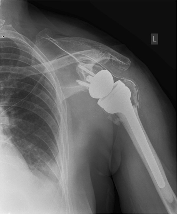
X-rayAnteroposterior shoulder radiograph after reverse total shoulder arthroplasty: glenosphere fixed on the glenoid, cup on the humeral stem.
Source : Mattiassich et al., BMC musculoskeletal disorders, 2013, figure 4 · CC BY
Both implants carry the name of total shoulder replacement and have nothing in common but occupying the same joint space. One restores a surface, the other replaces a function. Everything that follows in this article flows from that distinction.
The anatomical implant: giving a surface back to an intact force couple
The anatomical prosthesis reproduces the native geometry: a hemispherical metal head replaces the humeral head, a polyethylene glenoid replaces the worn glenoid surface. The convexity stays on the humeral side, the concavity on the scapular side, as in a healthy shoulder. The implant creates no intrinsic stability: it borrows it, exactly as the joint it replaces does, from the rotator cuff and the capsule.
This is the point to grasp before anything else. An anatomical replacement is a joint that is unstable by construction, held together by muscles. It therefore requires those muscles to exist and to work. The biomechanical review by Goetti and Lädermann puts it bluntly: the aim of anatomical arthroplasty is to reproduce pre-morbid kinematics, and failure to restore the anatomy, and therefore a stable fulcrum, leads to early failure of the implant through glenoid loosening, in a setting where joint reaction forces reach 2.4 times body weight (Goetti 2021,PMID 34760291).
That figure of 2.4 times body weight deserves a pause when prescribing exercise. It is not a carried load: it is the force that muscle contraction itself applies to the implant-bone interface. A shoulder working hard in a closed chain, or pushing against resistance at end of range, applies that stress to a cemented polyethylene component.
The reverse implant: replacing the force couple rather than restoring it
The reverse prosthesis swaps the surfaces over: a metal hemisphere, the glenosphere, is fixed to the glenoid; a concave polyethylene cup is fixed to the humerus. The convexity moves to the scapular side, the concavity to the humeral side. The joint becomes constrained: it has its own stability, independently of the cuff.
This is not merely a geometrical inversion. The Grammont design, which set the standard, medialises the centre of rotation and lowers the humerus. This double displacement allows an increase in the lever arm of the deltoid of up to 42 % (Goetti 2021). Measured directly on eight cadaveric upper limbs, implanting a reverse replacement increases the abductor lever arm of the anterior deltoid by 10.4 mm on average (95% CI 7.5 to 13.3) and that of the middle deltoid by 15.5 mm (95% CI 10.8 to 20.3), and additionally recruits the posterior deltoid as an abductor, which it was not (Ackland 2010,PMID 20439669).
In an anatomical shoulder, the deltoid needs the cuff so as not to simply drive the humeral head up under the acromion. In a reverse shoulder, the geometry spares it that: it can elevate the arm on its own.
The same cadaveric study yields an observation that is less often quoted and directly useful in rehabilitation: after a reverse replacement, the various portions of the subscapularis behave as extensors, and the adductor and extensor lever arms of teres major, latissimus dorsi and the lower and middle portions of pectoralis major increase substantially (Ackland 2010). In other words, the reverse implant does not merely strengthen the deltoid: it redistributes the roles of the whole girdle. A muscle you think you know no longer does the same thing.
Four numerical landmarks on the implant
What the mechanics impose, and what the registries observe in the long run.
Sources: Goetti 2021 (PMID 34760291), Ackland 2010 (PMID 20439669), Hole 2024 (PMID 39189259), Zhou 2023 (PMID 37178961).
How many patients, and following what trend?
France has no shoulder arthroplasty registry. The only national measurement available comes through the PMSI, the hospital stay database. In 2018, 234,612 primary shoulder surgery procedures were coded there, including 17,043 primary arthroplasties, that is 7.3 % of the total. That volume had risen by 47 % since 2012, and projections anticipate a rise of 31 % to 322 % by 2050 depending on the scenario chosen. Revisions numbered 1,508 in 2018, up 39 % over the same period (Villatte 2020,PMID 32863170).
Countries that keep a registry confirm the movement and show its composition. In Sweden, 28,632 primary shoulder arthroplasties were implanted between 2008 and 2023, with an incidence rising from 13.2 to 32.7 per 100,000 population. Over that period hemiarthroplasty declined until it became the least used method, while the reverse implant became the most frequent in people aged 65 years and over (Oro 2026,PMID 42443864).
The incidence of primary shoulder arthroplasty has more than doubled in fifteen years
Sweden, per 100,000 population, 2008 versus 2023. The relative rise is greater in men, the absolute rise in women.
Source: Oro 2026 (PMID 42443864). The percentage rises are calculated from the published incidences.
The table one would have wanted to read before the first session
Here is the line-by-line comparison of the two implants. It does not replace the operative report, which remains the source of truth for a given patient: it says what to look for in it.
| What is compared | Anatomical replacement | Reverse replacement |
|---|---|---|
| Geometry | Convexity on the humerus, concavity on the glenoid. Reproduces the native joint. | Convex glenosphere on the scapula, concave cup on the humerus. Constrained joint. |
| Reference indication | Centred glenohumeral osteoarthritis with a competent rotator cuff. | Cuff arthropathy, massive irreparable tear, complex proximal humeral fracture in the elderly, revision, glenoid bone loss. |
| Absolute prerequisite | A functional cuff, in particular the subscapularis, which will hold the implant. | A functional deltoid and an intact axillary nerve: it is the deltoid that will do the elevating. |
| Centre of rotation | Unchanged, or restored as close as possible to the pre-morbid position. | Medialised and lowered (Grammont design), or lateralised in the more recent designs. |
| Engine of elevation | The cuff-deltoid couple. The cuff centres the head, the deltoid elevates. | The deltoid alone, whose abductor lever arm increases by up to 42 % (Goetti 2021). |
| Fate of the subscapularis | Detached then reattached in the very large majority of cases (tenotomy, peel or lesser tuberosity osteotomy). | Often repaired when possible, sometimes not repairable. Repairing it cuts the instability rate (0.8 % versus 4.2 %; Bethell 2023). |
| Main restriction, weeks 0 to 6 | Passive external rotation beyond 30° in 7 published protocols out of 10, and any internal rotation against resistance (Bullock 2019). | The combination of adduction, internal rotation and extension, the position of posterior dislocation of the implant. No weight-bearing on the limb. |
| Sling | 3 to 8 weeks depending on the published protocols, with no consensus (Bullock 2019). | From “for comfort only” to 6 weeks, with no consensus either (Bullock 2019). |
| Point on which all protocols agree | Full passive mobility expected to be recovered at 12 weeks in 7 protocols out of 10. | Early deltoid isometrics: this is the only point on which all published protocols agree. |
| Expected active forward elevation | 149° ± 13° at 30 months in a matched cohort (Kirsch 2022). | 142° ± 15° in the same matched cohort. Mean gain of 52° at more than 10 years (Biner 2025). |
| Expected external rotation | 63° ± 14° (Kirsch 2022). This is the most consistent advantage of the anatomical implant. | 57° ± 18° in the matched cohort; mean gain of only 8° at 10 years in the cuff arthropathy series (Biner 2025). Depends on teres minor and on lateralisation. |
| Expected internal rotation | L3 or above in 68.7 % of patients (Kirsch 2022). | L3 or above in 37.3 % of patients. This is the widest functional gap between the two implants. |
| Its own specific complication | Glenoid loosening: asymptomatic radiolucent lines 7.3 % per year, symptomatic loosening 1.2 % per year (Papadonikolakis 2013). | Scapular notching (68 % in the historical Grammont series), instability (4.0 % pooled), acromial stress fracture (1.4 to 5 %). |
| Time to an acceptable symptom state | 6.1 months on average (Drager 2021). | 11.6 months on average, that is nearly twice as long (p = 0.009). |
| Lifetime revision risk, ages 46-50 | 35.8 % (95% CI 34.5 to 37.0), New Zealand registry. | 30.9 % (95% CI 29.9 to 32.0). The risk decreases with age at implantation in both groups. |
What this table changes at the first session
Three questions are enough to steer the whole management, and they arise before the patient has been touched: which implant, what was done to the subscapularis, and what the initial indication was. A reverse replacement implanted for cuff arthropathy and a reverse replacement implanted for a proximal humeral fracture carry neither the same risk of instability nor the same risk of nerve injury, as will be seen later: acute fracture triples the reported rate of nerve involvement (4.0 % versus 1.3 % on average; North 2023,PMID 36427756).
How does the rotator cuff decide the type of implant?
The surgeon does not choose the reverse implant because the patient is old, but because the cuff no longer holds the humeral head in place. Understanding the reasoning behind the indication means understanding what rehabilitation has to protect, and what it will not recover.
Two diseases that everyday language confuses
So-called centred glenohumeral osteoarthritis is glenohumeral arthritis in the classical sense: cartilage wear, osteophytes, joint space narrowing, with a humeral head that stays opposite the glenoid because the cuff keeps it there. Cuff arthropathy is something else. Massive, long-standing tearing of the tendons abolishes centring; the head migrates upwards, gradually articulates with the acromion, and the arthritis that follows is the consequence of the loss of the force couple, not of its own wear. This is what is called eccentric osteoarthritis.
This distinction can be read on a plain anteroposterior radiograph, and it is graded. The Hamada classification describes five stages, from a still preserved acromiohumeral space to acetabularisation of the acromion and complete glenohumeral degeneration; it is the common language of the surgical series on massive tears (Brolin 2017,PMID 28378277). On the centred osteoarthritis side, it is the Walch classification, based on the morphology and the wear of the glenoid, that structures the decision: concentric type A glenoid, retroverted and biconcave type B, dysplastic type C. It has been modified as three-dimensional imaging has refined its reading (Jawa 2021,PMID 33973964).
For the physiotherapist, the value of these two classifications is not in handling them, but in knowing that they exist and that they often appear in the operative report. A patient whose report mentions a high Hamada stage has lived for years without a functional cuff: their deltoid has compensated for a long time, and that is good news for what comes next. A patient whose report mentions a severely retroverted B3 glenoid has a bone problem, not a tendon problem: their rehabilitation will not be the same.
The decision tree, as it reads in the operative report
The prerequisite at the bottom applies to both branches: without a functional deltoid and axillary nerve, neither replacement gives a usable arm.
Sources: Walch classification (Jawa 2021,PMID 33973964), Hamada classification (Brolin 2017,PMID 28378277), widening of the indications for the reverse implant (Franceschi 2023,PMID 37301479). Reading diagram, not prescriptive: the decision rests with the surgeon.
The grey zone: osteoarthritis with an intact cuff, where the reverse implant is gaining ground
For some years now the reverse implant has been spilling beyond its original indication. It is increasingly offered for primary osteoarthritis with an intact cuff, a situation that by definition belonged to the anatomical implant. This shift is worth understanding, because it explains why the physiotherapist now meets reverse replacements in patients whose cuff is healthy.
A meta-analysis of 14 studies comparing the two implants in this precise indication finds, for the reverse implant, a lower complication rate (OR 0.54; p = 0.004) and a lower revision rate (OR 0.31; p < 0.001) at a mean follow-up of 3.4 years, with better SPADI, UCLA and Simple Shoulder Test scores. The price to be paid is explicit: external rotation is significantly poorer after a reverse replacement (p < 0.001), with no significant difference in the other ranges (Daher 2025,PMID 39142432).
In the most deformed glenoids, the type B2 and B3 glenoids with a preserved cuff, a systematic review of 36 studies covering 1,349 anatomical and 478 reverse replacements finds comparable range gains and comparable scores, but a complication rate of 7.8 % (95% CI 4.7 to 11.4) for the anatomical implant against 4.4 % (95% CI 2.8 to 6.9) for the reverse implant, and a revision rate of 5.2 % against 1.6 % (Aleisawi 2026,PMID 40689354). The authors themselves urge caution given the risk of bias in the included studies.
The most quoted propensity-score-matched series qualifies this shift on the functional side. In 134 patients operated on for primary osteoarthritis, 67 per implant, matched on age, sex, body mass index, preoperative ASES score, preoperative elevation and Walch glenoid morphology, patient-reported scores are identical at 30 months. What differs is the ranges of motion: the anatomical implant gives 149° ± 13° of elevation against 142° ± 15° (p = 0.003), 63° ± 14° of external rotation against 57° ± 18° (p = 0.02), and internal rotation reaching L3 or better in 68.7 % of patients against 37.3 % (p < 0.001). The overall complication rate was 4.5 %, with no difference between the groups (Kirsch 2022,PMID 35867705).
The sentence to remember from this controversy
In osteoarthritis with an intact cuff, the reverse implant buys mechanical reliability with rotation. It is a trade-off, not a one-sided advance, and it feeds straight through to what can be promised to the patient in rehabilitation. The movement that suffers most is reaching behind the back: getting dressed, fastening a garment, reaching a back pocket. Two patients in three manage it after an anatomical replacement, one in three after a reverse replacement.
Proximal humeral fracture, an indication of its own
A displaced three- or four-part fracture of the proximal humerus in the elderly is the third major gateway to the reverse implant, and it changes the prognosis. The Norwegian multicentre randomised DelPhi trial compared the reverse implant with plate fixation: the superiority of the reverse implant seen at two years (Fraser 2020,PMID 31977825) is maintained at five years (Fraser 2024,PMID 39303024). The most recent Cochrane review on proximal humeral fractures in adults, which includes 47 trials and 3,179 participants, stresses for its part the generally low methodological quality of the field, most comparisons resting only on small single-centre trials (Handoll 2022,PMID 35727196).
For rehabilitation, remember this above all: a reverse replacement implanted in a fracture setting does not have the risk profile of a reverse replacement implanted electively. The tuberosities have been reattached or they have not, the subscapularis is sometimes carried away by the fracture line, and the systematic review of 7,885 arthroplasties explicitly identifies proximal humeral fracture and its sequelae as risk factors for instability (Olson 2022,PMID 37588866). The reported rate of nerve involvement is also the highest there of all the indications, at 4.0 % (North 2023,PMID 36427756).
What should raise the alarm even before the first mobilisation
- No voluntary contraction of the deltoid on request, or reduced sensation over the shoulder cap. The axillary nerve is the most frequently affected after a reverse replacement (0.6 % of cases), and the reverse implant stresses it mechanically by lengthening the arm. A suspected injury is reported to the surgeon: it is not rehabilitated blind.
- An initial indication of acute fracture in the operative report, which triples the reported nerve risk and raises the risk of instability.
- Mention of a revision procedure: the instability rate rises from 1 to 5 % in primary surgery to 1 to 49 % across the revision series (Olson 2022), and the rate of nerve involvement from 1.3 % to 2.4 % (North 2023).
- Subscapularis recorded as not repairable on a reverse replacement: the instability rate rises from 0.8 % to 4.2 % when it is not repaired, in a meta-analysis of 17 studies and 2,620 patients, although the difference disappears with lateralised implants (Bethell 2023,PMID 37473906).
On the condition that leads to the reverse implant, the site's article devoted to symptomatic degenerative rotator cuff tear covers the upstream part of this story: the point at which the tear is still open to conservative treatment or to repair. This article begins where that one stops.
What does the reverse implant change in the mechanics of the shoulder?
A reverse replacement does not merely replace surfaces: it moves the centre of rotation, lengthens the arm, redistributes the lever arms and alters scapulohumeral rhythm. A physiotherapist who is unaware of these four changes is working with the map of a different shoulder.
First change: the centre of rotation moves down and closer to the axis of the body
In a native shoulder, the centre of rotation lies at the centre of the humeral head. The Grammont design moves it inwards, to the level of the glenoid surface, and lowers it. These two displacements have the same geometrical effect: they move the centre of rotation away from the line of action of the deltoid, which stays lateral. And the lever arm of a muscle is exactly that distance. The result is an increase in the lever arm of the deltoid of up to 42 % (Goetti 2021,PMID 34760291).
Why the deltoid becomes able to elevate the arm on its own
Diagram of principle, not to scale. Medialising and lowering the centre of rotation moves it away from the line of action of the deltoid, and it is that distance which defines the lever arm.
Sources: Ackland 2010 (PMID 20439669) for the cadaveric measurements, Goetti 2021 (PMID 34760291) for the relative gain of 42 %.
Second change: external rotation becomes the weak point
The deltoid elevates. It does not rotate. Active external rotation of a shoulder with a reverse replacement therefore depends on what is left of the external rotators, essentially infraspinatus and above all teres minor, and on the geometry of the implant.
The strongest demonstration comes from a French multicentre study by the French Society for Shoulder and Elbow Surgery, covering 501 shoulders reviewed at 2 to 5.5 years. External rotation with the elbow at the side improves there by 16.1° on average. Multivariate analysis identifies what makes it vary: it decreases with age (β = -0.35), increases with the lateralisation angle of the implant (β = 0.26), is better after an anterosuperior approach (β = 11.41), and is markedly poorer when teres minor is absent or atrophic (β = -10.06). The net gain is also smaller in patients operated on for osteoarthritis with a cuff tear or for a massive tear (Ducharne 2023,PMID 37318630).
Ten degrees less external rotation for an absent teres minor: that is a preoperative imaging finding, not a shortcoming of rehabilitation. Knowing it saves you from pushing relentlessly, and from letting the patient believe they are not doing enough.
When the external rotation deficit is major and combined with a loss of elevation, some teams add a latissimus dorsi tendon transfer, alone or with teres major, at the time of the replacement. That is a surgical decision, but it has a direct and immediate consequence for rehabilitation: the transferred tendon imposes its own protection timescales, and the operative report has to be read to the very end.
Third change: internal rotation is the movement most often lost
Reaching one's back is the function most frequently sacrificed. A multicentre study of 455 patients with the same implant system stratified glenoid lateralisation into four groups: 0 to 2 mm, 4 mm, 6 mm and 8 mm. Patients with 8 mm of lateralisation had significantly better active internal rotation than all the other groups, and those at 6 mm better than those at 0-2 and 4 mm, with no difference in elevation, external rotation or patient-reported scores. In multivariate analysis, glenoid lateralisation was the only implant-related variable associated with better internal rotation; neither body mass index, nor subscapularis repair, nor humeral lateralisation had a significant effect (Werner 2021,PMID 33753271).
In other words: the internal rotation of a reverse replacement is largely decided in the operating theatre. Rehabilitation can preserve what exists of it, maintain suppleness and work on compensation strategies at the trunk and the elbow. It cannot create a range that the geometry of the implant does not allow.
Fourth change: the scapula takes over, and exercise must take account of it
Scapulohumeral rhythm, the ratio between glenohumeral elevation and upward rotation of the scapula, changes after a reverse replacement. A meta-analysis of 27 studies covering 464 patients finds a mean rhythm of 1.6 in the plane of the scapula, against 3.2 in asymptomatic participants: the ratio becomes 1.9 against 3.2 in the direct comparison (p = 0.0238). The scapulothoracic contribution is therefore markedly greater after a reverse replacement, and all the more so as the arm goes higher, the rhythm being lowest between 60° and 90° of elevation (Johnson 2026,PMID 40976550).
This finding is not theoretical: it says that the scapula works more, hence that the muscles that move it, lower trapezius and serratus anterior first among them, are used more than they were before the operation, and that they are part of the programme.
A team in Ghent took the measurement as far as the exercise itself, recording three-dimensional scapular kinematics in 48 patients, 55 shoulders, twelve weeks after a reverse replacement, during three common rehabilitation exercises. The results transfer directly: the exercises performed in the vertical plane, wall slide and standing reach at 120°, produce significantly more upward rotation than the horizontal exercise performed seated at a table; and as regards rotation of the scapula, the vertical closed-chain exercise produces more scapular external rotation than the same movement in an open chain (Vandenbosch 2025,PMID 39842654).
The practical translation of the kinematic data
The choice of plane and of chain is not a detail of comfort: it changes what the scapula does. A standing wall slide, in a closed chain, calls on scapular upward rotation and external rotation more than a seated horizontal slide does. In a patient who has to rebuild a scapular contribution that has become the larger share, that is not a matter of indifference. In a patient one wants on the contrary to unload during the first weeks, the seated horizontal exercise is the most sparing.
Why does the subscapularis dictate the timescales of the anatomical implant?
There is no anatomical replacement without an anterior approach, and no anterior approach without crossing the subscapularis. This muscle, detached then repaired, is the only structure still healing in a shoulder that is otherwise metal. All the caution of the first six weeks is meant for it.
A tendon cut to get in, repaired to get out
The deltopectoral approach, the most widely used, reaches the joint from the front. The subscapularis bars the way. The surgeon must therefore detach it, insert the implant, then repair it. This step is invisible on a follow-up radiograph, and yet it explains every one of the early restrictions: no forced passive external rotation, which stretches the repair; no internal rotation against resistance, which puts it under active tension.
The systematic review of published protocols confirms it: seven protocols out of ten, after an anatomical replacement, limit external rotation to 30° during the initial phase, and expect full recovery of passive mobility at twelve weeks (Bullock 2019,PMID 31021690). Those thirty degrees are not a physiological value: they are a tissue-protection compromise, which the surgeon can adjust according to the quality of the repair and the tension observed in theatre. The exact limit is read in the operative report, not in a generic protocol.
Three techniques, three healing profiles
The tendon can be divided away from its insertion (tenotomy), detached flush with the bone (a subscapularis peel), or taken away with a bone fragment from the lesser tuberosity (a lesser tuberosity osteotomy). These three procedures do not heal in the same way, and the literature comparing them is of good quality.
| Technique | What heals | Measured healing rate | What the study says |
|---|---|---|---|
| Tenotomy | Tendon to tendon | 86.7 % with no full-thickness tear on ultrasound at 3 months (26 out of 30); 95 % healing beyond 12 months in another series | Levine's randomised trial: no difference in range or score at one year compared with osteotomy, but a shorter operating time (129.3 against 152.7 minutes) |
| Peel | Tendon to bone | 75 % on ultrasound beyond 12 months, against 95 % for tenotomy (p = 0.011) | Secondary analysis of a randomised trial of 100 patients: tenotomy heals better. No association between healing status and internal rotation function |
| Lesser tuberosity osteotomy | Bone to bone | 93.1 % radiological bone union (27 out of 29) | Heals the most reliably. In a comparative series, all tendons were normal on ultrasound after osteotomy, against 4 abnormal out of 32 after a peel |
Sources: Levine 2019, randomised controlled trial in 60 shoulders (PMID 30771825); Baisi 2023, secondary analysis of a randomised trial, 88 ultrasound scans (PMID 35973515); Buckley 2014, comparative series of 60 patients (PMID 24618191).
Two methodological points prevent over-reading this table. First, Levine's and Baisi's figures do not measure the same thing at the same moment: one looks for the absence of a full-thickness tear at three months, the other for healing beyond twelve months. Second, Levine's trial finds no clinical difference at one year between the two techniques compared, despite more reliable bone union after osteotomy. A difference in healing becomes a difference in function only if it is large enough, or frequent enough, to be seen.
What a failing subscapularis really costs
The Rochester series gives the clearest answer, because it cross-references imaging and functional scores. Among 60 patients, the four tendons abnormal on ultrasound, three thinned and one torn, were all in the peel group. Patients with an abnormal tendon had significantly lower scores: WOOS at 65 ± 18 against 88 ± 15, DASH at 25.9 ± 11.2 against 11.5 ± 11.4. Their strength on the belly press was significantly reduced, that on the bear hug tended to be, and their external rotation was paradoxically increased (Buckley 2014,PMID 24618191).
External rotation that gains quickly and abundantly after an anatomical replacement is not necessarily good news. It may signal a subscapularis that has given way.
This is the most useful signal in the whole chapter, and it is counter-intuitive. The physiotherapist who obtains without effort, at six weeks, an external rotation far greater than on the opposite side, and who notes alongside it a weakness on the belly press, should ask whether the construct is intact and raise it with the surgeon, rather than congratulate themselves on the progress made.
A word of caution about these tests: the belly press and the bear hug actively load the subscapularis. They are monitoring tools from the point at which the repair is protected onwards, not tests to be performed against resistance in the first weeks. The position of the elbow on the belly press, forward of or behind the plane of the trunk, showed in any case no significant association with healing status in Baisi's series, which invites us not to draw conclusions from that sign alone.
On the reverse implant, the same muscle does not play the same role
The subscapularis of a reverse replacement is no longer a driver of internal rotation: after a reverse replacement, cadaveric measurements show that it behaves as an extensor (Ackland 2010,PMID 20439669). Its repair, when it is possible, is debated for another reason: stability.
Two meta-analyses converge without overlapping. The first, on 7 studies and 1,306 patients, finds a markedly lower risk of dislocation when the subscapularis is repaired (OR 0.19; p < 0.001), and adds an important observation: in the absence of repair, a lateralised centre of rotation carries a lower risk of dislocation than a medialised centre (OR 0.24; p < 0.001) (Matthewson 2019,PMID 30827833). The second, more recent and larger (17 studies, 2,620 patients), puts instability at 0.8 % with repair against 4.2 % without (p = 0.04), but shows that this difference fades with lateralised implants (0.6 % against 1.6 %; p = 0.40). The functional gains associated with repair are statistically significant there but stay below the thresholds of clinical relevance: 3.4 ASES points, 3.7 Constant points, 3° of elevation (Bethell 2023,PMID 37473906).
What the physiotherapist does with this
With an anatomical replacement, repairing the subscapularis is a major functional issue: if it gives way, the patient loses strength in internal rotation and real score points. It is protected, and the two signs that betray its failure are watched for: weakness on the belly press and abnormally free external rotation.
With a reverse replacement, repair is a matter of stability, and its functional effect is below the threshold of clinical relevance. What is being protected is not the patient's strength, it is keeping the implant in place, and the risk is concentrated on medialised implants. The course of action is therefore not the same, because what one is trying to avoid is not the same thing.
What can the patient really hope for in range and strength?
The patient asks the question at the first session, and they are entitled to an answer in figures. Registries and matched cohorts give one, provided one distinguishes what the implant allows, what the initial pathology limits, and what time will still bring.
Ranges of motion: elevation catches up, the rotations do not
The cleanest comparison remains the propensity-score-matched cohort already quoted, because it neutralises age, sex, build, preoperative functional level and glenoid morphology: at 30 months of follow-up, the gap in forward elevation between the two implants is seven degrees, that in external rotation six degrees, but the proportion of patients able to reach L3 in internal rotation falls from 68.7 % to 37.3 % (Kirsch 2022,PMID 35867705).
What each implant returns, in matched patients
134 patients operated on for primary osteoarthritis, 67 per implant, matched on age, sex, BMI, preoperative ASES score and preoperative elevation, and glenoid morphology. Mean follow-up 30 months.
Source: Kirsch 2022, J Bone Joint Surg Am (PMID 35867705).
These figures describe a population operated on for osteoarthritis, that is to say the best possible situation for a reverse implant. When the reverse implant is used for its historical indication, cuff arthropathy, the gains are more modest and the weak point shifts squarely towards external rotation. The systematic review of series with more than ten years of follow-up, 469 reverse replacements, finds weighted mean gains of 54° of abduction and 52° of forward elevation, but of only 8° of external rotation (Biner 2025,PMID 40313685). The French multicentre study of 501 shoulders, more recent and covering more varied implants, finds for its part a mean gain of 16.1° of external rotation (Ducharne 2023,PMID 37318630). The gap between these two figures is not a contradiction: it comes down to the implants, the indications and the eras, and above all it says that announcing a precise range to a given patient is a risky exercise.
The timeline: what plateaus at six months, what is still improving at two years
This is perhaps the most useful piece of data in the whole article, because it contradicts a widespread intuition. A prospective cohort of 101 reverse replacements by a single operator, reviewed at six weeks then at three, six, twelve and twenty-four months, shows that forward elevation and the Constant score improve up to six months then plateau, whereas external and internal rotation keep improving beyond six months and up to two years (Collin 2017,PMID 28791445).
Two different clocks in the same shoulder
Elevation plateaus in the sixth month. The rotations, for their part, keep gaining for another eighteen months.
Sources: Collin 2017, prospective cohort of 101 reverse replacements (PMID 28791445); Drager 2021, institutional registry of 157 shoulders, Kaplan-Meier survival analysis (PMID 33711501).
The same study identifies what predicts poor elevation, and the timing of those predictors is instructive: insufficient elevation at six weeks is linked to preoperative deltoid weakness, whereas insufficient elevation at one year is linked to the operated side being the non-dominant one, to low preoperative elevation, to the preoperative activity level, to a low subjective shoulder value and to a low contralateral Constant score (Collin 2017). The determinants of the short term are muscular; those of the long term are contextual.
The survival analysis from the Chicago registry completes the picture on the experience side: 77.3 % of patients operated on with an anatomical replacement reach a substantial clinical benefit at six months, against 59.0 % after a reverse replacement (p = 0.024); at two years, 92.0 % against 79.5 % (p = 0.048). In both groups, clinically significant improvement continues throughout the two years of follow-up. One factor associated with earlier improvement, and it concerns physiotherapy directly, is regular physical activity before surgery (Drager 2021,PMID 33711501).
Reading a score, and knowing from what point improvement counts
Three thresholds structure the interpretation of a functional score: the minimal clinically important difference, the substantial clinical benefit, and the patient acceptable symptom state. The first says that a change is perceptible, the second that it is clear-cut, the third that the state reached is judged satisfactory regardless of the distance travelled.
| Measure | Minimal important difference | Substantial benefit | Acceptable state |
|---|---|---|---|
| ASES score | 13.9 points (20.9 in Tashjian's series) | 33.1 points | 79.5 points |
| Constant score | 7.2 points | 18.9 points | 64.2 points |
| Simple Shoulder Test | 2.1 points (2.4 in Tashjian's series) | 3.8 points | 9.2 points |
| Pain, visual analogue scale | 1.5 points (1.4 in Tashjian's series) | 3.3 points | 0.8 points |
| Forward elevation | 16° | 31° | 130° |
| Abduction | 13° | 30° | 104° |
| External rotation | 4° | 12° | 30° |
Overall thresholds, all implants combined, from an international multicentre database of 5,851 arthroplasties, 2,236 anatomical and 3,615 reverse (Simovitch 2024,PMID 38461936); Tashjian's values obtained by an anchor-based method in 326 patients (PMID 27545048). The authors of the multicentre database point out that these thresholds vary with the type of implant, the initial diagnosis and sex, and explicitly urge caution before transposing an acceptable-state threshold from one study to another.
Two practical lessons. First, four degrees of external rotation are enough to cross the threshold of perception: on this range, a small gain really counts. Second, the bar for the acceptable state in external rotation stands at 30°: that is an attainable target, and it is also exactly the limit that most protocols impose in the early phase after an anatomical replacement, a useful reminder that the constraint of the first weeks is not the prognosis.
Sport, driving, daily life
Return to sport is more frequent than one imagines. A systematic review of 23 studies and 2,199 patients, of mean age 68 years and mean follow-up 4.2 years, finds an overall return rate of 75.5 % with a mean delay of seven months: 77.4 % after an anatomical replacement, 75 % after a reverse replacement, 71.2 % after hemiarthroplasty, differences not significant (Küffer 2021,PMID 34667648). An earlier meta-analysis, restricted to recreational athletes, finds 80.7 % returning, with 79.2 % in golf (95% CI 62.9 to 89.5), 75.6 % in swimming (95% CI 61.3 to 85.8) and 63.5 % in tennis (95% CI 34.1 to 85.5); the reverse replacement subgroup returns at 76.5 % (Aim 2018,PMID 28719752).
One detail from this meta-analysis deserves to be repeated to the patient and to the doctor who refers them: every patient who returned to their sport was still practising it in the three months before the operation. It is a strong argument for maintaining activity before surgery, consistent with the predictor of earlier improvement identified by Drager.
For driving, a series of 406 patients, 214 anatomical and 192 reverse, finds a universal return: 100 % of the patients who drove before drove again afterwards. Those with an anatomical replacement return earlier (34 % within the first two weeks against 20 %), and the accident rate did not differ between those who had returned before two weeks and the others. Waiting until the sling was discontinued was associated with a later return after a reverse replacement (DeBernardis 2023,PMID 36528223). This study is retrospective and self-reported: it describes what patients did, not what they should be advised to do.
What rehabilitation after a shoulder replacement, and what does the evidence really say?
This is the most uncomfortable chapter of the article. The protocols in circulation are numerous, detailed, and for the most part unvalidated. The randomised trials that exist do not show what one would like them to show. Ignoring them would be more comfortable; looking at them is the only way to know where our work adds something.
The heterogeneity of the protocols is not a flaw in the literature, it is its result
The landmark systematic review catalogued the rehabilitation protocols published after anatomical and reverse arthroplasty. Sixteen studies met the inclusion criteria: only one of level I, one of level III, two of level IV, and twelve of level V, that is to say expert opinion. Ten described a protocol for the anatomical implant, six for the reverse implant (Bullock 2019,PMID 31021690).
The detail of the divergences speaks louder than the general observation.
| Parameter | After an anatomical replacement (10 protocols) | After a reverse replacement (6 protocols) |
|---|---|---|
| Sling duration | From 3 to 8 weeks depending on the protocol | From “for comfort only” to 6 weeks |
| Passive mobility | Full recovery expected at 12 weeks in 7 protocols out of 10 | From no passive mobilisation at all to varying precautionary limits |
| External rotation | Limited to 30° in 7 protocols out of 10 | No converging instruction |
| Exercise against resistance in the first 6 weeks | Present in 4 protocols out of 10 | Not specified in any converging way |
| Point of unanimous agreement | None | Early deltoid isometrics, in every one of the protocols |
Source: Bullock 2019, J Orthop Sports Phys Ther (PMID 31021690). The authors conclude that there is a high level of heterogeneity in the recommendations and the precautions, for both types of implant.
A critical review published the following year in JBJS Reviews reaches the same conclusion in blunter terms: after an anatomical replacement, comparative data are insufficient on the type and duration of the sling; there is no appreciable benefit of early mobilisation over a delayed protocol; the literature does not allow the superiority of formal physiotherapy over a physician-directed programme to be supported; and for the reverse implant, no high-quality data exist to guide postoperative rehabilitation (Kirsch 2020,PMID 32224631).
A protocol detailed to the day is not evidence. It is often a habit written up, and the heterogeneity between these protocols is the most reliable measure of our collective uncertainty.
Mobilise early or immobilise for six weeks after a reverse replacement?
A single-blind randomised controlled trial settled this question by assigning patients either to delayed rehabilitation, with no mobilisation at all for six weeks, or to immediate rehabilitation, passive and active. Of 107 shoulders included, 80.3 % were reviewed at one year: 44 in the delayed group, 42 in the immediate group.
Both groups improved comparably: 32° of forward elevation and 22° of abduction gained at three months, 9.4 ASES points as early as six weeks and 35.1 points at six months. No difference between groups on any postoperative parameter, with one exception, in favour of the delayed group: the ASES function subscore at six months improved by 26.3 points against 16.7. No difference in complications, in scapular notching or in opioid analgesic use. The authors conclude that early mobilisation may benefit the elderly population by sparing them the limitations of prolonged immobilisation (Hagen 2020,PMID 31924519).
The right reading of this trial is neither “mobilise early” nor “immobilise”. It is this: in an uncomplicated reverse replacement, the two strategies end up in the same place at one year, and the choice can therefore be made on other criteria, notably the risk of falling, independence and the patient's tolerance of immobilisation.
Supervised physiotherapy or a self-directed programme? Five trials, one constant answer
This is the question that unsettles, and it has been asked five times, independently, with the same result.
| Trial | Sample and comparison | Main result |
|---|---|---|
| Hagen 2020, JSES | 107 shoulders included, immediate mobilisation against 6 weeks of immobilisation | No difference in range or score at 1 year, apart from the ASES function subscore at 6 months favouring the delayed group |
| Chalmers 2023, JSES | 89 patients randomised, 2 centres: home programme (booklet and pulley) against supervised physiotherapy | No difference in score or range at 1 year. Complications 13 % against 17 % (p = 0.629). 20 % crossed over from home to physiotherapy |
| Schick 2023, JSES | 100 randomised, 70 analysed, mean follow-up 20.8 months | No difference in range or score. The only difference: external rotation strength higher by 0.8 kgf with formal physiotherapy (p = 0.04) |
| Rees 2025, JSES Int | 59 patients aged 60 to 85, supervised (n = 30) against manual-guided self-rehabilitation (n = 29) | ASES score at 1 year: 77.6 against 81.1 (p = 0.501). No difference in range at 3 months or at 1 year |
| SHORT trial, Garrigues 2026, JSES, 2025 Neer Award | 222 shoulders in 216 patients, 7 centres, 9 surgeons: outpatient physiotherapy (n = 117) against a surgeon-directed home programme (n = 105) | No difference in active or passive range at 1 and 2 years, no difference in score or quality of life, no difference in complications or revisions. Cost of care at 1 year: $17,837 against $11,285 (p < 0.01) |
Sources: Hagen 2020 (PMID 31924519), Chalmers 2023 (PMID 36690173), Schick 2023 (PMID 37178958), Rees 2025 (PMID 40959011), Garrigues 2026 (PMID 41177296).
What these trials say, and what they do not say
They say that, after an uncomplicated reverse shoulder replacement, in an independent patient, a structured and well explained programme achieves the same result as care in an outpatient clinic, at one and two years. It is a robust result: it has been reproduced by five teams, in two countries, with different designs, and the largest of the trials is multicentre with 93 % follow-up at one year.
They do not say that rehabilitation is useless. Five caveats deserve to be set out, not to save the profession, but because they are in the articles themselves.
- The “self-directed” groups are not groups without intervention. They receive a detailed booklet, equipment, and close follow-up by the surgeon at every consultation. What is compared is not “physiotherapy against nothing”, it is “physiotherapy against structured patient education”. The difference is crucial for what one concludes in practice: the patient who receives neither is represented in no arm of the trial.
- Crossovers are frequent and go in one direction only. In Chalmers's trial, 20 % of the patients in the home group switched to physiotherapy, against 4 % the other way. A subgroup therefore needed more, and intention-to-treat analysis dilutes it.
- The populations are selected. Rees's trial excluded a history of ipsilateral infection, autoimmune and neuromuscular disease, and patients needing a stay in a rehabilitation facility after discharge. The SHORT trial was conducted by nine specialist high-volume surgeons. This is not the average caseload of a community practice.
- The outcome measure is a group mean. A trial that finds no difference in means does not rule out that an identifiable fraction of patients derive a clear benefit, and another none at all. None of these trials was powered to find out who.
- The result concerns the reverse implant. None of these five trials concerns the anatomical replacement, where protecting a tendon repair creates an issue that the reverse implant does not have.
The honest conclusion is therefore a shift in the question. After a simple reverse replacement in an independent and well informed patient, the issue is no longer to justify sessions but to know which of them add something: education about at-risk positions, monitoring for complications, management of patients who are not progressing along the expected trajectory, and work on the rotations in the window where they are still improving, between six and twenty-four months, precisely where the protocols stop.
What is established, what is probable, and what is only a habit
Level of evidence by modality, graded on the design and the consistency of the available studies, not on how often the modality is used.
Grading established for this article from the design of the studies quoted: Bullock 2019 (PMID 31021690), Hagen 2020, Chalmers 2023, Schick 2023, Rees 2025, Garrigues 2026, Matthewson 2019, Bethell 2023, Su 2024, Drager 2021, Aim 2018. This is not a formal GRADE assessment: none exists to date on this question.
The consequence, for the patient in front of you
An independent, well informed patient with a simple reverse replacement and a trajectory that fits needs a clear programme, monitoring, and few sessions. An isolated, anxious patient, with a reverse replacement for a fracture, an unrepaired subscapularis, a history of revision or an off-track course, is represented in none of these five trials. That is where the usefulness of physiotherapy is concentrated, and that is where the sessions should go.
How to progress on criteria rather than on a calendar?
Since the published timescales vary threefold for the same situation, the calendar cannot be the decision rule. What is left are passage criteria: states to be reached before adding a constraint. The framework that follows is a proposed synthesis, built from the data quoted in this article. There is no protocol validated by a trial for shoulder arthroplasty.
Why the calendar alone fails
Three reasons, all documented above. First, the heterogeneity of the published protocols indicates that no date has been validated: sling duration varies from three to eight weeks after an anatomical replacement, and from “for comfort only” to six weeks after a reverse replacement (Bullock 2019,PMID 31021690). Second, the two implants do not move at the same speed: 6.1 months against 11.6 months to reach an acceptable symptom state (Drager 2021,PMID 33711501). Third, within one and the same shoulder, the ranges do not run on the same clock: elevation plateaus at six months while the rotations improve up to two years (Collin 2017,PMID 28791445).
A single calendar applied to these three sources of variation mechanically produces two symmetrical errors: it rushes the slow patients and holds back the fast ones.
Four phases, defined by what has to be achieved before moving on to the next
| Phase | Aim | Criteria for moving to the next phase | Restrictions by implant |
|---|---|---|---|
| 1. Protection | Protect the construct, preserve the elbow, the hand and the scapula, obtain a voluntary contraction of the deltoid | Pain controlled at rest and at night; full painless range at the elbow, wrist and hand; voluntary and painless isometric deltoid contraction; no sign of instability; sling weaning authorised by the surgeon | Anatomical: no passive external rotation beyond the limit set in the operative report, often 30°; no internal rotation against resistance; no weight-bearing on the limb. Reverse: no combination of adduction, internal rotation and extension; no weight-bearing; no forced passive movement into rotation. |
| 2. Mobility | Restore passive then active-assisted mobility, within the limits allowed | Comfortable passive elevation beyond 120°; passive external rotation to the permitted limit without pain or apprehension; active elevation against gravity initiated and controlled; visible scapular control in the plane of the scapula | Anatomical: the external rotation limit stays in force until the surgeon lifts it. Reverse: avoid repeated constrained adduction, which is the mechanism of scapular notching. |
| 3. Active control and strength | Build controlled active elevation and the strength of the deltoid and the scapular stabilisers | Active elevation above 120° without major trunk compensation; active abduction close to 100°; active external rotation beyond 30°; ability to repeat a movement above the horizontal without secondary pain the next day | Anatomical: progressive loading, bearing in mind that contraction itself applies up to 2.4 times body weight to the implant-bone interface. Reverse: in the osteoporotic patient, caution with load and with how early it starts (see the complications chapter). |
| 4. Consolidation and activities | Reach the acceptable-state thresholds and resume the chosen activities | Active elevation towards 130°, abduction towards 104°, external rotation towards 30°: acceptable symptom state thresholds measured in 5,851 arthroplasties; full independence for dressing and hygiene; return to the sporting or occupational task aimed at | Work on the rotations continues: it is between 6 and 24 months that they are still gaining, once elevation has plateaued. |
Synthesis framework proposed for this article. The acceptable-state thresholds of phase 4 are those of Simovitch 2024 (PMID 38461936). The restrictions take up the most frequent precautions from the protocols catalogued by Bullock 2019 (PMID 31021690) and the risk factors for instability from Olson 2022 (PMID 37588866). None of these passage criteria has been validated by a trial: they organise a decision, they do not prove it. The operative report takes precedence over this table in case of disagreement.
Three questions worth more than a date
Is the trajectory on track? After a reverse replacement, elevation that fails to get off the ground at six weeks points to preoperative deltoid weakness, a predictor identified in Collin's cohort. After six months, elevation will not gain much more, but the rotations will: an elevation plateau at that stage is expected, it is not a failure.
Does the observed gain exceed the threshold of perception? Four degrees of external rotation and sixteen degrees of elevation are enough to cross the minimal important difference. Below that the patient will feel nothing, and insisting on the measurement is counterproductive.
Is anything getting worse? This is the question that outranks the other two. A range that regresses, night pain that returns, strength that gives way: none of these three observations calls for stepping up the programme. They call for the next chapter.
The principle that sums up the chapter
A timescale says when you may try something. A criterion says when you must try it, and above all when to hold back. In a condition where the literature validates no calendar, the criterion is the only rule that can be defended in front of the patient and in front of the surgeon.
Which complications must you be able to recognise in a session?
The physiotherapist sees the patient more often than the surgeon does, and sees them moving. Five complications first show themselves as a change in the shoulder's behaviour, long before the next consultation. Recognising them is an act of care; treating them is not.
The reported frequencies, with their denominator
The rates below are not comparable with one another without reading their base: some are cumulative at more than ten years, others are annual, others are cross-sectional. That is why the base is written on every line.
Sources: Lévigne 2011 (PMID 21116754), Biner 2025 (PMID 40313685), Lau 2020 (PMID 33281942), Loucas 2022 (PMID 35394378), Su 2024 (PMID 37454923), North 2023 (PMID 36427756), Kirsch 2022 (PMID 35867705), Papadonikolakis 2013 (PMID 24352774).
Instability, the only one visible to the naked eye
Dislocation of a reverse replacement is not rare. The most quoted meta-analysis finds a pooled rate of 4.0 % in 3,810 patients of mean age 68.8 years (Loucas 2022,PMID 35394378). The largest systematic review, 17 studies and 7,885 arthroplasties, finds 204 dislocations, that is 2.5 % overall, but above all a considerable spread: from 0.4 % to 49 % depending on the series, with 1 to 5 % in primary surgery and 1 to 49 % in revision. The risk factors identified are subscapularis insufficiency, proximal humeral fracture and its sequelae, malunion and non-union (Olson 2022,PMID 37588866).
The mechanism to make known to the patient is the combination of adduction, internal rotation and extension: putting a hand behind the back, getting up from an armchair by pushing on the armrest, or pulling up a pair of trousers. Treatment is closed reduction and immobilisation; the review reports stabilisation rates of 28 to 100 % after reduction and bracing, and of 55 to 100 % after revision, and points out that hemiarthroplasty or resection arthroplasty are not rare after two or more episodes.
Scapular notching, specific to the reverse implant
This is erosion of the scapular neck, caused by impingement of the medial rim of the humeral cup during adduction. In 461 Grammont-design shoulders reviewed at 51 months on average, it occurred in 68 % of cases, appeared early, progressed variably, and was associated with the duration of follow-up, with strength, with passive and active elevation, and with the presence of humeral and glenoid radiolucent lines. The authors stress the role of preoperative glenoid erosion and the importance of avoiding a cranial position and a superior tilt of the baseplate (Lévigne 2011,PMID 21116754). The review in JAAOS confirms that its clinical importance remains uncertain, while recalling that it can lead to functional deterioration and to loosening of the glenoid implant, and that lateral offset, inferior overhang of the glenosphere and preoperative analysis of glenoid morphology all help to prevent it (Friedman 2019,PMID 30260909).
In the most recent series beyond ten years, the frequency remains highly variable: Nerot-Sirveaux grade I and II notching in 15 to 59 % of patients, grades III and IV in 7 to 47 % (Biner 2025,PMID 40313685).
This is not a complication that the physiotherapist treats. But its mechanism, impingement in adduction, says something useful: repeated constrained adduction with the arm at the side is not a neutral movement on this replacement.
Acromial stress fracture, the only one where our calendar has been called into question
This is the chapter most directly addressed to our profession, and it deserves careful reading. A systematic review of 25 articles catalogued 208 fractures of the acromion or the scapular spine after a reverse replacement, for an overall incidence of 5 %, stress fractures being more frequent than post-traumatic fractures. Functional results deteriorated after the fracture, whether it was operated on or not (Lau 2020,PMID 33281942).
An analysis of an administrative database, with a minimum follow-up of two years, finds an incidence of 1.4 % and identifies the independent factors: osteoporosis, rheumatological disease, corticosteroid injection into the shoulder in the three months before surgery, and long-term oral corticosteroid therapy. Then comes the sentence that concerns us directly: in osteoporotic patients, starting physiotherapy within six weeks of surgery also increased the risk of acromial stress fracture. The authors conclude that this risk can be reduced by avoiding injections in the three preoperative months and by delaying exercise in osteoporotic patients. Surgical treatment of these fractures carried a prosthetic revision rate of 7.0 % against 3.2 % with conservative treatment (Su 2024,PMID 37454923).
These data come from an administrative database, with the limits that implies: coding says nothing about the intensity or the content of the sessions, and an association does not establish causation. It is, however, the only finding in this whole literature that names a physiotherapy practice as a risk factor, and it concerns a subgroup identifiable in advance. Spotting osteoporosis before starting, and adjusting how early and how heavily one loads in these patients, costs little and answers a finding that exists.
Glenoid loosening, a complication specific to the anatomical implant
The systematic review of 27 articles and 3,853 arthroplasties remains the reference: asymptomatic radiolucent lines at a rate of 7.3 % per year, symptomatic glenoid loosening 1.2 % per year, revision surgery 0.8 % per year, with no evidence of a fall in the rate of symptomatic loosening over time. Keeled components showed more radiolucent lines than pegged components in direct comparisons (Papadonikolakis 2013,PMID 24352774). In the shorter term, the matched cohort finds 14.9 % of radiolucent lines at 30 months, with no gross loosening (Kirsch 2022,PMID 35867705).
The clinical signal is mechanical pain that reappears after a pain-free period, often months or years after the operation, in a patient who was doing well. It is grounds for a surgical opinion, not for stepping up strengthening.
Infection, which does not present like an infection
The shoulder has a microbiological peculiarity that traps clinical reasoning. In 87 first-stage revisions analysed according to the 2018 international consensus criteria, Cutibacterium acnes accounted for 77.3 % of culture-positive cases and was present in 39.1 % of cases overall. Loosening of the humeral stem multiplied the probability of definite or probable infection by seven (OR 7.2; 95% CI 2.67 to 19.37), and a raised erythrocyte sedimentation rate or C-reactive protein was also associated with this diagnosis (Patel 2021,PMID 33895298).
C. acnes is an indolent organism: it produces neither fever, nor redness, nor discharge in most cases. The picture is a shoulder that stays painful and stiff with no obvious mechanical reason, or that deteriorates after a period of normal progress. The recent summary in JBJS sets out the principles of prevention, diagnosis and treatment of these periprosthetic shoulder infections (Nazzal 2024,PMID 39475925).
Nerve injuries
The systematic review of 188 articles and 40,146 patients establishes a rate of 1.3 % after primary reverse replacement and 2.4 % after revision. The axillary nerve is most often affected (0.6 %), ahead of the ulnar nerve (0.26 %) and the median nerve (0.23 %), brachial plexus injuries accounting for 0.19 % of the total. The initial diagnosis most strongly associated is acute proximal humeral fracture, at 4.0 %, ahead of cuff arthropathy at 3.0 % (North 2023,PMID 36427756).
| Complication | Implant involved | What raises the alarm in a session | Course of action |
|---|---|---|---|
| Dislocation | Reverse above all | Sudden pain on a movement of adduction, internal rotation and extension; deformity; immediate loss of function | Surgical emergency. Immobilisation, same-day opinion |
| Acromial stress fracture | Reverse | Posterosuperior pain of gradual onset, often after several weeks or months; loss of active elevation that regresses after having improved; tenderness on palpation of the scapular spine | Stop progression, surgical opinion with imaging. One reported case united under conservative treatment with teriparatide |
| Glenoid loosening | Anatomical | Return of mechanical pain after a pain-free period, sometimes years later | Surgical opinion. Do not step up strengthening |
| Periprosthetic infection | Both | Persistent painful stiffness or unexplained deterioration, most often without fever or local signs | Surgical opinion with blood tests. The commonest organism is indolent |
| Nerve injury | Both, more frequent after fracture or revision | Loss of deltoid contraction, reduced sensation over the shoulder cap, distal deficit | Immediate reporting, do not rehabilitate a deficit that has not been investigated |
| Rupture of the repaired subscapularis | Anatomical | External rotation that gains abnormally fast and exceeds the healthy side; weakness on the belly press | Surgical opinion with ultrasound. Do not pursue further gains in external rotation |
| Scapular notching | Reverse | Radiological, not symptomatic in itself | Nothing to be done in the session. Avoid repeated constrained adduction |
The three situations that stop the session
- A range or a strength that regresses after having improved. In a normal recovery, elevation plateaus; it does not go backwards. A backward step suggests a stress fracture, a failed repair or an infection.
- Night pain that reappears after a phase of restored sleep, with no injury and no change of programme. It is the shared signal of loosening and of low-grade infection.
- A new neurological deficit: no deltoid contraction on request, reduced sensation over the shoulder cap, a deficit in the hand. The axillary nerve is the most exposed, and the reverse implant puts it under tension by lengthening the limb.
In all three cases the course of action is the same: progression is suspended, exactly what was measured and on what date is documented, and the patient is referred. A complication recognised early and a complication recognised late do not carry the same treatment or the same prognosis.
What do published case reports teach us?
Three published and referenced observations, chosen because each illustrates one of the three main ways in which a shoulder replacement deteriorates: instability that recurs, bone that gives way, and a tendon repair that lets go. None is a textbook case: these are real patients, with real outcomes.
First case: a reverse replacement that dislocates, and does so again
A right-handed man of 65 had chronic pain and weakness of the right shoulder, with advanced glenohumeral arthritis and massive irreparable cuff tears. He had a reverse replacement with a glenoid bone autograft, known as BIO-RSA, combined with a subscapularis repair. The postoperative course was marked by pain and a sensation of instability, then by an atraumatic anterior dislocation at four months. Conservative treatment was not enough: the dislocations recurred. A revision was performed with an increase in the size of the polyethylene component to improve containment. One year after that revision, a further dislocation occurred. A second revision then combined a pectoralis major transfer, because of subscapularis insufficiency from re-rupture caused by the repeated dislocations, with an increase in the size of the humeral tray. Eighteen months after that procedure, the patient had a stable shoulder, improved ranges of motion, and declared himself satisfied (Baek 2024,PMID 38910979).
What this case teaches the physiotherapist
The patient reported a sensation of instability before the first dislocation. This is the sign not to file under ordinary apprehension. In a reverse replacement, a complaint of instability, a feeling that the shoulder “slips out” on certain movements, or a reproducible discomfort in adduction and internal rotation, calls for a message to the surgeon rather than desensitisation work. The case also shows that a recurring dislocation progressively destroys the repaired subscapularis, which in turn feeds the instability: the spiral closes all the faster if the first episode goes unnoticed.
Second case: an acromial fracture at nine months, in a patient who was doing well
A woman of 78 had had a reverse replacement for a proximal humeral fracture. Nine months after the operation she developed a type 3 acromial stress fracture, with increasing pain and declining function. She was managed without surgery, with the addition of teriparatide. After four months of this treatment she had regained excellent ranges of motion and the fracture had united (Lipof 2020,PMID 32649098). Another published observation describes a fracture of the base of the acromion presenting late after a reverse replacement, which confirms that the time to onset can be long (Khwaja 2021,PMID 33681847).
What this case teaches the physiotherapist
Nine months is well beyond the window in which anyone is still watching. The patient was probably no longer being followed in rehabilitation. The lesson is not to prolong the sessions indefinitely, but to explain to the patient, at the point where you let them go, what should bring them back: posterosuperior pain of gradual onset and a loss of active elevation that had been gained. This patient's risk profile was in other respects typical: an elderly woman, operated on for a fracture, therefore probably fragile in bone terms, exactly the subgroup in which osteoporosis is an independent factor.
Third case: an anatomical replacement that dislocates at eight weeks, with the subscapularis gone
A woman of 54 had had a stemless anatomical replacement for primary osteoarthritis. Eight weeks after the operation she presented with a dislocation of the implant with failure of the subscapularis tendon. The quality of the metaphyseal bone and the sound fixation of the stemless humeral component, of convertible design, allowed conversion to a stemless reverse replacement with the humeral component left in place. At two years of follow-up, the patient had an excellent functional result, improved ranges of motion and a stable radiological appearance (Juma 2026,PMID 41541507).
What this case teaches the physiotherapist
Eight weeks is precisely the moment when the protocols lift the external rotation restrictions and allow active work. It is also the moment when a repair that has not held reveals itself, because something is finally being asked of it. The combination described here, subscapularis failure and anterior instability, is exactly the picture set out in chapter 4: the subscapularis is the anterior lock of an anatomical replacement, and its failure translates not only into a loss of internal rotation strength, but into a loss of stability. Lifting the restrictions at six or eight weeks is not a calendar formality: it is a test of the repair, and it is carried out gradually, watching the belly press and the external rotation range.
What three cases do not prove
A case report is an isolated observation, with no control group and no denominator. These three say nothing about the frequency of the events described: for that, one has to go back to the series in the previous chapter. Their value lies elsewhere. They show the real sequence of events, in the patient's own time, and that sequence is what the physiotherapist observes. A sensation of instability that precedes the dislocation, posterosuperior pain that appears nine months later, a repair that gives way at the moment it is asked to work: these are three reasons for consultation, not three statistics.
How to apply all this from the first session?
This chapter is deliberately operational. It contains no new figures: only the order in which to use those that come before.
Before touching the patient: five lines to find in the operative report
- The type of implant. Anatomical or reverse. Without that information no decision is possible, and the patient almost never knows it. In case of doubt, a plain anteroposterior radiograph removes the ambiguity immediately.
- What was done to the subscapularis. Tenotomy, peel, lesser tuberosity osteotomy, repair or non-repair. With an anatomical replacement, this line sets the restrictions of the first six weeks. With a reverse replacement, it modulates the risk of instability.
- The initial indication. Osteoarthritis, cuff arthropathy, acute fracture, fracture sequelae, revision. It changes the nerve risk, the risk of instability, and the prognosis for external rotation.
- The range limits imposed by the surgeon, with their end date. They take precedence over any generic protocol. If they do not appear in the operative report, they should be asked for rather than deduced.
- Any associated tendon transfer, of latissimus dorsi or teres major. It imposes its own protection timescales, distinct from those of the replacement.
At the first session: four measurements that will serve as a baseline
What is not measured at the outset cannot be compared later, and it is comparison that detects complications. Four markers are enough: active and passive forward elevation, external rotation with the elbow at the side, the vertebral level reached in internal rotation, and the presence or absence of a voluntary deltoid contraction. That last point is not a formality: preoperative deltoid weakness predicts insufficient elevation at six weeks, and an absent contraction raises the suspicion of axillary nerve injury.
What to tell the patient, which counts as much as the exercises
- The real duration. After a reverse replacement, the average time to feeling well is eleven to twelve months, against six after an anatomical replacement. A patient who does not know this experiences their sixth month as a failure.
- The two clocks. Elevation plateaus at around six months, the rotations improve up to two years. The patient who understands this does not stop working when elevation stops gaining.
- The rotation ceiling, if it can be predicted. A medialised reverse replacement with an atrophic teres minor will not give back a normal external rotation, and a barely lateralised reverse replacement will rarely give back the movement of reaching one's back. Saying so in advance turns a disappointment into information.
- The movements to avoid, and why. With a reverse replacement: the combination of adduction, internal rotation and extension, which is the mechanism of dislocation. With an anatomical replacement: internal rotation against resistance and forced external rotation, which load the repair.
- What should bring them back after the sessions have ended: a range that goes backwards, night pain that returns, new posterosuperior pain. The case of the acromial fracture at nine months shows that this message keeps its value long after the last session.
- Maintaining activity before the operation, when the patient is seen preoperatively. It is one of the few modifiable factors associated with earlier improvement, and every patient who returned to their sport was still practising it in the three months before surgery.
Choosing your exercises knowingly
The three-dimensional kinematic measurements at twelve weeks after a reverse replacement give a simple principle: plane and chain change the scapular contribution. A vertical exercise, wall slide or standing reach, produces more upward rotation than a seated horizontal slide; the same vertical movement in a closed chain produces more scapular external rotation than in an open chain. Since scapulohumeral rhythm shifts towards the scapula after a reverse replacement, with the ratio falling from 3.2 to 1.9, this contribution is not a detail: it is a substantial part of the movement to be rebuilt.
In concrete terms: the seated horizontal exercise is the most sparing to start with, the standing closed-chain wall slide is the richest in scapular work, and the standing open-chain reach is the closest to the functional movement. This is not a hierarchy of quality, it is a gradation of demand.
The operational summary in one sentence per implant
Anatomical: you protect a tendon repair for six weeks, you watch for the two signs of its failure, and you know you are aiming at a high functional ceiling, reached at around six months.
Reverse: you protect stability, you build a deltoid and a scapula, you accept a lower rotation ceiling and a timescale twice as long, and you adjust how early you start in the osteoporotic patient.
Frequently asked questions
The questions patients ask most often, and those colleagues ask on courses. Each answer points to the source that supports it.
How do I know whether my patient has an anatomical or a reverse replacement?
The operative report says so. Failing that, a plain anteroposterior radiograph settles the doubt immediately: with an anatomical replacement, the sphere is on the humeral side; with a reverse replacement, it is fixed to the scapula and the humerus carries a cup. The question is not cosmetic: the precautions, the timescales and the functional ceiling differ all the way down the line, as the comparison table in the first chapter shows. Asking the surgeon for the information is part of the job.
What ranges of motion can I promise my patient?
In matched patients operated on for osteoarthritis, the anatomical implant gives 149° ± 13° of active forward elevation, 63° ± 14° of external rotation, and internal rotation reaching L3 or better in 68.7 % of patients; the reverse implant gives 142° ± 15°, 57° ± 18°, and 37.3 % (Kirsch 2022,PMID 35867705). These figures describe the best possible situation. When the reverse implant is used for cuff arthropathy, the gain in external rotation is far more modest: 8° on average in the series beyond ten years (Biner 2025,PMID 40313685), 16.1° in a more recent French multicentre series (Ducharne 2023,PMID 37318630).
How long does recovery really take?
The average time to reach an acceptable symptom state is 6.1 months after an anatomical replacement and 11.6 months after a reverse replacement (p = 0.009), and clinically significant improvement continues throughout the two years of follow-up in both groups (Drager 2021,PMID 33711501). Within that period, forward elevation and the Constant score plateau at around six months, whereas external and internal rotation improve up to two years (Collin 2017,PMID 28791445).
Is supervised physiotherapy really necessary after a reverse replacement?
Five independent randomised trials, including the multicentre SHORT trial in 222 shoulders which won the 2025 Neer Award, find no difference in range of motion or in functional score at one and two years between outpatient physiotherapy and a surgeon-directed programme, at a markedly lower cost of care ($11,285 against $17,837; p < 0.01) (Garrigues 2026,PMID 41177296).
Three qualifications matter before drawing a general conclusion. The “self-directed” groups received a detailed booklet and close surgical follow-up: what is compared is not physiotherapy against nothing. Twenty per cent of the patients in the home group switched to physiotherapy in Chalmers's trial, against 4 % the other way. Lastly, none of these trials concerns the anatomical replacement, nor the at-risk situations.
Why is external rotation so limited after a reverse replacement?
Because the deltoid elevates but does not rotate. Active external rotation depends on what is left of the external rotators, and the French multicentre study of 501 shoulders identifies atrophy or absence of teres minor as a factor associated with a loss of about ten degrees, along with age and the degree of lateralisation of the implant (Ducharne 2023,PMID 37318630). These are anatomical and surgical determinants: rehabilitation preserves what exists of them, it does not create them.
My patient cannot fasten their bra or reach their back pocket. Is that recoverable?
It is the most frequent limitation after a reverse replacement, and it is largely decided in the operating theatre. A study of 455 patients shows that glenoid lateralisation of 6 to 8 mm is associated with better active internal rotation at one year, and that it is the only implant-related variable that is; neither subscapularis repair nor humeral lateralisation had a significant effect (Werner 2021,PMID 33753271). The useful work is on maintaining suppleness, trunk mobility and adapting the movement, rather than on gaining pure range.
Should one avoid starting rehabilitation too early?
In an uncomplicated reverse replacement, no: the randomised trial comparing immediate mobilisation with six weeks of immobilisation finds no difference at one year, in range, in scores, or in complications or notching (Hagen 2020,PMID 31924519).
There is one documented exception, and it is important: in osteoporotic patients, starting physiotherapy within six weeks of a reverse replacement was associated with an increased risk of acromial stress fracture, in a database analysis (Su 2024,PMID 37454923). Spotting osteoporosis before starting is a simple step that answers a real finding.
What is scapular notching, and should I worry about it?
It is erosion of the scapular neck from impingement of the humeral cup in adduction, specific to the reverse implant. In 461 Grammont-design shoulders it occurred in 68 % of cases and was associated with strength, with elevation and with the appearance of radiolucent lines (Lévigne 2011,PMID 21116754). Current lateralised designs reduce its frequency. The physiotherapist does not treat it, but its mechanism explains why repeated constrained adduction with the arm at the side is not a neutral movement on this replacement.
What signs should make me stop and refer?
Three, and they overlap little: a range or a strength that regresses after having improved; night pain that reappears with no mechanical cause; a new neurological deficit, in particular an absent deltoid contraction or reduced sensation over the shoulder cap. With an anatomical replacement, a fourth, counter-intuitive sign is added: external rotation that gains abnormally fast and exceeds the healthy side, together with weakness on the belly press, should raise the suspicion of a rupture of the subscapularis repair (Buckley 2014,PMID 24618191).
Can my patient go back to sport? And drive?
The overall rate of return to sport is 75.5 % with a mean delay of seven months, with no significant difference between the types of implant (Küffer 2021,PMID 34667648). Among recreational athletes it rises to 80.7 %, with 79.2 % in golf and 63.5 % in tennis; above all, every patient who returned was still practising their sport in the three months before surgery (Aim 2018,PMID 28719752). For driving, a series of 406 patients finds a universal return, earlier after an anatomical than after a reverse replacement, with no excess of accidents among those who returned before two weeks (DeBernardis 2023,PMID 36528223). This last study is retrospective and self-reported: the decision rests with the surgeon.
How long will the replacement last?
The Norwegian registry, on 5,494 reverse replacements followed for up to fifteen years, finds revision-free survival of 94 % at ten years (95% CI 93 to 95), instability and deep infection being the most frequent reasons for revision (Hole 2024,PMID 39189259). Expressed from the patient's point of view rather than the implant's, the risk of undergoing a revision during the rest of their life is 35.8 % for an anatomical replacement implanted between the ages of 46 and 50, and 30.9 % for a reverse replacement at the same age; it falls markedly with age at implantation (Zhou 2023,PMID 37178961).
To place this article in the series on the major arthroplasties, the site's two other summaries are available: rehabilitation after total hip replacement and rehabilitation after total knee replacement. On the upstream part of the indication for a reverse replacement, see symptomatic degenerative rotator cuff tear; on the differential diagnoses of a painful, stiff shoulder, adhesive capsulitis and rotator cuff tendinopathy.

