















In brief
A symptomatic degenerative tear of the rotator cuff is a multifactorial, age-related condition, distinct from the acute traumatic tears of younger patients; the tendon most often affected is supraspinatus. It combines anterolateral shoulder pain, often at night, functional weakness and a progressive loss of active movement, arising above all after the age of 60. First-line management is conservative, with progressive exercise the most effective intervention: the MOON cohort shows that 75 % of patients avoid surgery at 2 years. Prevalence rises with age, from 9.7 % before 30 to 62 % after 80.
Clinical synthesis based on the most recent meta-analyses and CPGs: JOSPT 2025 Desmeules (the rotator cuff CPG), JOSPT CPG 2022 Lafrance (return to work), the BMJ Rapid Recommendation Vandvik 2019 (decompression), the meta-analyses Longo 2021 and Ryösä 2017, and the MOON cohort Kuhn 2013 / Kuhn 2024 (10-year).
Clinical summary
- A degenerative rotator cuff tear is a multifactorial, age-related condition, distinct from acute traumatic tears. The tendon most often affected is supraspinatus, in its critical vascular zone.
- Age-dependent prevalence (Teunis 2014, SR pooled n=6,112 shoulders): 9.7 % before 30 years, 62 % after 80. Yamamoto 2010 (n=683, Japan) finds 20.7 % of tears in the general population, of which about 65 % are asymptomatic.
- Key risk factors: age, smoking, diabetes (OR 1.49), dyslipidaemia (OR 1.48), hypertension (OR 1.40) (Giri 2023, SR/MA), repetitive overhead movement, arm dominance.
- Natural history: 40-50 % of full-thickness tears enlarge within 5 years (Keener 2015). Enlargement correlates with progressive atrophy and fatty infiltration (Goutallier).
- Diagnosis combines the history (age > 60 years, anterolateral pain, night pain) with clinical examination. No single test is sufficient: a combination of tests (painful arc + drop arm + resisted external rotation) increases diagnostic accuracy (Hegedus 2012 BJSM; Lädermann 2020-2021 KSSTA).
- The drop arm sign has the best specificity (about 97 %) for a full-thickness supraspinatus tear. The lift-off and bear hug tests detect subscapularis tears.
- Imperative differential diagnoses : C5-C6 cervical radiculopathy (Spurling test), acromioclavicular joint disease, frozen shoulder, Parsonage-Turner syndrome, neoplasia.
- The Patte classification (tendon retraction) and the Goutallier classification (muscle fatty infiltration) guide the prognosis. A Goutallier grade of 3 or more carries a poor prognosis, conservatively as much as surgically.
- First-line treatment is conservative : the prospective multicentre MOON cohort (Kuhn 2013, n=433, JSES) showed that 75 % of patients avoid surgery at 2 years with a structured programme. The 10-year predictors (Kuhn 2024, JBJS, n=311) confirm that the conservative response lasts.
- The meta-analyses comparing surgery with physiotherapy (Ryösä 2017, Longo 2021 BMC) show no clinical superiority for surgery at 1, 2 and 5 years in degenerative tears.
- Progressive exercise is the most effective intervention. The JOSPT 2025 Desmeules CPG confirms that no type of exercise (eccentric, concentric, isometric) is superior to another: adherence and load progression matter more than the choice of exercise.
- And isolated subacromial decompression is no longer recommended for chronic shoulder pain (BMJ Rapid Recommendation Vandvik 2019, PMID 30728120; Beard 2018 Lancet, the CSAW trial).
- Patient education about the weak correlation between imaging and symptoms is crucial. Self-efficacy and kinesiophobia are the most powerful psychological predictors of outcome (Chester 2018-2019 BJSM).
- The return to activity and sport rests on functional criteria (strength ≥ 80 % of the healthy side) and psychological ones, not on a fixed calendar (Migliorini 2023, Lafrance CPG JOSPT 2022).
- Red flags: acute trauma, a progressive neurological deficit, non-mechanical night pain with constitutional symptoms, weight loss, a palpable mass. Surgical referral after 3-6 months of conservative treatment well conducted and without improvement.
Contents
- What are the fundamentals to know about symptomatic (degenerative) cuff tears?
- How do you assess and diagnose a symptomatic cuff tear with certainty?
- Which treatment strategies are the most effective for a symptomatic cuff tear?
- How do you secure lasting recovery and prevent recurrence of a symptomatic cuff tear?
- What do real clinical cases teach us about symptomatic cuff tears?
- How do you apply these recommendations concretely in your practice?
What are the fundamentals to know about symptomatic (degenerative) cuff tears?
How is this condition defined, who does it affect and what are the risk factors?
The contemporary nomenclature, harmonised by the JOSPT 2025 Desmeules CPG on rotator cuff tendinopathy⁴ and the JOSPT 2022 Lafrance CPG on return to work⁵, distinguishes several entities within one continuum:- Rotator cuff-related shoulder pain (RCRSP) : an umbrella term covering tendinopathy, partial tears and symptomatic degenerative full tears (Lewis 2018).⁶
- Partial tear : an intratendinous, bursal-side or articular-side lesion, classified after Ellman.
- Full-thickness tear : the loss of continuity spans the whole thickness of the tendon, from the articular to the bursal side.
- Massive tear : at least 2 tendons involved, or retraction > 5 cm (the DeOrio and Cofield criteria).
📊 Age-dependent prevalence of cuff tears (Teunis 2014, SR/MA n=6,112 shoulders)
A cumulative prevalence curve: a pooled meta-analysis of 30 studies, symptomatic and asymptomatic populations combined.
Source: Teunis T, Lubberts B, Reilly BT, Ring D. J Shoulder Elbow Surg. 2014;23(12):1913-1921. PMID 25441568.
The risk factors for degenerative tears are now well characterised. Age is the dominant non-modifiable factor. Among the metabolic factors, the meta-analysis Giri 2023 (Ann Phys Rehabil Med, SR/MA, PMID 35257948) is the modern reference source: it finds an OR of 1.49 [95 % CI 1.43-1.55] for diabetes, an OR of 1.40 [1.19-1.65] for hypertension and an OR of 1.48 [1.42-1.55] for dyslipidaemia in association with the tear.¹⁰ Smoking remains a major factor: Carbone 2012 (J Shoulder Elbow Surg, n=408, PMID 21524922) showed a dose-response relationship between the intensity and duration of smoking and the size of the tear.¹¹ The twin study Gwilym 2009 (J Bone Joint Surg Br) establishes a genetic component , with an increased risk in first-degree relatives.¹²
📊 Hierarchy of the modifiable risk factors (Giri 2023 SR/MA, n=25 studies)
Pooled odds ratios with 95 % confidence intervals.
* Smoking: a quantitative effect on the SIZE of the tear rather than a dichotomous one. Sources: Giri 2023 (PMID 35257948), Carbone 2012 (PMID 21524922), Sayampanathan 2017, Gwilym 2009.
What happens in the cuff, and how does it evolve naturally?
🧬 The pathophysiology of the degenerative tear brings together intrinsic factors (biological change in the tendon: collagen disorganisation, fewer tenocytes, more matrix metalloproteinases) and extrinsic ones (repeated mechanical stress, secondary subacromial impingement).¹³ The supraspinatus tendon has a critical zone of reduced vascularity, described by Rathbun & Macnab 1970 and Lohr & Uhthoff 1990, about 1 cm proximal to its insertion on the greater tuberosity: the preferential site of degenerative tears.¹⁴ The place of subacromial impingement as a primary cause is now widely questioned: it is seen instead as a secondary aggravating factor on an already weakened tendon.¹⁵natural history has been clarified by several longitudinal cohorts and recent meta-analyses. The SR/MA Keener 2015 (J Bone Joint Surg Am) and the SR Khatri 2019 (Bone Joint Res, PMID 31037208) establish the following trajectory:¹⁶,¹⁷
- 40-50 % of full-thickness tears enlarge over 2-5 years of follow-up.
- Enlargement correlates with worsening symptoms.
- Enlargement generally precedes the appearance of fatty degeneration (Goutallier 2 or more).
- A Goutallier grade of 3 or more of fatty infiltration is considered irreversible and marks a point of no return for effective surgical repair.
Key points
- The degenerative tear is a shoulder condition that is strongly age-dependent (9.7 % before 30 → 62 % after 80, Teunis 2014).
- A substantial share of tears (estimated at 65 % in Yamamoto 2010) is asymptomatic: a tear on imaging does not confirm the cause of the symptoms.
- Major metabolic risk factors (Giri 2023 SR/MA): diabetes (OR 1.49), dyslipidaemia (OR 1.48), hypertension (OR 1.40), smoking (a dose-dependent effect on size).
- The natural history shows that 40-50 % of tears enlarge within 5 years (Keener 2015), progressing towards atrophy and irreversible fatty degeneration.
Bibliography, chapter 1
- Longo UG, Risi Ambrogioni L, Candela V, et al. Conservative versus surgical management for patients with rotator cuff tears: a systematic review and META-analysis. BMC Musculoskelet Disord. 2021;22(1):50. PMID 33419401.
- Lewis J. Rotator cuff related shoulder pain: Assessment, management and uncertainties. Man Ther. 2016;23:57-68. PMID 27083390.
- Powell JK, Lewis JS. Rotator Cuff-Related Shoulder Pain: Is It Time to Reframe the Advice, "You Need to Strengthen Your Shoulder"? J Orthop Sports Phys Ther. 2021;51(4):156-158. PMID 33789431.
- Desmeules F, Roy JS, Lafrance S, et al. Rotator Cuff Tendinopathy Diagnosis, Nonsurgical Medical Care, and Rehabilitation: A Clinical Practice Guideline. J Orthop Sports Phys Ther. 2025. DOI 10.2519/jospt.2025.13182.
- Lafrance S, Charron M, Roy JS, et al. Diagnosing, Managing, and Supporting Return to Work of Adults With Rotator Cuff Disorders: A Clinical Practice Guideline. J Orthop Sports Phys Ther. 2022;52(10):647-664. PMID 35881707.
- Powell JK, Lewis J, Schram B, Hing W. Is exercise therapy the right treatment for rotator cuff-related shoulder pain? Uncertainties, theory, and practice. Musculoskeletal Care. 2024;22(2):e1879. PMID 38563603.
- Teunis T, Lubberts B, Reilly BT, Ring D. A systematic review and pooled analysis of the prevalence of rotator cuff disease with increasing age. J Shoulder Elbow Surg. 2014;23(12):1913-1921. PMID 25441568.
- Yamamoto A, Takagishi K, Osawa T, et al. Prevalence and risk factors of a rotator cuff tear in the general population. J Shoulder Elbow Surg. 2010;19(1):116-120. PMID 19540777.
- Minagawa H, Yamamoto N, Abe H, et al. Prevalence of symptomatic and asymptomatic rotator cuff tears in the general population: From mass screening in one village. J Orthop. 2013;10(1):8-12. PMID 24403741.
- Giri A, O'Hanlon D, Jain NB. Risk factors for rotator cuff disease: A systematic review and meta-analysis of diabetes, hypertension, and hyperlipidemia. Ann Phys Rehabil Med. 2023;66(1):101631. PMID 35257948.
- Carbone S, Gumina S, Arceri V, et al. The impact of preoperative smoking habit on rotator cuff tear: cigarette smoking influences rotator cuff tear sizes. J Shoulder Elbow Surg. 2012;21(1):56-60. PMID 21524922.
- Gwilym SE, Watkins B, Cooper CD, et al. Genetic influences in the progression of tears of the rotator cuff. J Bone Joint Surg Br. 2009;91(7):915-917. PMID 19567856.
- Millar NL, Murrell GA, McInnes IB. Inflammatory mechanisms in tendinopathy - towards translation. Nat Rev Rheumatol. 2017;13(2):110-122. PMID 28119539.
- Lohr JF, Uhthoff HK. The microvascular pattern of the supraspinatus tendon. Clin Orthop Relat Res. 1990;254:35-38. PMID 2323147. [The founding paper on the critical vascular zone].
- Vandvik PO, Lähdeoja T, Ardern C, et al. Subacromial decompression surgery for adults with shoulder pain: a clinical practice guideline. BMJ. 2019;364:l294. PMID 30728120.
- Keener JD, Galatz LM, Teefey SA, et al. A prospective evaluation of survivorship of asymptomatic degenerative rotator cuff tears. J Bone Joint Surg Am. 2015;97(2):89-98. PMID 25609434.
- Khatri C, Ahmed I, Parsons H, et al. The Natural History of Full-Thickness Rotator Cuff Tears in Randomized Controlled Trials: A Systematic Review and Meta-analysis. Am J Sports Med. 2019;47(7):1734-1743. PMID 29963905.
How do you assess and diagnose a symptomatic cuff tear with certainty?
Which questions should you ask to understand the patient and their history?
🤔 The history remains the most discriminating diagnostic tool. The relevant elements are:- Age : pre-test probability is high beyond 60 years, and low before 40 in the absence of trauma.⁴
- Location : the pain is typically anterolateral in the arm, often radiating towards the deltoid insertion, rarely below the elbow. Radiation below the elbow should raise the suspicion of a cervical origin.⁵
- Character of the pain : dull and deep at rest; triggered by overhead elevation, carrying loads, dressing, doing one's hair.⁶
- Night pain : extremely characteristic. Lying on the affected side typically wakes the patient. Its presence markedly raises the pre-test probability of a symptomatic tear.⁷
- Onset : insidious and gradual in the degenerative tear; an abrupt onset after an ordinary movement can signal the decompensation of a pre-existing asymptomatic tear. A frank traumatic episode (a heavy fall, an abrupt traction) should reclassify the case as an acute traumatic tear, which is managed differently (often surgically, particularly in younger patients).⁸
- Functional impact : assess with a validated PROM (SPADI, DASH, Constant-Murley, the Oxford Shoulder Score). The SPADI is particularly relevant in physiotherapy (MCID 8-13 points after Roy 2009; Tveita 2018).⁹,¹⁰
- Systemic comorbidities : diabetes, dyslipidaemia, hypertension, current smoking (see Giri 2023 SR/MA, OR 1.40-1.49).¹¹
- Psychosocial and occupational factors : kinesiophobia, self-efficacy, expectations (Chester 2018 BJSM), working conditions, exposure to overhead movement (screened with the Lafrance 2022 CPG).¹²,¹³
Which clinical tests should you perform, and which other conditions should you rule out?
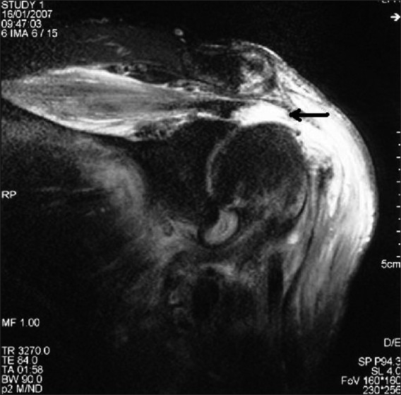
MRIOblique coronal shoulder MRI: full-thickness rotator cuff tear, arrow on fluid leaking through the torn tendon.
Source : Naqvi et al., International Journal of Shoulder Surgery, 2009, figure 3 · CC BY
Inspection & palpation : look for wasting of the fossae above and below the scapular spine, a sign both sensitive and highly specific (> 90 %) of an extensive chronic tear (Walch; Codman).¹⁴ Palpating the deltopectoral groove can reveal swelling of the long head of biceps where there is an intra-articular tear.
Specific cuff tests: their diagnostic properties : the meta-analyses Hegedus 2012 (BJSM, PMID 22773322)¹⁵ and Lädermann 2020-2021 (KSSTA, PMID 32725446)¹⁶ summarise the pooled performance:
| Test (region) | Sensitivity | Specificity | Level of evidence |
|---|---|---|---|
| Painful arc (supraspinatus + impingement), pathology | 0.74 | 0.81 | High SR/MA Hegedus |
| Empty can / Jobe (supraspinatus) | 0.77 | 0.53 | High Lädermann 2021 |
| Drop arm sign (full-thickness tear) | 0.38 | 0.97 | High Lädermann 2021 |
| External rotation lag sign (infraspinatus) | 0.46 | 0.94 | Moderate |
| ER resistance test (infraspinatus) | 0.76 | 0.57 | Moderate |
| Lift-off test (subscapularis) | 0.42 | 0.97 | Moderate |
| Belly-press test (subscapularis) | 0.40 | 0.98 | Moderate |
| Bear hug test (upper subscapularis) | 0.60 | 0.92 | Moderate |
| Neer / Hawkins-Kennedy (impingement) | 0.79 / 0.74 | 0.53 / 0.57 | Low provocation tests |
| A cluster of 3 or more positive tests | 0.74 | 0.86 | High a Bayesian approach |
In practice, combining tests (for example painful arc + drop arm + resisted external rotation) discriminates better than any test alone.¹⁵,¹⁶ The drop arm and the lift-off are SpIN tests (Specific, when Positive rules IN), very useful for confirming a suspicion; the painful arc and Jobe are SnNOUT (Sensitive, when Negative rules OUT), more useful for excluding.
Red flags (immediate medical referral)
- Acute trauma with deformity, extensive bruising, an abrupt loss of active elevation (suspect an acute tear, a fracture, a dislocation).
- A progressive neurological deficit : weakness not explained by pain, a sensory deficit, fasciculation (Parsonage-Turner, neoplasia, compressive radiculopathy).
- Non-mechanical night pain plus constitutional symptoms (fever, sweats, unexplained weight loss) → urgent oncological or infectious referral.
- A palpable mass at the shoulder or in the supraclavicular fossa.
- A history of cancer or immunosuppression plus resistant shoulder pain: think humeral metastasis, or brachial plexus involvement.
- Failure or rapid worsening after 4-6 weeks of well-conducted management.
Imperative differential diagnosis :
- C5-C6 cervical radiculopathy : Spurling test, neurological examination of dermatomes and myotomes, biceps reflexes. It can mimic a Jobe deficit or a positive lift-off through neurogenic weakness.¹⁷
- Acromioclavicular joint disease : localised palpation of the AC joint, the cross-body adduction test, pain localised to the lateral end of the clavicle (Hermans 2013).¹⁸
- Frozen shoulder (adhesive capsulitis) : a global loss of both active AND passive range, external rotation in particular, a restriction typically absent in an isolated cuff tear.¹⁹
- Parsonage-Turner syndrome (acute brachial neuritis): intense acute pain rapidly followed by profound weakness and rapid wasting; EMG directs the diagnosis (see the Cureus 2020 case, PMID 32742859).²⁰
- Calcific tendinitis, bursitis, tumour (rare).
Should patients with a symptomatic cuff tear be classified, and what are the benefits?
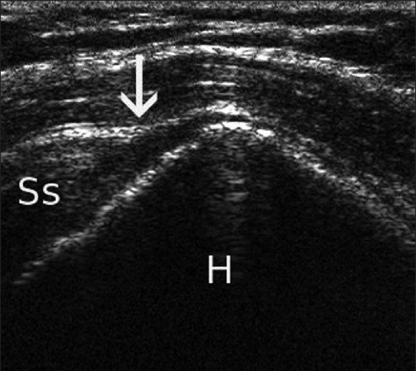
UltrasoundLongitudinal shoulder ultrasound: full-thickness tear of the supraspinatus (Ss), arrow on the tendon interruption adjacent to the humeral head (H).
Source : de Beer et al., International Journal of Shoulder Surgery, 2010, figure 1 · CC BY
- The Patte 1990 classification (PMID 2323151): it assesses tendon retraction on MRI or CT arthrography: Stage I (little retraction, as far as the humeral apex), Stage II (as far as the top of the humerus), Stage III (beyond that, massive retraction).²¹
- The Goutallier 1994 / Fuchs 1999 classification : it assesses fatty degeneration of the muscle (initially on CT, adapted to MRI by Fuchs): Stage 0 (normal), Stage 1 (a few fatty streaks), Stage 2 (< 50 % fat), Stage 3 (about 50 %), Stage 4 (> 50 % fat).²² A stage of 3 or more is considered a point of no return for surgical repair.
- The Ellman classification for partial tears: grade 1 (< 3 mm deep), 2 (3-6 mm), 3 (> 6 mm), specifying the side (bursal / articular / intratendinous).²³
- The size of the full-thickness tear (DeOrio & Cofield 1984, taken up by Snyder): small (< 1 cm), medium (1-3 cm), large (3-5 cm), massive (> 5 cm or 2 or more tendons).²⁴
📊 Estimated prognosis by Goutallier stage (based on Gladstone 2007, Burkhart 2007)
Cumulative functional success rate at 2 years, conservative and surgical treatment combined.
Sources: Gladstone JN, et al. Am J Sports Med. 2007;35(5):719-728. PMID 17337727. Burkhart 2007. Indicative estimates; the real figures vary with the size of the associated tear.
The clinical benefits of classification: (1) it informs the conservative-versus-surgical decision, (2) it sets realistic expectations to share with the patient, (3) it guides rehabilitation (precautions vs rapid progression), (4) it standardises communication between physiotherapist, physician, surgeon and radiologist.
Key points
- The history is the best diagnostic tool: age > 60 years + anterolateral pain + night pain = a high pre-test probability.
- No clinical test is perfect. A combination of tests (painful arc + drop arm + resisted external rotation) increases accuracy (Hegedus 2012, Lädermann 2020-2021).
- The drop arm and the lift-off have excellent specificity (about 97 %): they confirm rather than exclude.
- Imperative differential diagnosis: cervical radiculopathy, the acromioclavicular joint, frozen shoulder, Parsonage-Turner syndrome.
- The Patte (retraction) and Goutallier (fatty infiltration) classifications are the prognostic standards. A Goutallier of 3 or more is roughly the point of no return for repair.
- A tear on imaging is not enough to confirm the cause of the symptoms: clinical-radiological correlation is imperative.
Bibliography, chapter 2
- Yamamoto A, Takagishi K, Osawa T, et al. Prevalence and risk factors of a rotator cuff tear in the general population. J Shoulder Elbow Surg. 2010;19(1):116-120. PMID 19540777.
- Sher JS, Uribe JW, Posada A, Murphy BJ, Zlatkin MB. Abnormal findings on magnetic resonance images of asymptomatic shoulders. J Bone Joint Surg Am. 1995;77(1):10-15. PMID 7822341.
- Desmeules F, Roy JS, Lafrance S, et al. Rotator Cuff Tendinopathy Diagnosis, Nonsurgical Medical Care, and Rehabilitation: A Clinical Practice Guideline. J Orthop Sports Phys Ther. 2025. DOI 10.2519/jospt.2025.13182.
- Teunis T, Lubberts B, Reilly BT, Ring D. A systematic review and pooled analysis of the prevalence of rotator cuff disease with increasing age. J Shoulder Elbow Surg. 2014;23(12):1913-1921. PMID 25441568.
- Slaven EJ, Mathers J. Differential diagnosis of shoulder and cervical pain: a case report. J Man Manip Ther. 2010;18(4):191-196. PMID 22131792.
- Kuhn JE, Dunn WR, Sanders R, et al. Effectiveness of physical therapy in treating atraumatic full-thickness rotator cuff tears: a multicenter prospective cohort study. J Shoulder Elbow Surg. 2013;22(10):1371-1379. PMID 23540577.
- Mulligan EP, Brunette M, Shirley Z, Khazzam M. Sleep quality and nocturnal pain in patients with shoulder disorders. J Shoulder Elbow Surg. 2015;24(9):1452-1457. PMID 25842028.
- Mazuquin BF, Wright AC, Russell S, et al. Effectiveness of early compared with conservative rehabilitation for patients having rotator cuff repair surgery: an overview of systematic reviews. Br J Sports Med. 2018;52(2):111-121. PMID 28039127.
- Roy JS, MacDermid JC, Woodhouse LJ. Measuring shoulder function: a systematic review of four questionnaires. Arthritis Rheum. 2009;61(5):623-632. PMID 19405008.
- Tveitå EK, Ekeberg OM, Juel NG, Bautz-Holter E. Responsiveness of the shoulder pain and disability index in patients with adhesive capsulitis. BMC Musculoskelet Disord. 2008;9:161. PMID 19055757.
- Giri A, O'Hanlon D, Jain NB. Risk factors for rotator cuff disease (diabetes, hypertension, dyslipidaemia): SR/MA. Ann Phys Rehabil Med. 2023;66(1):101631. PMID 35257948.
- Chester R, Jerosch-Herold C, Lewis J, Shepstone L. Psychological factors are associated with the outcome of physiotherapy for people with shoulder pain. Br J Sports Med. 2018;52(4):269-275. PMID 27445360.
- Lafrance S, Charron M, Roy JS, et al. Diagnosing, Managing, and Supporting Return to Work of Adults With Rotator Cuff Disorders: A Clinical Practice Guideline. J Orthop Sports Phys Ther. 2022;52(10):647-664. PMID 35881707.
- Codman EA. The Shoulder: Rupture of the Supraspinatus Tendon and Other Lesions in or about the Subacromial Bursa. Boston: Thomas Todd Co; 1934 [the founding reference for the clinical examination of the shoulder].
- Hegedus EJ, Goode AP, Cook CE, et al. Which physical examination tests provide clinicians with the most value when examining the shoulder? Update of a SR with MA. Br J Sports Med. 2012;46(14):964-978. PMID 22773322.
- Lädermann A, Meynard T, Denard PJ, et al. Reliable diagnosis of posterosuperior rotator cuff tears requires a combination of clinical tests. Knee Surg Sports Traumatol Arthrosc. 2021;29(7):2118-2133. PMID 32725446.
- Thoomes EJ, Scholten-Peeters W, Koes B, Falla D, Verhagen AP. The effectiveness of conservative treatment for patients with cervical radiculopathy: a systematic review. Clin J Pain. 2013;29(12):1073-1086. PMID 23446070.
- Hermans J, Luime JJ, Meuffels DE, Reijman M, Simel DL, Bierma-Zeinstra SM. Does this patient with shoulder pain have rotator cuff disease? The Rational Clinical Examination systematic review. JAMA. 2013;310(8):837-847. PMID 23982370.
- Hand C, Clipsham K, Rees JL, Carr AJ. Long-term outcome of frozen shoulder. J Shoulder Elbow Surg. 2008;17(2):231-236. PMID 17993282.
- Ibrahim R, Krivitsky M, Nicola M, Zarour CC. Atypical Presentation of Parsonage-Turner Syndrome. Cureus. 2020;12(6):e8892. PMID 32742859.
- Patte D. Classification of rotator cuff lesions. Clin Orthop Relat Res. 1990;254:81-86. PMID 2323151.
- Goutallier D, Postel JM, Bernageau J, Lavau L, Voisin MC. Fatty muscle degeneration in cuff ruptures. Pre- and postoperative evaluation by CT scan. Clin Orthop Relat Res. 1994;304:78-83. PMID 8020238.
- Ellman H. Diagnosis and treatment of incomplete rotator cuff tears. Clin Orthop Relat Res. 1990;254:64-74. PMID 2182260.
- DeOrio JK, Cofield RH. Results of a second attempt at surgical repair of a failed initial rotator-cuff repair. J Bone Joint Surg Am. 1984;66(4):563-567. PMID 6707035.
Which treatment strategies are the most effective for a symptomatic cuff tear?
Where do you start? What is the recommended hierarchy of interventions?
The evidence-based hierarchy of interventions for the symptomatic degenerative tear rests on the most recent CPGs: JOSPT 2025 Desmeules CPG (rotator cuff tendinopathy and partial tears)³, JOSPT 2022 Lafrance CPG (return to work)⁴ and the BMJ Rapid Recommendation Vandvik 2019 on subacromial surgery.⁵- Patient education + therapeutic exercise (a strong recommendation, high level of evidence).
- Adjunctive manual therapy : can be added for short-term pain management (a moderate recommendation).
- Physical modalities and shockwave : optional adjuncts depending on the clinical profile (low to moderate evidence).
- Injections (corticosteroid, PRP, and so on) : to be discussed case by case, never first line (variable evidence, transient effects).
- Surgical referral : considered after the failure of at least 12 weeks (and more generally 3-6 months) of well-conducted conservative management, or from the outset in specific situations (an acute traumatic tear in a young active patient, a major functional deficit that is not improving).
The meta-analyses comparing surgery with conservative treatment (Ryösä 2017 Disabil Rehabil, Longo 2021 BMC Musculoskelet Disord) demonstrate no clinically significant superiority for surgery in the degenerative tear at 1, 2 or 5 years for pain and function.⁶,⁷ The randomised FINO trial (Kukkonen 2014/2015) compared physiotherapy, acromioplasty + PT, and repair + acromioplasty + PT in patients over 55 with a non-traumatic tear: no significant difference between the three groups at 1 and 2 years.⁸,⁹ The CSAW trial (Beard 2018, Lancet) on isolated subacromial decompression against placebo (investigational arthroscopy with no procedure) showed no clinically significant difference, supporting the BMJ Rapid Recommendation Vandvik 2019 against isolated decompression for chronic shoulder pain.¹⁰,⁵
What is the place of exercise, and is there a superior approach? 💪
Therapeutic exercise is the cornerstone of management. Its superiority over no treatment is established; the contemporary question is: is one type of exercise superior to another?The JOSPT 2025 Desmeules CPG concludes that no specific protocol has demonstrated clinically significant superiority in the current literature.³ Recent systematic reviews (Naunton 2020, Dominguez-Romero 2021, Pieters 2020 JOSPT update) confirm that the different formats are equivalent: eccentric, concentric, isometric, heavy slow loading (HSL), functional or scapula-focused exercise.¹¹,¹²,¹³ The principles of progression and individualisation matter more than the choice of any particular format.
The RoCTEx trial of Ingwersen 2017 (Orthopaedic Journal of Sports Medicine, and not BJSM 2021 as some sources wrongly state) compared progressive heavy loading with traditional low-load exercise in 100 patients with cuff tendinopathy: it demonstrated no superiority for the heavy-load protocol.¹⁴
Several practical principles emerge:
- Start with exercise below the pain threshold, prioritising the quality of scapulohumeral movement.
- Include exercises targeting the scapular stabilisers (serratus anterior, middle and lower trapezius).
- Progress in load, complexity and range as tolerance allows (Mertens 2022 narrative review).¹⁵
- Encourage simple home programmes (2-4 exercises) to support long-term adherence (Littlewood 2014, Edwards 2016).¹⁶,¹⁷
- Pain of 3-4/10 or less during or after the exercise, which does not worsen within 24 h, is acceptable.
Adjunctive therapies: what are manual therapy, shockwave and injections worth?
| Modality | Effect on pain | Effect on function | Level of evidence | Recommendation |
|---|---|---|---|---|
| Education + progressive exercise | A moderate short-term effect, lasting at 12 months and beyond | Significant lasting improvement (Kuhn MOON) | High GRADE | Strong (first line) |
| Adjunctive manual therapy | Demonstrated short-term relief (Desjardins-Charbonneau 2015 SR/MA, Pieters 2022) | A small additional effect when combined with exercise | Moderate | Conditional, as an adjunct |
| Shockwave (ESWT) | A benefit mainly in calcific tendinopathy (Bannuru 2014) | Limited data in non-calcific tears | Low (non-calcific tears) | Conditional, where 12 weeks or more have failed |
| Subacromial corticosteroid injection | Short-term relief (4-6 weeks, MOA) | No lasting functional improvement, and a risk to the tendon if repeated | Low in the long term | Conditional, a maximum of 2-3 injections a year |
| PRP (Platelet-Rich Plasma) | A variable effect, no better than sham in recent SRs | Mixed data | Low | Not recommended as routine |
| Subacromial decompression (isolated) | No better than placebo (CSAW Beard 2018) | No difference against PT (FINO Kukkonen) | A strong recommendation AGAINST | BMJ Vandvik 2019 |
| Surgical repair (selected) | Results similar to PT at 1-5 years in degenerative tears | Remaining indications: an acute traumatic tear in a young patient, PT failure at 3-6 months in an active patient | Moderate | Selected cases only |
As for therapeutic ultrasound, low-level laser and heat or cold modalities, the current recommendations converge: there is not enough evidence of efficacy to recommend them as routine (Page 2016 Cochrane, Desmeules 2025 CPG).¹⁸
Beyond the physical: how do you educate the patient and address psychological factors? 🧠
Patient education is a major determinant of therapeutic success, and is no longer regarded as an “extra” but as a central intervention.¹⁹ The key messages to convey:- A tear on imaging does not necessarily mean the tear is the cause of the pain (the corollary of the data in Yamamoto 2010, Sher 1995, Teunis 2014).
- “Hurt does not equal harm”: moderate pain during exercise is not a signal of further tissue damage.
- Function can be restored without structural healing of the tear: that is the explicit aim of conservative treatment.
- Movement is safe and beneficial; inactivity is harmful.
The psychological factors (kinesiophobia, catastrophising, self-efficacy, expectations) are powerful predictors of outcome, independent of the size of the tear or its biomechanical properties:
- Chester 2018 BJSM (PMID 27445360): in a multicentre cohort of n=1,030 patients with shoulder pain, the psychological factors (kinesiophobia, low expectations, catastrophising) are the most powerful predictors of outcome at 6 months.²⁰
- Chester 2019 BJSM (PMID 30626599): a CART analysis identifies self-efficacy (the PSEQ questionnaire) as the leading predictor of persistent pain at 6 months.²¹
- Lafrance 2022 CPG : explicitly recommends screening for psychosocial factors as an integral part of the initial assessment.⁴
Key points
- The first line is conservative (education + exercise + adjunctive manual therapy): 75 % of patients avoid surgery at 2 years (Kuhn MOON 2013).
- Surgery is not superior to physiotherapy in the degenerative tear (Ryösä 2017, Longo 2021, Kukkonen FINO 2014/2015).
- And isolated subacromial decompression is no longer recommended for chronic shoulder pain (BMJ Rapid Recommendation Vandvik 2019; the CSAW trial, Beard 2018).
- No exercise protocol (eccentric, concentric, isometric, heavy loading) is superior to another:adherence and progression matter more than the format (JOSPT 2025 CPG).
- The psychological factors (kinesiophobia, self-efficacy) are the most powerful predictors of outcome (Chester 2018-2019 BJSM).
- Patient education about the imaging-symptom dissociation and about the safety of movement is a central therapeutic act.
Bibliography, chapter 3
- Kuhn JE, Dunn WR, Sanders R, et al. Effectiveness of physical therapy in treating atraumatic full-thickness rotator cuff tears: a multicenter prospective cohort study. J Shoulder Elbow Surg. 2013;22(10):1371-1379. PMID 23540577.
- Kuhn JE, Dunn WR, Sanders R, et al. The Predictors of Surgery for Symptomatic, Atraumatic Full-Thickness Rotator Cuff Tears Change Over Time: Ten-Year Outcomes of the MOON Shoulder Prospective Cohort. J Bone Joint Surg Am. 2024;106(17):1563-1572. PMID 38980920.
- Desmeules F, Roy JS, Lafrance S, et al. Rotator Cuff Tendinopathy Diagnosis, Nonsurgical Medical Care, and Rehabilitation: A Clinical Practice Guideline. J Orthop Sports Phys Ther. 2025. DOI 10.2519/jospt.2025.13182.
- Lafrance S, Charron M, Roy JS, et al. Diagnosing, Managing, and Supporting Return to Work of Adults With Rotator Cuff Disorders: A Clinical Practice Guideline. J Orthop Sports Phys Ther. 2022;52(10):647-664. PMID 35881707.
- Vandvik PO, Lähdeoja T, Ardern C, et al. Subacromial decompression surgery for adults with shoulder pain: a clinical practice guideline (BMJ Rapid Recommendation). BMJ. 2019;364:l294. PMID 30728120.
- Ryösä A, Laimi K, Äärimaa V, Lehtimäki K, Kukkonen J, Saltychev M. Surgery or conservative treatment for rotator cuff tear: a meta-analysis. Disabil Rehabil. 2017;39(14):1357-1363. PMID 27385156.
- Longo UG, Risi Ambrogioni L, Candela V, et al. Conservative versus surgical management for patients with rotator cuff tears: a systematic review and meta-analysis. BMC Musculoskelet Disord. 2021;22(1):50. PMID 33419401.
- Kukkonen J, Joukainen A, Lehtinen J, et al. Treatment of non-traumatic rotator cuff tears: A randomised controlled trial with one-year clinical results. Bone Joint J. 2014;96-B(1):75-81. PMID 24395315.
- Kukkonen J, Joukainen A, Lehtinen J, et al. Treatment of Nontraumatic Rotator Cuff Tears: A Randomized Controlled Trial with Two Years of Clinical and Imaging Follow-up. J Bone Joint Surg Am. 2015;97(21):1729-1737. PMID 26537160.
- Beard DJ, Rees JL, Cook JA, et al. Arthroscopic subacromial decompression for subacromial shoulder pain (CSAW): a multicentre, pragmatic, parallel group, placebo-controlled, three-group, randomised surgical trial. Lancet. 2018;391(10118):329-338. PMID 29169668.
- Naunton J, Street G, Littlewood C, Haines T, Malliaras P. Effectiveness of progressive and resisted and non-progressive or non-resisted exercise in rotator cuff related shoulder pain: a SR. Clin Rehabil. 2020;34(9):1198-1216. PMID 32571081.
- Dominguez-Romero JG, Jiménez-Rejano JJ, Ridao-Fernández C, Chamorro-Moriana G. Exercise-Based Muscle Development Programmes and Their Effectiveness in the Functional Recovery of Rotator Cuff Tendinopathy: A Systematic Review. Diagnostics (Basel). 2021;11(3):529. PMID 33809604.
- Pieters L, Lewis J, Kuppens K, et al. An Update of Systematic Reviews Examining the Effectiveness of Conservative Physical Therapy Interventions for Subacromial Shoulder Pain. J Orthop Sports Phys Ther. 2020;50(3):131-141. DOI 10.2519/jospt.2020.8498.
- Ingwersen KG, Jensen SL, Sørensen L, et al. Three Months of Progressive High-Load Versus Traditional Low-Load Strength Training Among Patients With Rotator Cuff Tendinopathy: Primary Results From the Double-Blind Randomized Controlled RoCTEx Trial. Orthop J Sports Med. 2017;5(8):2325967117723292. PMID 28875153.
- Mertens MGCAM, Meert L, Struyf F, et al. Exercise therapy is effective for improvement in range of motion, function, and pain in patients with frozen shoulder: a systematic review and meta-analysis. Arch Phys Med Rehabil. 2022;103(5):998-1012.e14. PMID 34425089.
- Littlewood C, Malliaras P, Mawson S, May S, Walters SJ. Self-managed loaded exercise versus usual physiotherapy treatment for rotator cuff tendinopathy: a pilot randomised controlled trial. Physiotherapy. 2014;100(1):54-60. PMID 23954024.
- Edwards P, Ebert J, Joss B, Bhabra G, Ackland T, Wang A. Exercise rehabilitation in the non-operative management of rotator cuff tears: a review of the literature. Int J Sports Phys Ther. 2016;11(2):279-301. PMID 27104061.
- Page MJ, Green S, Mrocki MA, et al. Electrotherapy modalities for rotator cuff disease. Cochrane Database Syst Rev. 2016;6:CD012225. PMID 27283591.
- Powell JK, Lewis JS. Rotator Cuff-Related Shoulder Pain: Is It Time to Reframe the Advice, "You Need to Strengthen Your Shoulder"? J Orthop Sports Phys Ther. 2021;51(4):156-158. PMID 33789431.
- Chester R, Jerosch-Herold C, Lewis J, Shepstone L. Psychological factors are associated with the outcome of physiotherapy for people with shoulder pain. Br J Sports Med. 2018;52(4):269-275. PMID 27445360.
- Chester R, Khondoker M, Shepstone L, Lewis JS, Jerosch-Herold C. Self-efficacy and risk of persistent shoulder pain: results of a Classification and Regression Tree (CART) analysis. Br J Sports Med. 2019;53(13):825-834. PMID 30626599.
How do you secure lasting recovery and prevent recurrence of a symptomatic cuff tear?
How do you make the patient an active participant in their recovery through self-management?
👍 Self-management is the process by which the patient, guided by the clinician, becomes the main agent of their own rehabilitation and of the future management of their condition. The key elements:- Structured therapeutic education : the nature of the condition, the role of loading, the imaging-symptom dissociation, the safety of movement.
- A simple home programme (2-4 exercises) : Littlewood 2014 showed efficacy comparable to a complex supervised programme.⁴ Edwards 2016 and Pieters 2020 confirm that long-term adherence is inversely correlated with the complexity of the programme.⁵,⁶
- Load progression as tolerance allows (pain of 3-4/10 or less is acceptable during the exercise if there is no worsening at 24 h).
- Self-monitoring : teach the patient to identify the signs of improvement (strength, function, sleep quality) and the signs of worsening that warrant reassessment.
- Strategies for painful flares : temporarily reducing the load without stopping altogether, managing sleep, occasional anti-inflammatory measures.
Self-efficacy (measured by the Pain Self-Efficacy Questionnaire, PSEQ) is one of the most powerful predictors of long-term outcome (Chester 2019).⁷ Strengthening self-efficacy is therefore an explicit goal for the clinician.
When and how should a safe return to activity and to sport be planned?
Planning the return to activity and sport rests on functional and psychological criteria rather than on a fixed calendar.⁸,⁹ The systematic review Migliorini 2023 (and the earlier SR of Klouche 2016) establishes an overall return-to-sport rate of the order of 60-85 % depending on the sport, the level of play and the type of management (conservative or surgical).¹⁰The recommended sequence:
- Prerequisites : full, pain-free active mobility, strength at 80 % or more of the healthy side (measured on isometric dynamometry).
- Functional tests : press-ups against a wall, an incline or the floor as progression allows, graded throwing (overhead sports), scapulohumeral endurance, scapular control in movement.
- A progressive return : intensity and volume rising, the sport-specific movement first in a controlled then in an open environment.
- Psychological assessment : confidence in movement, the absence of residual kinesiophobia (PSEQ, Tampa Scale for Kinesiophobia).
- Communicating realistic expectations : in the degenerative tear, a return to the pre-symptomatic level is not automatic; the main goal is satisfactory function in daily life and in the activities that matter to the patient.
The MOON cohort followed to 10 years (Kuhn 2024) shows that among patients whose initial conservative treatment succeeded, most remain satisfied at 10 years without surgery, even though a minority convert to surgery during follow-up (typically for worsening symptoms or loss of function).¹¹
Key points
- Empowering the patient through therapeutic education and a simple home programme (2-4 exercises) is more effective in the long term than passive approaches.
- Self-efficacy (PSEQ) is one of the most powerful predictors of outcome (Chester 2019 BJSM).
- The return to activity and sport should be based on functional and psychological criteria, not on a fixed calendar (Lafrance 2022 CPG).
- The return-to-sport rate is 60-85 % depending on the sport and the type of management (Migliorini 2023).
- The MOON cohort at 10 years (Kuhn 2024) confirms that the conservative response lasts in most patients who responded initially.
Bibliography, chapter 4
- Lafrance S, Charron M, Roy JS, et al. Diagnosing, Managing, and Supporting Return to Work of Adults With Rotator Cuff Disorders: A Clinical Practice Guideline. J Orthop Sports Phys Ther. 2022;52(10):647-664. PMID 35881707.
- Desmeules F, Roy JS, Lafrance S, et al. Rotator Cuff Tendinopathy Diagnosis, Nonsurgical Medical Care, and Rehabilitation: A Clinical Practice Guideline. J Orthop Sports Phys Ther. 2025. DOI 10.2519/jospt.2025.13182.
- Powell JK, Lewis JS. Rotator Cuff-Related Shoulder Pain: Is It Time to Reframe the Advice, "You Need to Strengthen Your Shoulder"? J Orthop Sports Phys Ther. 2021;51(4):156-158. PMID 33789431.
- Littlewood C, Malliaras P, Mawson S, May S, Walters SJ. Self-managed loaded exercise versus usual physiotherapy treatment for rotator cuff tendinopathy: a pilot randomised controlled trial. Physiotherapy. 2014;100(1):54-60. PMID 23954024.
- Edwards P, Ebert J, Joss B, et al. Exercise rehabilitation in the non-operative management of rotator cuff tears: a review of the literature. Int J Sports Phys Ther. 2016;11(2):279-301. PMID 27104061.
- Pieters L, Lewis J, Kuppens K, et al. An Update of Systematic Reviews Examining the Effectiveness of Conservative Physical Therapy Interventions for Subacromial Shoulder Pain. J Orthop Sports Phys Ther. 2020;50(3):131-141. DOI 10.2519/jospt.2020.8498.
- Chester R, Khondoker M, Shepstone L, Lewis JS, Jerosch-Herold C. Self-efficacy and risk of persistent shoulder pain: results of a Classification and Regression Tree (CART) analysis. Br J Sports Med. 2019;53(13):825-834. PMID 30626599.
- Klouche S, Lefevre N, Herman S, Gerometta A, Bohu Y. Return to Sport After Rotator Cuff Tear Repair: A Systematic Review and Meta-analysis. Am J Sports Med. 2016;44(7):1877-1887. PMID 26316611.
- Migliorini F, Asparago G, Cuozzo F, Oliva F, Hildebrand F, Maffulli N. Patient outcomes and return to play after arthroscopic rotator cuff repair in overhead athletes: a systematic review. J Orthop Traumatol. 2023;24(1):3. PMID 36656423. PMC9852377.
- Klouche S, et al. Return to Sport After Rotator Cuff Tear Repair: A Systematic Review and Meta-Analysis. Am J Sports Med. 2016;44(7):1877-1887. See reference 8.
- Kuhn JE, Dunn WR, Sanders R, et al. The Predictors of Surgery for Symptomatic, Atraumatic Full-Thickness Rotator Cuff Tears Change Over Time: Ten-Year Outcomes of the MOON Shoulder Prospective Cohort. J Bone Joint Surg Am. 2024;106(17):1563-1572. PMID 38980920.
What do real clinical cases teach us about symptomatic cuff tears?
Analysis of a “classic” case: the MOON cohort and its lessons
The MOON cohort (Multicenter Orthopaedic Outcomes Network, Kuhn 2013, J Shoulder Elbow Surg, n=433, PMID 23540577) is the archetypal case mix, in which every patient with a symptomatic degenerative tear shares certain features:¹- A mean age of 62, with no identified acute trauma.
- A full-thickness supraspinatus tear ± infraspinatus, mean size 1.5-2 cm.
- Insidious pain, night pain, weakness on elevation.
- A structured 6-12 week programme: phase 1 pain control plus recovery of range; phase 2 progressive strengthening of the stabilisers; phase 3 functional reintegration.
- The result at 2 years : 75 % avoid surgery, with significant improvement in the ASES and SST.
- The result at 10 years (Kuhn 2024): most of the initial responders remain satisfied with function maintained.²
The key lesson: structural healing of the tear is not necessary to obtain an excellent functional result. The proposed mechanism is neuromuscular optimisation of the intact muscles (deltoid, scapular muscles, the healthy parts of the cuff).
The diagnostic challenge: when a symptomatic cuff tear mimics another condition
🕵️♂️ Published cases of diagnostic “mimics” are valuable reminders. Several verified PMC publications are representative:- Parsonage-Turner syndrome presenting initially as a cuff tear: Ibrahim 2020 (Cureus, PMID 32742859, PMC7388810) describes an atypical presentation 10 weeks after surgical repair of a cuff tear: the EMG finds denervation of biceps, deltoid, supraspinatus and infraspinatus with no cervical involvement, the electrophysiological signature of the syndrome.³
- Atypical presentation confounded by surgical rotator cuff injury (PMC5623205): cuff surgery preceded the onset of a Parsonage-Turner syndrome, delaying the diagnosis and compromising rehabilitation.⁴
- C5-C6 cervical radiculopathy mimicking a supraspinatus deficit: neurogenic weakness can be clinically indistinguishable from a full-thickness tear without a targeted EMG or cervical MRI (Slaven 2010 J Man Manip Ther, PMID 22131792).⁵
- Frozen shoulder in the early painful stage: the loss of passive external rotation (typically absent in an isolated cuff tear) should raise the alarm (Hand 2008, JSES).⁶
A complex case: a massive tear with Goutallier 3-4
Massive tears with advanced fatty degeneration (Goutallier 3 or more) are a therapeutic challenge. Several studies have documented that these patients can achieve significant functional improvement with conservative treatment, although the prognosis is more guarded than with a Goutallier of 2 or less.- Collin 2014 (J Shoulder Elbow Surg, n=66 patients with a massive tear not treated surgically) : a retrospective analysis of the factors predicting pseudoparalysis. The loss of active shoulder function is determined not by size alone, but by the combination of a massive tear with advanced degeneration.⁷
- Levy 2008 (Tendon transfers in irreparable rotator cuff tears, review) : even with a massive tear, optimising the remaining muscles (deltoid, pectoralis major, latissimus dorsi) can restore useful function.⁸
- The rehabilitation programme prioritises: humeral head centring, scapular control, strengthening of the anterior and middle deltoid (which becomes the functional pivot), and endurance training.
Refractory cases despite well-conducted conservative management can be referred for salvage surgery : partial repair, tendon transfer (latissimus dorsi, pectoralis major, lower trapezius), or reverse shoulder arthroplasty in older patients. These options are to be discussed case by case with a shoulder surgeon.
Key points
- The “classic case” from the MOON cohort confirms that 75 % of symptomatic degenerative tears achieve functional success with physiotherapy, without structural healing.
- The diagnostic mimics (Parsonage-Turner, cervical radiculopathy, frozen shoulder) must be looked for actively: a tear on imaging is not always the cause of the symptoms.
- Even massive tears with Goutallier 3-4 can respond to rehabilitation targeted at centring and motor control (Collin 2014).
- The surgical options for refractory cases (partial repair, tendon transfer, reverse arthroplasty) are to be discussed case by case with a specialist shoulder surgeon.
Bibliography, chapter 5
- Kuhn JE, Dunn WR, Sanders R, et al. Effectiveness of physical therapy in treating atraumatic full-thickness rotator cuff tears: a multicenter prospective cohort study (MOON). J Shoulder Elbow Surg. 2013;22(10):1371-1379. PMID 23540577.
- Kuhn JE, Dunn WR, Sanders R, et al. The Predictors of Surgery for Symptomatic, Atraumatic Full-Thickness Rotator Cuff Tears Change Over Time: Ten-Year Outcomes of the MOON Shoulder Prospective Cohort. J Bone Joint Surg Am. 2024;106(17):1563-1572. PMID 38980920.
- Ibrahim R, Krivitsky M, Nicola M, Zarour CC. Atypical Presentation of Parsonage-Turner Syndrome. Cureus. 2020;12(6):e8892. PMID 32742859. PMC7388810.
- Greenhill DA, Abdelfattah H, Torg JS, Sewards JM. Atypical presentation of Parsonage-Turner syndrome confounded by surgical rotator cuff injury. BMJ Case Rep. 2017;2017:bcr-2017-220532. PMID 28739618. PMC5623205.
- Slaven EJ, Mathers J. Differential diagnosis of shoulder and cervical pain: a case report. J Man Manip Ther. 2010;18(4):191-196. PMID 22131792.
- Hand C, Clipsham K, Rees JL, Carr AJ. Long-term outcome of frozen shoulder. J Shoulder Elbow Surg. 2008;17(2):231-236. PMID 17993282.
- Collin P, Matsumura N, Lädermann A, Denard PJ, Walch G. Relationship between massive chronic rotator cuff tear pattern and loss of active shoulder range of motion. J Shoulder Elbow Surg. 2014;23(8):1195-1202. PMID 24433628.
- Levy O, Mullett H, Roberts S, Copeland S. The role of anterior deltoid reeducation in patients with massive irreparable degenerative rotator cuff tears. J Shoulder Elbow Surg. 2008;17(6):863-870. PMID 18718765.
How do you apply these recommendations concretely in your practice?
When and to which other health professionals should you refer?
🧭 The decision to refer depends on the following criteria:- Red flags (see the diagnostic section): immediate medical referral (general practitioner, emergency department, specialist as the context requires).
- Failure of conservative treatment after at least 12 weeks (and generally 3-6 months) well conducted, with no significant functional improvement: refer to a shoulder surgeon to discuss imaging and options.
- Young patients with an acute traumatic tear (a clear mechanism, MRI showing a recent full-thickness tear with little retraction and good muscle quality): early surgical referral is often recommended.
- Dominant psychosocial factors (severe kinesiophobia, catastrophising, depression, anxiety): refer to a pain psychologist or psychotherapist (Coronado 2020).¹
- Adapting the workstation or activities of daily living: refer to an occupational therapist or occupational physician.²
- Pharmacology : where NSAIDs, step 1-2 analgesics or an injection are being considered, work with the general practitioner or rheumatologist.
How do you measure outcomes and overcome barriers to implementation?
📊 Measuring outcomes objectively is essential. The validated tools recommended for the shoulder:- SPADI (Shoulder Pain and Disability Index) : 13 items, 5 pain + 8 function, MCID 8-13 points. The pivotal tool in physiotherapy.³
- DASH (Disabilities of the Arm, Shoulder and Hand) or its short version QuickDASH : 30 or 11 items, DASH MCID about 10 points.⁴
- Constant-Murley Score : combines pain (15 pts), ADLs (20 pts), mobility (40 pts), strength (25 pts). MCID about 10-15 points depending on the study.⁵
- Oxford Shoulder Score (OSS) : 12 items, MCID about 4-5 points (Tveita 2018).⁶
- ASES (American Shoulder and Elbow Surgeons Score) : used in research and surgery.
- PSEQ (Pain Self-Efficacy Questionnaire) for screening the psychological factors.
Assessment at baseline, 6 weeks, 12 weeks and 6 months makes it possible to quantify the trajectory and to make progress objective.
📊 Contemporary management algorithm for the symptomatic degenerative tear
A synthesis of the JOSPT 2025 Desmeules CPG, the JOSPT 2022 Lafrance CPG and the BMJ Rapid Recommendation Vandvik 2019.
A synthesis of: Desmeules 2025 JOSPT CPG, Lafrance 2022 JOSPT CPG, Vandvik 2019 BMJ. The diagram is simplified for the article; individual decisions depend on the clinical context.
Overcoming the barriers to implementation
🚧 The main barriers to implementing EBP in physiotherapy practice have been identified in several systematic reviews:- Scurlock-Evans 2014 (J Contin Educ Health Prof, SR): lack of time, limited critical appraisal skills, poor access to databases, lack of institutional support.⁷
- Bork 2023 (BMC Health Serv Res, a qualitative study of German physiotherapists): confirms that these barriers persist, and adds the lack of mentoring and of clinical leadership.⁸
- Tools for overcoming them: pre-appraised resources (Cochrane Summaries, CPGs such as JOSPT's), continuing education, mentoring, journal clubs, a working environment that supports learning.
Successfully implementing EBP is not only an individual matter but a systemic question : funding models, resources allocated to continuing education, access to the scientific literature.⁹
Key points
- Surgical referral after at least 3-6 months of well-conducted conservative treatment with no improvement; immediately where there are red flags or an acute traumatic tear in a young patient.
- Measure outcomes with validated PROMs : SPADI (MCID 8-13), DASH (MCID about 10), Constant-Murley (MCID 10-15), OSS, ASES.
- And isolated subacromial decompression is not recommended for chronic shoulder pain (BMJ Rapid Recommendation Vandvik 2019).
- The barriers to implementing EBP are individual (time, training) and systemic (funding, access to resources), Scurlock-Evans 2014, Bork 2023.
- Structured interprofessional collaboration (general practitioner, shoulder surgeon, psychologist, occupational therapist, occupational physician) is essential in complex cases.
Bibliography, chapter 6
- Coronado RA, Brintz CE, McKernan LC, et al. Psychologically informed physical therapy for musculoskeletal pain: current approaches, implications, and future directions from recent randomized trials. Pain Rep. 2020;5(5):e847. PMID 33490842.
- Lafrance S, Charron M, Roy JS, et al. Diagnosing, Managing, and Supporting Return to Work of Adults With Rotator Cuff Disorders: A Clinical Practice Guideline. J Orthop Sports Phys Ther. 2022;52(10):647-664. PMID 35881707.
- Roach KE, Budiman-Mak E, Songsiridej N, Lertratanakul Y. Development of a shoulder pain and disability index. Arthritis Care Res. 1991;4(4):143-149. PMID 11188601.
- Hudak PL, Amadio PC, Bombardier C. Development of an upper extremity outcome measure: the DASH. Am J Ind Med. 1996;29(6):602-608. PMID 8773720.
- Constant CR, Gerber C, Emery RJ, et al. A review of the Constant score: modifications and guidelines for its use. J Shoulder Elbow Surg. 2008;17(2):355-361. PMID 18218327.
- Tveitå EK, Ekeberg OM, Juel NG, Bautz-Holter E. Responsiveness of the shoulder pain and disability index in patients with adhesive capsulitis. BMC Musculoskelet Disord. 2008;9:161. PMID 19055757.
- Scurlock-Evans L, Upton P, Upton D. Evidence-based practice in physiotherapy: a systematic review of barriers, enablers and interventions. Physiotherapy. 2014;100(3):208-219. PMID 24780633.
- Paci M, Faedda G, Ugolini A, Pellicciari L. Barriers to evidence-based practice implementation in physiotherapy: a systematic review and meta-analysis. Int J Qual Health Care. 2021;33(2):mzab093. PMID 34110410.
- Vandvik PO, Lähdeoja T, Ardern C, et al. Subacromial decompression surgery for adults with shoulder pain: a clinical practice guideline. BMJ. 2019;364:l294. PMID 30728120.
Would you like to go further with this article? See the other clinical resources from Physio Learning on evidence-based shoulder physiotherapy.
Discover Physio Learning
