















In brief
Frozen shoulder, or adhesive capsulitis, is an inflammatory fibrosis of the glenohumeral capsule causing a progressive, painful loss of both active and passive shoulder movement. It affects 2 to 5 % of the population, above all women aged 40 to 60, with diabetes the main systemic risk factor. It runs through three phases (freezing, frozen, thawing) over one to three years. Diagnosis is clinical, the cardinal sign being a loss of passive external rotation greater than 50 % of the healthy side. Treatment, matched to the phase, combines education, gentle exercise, manual therapy and intra-articular corticosteroid injections.
Clinical synthesis based on the landmark review Millar 2022 Nature Reviews, the UK FROST trial 2020 Lancet, the network meta-analysis Challoumas 2020 JAMA Network Open and the 2023-2026 data on the diabetic subgroup.
Clinical summary
- Frozen shoulder is an inflammatory fibrosis of the glenohumeral capsule affecting 2 to 5 % of the population, mainly women between 40 and 60. It may be primary and idiopathic, or secondary (after trauma, surgery or immobilisation).
- Above all, diabetes mellitus is the main systemic risk factor (prevalence 10-38 %, with more severe and more treatment-resistant forms). Thyroid disorders (both hyper- and hypothyroidism) are the second major axis, confirmed by the Chuang JSES 2023 meta-analysis.
- The condition runs through 3 overlapping phases : a painful phase (freezing, 2-9 months), a stiff phase (frozen, 4-12 months), and a thawing phase (5 months to more than 2 years). The natural history is long (1-3 years), but 30-40 % of patients keep long-term residual impairment.
- Diagnosis is clinical. The cardinal sign: a symmetrical loss of both active AND passive range, in particular passive external rotation reduced by more than 50 % against the healthy side (Kelley 2013).
- Plain radiography serves above all for the differential diagnosis (glenohumeral osteoarthritis, calcific tendinopathy, the sequelae of a fracture). MRI or ultrasound, after Suh 2019, can confirm thickening of the axillary capsule and of the coracohumeral ligament.
- Treatment is progressive and matched to the phase. Patient education and reassurance are fundamental. Corticosteroid injections into the joint are effective on pain in the short term (Challoumas 2020 NMA, Sun 2017 MA).
- Gentle, progressive exercise is the basis of treatment (Cochrane Page 2014). Manual therapy alongside it improves mobility and function (Noten 2016, Yang 2007).
- The major randomised trial UK FROST (Rangan 2020 Lancet) shows that early structured physiotherapy, manipulation under anaesthetic and arthroscopic capsular release give comparable functional results at 12 months, with physiotherapy remaining the most cost-effective.
- For resistant cases (particularly in people with diabetes), arthrographic hydrodilatation under ultrasound guidance (Dimitri-Pinheiro 2022) can create a window of opportunity to restart rehabilitation.
- Recurrence on the same side is rare (under 5 %), but the other shoulder is affected in 6 to 17 % of patients within 5 years, with an excess risk in people with diabetes.
- The return to sport should rest on objective functional criteria (range and strength above 90 % of the healthy side, no pain, scapulohumeral control) rather than on a fixed calendar.
- Medical referral is required where red flags are present (Finucane 2020): non-mechanical night pain, disproportionate motor weakness, a history of cancer, a neurological sign.
- Measuring outcomes with PROMs (SPADI, DASH, ASES, Constant-Murley) is essential for making progress objective and adapting management.
Contents
- What are the fundamentals to know about frozen shoulder (adhesive capsulitis)?
- How do you assess and diagnose frozen shoulder (adhesive capsulitis) with certainty?
- Which treatment strategies are the most effective for frozen shoulder (adhesive capsulitis)?
- Frozen shoulder and diabetes: why does this subgroup deserve particular attention?
- How do you secure lasting recovery and prevent recurrence of frozen shoulder (adhesive capsulitis)?
- What do real clinical cases teach us about frozen shoulder (adhesive capsulitis)?
- How do you apply these recommendations concretely in your practice?
What are the fundamentals to know about frozen shoulder (adhesive capsulitis)?
How is this condition defined, who does it affect and what are the risk factors?
Frozen shoulder is defined as an inflammatory fibrosis of the glenohumeral joint capsule, causing thickening and retraction of the capsule and a painful restriction of movement.1,2 Its prevalence in the general population is estimated at between 2 % and 5 %, affecting women preferentially (OR about 1.6) between 40 and 60 years.1 Idiopathic forms are the most frequent, but secondary forms occur after trauma, shoulder surgery (particularly of the cuff) or prolonged immobilisation.3Prevalence compared: general population vs people with diabetes
Diabetes multiplies the risk of frozen shoulder by 5 to 8 (Zreik 2016 MA, Chen 2024 review)
Source: Zreik NH et al. Muscles Ligaments Tendons J 2016;6(1):26-34. PMID 27331029 - a meta-analysis of 12 studies. Chen MH, Chen WS. J Clin Med 2024;13(19):5696. PMID 39407755.
Systemic risk factors for frozen shoulder (adjusted OR/RR)
A synthesis of the 2016-2024 meta-analyses - the values are pooled estimates
Sources: Zreik 2016 MLTJ - Chuang 2023 JSES - Cucchi 2017 Joints - Millar 2022 NRDP.
What happens in the joint capsule, and how does the condition evolve naturally?
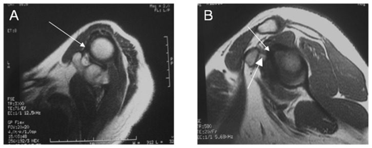
MRIAxial and sagittal shoulder MRI: thickened and twisted coracohumeral ligament, arrows on the ligament, adhesive capsulitis.
Source : Zhao et al., PloS one, 2012, figure 2 · CC BY
The natural course in 3 phases (Reeves 1975, updated Millar 2022)
Overlapping phases - a typical total duration of 1 to 3 years
Source: Reeves B 1975, Hannafin JA, Chiaia TA 2000, updated Millar NL et al. Nat Rev Dis Primers 2022;8:59. PMID 36075904.
Key points
- Frozen shoulder is an inflammatory fibrosis of the glenohumeral capsule affecting 2 to 5 % of the population (Millar 2022 NRDP).
- Above all, diabetes is the main systemic risk factor (OR ~5, prevalence 10 to 38 %); thyroid dysfunction and Dupuytren's disease are well-documented secondary factors.
- The pathophysiology combines hypervascular synovitis and TGF-beta mediated fibrosis.
- The course runs through 3 phases (freezing, frozen, thawing) over 1 to 3 years, but 30 to 40 % of patients keep residual impairment beyond 4 years, above all patients with diabetes.
Bibliography
- Millar NL, Meakins A, Struyf F, et al. Frozen shoulder. Nat Rev Dis Primers. 2022;8(1):59. PMID 36075904.
- Cho CH, Bae KC, Kim DH. Treatment Strategy for Frozen Shoulder. Clin Orthop Surg. 2019;11(3):249-257. PMID 31475043.
- Lewis J. Frozen shoulder contracture syndrome - Aetiology, diagnosis and management. Man Ther. 2015;20(1):2-9. PMID 25107826.
- Huang YP, Fann CY, Chiu YH, et al. Association of diabetes mellitus with the risk of developing adhesive capsulitis. Arthritis Care Res. 2013;65(7):1197-1202. DOI 10.1002/acr.21938.
- Zreik NH, Malik RA, Charalambous CP. Adhesive capsulitis of the shoulder and diabetes: a meta-analysis of prevalence. Muscles Ligaments Tendons J. 2016;6(1):26-34. PMID 27331029.
- Chuang SH, Chen YP, Huang SW, Kuo YJ. Association between adhesive capsulitis and thyroid disease: a meta-analysis. J Shoulder Elbow Surg. 2023;32(6):1314-1322. PMID 36871608.
- Cucchi D, Marmotti A, De Giorgi S, et al. Risk Factors for Shoulder Stiffness: Current Concepts. Joints. 2017;5(4):217-223. PMID 29270559.
- Ryan V, Brown H, Minns Lowe CJ, Lewis JS. The pathophysiology associated with primary (idiopathic) frozen shoulder: A systematic review. BMC Musculoskelet Disord. 2016;17:340. PMID 27527912.
- Rodeo SA, Hannafin JA, Tom J, Warren RF, Wickiewicz TL. Immunolocalization of cytokines and their receptors in adhesive capsulitis of the shoulder. J Orthop Res. 1997;15(3):427-436. PMID 9246090.
- Hagiwara Y, Ando A, Onoda Y, et al. Coexistence of fibrotic and chondrogenic process in the capsule of idiopathic frozen shoulders. Osteoarthritis Cartilage. 2012;20(3):241-249. PMID 22233812.
- Chen MH, Chen WS. A Narrative Review of Adhesive Capsulitis with Diabetes. J Clin Med. 2024;13(19):5696. PMID 39407755.
- Vastamaki H, Kettunen J, Vastamaki M. The natural history of idiopathic frozen shoulder: a 2- to 27-year followup study. Clin Orthop Relat Res. 2012;470(4):1133-1143. PMID 22090356.
- Hand C, Clipsham K, Rees JL, Carr AJ. Long-term outcome of frozen shoulder. J Shoulder Elbow Surg. 2008;17(2):231-236. PMID 17993282.
- Wong CK, Levine WN, Deo K, et al. Natural history of frozen shoulder: fact or fiction? A systematic review. Physiotherapy. 2017;103(1):40-47. DOI 10.1016/j.physio.2016.05.009.
How do you assess and diagnose frozen shoulder (adhesive capsulitis) with certainty?
Which questions should you ask to understand the patient and their history?
The history points strongly towards the diagnosis from the first consultation. The cardinal elements to look for are:- Chronology : typically an insidious onset, with no triggering trauma, worsening gradually over several weeks to months.1,3
- Pain profile : diffuse shoulder pain, often referred to the deltoid, worse at night and on rapid movement. Night pain is very frequent in the early phase but should not cause concern in the absence of other red flags.3
- Progressive functional limitation : difficulty doing one's hair, fastening a bra, reaching a back pocket or an object overhead.4
- Comorbidities to look for actively : diabetes mellitus (HbA1c, duration), thyroid dysfunction, Dupuytren's disease, a history of immobilisation, chest or breast surgery.5,6
- Medication review : statins, protease inhibitors, fluoroquinolones and aromatase inhibitors have all been described.5
Which clinical tests should you perform, and which other conditions should you rule out?
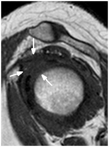
MRIOblique sagittal shoulder MRI, T1 sequence: complete obliteration of the fat surrounding the coracohumeral ligament (arrows), a sign of frozen shoulder.
Source : Li et al., PloS one, 2011, figure 4 · CC BY
- A loss of passive range of at least 25 % in at least 2 planes of movement compared with the healthy side.
- A loss of passive external rotation greater than 50 % (or less than 30 degrees in absolute terms) with the elbow at the side, the most sensitive and specific sign.
- A symmetrical limitation of active and passive movement (unlike cuff conditions).
Diagnostic decision algorithm
After the Kelley 2013 JOSPT CPG and the Walmsley 2009 criteria
Source: Kelley MJ et al. JOSPT 2013;43(5):A1-A31 (APTA CPG) PMID 23636125. Walmsley S et al. Phys Ther 2009;89:906-917 PMID 19589853.
- Rotator cuff conditions : tendinopathy, partial or massive tears. Passive movement is generally preserved, unlike in frozen shoulder. Jobe test, lift-off, bear hug, a painful arc in abduction (60-120 degrees).1
- Glenohumeral osteoarthritis : similar stiffness, but the radiograph reveals joint space narrowing and osteophytes. The population is often older (> 60 years).2
- Calcific tendinopathy : very acute pain, calcification visible on the radiograph. An intense resorptive phase is possible.8
- Parsonage-Turner syndrome (neuralgic amyotrophy): disproportionate motor weakness, early muscle wasting, an abnormal EMG.9
- Cervical radicular conditions : neurological signs (paraesthesia, motor deficit in a root distribution), a positive Spurling test.1
- Rare causes still worth considering : primary or metastatic tumour of the shoulder, septic arthritis, polymyalgia rheumatica (in older patients).10
Red flags - immediate medical referral
- Non-mechanical night pain, not relieved by changing position.
- Unexplained weight loss, fever, night sweats, marked fatigue.
- A personal history of cancer (breast, lung, prostate, kidney, melanoma, thyroid).
- Motor weakness out of proportion to the loss of passive movement (think Parsonage-Turner, neurological involvement).
- Visible, rapid muscle wasting.
- Intense local inflammatory signs (heat, erythema, swelling) - rule out septic arthritis.
- Complete failure after 6 months of well-conducted management - a diagnostic reassessment is imperative.
Should patients be classified into stages, and what are the benefits?
Clinical staging by Reeves's phases (freezing, frozen, thawing), updated by Hannafin & Chiaia 2000 and then Millar 2022, remains useful for guiding treatment and managing the patient's expectations, even though this is a continuum rather than a set of discrete stages.1,11| Phase | Duration | Dominant feature | Treatment strategy |
|---|---|---|---|
| Phase 1 - Freezing (painful) | 2-9 months | Intense pain, above all at night. Progressive stiffness. | Education, pain control, gentle mobilisations, intra-articular corticosteroids if severe (Challoumas 2020). |
| Phase 2 - Frozen (stiff) | 4-12 months | Maximal stiffness. Pain at end range. ER < 30 degrees. | End-range manual therapy + progressive stretching + a home exercise programme. |
| Phase 3 - Thawing (recovery) | 5-24+ months | Progressive improvement in ROM and function. Pain has almost gone. | Functional strengthening, return to activity, prevention of contralateral recurrence. |
Key points
- Diagnosis is essentially clinical, based on a symmetrical loss of active and passive range.
- The cardinal sign is a loss of passive external rotation with the elbow at the side greater than 50 % of the other side.
- The Walmsley 2009 consensus criteria and the Kelley 2013 JOSPT (APTA) recommendations are the references.
- Imaging (plain radiography, ultrasound after Suh 2019, MRI where the presentation is atypical) serves above all for the differential diagnosis.
- Actively look for the comorbidities: diabetes, thyroid dysfunction, Dupuytren's.
- Know the red flags (non-mechanical night pain, a history of cancer, disproportionate motor weakness) so that referral is prompt.
Bibliography
- Lewis J. Frozen shoulder contracture syndrome - Aetiology, diagnosis and management. Man Ther. 2015;20(1):2-9. PMID 25107826.
- Cho CH, Bae KC, Kim DH. Treatment Strategy for Frozen Shoulder. Clin Orthop Surg. 2019;11(3):249-257. PMID 31475043.
- Kelley MJ, Shaffer MA, Kuhn JE, et al. Shoulder pain and mobility deficits: adhesive capsulitis. J Orthop Sports Phys Ther. 2013;43(5):A1-A31. PMID 23636125.
- Le HV, Lee SJ, Nazarian A, Rodríguez EK. Adhesive capsulitis of the shoulder: review of pathophysiology and current clinical treatments. Shoulder Elbow. 2017;9(2):75-84. PMID 28405218.
- Cucchi D, Marmotti A, De Giorgi S, et al. Risk Factors for Shoulder Stiffness: Current Concepts. Joints. 2017;5(4):217-223. PMID 29270559.
- Chuang SH, Chen YP, Huang SW, Kuo YJ. Association between adhesive capsulitis and thyroid disease: a meta-analysis. J Shoulder Elbow Surg. 2023;32(6):1314-1322. PMID 36871608.
- Walmsley S, Rivett DA, Osmotherly PG. Adhesive capsulitis: establishing consensus on clinical identifiers for stage 1 using the Delphi technique. Phys Ther. 2009;89(9):906-917. PMID 19589853.
- Kim MS, Kim IW, Lee S, Shin SJ. Diagnosis and treatment of calcific tendinitis of the shoulder. Clin Shoulder Elb. 2020;23(4):210-216. PMID 33330261.
- Ohta R, Shimabukuro A. Parsonage-Turner syndrome in a patient with bilateral shoulder pain: A case report. J Rural Med. 2017;12(2):135-138. PMID 29255532.
- Mezian K, Coffey R, Chang KV. Frozen Shoulder. In: StatPearls [Internet]. StatPearls Publishing; 2023. PMID 29489251.
- Suh CH, Yun SJ, Jin W, et al. Systematic review and meta-analysis of magnetic resonance imaging features for diagnosis of adhesive capsulitis of the shoulder. Eur Radiol. 2019;29(2):566-577. PMID 29978436.
- Hollmann L, Halaki M, Kamper SJ, et al. Does muscle guarding play a role in range of motion loss in patients with frozen shoulder? Musculoskelet Sci Pract. 2018;37:64-68. PMID 29986193.
Which treatment strategies are the most effective for frozen shoulder (adhesive capsulitis)?
Where do you start? What is the recommended hierarchy of interventions?
The initial approach rests on the triad of education + pain management + physiotherapy. The network meta-analysis Challoumas 2020 (JAMA Network Open, 65 RCTs, 4,097 participants) is the current reference for comparing the available interventions.3 It identifies as the most effective short-term intervention the intra-articular corticosteroid injection combined with physiotherapy, followed by surgery (manipulation under anaesthetic or arthroscopic release) and then by physiotherapy alone.Hierarchy of the interventions by level of evidence (GRADE)
A synthesis of Challoumas 2020 NMA + UK FROST Rangan 2020 + Cochrane Page 2014
Sources: Challoumas D et al. JAMA Netw Open 2020;3(12):e2029581 - Rangan A et al. Lancet 2020;396:977-989 - Page MJ et al. Cochrane 2014 - Kelley 2013 JOSPT CPG.
What is the place of exercise and of manual therapy?
Supervised exercise plus a home exercise programme (HEP) is the cornerstone of conservative treatment. The Cochrane review Page 2014 (manual therapy + exercise, 32 RCTs) shows modest but consistent benefits on pain and function, particularly when exercise and manual therapy are combined.5 The principles of application are:6,7- Painful phase (freezing) : gentle pendulum exercises, active-assisted mobilisation within pain-free range. Avoid forced stretching, which can reactivate the inflammation.
- Stiff phase (frozen) : low-load, long-duration stretching (Russell 2014), end-range joint mobilisation (Maitland grade III-IV), a home exercise programme several times a day.
- Thawing phase : progressive strengthening of the cuff and the scapular stabilisers, a return to functional activities, prevention of contralateral recurrence.
| Modality | Level of evidence | Expected effect | Landmark reference |
|---|---|---|---|
| Education + reassurance | GRADE high | Adherence up, anxiety down, catastrophising prevented | Chester 2018 BJSM; Coronado 2020 Pain Rep |
| Supervised exercise + HEP | GRADE high | Pain, function, ROM | Page 2014 Cochrane |
| End-range manual therapy | GRADE moderate | ROM (synergistic with exercise) | Noten 2016 APMR; Yang 2007 Phys Ther |
| Intra-articular corticosteroids | GRADE high (short term), moderate (long term) | Pain, function at 6-12 weeks | Sun 2017 AJSM MA; Challoumas 2020 NMA |
| Hydrodilatation | GRADE moderate | ROM + function, resistant cases | Dimitri-Pinheiro 2022 PMC9465918 |
| Manipulation under anaesthetic | GRADE moderate | Rapid ROM gain, a risk of fracture or dislocation | UK FROST Rangan 2020 |
| Arthroscopic capsular release | GRADE moderate | Equivalent to MUA and PT at 12 months | UK FROST Rangan 2020 |
| ESWT | GRADE low | A possible adjunct | Vahdatpour 2014 IJPM |
| LLLT | GRADE low | Short-term pain (heterogeneous data) | Mixed data |
| Therapeutic ultrasound | GRADE very low | No demonstrated benefit | Page 2014 Cochrane |
Injections, hydrodilatation, surgery: when should you escalate?
The intra-articular corticosteroid injections are strongly recommended in phase 1 (painful). The Sun 2017 meta-analysis (AJSM, 8 RCTs, 416 patients) confirms a significant reduction in pain at 6 weeks against placebo and against physiotherapy alone.10 The optimal injection technique (ultrasound-guided vs anatomical, dose, repetition) remains debated, but a dose of 40 mg of triamcinolone or 7-10 mg of dexamethasone, as a single posterior ultrasound-guided injection, is the most widely agreed approach.3 Arthrographic hydrodilatation (distension with a large volume of saline plus corticosteroid plus anaesthetic under ultrasound or fluoroscopic guidance) is an option for patients who do not respond to 3-6 months of conservative treatment, particularly patients with diabetes. The retrospective study Dimitri-Pinheiro 2022 (PMC9465918) shows reduced but real efficacy in patients with diabetes compared with those without.11 Any surgery (manipulation under anaesthetic or arthroscopic release) remains reserved for failures of maximal conservative management (beyond 6-9 months). UK FROST shows that the three arms (PT, MUA, arthroscopy) are equivalent in function at 12 months, which justifies favouring physiotherapy on grounds of cost-effectiveness and lower invasiveness.4Beyond the physical: how do you educate the patient and address psychological factors?
Therapeutic education is the indispensable first step. It aims to:2,12- Explain the natural history (3 phases, a typical duration of 1-3 years, the possibility of minor residual impairment).
- Reassure the patient that the condition is not serious (in the absence of red flags).
- Counter the kinesiophobia and catastrophising that worsen disability (Chester 2018 BJSM).
- Promote self-management (the home exercise programme, energy management, adapting ADLs).
Key points
- Treatment follows a stepped approach : education + exercise + manual therapy first, intra-articular corticosteroids in the painful phase 1, escalation to hydrodilatation or surgery where it fails after 6-9 months.
- The network meta-analysis Challoumas 2020 (JAMA Netw Open) identifies intra-articular corticosteroids combined with PT as the most effective intervention in the short term.
- The trial UK FROST (Rangan 2020 Lancet) shows that early structured PT, MUA and arthroscopic release are equivalent at 12 months: PT is the most cost-effective.
- Exercise should be gentle and progressive, matched to the phase, without triggering significant pain that would reactivate the inflammation.
- And end-range manual therapy alongside exercise is superior to low-grade passive mobilisation (Noten 2016).
- Education and attention to psychological factors are essential (Chester 2018, Coronado 2020).
Bibliography
- Lewis J. Frozen shoulder contracture syndrome. Man Ther. 2015;20(1):2-9. PMID 25107826.
- Cho CH, Bae KC, Kim DH. Treatment Strategy for Frozen Shoulder. Clin Orthop Surg. 2019;11(3):249-257. PMID 31475043.
- Challoumas D, Biddle M, McLean M, Millar NL. Comparison of Treatments for Frozen Shoulder: A Systematic Review and Meta-analysis. JAMA Netw Open. 2020;3(12):e2029581. DOI 10.1001/jamanetworkopen.2020.29581.
- Rangan A, Brealey SD, Keding A, et al. Management of adults with primary frozen shoulder in secondary care (UK FROST): a multicentre, pragmatic, three-arm, superiority randomised clinical trial. Lancet. 2020;396(10256):977-989. PMID 33010843.
- Page MJ, Green S, Kramer S, et al. Manual therapy and exercise for adhesive capsulitis (frozen shoulder). Cochrane Database Syst Rev. 2014;(8):CD011275. PMID 25157702.
- Kelley MJ, Shaffer MA, Kuhn JE, et al. Shoulder pain and mobility deficits: adhesive capsulitis. J Orthop Sports Phys Ther. 2013;43(5):A1-A31. PMID 23636125.
- Russell S, Jariwala A, Conlon R, et al. A blinded, randomized, controlled trial assessing conservative management strategies for frozen shoulder. J Shoulder Elbow Surg. 2014;23(4):500-507. PMID 24630545.
- Noten S, Meeus M, Stassijns G, et al. Efficacy of Different Types of Mobilization Techniques in Patients With Primary Adhesive Capsulitis of the Shoulder: A Systematic Review. Arch Phys Med Rehabil. 2016;97(5):815-825. PMID 26284892.
- Yang JL, Chang CW, Chen SY, Wang SF, Lin JJ. Mobilization techniques in subjects with frozen shoulder syndrome: randomized multiple-treatment trial. Phys Ther. 2007;87(10):1307-1315. PMID 17684085.
- Sun Y, Zhang P, Liu S, et al. Intra-articular Steroid Injection for Frozen Shoulder: A Systematic Review and Meta-analysis of Randomized Controlled Trials. Am J Sports Med. 2017;45(9):2171-2179. PMID 28298050.
- Dimitri-Pinheiro S, Pinto BS, Pimenta M, Neves JS, Carvalho D. Influence of diabetes on response to ultrasound guided hydrodistension treatment of adhesive capsulitis: a retrospective study. BMC Endocr Disord. 2022;22(1):227. PMC9465918.
- Maund E, Craig D, Suekarran S, et al. Management of frozen shoulder: a systematic review and cost-effectiveness analysis. Health Technol Assess. 2012;16(11):1-264. PMID 22405512.
- Chester R, Jerosch-Herold C, Lewis J, Shepstone L. Psychological factors are associated with the outcome of physiotherapy for people with shoulder pain. Br J Sports Med. 2018;52(4):269-275. PMID 27445360.
- Coronado RA, Brintz CE, McKernan LC, et al. Psychologically informed physical therapy for musculoskeletal pain. Pain Rep. 2020;5(5):e847. PMID 33490842.
- Vahdatpour B, Taheri P, Zare Zadeh A, Moradian S. Efficacy of extracorporeal shockwave therapy in frozen shoulder. Int J Prev Med. 2014;5(7):875-881. PMID 25104999.
- Ramirez J. Adhesive Capsulitis: Diagnosis and Management. Am Fam Physician. 2019;99(5):297-300. PMID 30811157.
Frozen shoulder and diabetes: why does this subgroup deserve particular attention?
What are the biological mechanisms linking diabetes and frozen shoulder?
The Zreik 2016 meta-analysis (MLTJ, 12 studies) establishes that the prevalence of frozen shoulder reaches 13.4 % in people with diabetes (against 2-5 % in the general population) and, conversely, that up to 30 % of frozen shoulder patients have diabetes.3 The narrative review Chen 2024 (J Clin Med, PMID 39407755) synthesises the biological mechanisms identified to date:1- Advanced glycation end-products (AGEs) : chronic hyperglycaemia leads to the formation of AGEs that bind to the collagen of the joint capsule. These AGE cross-links stiffen the extracellular matrix and resist normal degradation mechanisms.
- Overexpression of TGF-beta and amplification of the profibrotic signalling pathway in people with diabetes, comparable to what is seen in Dupuytren's disease (another fibrosis associated with diabetes).
- Microangiopathy : impaired periarticular microcirculation, with tissue hypoxia favouring fibrosis.
- Chronic low-grade inflammation linked to diabetes (raised CRP, IL-6), which prolongs the synovitic phase.
- Partial resistance to corticosteroids : a poorer response to intra-articular corticosteroids than in people without diabetes (Dimitri-Pinheiro 2022).
Clinical features specific to the patient with diabetes
A synthesis of Chen 2024 J Clin Med + Mertens 2021 SR
Sources: Chen MH, Chen WS. J Clin Med 2024;13(19):5696 PMID 39407755 - Zreik NH et al. MLTJ 2016 PMID 27331029 - Dimitri-Pinheiro 2022 PMC9465918.
How do you stratify risk and adapt management in the patient with diabetes?
Clinical stratification helps personalise the prognosis and the treatment plan. Several predictors of severity and resistance are documented:1,4,5- Duration of diabetes : the risk rises beyond 10 years.
- HbA1c > 7.5 % : poor glycaemic control is associated with slower recovery and a higher failure rate.
- Type 1 diabetes : a slightly stronger association with frozen shoulder than T2DM (Huang YP 2013).
- The presence of other microvascular complications : retinopathy, nephropathy, neuropathy.
- A bilateral or recurrent form.
| Risk profile | Characteristics | Adapted treatment strategy |
|---|---|---|
| Low risk | T2DM < 5 years, HbA1c < 7 %, no complications | The standard protocol (PT + HEP + intra-articular corticosteroids in phase 1). A prognosis close to the general population's. |
| Intermediate risk | 5-10 years of diabetes, HbA1c 7-8 %, no major complications | Strengthen education, intensify the HEP, follow up closely, use intra-articular corticosteroids earlier, consider hydrodilatation at 4-6 months if progress plateaus. |
| High risk | T1DM or T2DM > 10 years, HbA1c > 8 %, microvascular complications, bilateral | A multidisciplinary approach (diabetologist + physiotherapist + rheumatologist), early hydrodilatation, escalation to MUA or arthroscopy if progress stalls at 6-9 months. |
Red flags specific to the patient with diabetes
- Marked local inflammatory signs plus fever: think septic arthritis (people with diabetes are at increased risk).
- Associated calcific tendinopathy : more frequent in people with diabetes, and it can complicate the picture.
- Simultaneous bilateral frozen shoulder : rule out an underlying systemic condition (in particular associated thyroid dysfunction, late-onset JIA).
- Paradoxical worsening after intra-articular corticosteroids : an infection risk, but also the possibility of avascular necrosis (rare).
- Persistently poor glycaemic control : to be addressed actively with the general practitioner or diabetologist (hyperglycaemia can be transiently worsened by intra-articular corticosteroids).
Key points
- Diabetes multiplies 5 - 8 the risk of frozen shoulder (Zreik 2016 MA).
- The specific biological mechanisms include advanced glycation end-products (AGEs), overexpression of TGF-beta and microangiopathy.
- The clinical picture in diabetes is more severe, prolonged and resistant to standard treatment.
- Stratifying by HbA1c and duration of diabetes guides the escalation of treatment.
- Ultrasound-guided hydrodilatation is an option of choice where progress plateaus at 3-6 months of PT.
- Coordinate with the diabetologist about glycaemic adjustment around the injection.
Bibliography
- Chen MH, Chen WS. A Narrative Review of Adhesive Capsulitis with Diabetes. J Clin Med. 2024;13(19):5696. PMID 39407755.
- Millar NL, Meakins A, Struyf F, et al. Frozen shoulder. Nat Rev Dis Primers. 2022;8(1):59. PMID 36075904.
- Zreik NH, Malik RA, Charalambous CP. Adhesive capsulitis of the shoulder and diabetes: a meta-analysis of prevalence. Muscles Ligaments Tendons J. 2016;6(1):26-34. PMID 27331029.
- Dimitri-Pinheiro S, Pinto BS, Pimenta M, Neves JS, Carvalho D. Influence of diabetes on response to ultrasound guided hydrodistension treatment of adhesive capsulitis: a retrospective study. BMC Endocr Disord. 2022;22(1):227. PMC9465918.
- Huang YP, Fann CY, Chiu YH, et al. Association of diabetes mellitus with adhesive capsulitis. Arthritis Care Res. 2013;65(7):1197-1202. DOI 10.1002/acr.21938.
- Mezian K, Coffey R, Chang KV. Frozen Shoulder. StatPearls. 2023. PMID 29489251.
- Le HV, Lee SJ, Nazarian A, Rodríguez EK. Adhesive capsulitis of the shoulder. Shoulder Elbow. 2017;9(2):75-84. PMID 28405218.
How do you secure lasting recovery and prevent recurrence of frozen shoulder (adhesive capsulitis)?
How do you make the patient an active participant in their recovery through self-management?
Self-management (self-management) guided by the physiotherapist is the pillar of durable recovery. It rests on three pillars:3,4- Education about the natural history : explain the 3 phases, the typical duration (1-3 years), how rare recurrence on the same side is (under 5 %) but the risk of contralateral involvement (6-17 % according to Wong 2017, above all in people with diabetes).
- A home exercise programme (HEP) :
- Done several times a day (3-5 sessions of 10-15 minutes).
- Controlled progression, respecting the pain threshold (the 24 h rule: if pain increases the next day, reduce the intensity).
- Adapt to the phase: pendulum exercises and gentle mobilisation in phase 1; low-load, long-duration stretching in phase 2; progressive strengthening in phase 3.
- Adaptive management of daily activities :
- Adapt the provocative movements without giving up movement altogether.
- Support the painful arm at night (a pillow under the elbow).
- Apply heat before the exercises, ice afterwards if there is post-exertional pain.
When and how should a safe return to sport and to activity be planned?
The return to sport (RTS) or to demanding occupational activity (lifting, overhead work) should be based on objective functional criteria, not on a fixed timescale. The consensus criteria are:5,6- Range : passive and active ROM above 90 % of the healthy side, particularly ER and elevation.
- Strength : isometric cuff strength (Jobe, ER in abduction, lift-off) above 90 % of the other side.
- Pain : VAS < 2/10 for the movements specific to the sport or the job.
- Neuromuscular control : good scapular mechanics, proprioceptive control of complex movements.
- Confidence and self-efficacy : assessed by questionnaire (the TSK for fear of movement, for example).
Return-to-sport progression - a criterion-based approach
Adapted from the Kelley 2013 JOSPT CPG and the principles of musculoskeletal RTS
Inspired by Kelley MJ et al. JOSPT 2013;43(5):A1-A31 and the principles of musculoskeletal RTS - a criterion-based approach.
Key points
- Up to 40 % of patients keep functional impairment in the long term (Vastamaki 2012, Hand 2008).
- Guided self-management rests on education about the natural history + a regular HEP + adapting ADLs.
- Recurrence on the same side is rare (< 5 %), but the other shoulder is affected in 6 to 17 % of patients, above all in people with diabetes (Wong 2017).
- The return to sport should be based on objective functional criteria (ROM, strength, pain, motor control) and not on a fixed timescale.
- The stepped approach (unloaded movement -> progressive load -> intensity and speed -> competitive return) is validated.
Bibliography
- Vastamaki H, Kettunen J, Vastamaki M. The natural history of idiopathic frozen shoulder. Clin Orthop Relat Res. 2012;470(4):1133-1143. PMID 22090356.
- Hand C, Clipsham K, Rees JL, Carr AJ. Long-term outcome of frozen shoulder. J Shoulder Elbow Surg. 2008;17(2):231-236. PMID 17993282.
- Lewis J. Frozen shoulder contracture syndrome. Man Ther. 2015;20(1):2-9. PMID 25107826.
- Russell S, Jariwala A, Conlon R, et al. A blinded, randomized, controlled trial. J Shoulder Elbow Surg. 2014;23(4):500-507. PMID 24630545.
- Kelley MJ, Shaffer MA, Kuhn JE, et al. Shoulder pain and mobility deficits: adhesive capsulitis. J Orthop Sports Phys Ther. 2013;43(5):A1-A31. PMID 23636125.
- Wong CK, Levine WN, Deo K, et al. Natural history of frozen shoulder: fact or fiction? Physiotherapy. 2017;103(1):40-47. DOI 10.1016/j.physio.2016.05.009.
- Rangan A, Brealey SD, Keding A, et al. UK FROST. Lancet. 2020;396:977-989. PMID 33010843.
What do real clinical cases teach us about frozen shoulder (adhesive capsulitis)?
Analysis of a “classic” case: from assessment to resolution.
The case report Phansopkar 2023 (Cureus, PMC10387580) illustrates a typical trajectory. An adult patient presents with progressive shoulder pain and stiffness of non-traumatic onset over several months. Examination finds the consensus criteria: a significant, symmetrical loss of active and passive range, particularly in ER (elbow at the side) and in abduction.1 The treatment plan deployed combined:- Therapeutic education : explaining the nature of the condition, the 3 phases, and the generally favourable but slow long-term outlook.
- Manual therapy : the Spencer technique (rhythmic low-amplitude oscillations of the glenohumeral and acromioclavicular joints, combining mobilisation and myofascial release).
- Stretching and mobility exercises : pendulum work, closed-chain stretching, rope and pulley exercises, progressively intensified.
- A structured HEP several times a day.
The diagnostic challenge: when frozen shoulder mimics another condition.
The case report Ohta 2017 (J Rural Med, PMID 29255532) illustrates a classic diagnostic trap : a patient initially referred with suspected frozen shoulder who does not respond to standard treatment and develops disproportionate muscle weakness. Further assessment (EMG, MRI) reveals Parsonage-Turner syndrome (neuralgic amyotrophy of the brachial plexus).2 The warning signs that should prompt the diagnosis of frozen shoulder to be reconsidered:2,3- Very intense, abrupt initial pain, sometimes preceded by a viral illness.
- Marked motor weakness, greater than the loss of passive range.
- Early, visible muscle wasting (deltoid, supraspinatus/infraspinatus, serratus anterior).
- Failure of well-conducted PT after 6-8 weeks.
- A territorial distribution of the weakness (suprascapular nerve, long thoracic nerve).
A complex case: resistant frozen shoulder in a patient with diabetes.
The retrospective study Dimitri-Pinheiro 2022 (PMC9465918) is an excellent illustration of managing resistant frozen shoulder in the patient with diabetes. A series of patients with diabetes and frozen shoulder refractory to 3-6 months of conventional PT received ultrasound-guided hydrodilatation (injection of 30-40 mL of saline plus corticosteroid plus lidocaine under posterior ultrasound guidance).5 The results show:5- An immediate mobility gain (ER, ABD) of the order of 20-30 degrees.
- Progressive improvement over 6 months with PT continuing.
- A response that is smaller than in people without diabetes, but clinically significant.
- A favourable safety profile, provided insulin is adjusted around the procedure.
Red flags from the cases presented
- Disproportionate motor weakness plus early wasting: rule out Parsonage-Turner (EMG).
- Very acute pain with calcification on the radiograph: calcific tendinopathy in the resorptive phase.
- Stalled progress at 3-6 months in the patient with diabetes: consider hydrodilatation before surgical escalation.
- Any simultaneous bilateral frozen shoulder : a wider aetiological work-up (thyroid, late-onset JIA, other systemic fibroses).
Key points
- The Phansopkar 2023 case (Cureus PMC10387580) illustrates the value of an early conservative multimodal approach (education + manual therapy + HEP).
- The Ohta 2017 case (Parsonage-Turner) shows the importance of reconsidering the diagnosis when faced with disproportionate motor weakness or treatment failure.
- The Dimitri-Pinheiro 2022 case series validates ultrasound-guided hydrodilatation in the resistant patient with diabetes.
- The treatment escalation strategy (PT -> intra-articular corticosteroids -> hydrodilatation -> MUA/arthroscopy) is validated by UK FROST.
Bibliography
- Phansopkar P, Qureshi MI. An Integrated Physical Therapy Using Spencer's Technique in the Rehabilitation of a Patient With a Frozen Shoulder: A Case Report. Cureus. 2023;15(6):e41233. PMC10387580.
- Ohta R, Shimabukuro A. Parsonage-Turner syndrome in a patient with bilateral shoulder pain: A case report. J Rural Med. 2017;12(2):135-138. PMID 29255532.
- Mezian K, Coffey R, Chang KV. Frozen Shoulder. StatPearls. 2023. PMID 29489251.
- Wu YC, Tsai WC, Tu YK, Yu TY. Comparative Effectiveness of Nonoperative Treatments for Chronic Calcific Tendinitis of the Shoulder. Arch Phys Med Rehabil. 2017;98(8):1678-1692. PMID 28400182.
- Dimitri-Pinheiro S, Pinto BS, Pimenta M, Neves JS, Carvalho D. Influence of diabetes on response to ultrasound guided hydrodistension treatment of adhesive capsulitis: a retrospective study. BMC Endocr Disord. 2022;22(1):227. PMC9465918.
- Rangan A, Brealey SD, Keding A, et al. UK FROST. Lancet. 2020;396:977-989. PMID 33010843.
- Challoumas D, Biddle M, McLean M, Millar NL. Comparison of Treatments for Frozen Shoulder: A SR and MA. JAMA Netw Open. 2020;3(12):e2029581. DOI 10.1001/jamanetworkopen.2020.29581.
- Sun Y, Zhang P, Liu S, et al. Intra-articular Steroid Injection for Frozen Shoulder: A SR and MA of RCTs. Am J Sports Med. 2017;45(9):2171-2179. PMID 28298050.
- Chen MH, Chen WS. A Narrative Review of Adhesive Capsulitis with Diabetes. J Clin Med. 2024;13(19):5696. PMID 39407755.
How do you apply these recommendations concretely in your practice?
When and to which other health professionals should you refer?
Collaborative referral is an essential dimension of practice. The situations calling for medical referral are:- The presence of red flags (Finucane 2020 JOSPT, the international consensus framework): non-mechanical night pain, weight loss, persistent fever, a history of cancer, progressive neurological involvement, marked inflammatory signs.1
- Stalled progress at 3-6 months despite well-conducted PT: refer to an orthopaedic surgeon to consider intra-articular corticosteroids, hydrodilatation or surgery.2
- A suspected alternative diagnosis (Parsonage-Turner, refractory calcific tendinopathy, early inflammatory arthritis): rheumatologist or neurologist.
- A patient with poorly controlled diabetes (HbA1c > 8 %): diabetologist, for glycaemic optimisation.
- Significant psychological distress (catastrophising, depression): psychologist or psychotherapist (CBT).3,4
How do you measure outcomes and overcome barriers to implementation?
Standardised outcome measurement is indispensable for making progress objective and adapting management. For frozen shoulder, the PROMs validated in French and most widely used are:6| PROM | Items | Score | MCID | Indication |
|---|---|---|---|---|
| SPADI | 13 (5 pain + 8 disability) | 0-100 (100 = worst) | ~8-13 points | The shoulder reference, sensitive to change |
| QuickDASH | 11 | 0-100 | ~10 points | Upper limb, short version |
| ASES | 10 | 0-100 (100 = best) | ~6-12 points | Mainly surgical, for postoperative follow-up |
| Constant-Murley | 4 domains (pain, ADLs, ROM, strength) | 0-100 (100 = best) | ~10 points | The surgical standard, requires an examiner |
| VAS / NRS for pain | 1 | 0-10 | ~2 points | Quick day-to-day pain tracking |
Overcoming the barriers to implementing the evidence
The Scurlock-Evans 2014 meta-analysis (Physiotherapy, PMID 24780633) identifies the main barriers to implementing evidence-based practice in physiotherapy:7- Lack of time in clinical practice.
- Difficulty accessing and critically appraising the literature.
- Lack of organisational support.
- The gap between the reality of complex patients and the often homogeneous populations of RCTs.
- Structured audit and feedback (Ivers 2012 Cochrane): comparing one's practice with evidence-based indicators, followed by constructive feedback.
- Identifying local leaders (Flodgren 2019 Cochrane) who promote and support the change.
- Interactive meetings and workshops as continuing education.
- Decision-support tools built into the practice software.
Key points
- Refer to a physician where there are red flags (Finucane 2020) or where progress stalls at 3-6 months.
- Collaborate with a diabetologist where control is poor, and with a psychologist where there is catastrophising or depression.
- Use validated PROMs : the SPADI and QuickDASH are the most practical in the clinic, the ASES and Constant for postoperative follow-up.
- The barriers to EBP (time, access to the literature) are overcome by audit and feedback (Ivers 2012) and local leaders (Flodgren 2019).
- EBP is a critical synthesis of evidence, expertise and the patient's values - not the mechanical application of protocols.
Bibliography
- Finucane LM, Downie A, Mercer C, et al. International Framework for Red Flags for Potential Serious Spinal Pathologies. J Orthop Sports Phys Ther. 2020;50(7):350-372. DOI 10.2519/jospt.2020.9971.
- Rangan A, Brealey SD, Keding A, et al. UK FROST. Lancet. 2020;396:977-989. PMID 33010843.
- Chester R, Jerosch-Herold C, Lewis J, Shepstone L. Psychological factors and physiotherapy outcome shoulder pain. Br J Sports Med. 2018;52(4):269-275. PMID 27445360.
- Coronado RA, Brintz CE, McKernan LC, et al. Psychologically informed physical therapy. Pain Rep. 2020;5(5):e847. PMID 33490842.
- Greenhalgh T, Howick J, Maskrey N. Evidence based medicine: a movement in crisis? BMJ. 2014;348:g3725. PMID 24927763.
- Kelley MJ, Shaffer MA, Kuhn JE, et al. Shoulder pain and mobility deficits: adhesive capsulitis. JOSPT. 2013;43(5):A1-A31. PMID 23636125.
- Scurlock-Evans L, Upton P, Upton D. Evidence-based practice in physiotherapy: a systematic review of barriers, enablers and interventions. Physiotherapy. 2014;100(3):208-219. PMID 24780633.
- Ivers N, Jamtvedt G, Flottorp S, et al. Audit and feedback: effects on professional practice and healthcare outcomes. Cochrane Database Syst Rev. 2012;(6):CD000259. PMID 22696318.
- Flodgren G, O Brien MA, Parmelli E, Grimshaw JM. Local opinion leaders: effects on professional practice and healthcare outcomes. Cochrane Database Syst Rev. 2019;(6):CD000125. PMID 31232458.
- Geneen LJ, Moore RA, Clarke C, et al. Physical activity and exercise for chronic pain in adults. Cochrane Database Syst Rev. 2017;4(4):CD011279. PMID 28436583.
- Hoffmann TC, Légaré F, Simmons MB, et al. Shared decision making: what do clinicians need to know? Med J Aust. 2014;201(1):35-39. DOI 10.5694/mja14.00002.
And after this article?
This article is part of a collection of evidence-based clinical syntheses. A question, a comment, a correction to suggest? Contact us directly through the WhatsApp button at the bottom right of the screen.

