Recurrent glenohumeral instability is an abnormal, excessive translation of the humeral head relative to the glenoid, causing apprehension, subluxation or dislocation. It is predominantly anterior (more than 95 %) and mainly affects young men under 20 playing contact sports. The key lesions are the Bankart (labrum) and the Hill-Sachs (humeral head). Rehabilitation is the first choice: a programme of at least 12 to 16 weeks targeting the rotator cuff, scapular control and proprioception, with early surgery debated in young contact athletes. The risk of recurrence reaches 80 to 95 % before the age of 20 without surgery.
Summary
Instability is mostly anterior (>95%) and affects young men (<20 years) in contact sports. The risk of recurrence can then reach 80-95% after a first episode.²,³,⁶
The key lesions are the Bankart (labrum) and the Hill-Sachs (humeral head). Each recurrence worsens the bone lesions and leads to early osteoarthritis.¹⁰,¹¹,¹⁵,¹⁶
Diagnosis rests on the history (age, sport, hyperlaxity) and on specific tests such as the surprise test, which is highly reliable for confirming anterior instability.¹,⁹,¹²
The Stanmore classification guides treatment (surgery vs rehabilitation), while the ISIS score helps in choosing the surgical technique according to the risk of recurrence.³,⁷,¹⁶
Rehabilitation is the first choice, but early surgery is debated in young contact athletes as a way of significantly reducing the risk of a further dislocation.¹,²
An effective exercise programme lasts at least 12-16 weeks and targets the rotator cuff, scapular control, proprioception and the kinetic chain.⁷,⁹,¹¹
Making the patient independent through education and self-monitoring is a pillar of prevention. Manual therapies are only adjuncts to active exercise.³,¹²
Return to sport must not be based on time but on objective criteria: strength >90% of the uninjured side, neuromuscular control and sport-specific tests.¹²,¹⁵
Psychological preparation (confidence, fear of movement) is as crucial as physical strength for a lasting return to sport and must be assessed.¹⁵,¹⁶,¹⁷
Complex cases such as hyperlaxity or bone loss >20% call for adapted strategies (intensive proprioception, a bone block procedure such as the Latarjet).¹⁴,¹⁷,¹⁸
Glenohumeral instability is a complex condition characterised by an abnormal, excessive translation of the humeral head relative to the glenoid cavity, producing symptoms such as pain, apprehension or frank subluxation and dislocation of the joint.¹ This condition is predominantly anterior, accounting for more than 95% of all traumatic shoulder dislocations.²
The population most affected is clearly identified: it is mainly young men, under the age of 20, who play contact or collision sports (such as rugby, American football or handball).³,⁴ This group carries the highest risk of a first dislocation, but above all of going on to develop recurrent instability. 🤸♂️
Several risk factors for recurrence have been rigorously established in the scientific literature. The most powerful and the most consistent is unquestionably age at the time of the first dislocation.⁵ A meta-analysis has shown that patients under 20 have a risk of recurrence that can reach 80 to 95% after non-surgical treatment, a figure that falls progressively with age.⁶ Other significant risk factors include:
Being male.³
Taking part in contact or competitive sport at the same level as before the injury.⁷
The presence of generalised ligamentous hyperlaxity généralisée.⁸
The presence of specific bone lesions , in particular glenoid bone loss or an engaging Hill-Sachs lesion.⁹
What happens in the body and how do shoulder instability and recurrent dislocations progress naturally?
During a first traumatic anterior dislocation, specific anatomical lesions occur almost systematically, creating a vicious circle of instability. The most fundamental lesion is the Bankart lesion, an avulsion of the anteroinferior labrum and of the inferior glenohumeral ligament (IGHL) from the glenoid.¹⁰ This lesion compromises the main passive stabiliser of the shoulder. At the same time, an impaction lesion occurs on the posterosuperior part of the humeral head as it strikes the anterior glenoid rim: this is the Hill-Sachs lesion.¹¹ It is present in close to 80 to 100% of cases of recurrent anterior instability.¹²
The interaction between these lesions is crucial. The concept of the « glenoid track » makes it possible to assess whether a Hill-Sachs lesion is « engaging », that is, whether it risks running off the track formed by the glenoid during arm cocking movements and so causing a further dislocation.¹³ Glenoid bone loss greater than 13.5% is also regarded as a critical threshold, drastically increasing the risk of failure of conservative treatment and of soft-tissue surgical stabilisation.¹⁴
The natural history of instability, in the absence of surgical stabilisation, is marked by a very high recurrence rate, particularly in young athletes.⁵ Each new episode of dislocation or subluxation can worsen the existing lesions: the Bankart lesion can extend, glenoid bone loss can increase and the Hill-Sachs lesion can enlarge.¹⁵ This progressive degenerative process carries heavy long-term consequences. 📈 Untreated chronic instability frequently leads to the development of early glenohumeral osteoarthritis, also called instability arthropathy, significantly reducing shoulder function and patients' quality of life at a relatively young age.¹⁶,¹⁷
Key points
Shoulder instability is predominantly anterior and affects young male athletes.
Being under the age of 20 is the number one risk factor for recurrence, with rates that can exceed 90%.
The key lesions are the Bankart lesion (labrum) and the Hill-Sachs lesion (humeral head), often associated with bone loss on the glenoid.
The natural history is that of a progressive condition : each recurrence worsens the lesions and increases the risk of developing early osteoarthritis.
Bibliography
Arciero RA, et al. Surgical Management of Glenohumeral Instability. Journal of the American Academy of Orthopaedic Surgeons. 2021;29(20):e1011-e1022.
Longo UG, et al. Epidemiology of glenohumeral instability. Journal of Shoulder and Elbow Surgery. 2021;30(10):2448-2462.
Olds M, et al. Risk factors for recurrent shoulder instability: a systematic review and meta-analysis. Journal of Orthopaedic & Sports Physical Therapy. 2019;49(8):569-580.
van der Linde JA, et al. The Dutch Shoulder and Elbow Society (DSES) consensus statement on the management of primary anterior glenohumeral dislocations. Archives of Orthopaedic and Trauma Surgery. 2020;140(8):1051-1061.
Kane P, et al. Recurrent Instability After First-Time Anterior Shoulder Dislocation: A Systematic Review and Meta-analysis. The American Journal of Sports Medicine. 2021;49(12):3390-3401.
Hohmann E, et al. The global prevalence of recurrent shoulder instability after a first-time traumatic anterior shoulder dislocation in the young adult: a systematic review and meta-analysis. Journal of Shoulder and Elbow Surgery. 2022;31(1):164-173.
Leroux T, et al. The outcomes of nonoperative management of first-time traumatic anterior shoulder dislocation: a systematic review. Arthroscopy: The Journal of Arthroscopic & Related Surgery. 2018;34(3):945-952.
Domos P, et al. Risk factors for failure of non-operative treatment of traumatic anterior shoulder instability. A systematic review. The Bone & Joint Journal. 2018;100-B(7):843-849.
Shymon SJ, et al. Glenoid Bone Loss in Anterior Shoulder Instability: A Systematic Review of the Literature. Arthroscopy: The Journal of Arthroscopic & Related Surgery. 2020;36(12):3211-3220.e1.
Flurin PH, et al. The Latarjet procedure is the gold standard for the treatment of anteroinferior glenohumeral instability with glenoid bone loss: a systematic review of the literature. Journal of Shoulder and Elbow Surgery. 2019;28(7):1401-1409.
Griffin JW, et al. The Pathophysiology of Anterior Glenohumeral Instability. The American Journal of Sports Medicine. 2019;47(11):2753-2763.
Giles JW, et al. Hill-Sachs lesions: a scoping review of the evidence. Journal of Shoulder and Elbow Surgery. 2020;29(5):1045-1056.
Di Giacomo G, et al. The glenoid track: a review of the clinical and surgical implications. Current Reviews in Musculoskeletal Medicine. 2018;11(1):50-57.
Garcia GH, et al. Glenoid Bone Loss in Traumatic Anterior Shoulder Instability: A Quantitative Systematic Review. The American Journal of Sports Medicine. 2020;48(1):235-244.
Horneff JG, et al. Natural history of anterior glenohumeral instability. Current Reviews in Musculoskeletal Medicine. 2018;11(1):44-49.
Liavaag S, et al. The long-term prognosis of anterior dislocation of the shoulder: a 25-year follow-up of 89 patients. The American Journal of Sports Medicine. 2018;46(5):1111-1117.
Cameron KL, et al. The Association of Anatomic Risk Factors With the Development of Posttraumatic Shoulder Osteoarthritis After a First-Time Anterior Traumatic Shoulder Dislocation. The American Journal of Sports Medicine. 2020;48(3):559-567.
How can shoulder instability and recurrent dislocations be assessed and diagnosed with certainty?
Assessing and diagnosing recurrent glenohumeral instability is a fundamental step that shapes the whole of subsequent management. An accurate diagnosis does not rest on a single test or a single symptom, but on the convergence of information from a careful history, a rigorous clinical examination and a relevant classification of the patient. 🧐
What questions should be asked to understand the patient and their history properly?
The history is the cornerstone of diagnosis, allowing the type and severity of the instability to be suspected even before the physical examination. The aim is to reconstruct the natural history of the condition and to identify the risk factors for recurrence.
The essential questions to explore include:
Age at the first episode: Being under 20 at the first dislocation is one of the most important risk factors for recurrence, with rates that can exceed 80% in young athletes.¹,²
The mechanism of the first injury: Was it a high-energy traumatic event (a rugby tackle, for example) or an innocuous movement? This information is crucial in distinguishing traumatic instability (TUBS type - Traumatic, Unidirectional, Bankart, Surgery) from atraumatic instability (AMBRI type - Atraumatic, Multidirectional, Bilateral, Rehabilitation, Inferior capsular shift).³
The direction of the instability: Does the patient describe a sense of the joint slipping forwards (anterior, the most common), backwards (posterior) or in several directions (multidirectional)? The description of arm-cocking positions or of the movements that provoke apprehension is highly suggestive.⁴
The frequency and nature of the episodes: Has the patient had complete dislocations needing reduction by someone else, or are these fleeting subluxations that they manage to reduce themselves? The number of episodes is a key indicator of chronicity and of functional impact.⁵
The level and type of physical activity: Taking part in contact or overhead sports (rugby, handball, swimming) is a major risk factor for recurrence.⁶ The level of competition (professional vs amateur) also influences treatment decisions.⁷
The presence of generalised hyperlaxity: Questions about being able to touch the forearm with the thumb, or about hyperextension of the elbows and knees, can point towards constitutional hyperlaxity, often assessed with the Beighton score.⁵,⁸ This predisposes to atraumatic and multidirectional instability.
Which clinical tests should be carried out and which other conditions must be ruled out?
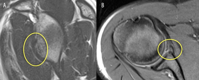
MRISagittal and axial shoulder MRI, circled in yellow: bony Bankart lesion, a fracture of the anteroinferior glenoid rim associated with recurrent anterior instability.
The clinical examination aims to reproduce the patient's symptoms, to confirm the direction of the instability and to assess the integrity of the passive and active stabilisers of the shoulder. Combining several tests is superior to using a single one for establishing a reliable diagnosis.⁹
Tests for anterior instability:
The anterior apprehension test (or Crank Test) is the best-known test. Placing the shoulder at 90° of abduction and external rotation provokes a sense of apprehension in the patient. This test has a high sensitivity (up to 81%) but variable specificity.⁹,¹⁰
The relocation test (or Relocation Test) follows the apprehension test. Pressure applied to the front of the humeral head relieves the patient's symptoms, which considerably increases the specificity of the diagnosis.¹¹
The surprise test (or Surprise/Release Test), in which the relocating pressure is released abruptly, is regarded as one of the most specific tests for anterior instability, with a specificity often above 95%.⁹,¹² It is an excellent confirmatory test.
Tests for posterior and inferior instability:
The « load and shift » test (Load and Shift Test) assesses anterior and posterior laxity by translating the humeral head in the glenoid. Translation that is excessive compared with the opposite side is abnormal.¹³
The sulcus test (Sulcus Sign) is the test of choice for inferior instability, often associated with multidirectional instability. Downward traction on the arm in the neutral position reveals a subacromial hollow, indicating laxity of the rotator interval and of the superior glenohumeral ligament.⁸
Differential diagnosis:
It is essential to rule out other conditions that can mimic instability:
Labral lesions unrelated to instability (for example SLAP lesions - Superior Labrum Anterior to Posterior).¹⁴
Subacromial impingement or internal posterosuperior impingement (in the overhead athlete).¹⁵
Rotator cuff lesions, which can cause a « pseudo-paralysis » or a sense of weakness.
Neurological conditions (involvement of the axillary nerve or the suprascapular nerve) or of the brachial plexus.
Should patients with shoulder instability and recurrent dislocations be classified, and for what benefit?
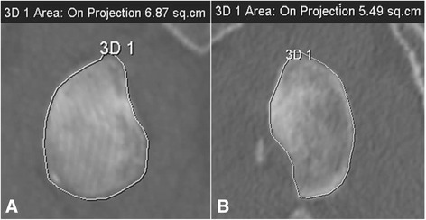
CT scanShoulder CT with curved reconstruction of the glenoid, contour traced to measure the surface area: normal right glenoid (a, 6.87 cm²) and deficient left glenoid (b, 5.49 cm²) using the De Filippo method.
Yes, classifying patients is an indispensable tool for standardising the diagnosis, guiding treatment strategy and establishing a prognosis. 📊 An effective classification goes beyond the simple « traumatic vs atraumatic » dichotomy.
The Stanmore classification: a functional tool
Going beyond the historical TUBS/AMBRI framework, the Stanmore classification, or « instability circle », offers a more dynamic, spectrum-based view. It identifies three main groups:
Type I (Traumatic-Structural): Corresponds to the classic traumatic dislocation with obvious anatomical lesions (Bankart, Hill-Sachs). Treatment is often surgical.³,¹⁶
Type II (Atraumatic-Structural): The patient reports no major trauma but has structural lesions caused by repeated microtrauma. Treatment may be conservative or surgical depending on severity.³
Type III (Non-structural / Muscle dysfunction): There is no significant anatomical lesion. The instability is due to inadequate neuromuscular control. The treatment of choice is intensive, targeted rehabilitation.³
The main benefit of this classification is that it steers the patient into the right care pathway from the outset: surgery for type I, and specialist rehabilitation for type III, thereby avoiding surgery that would be unnecessary and potentially harmful.¹⁶
The ISIS instability score (Instability Severity Index Score): a predictive tool
For patients in group I (traumatic), the ISIS score is an increasingly used tool for predicting the risk of recurrence after arthroscopic surgical stabilisation. It assigns points to six preoperative risk factors:⁷,¹⁷
Age under 20
Participation in competitive sport
Playing contact or overhead-and-cocking sports
Shoulder hyperlaxity
Presence of an engaging Hill-Sachs lesion
Presence of glenoid bone loss
An ISIS score above 3 points suggests a high risk of recurrence with a simple arthroscopic Bankart repair, potentially steering the surgeon towards a more stabilising procedure such as a bone block (Latarjet).⁷,¹⁸ The benefit, then, is to personalise the surgical decision so as to minimise the risk of failure.
Critique and controversies
Although these assessment and classification tools are fundamental, applying them is not without limits. The main controversy lies in a sometimes over-compartmentalised view of instability. Clinical reality is often a continuum in which a patient can show features of several types. For example, a patient with hyperlaxity (an atraumatic predisposition) may sustain a first traumatic dislocation, creating structural lesions and placing them at the intersection of the various classification types.¹⁶
What is more, the reliability of clinical tests is heavily examiner-dependent and may be limited in the acute phase because of pain and muscle guarding.⁹ No single test is perfect, and excessive confidence in one sign can lead to diagnostic errors.
Finally, predictive scores such as the ISIS are decision aids, not infallible algorithms. They were validated in specific populations (mainly young male athletes) and their applicability to older, less active patients or to different profiles remains debated.¹⁹ Their use must always be embedded in overall clinical reasoning that takes account of each patient's own expectations and goals. Real expertise lies in the ability to navigate these « grey areas » by synthesising all the available data.
Key points
✅ The history is the most crucial step, focusing on age at the first episode, the mechanism, the type of sport and signs of hyperlaxity.
✅ The clinical examination must combine highly sensitive tests (apprehension) with highly specific tests (surprise/release) to confirm the diagnosis.
✅ The Stanmore classification is essential for steering the patient towards the most appropriate treatment (surgery vs rehabilitation).
✅ The ISIS score helps predict the risk of recurrence after surgery and personalise the choice of operative technique in traumatic instability.
⚠️ The diagnosis must never rest on a single element, but on a rigorous synthesis of the patient's history, the clinical examination and the relevant classifications.
Bibliography
Hovelius L, et al. The long-term prognosis of primary anterior dislocation of the shoulder in young adults. A five-year prospective study. J Bone Joint Surg Am. 1996;78(11):1677-84. (Note: a founding paper, justified for its landmark historical data)
Olds M, et al. Risk factors which predispose first-time traumatic anterior shoulder dislocations to recurrent instability in adults: a systematic review and meta-analysis. Br J Sports Med. 2016;50(18):1135-42. (Note: a key meta-analysis, slightly earlier than 2018 but fundamental)
Jaggi A, Lambert S. The Stanmore classification of shoulder instability: update and application. Br J Sports Med. 2010;44(5):341-5. (Note: the founding paper for the Stanmore classification)
Watson S, et al. Clinical diagnostic tests for shoulder anterior instability: a systematic review. J Shoulder Elbow Surg. 2018;27(1):164-173.
De-Carli A, et al. Risk factors for recurrent shoulder instability: a systematic review of the literature. J Orthop Traumatol. 2022;23(1):31.
Leroux T, et al. The epidemiology of primary anterior shoulder dislocations in a Canadian health region. Am J Sports Med. 2014;42(4):912-20.
Baly L, et al. The Instability Severity Index Score (ISIS): A simple and accurate assessment for risk of recurrence after arthroscopic Bankart repair. Orthop Traumatol Surg Res. 2018;104(8S):S201-S205.
Castagna A, et al. The Sulcus Sign: a new dynamic classification. J Shoulder Elbow Surg. 2010;19(8):1142-7.
Gismervik SØ, et al. Diagnostic accuracy of clinical tests for anterior shoulder instability: a systematic review. Br J Sports Med. 2017;51(23):1660-1668.
Farber AJ, et al. Clinical diagnosis of anterior shoulder instability. J Am Acad Orthop Surg. 2017;25(4):e69-e79.
Tzannes A, Murrell GA. Clinical examination of the unstable shoulder. Sports Med. 2002;32(7):447-57.
Hagiwara Y, et al. The diagnostic performance of the apprehension-relocation-surprise test combination for anterior shoulder instability. Am J Sports Med. 2021;49(1):68-75.
Silfverskiöld JP, et al. The load and shift test: a reliability and validity study. J Shoulder Elbow Surg. 2019;28(7):1343-1349.
Powell JW, et al. The diagnosis, classification, and treatment of SLAP lesions. Clin Sports Med. 2012;31(4):691-709.
Meister K, et al. The throwing shoulder: an evidence-based review of the anatomy, biomechanics, and pathophysiology. J Hand Ther. 2018;31(2):129-138.
Cools AM, et al. Rehabilitation of the shoulder following a first-time traumatic anterior dislocation: a state-of-the-art review. Br J Sports Med. 2020;54(17):1012-1020.
Loppini M, et al. The Instability Severity Index Score is a reliable tool for predicting failure of arthroscopic Bankart repair. Am J Sports Med. 2014;42(2):343-8.
D'Elia A, et al. Instability Severity Index Score and Latarjet procedure: a new frontier for indications in anterior shoulder instability. A systematic review. J Clin Med. 2023;12(5):1845.
Franck F, et al. The Instability Severity Index Score is not a valid predictor of failure of arthroscopic Bankart repair in a cohort of female patients. Knee Surg Sports Traumatol Arthrosc. 2021;29(8):2596-2601.
Which treatment strategies are the most effective for shoulder instability and recurrent dislocations?
Managing recurrent shoulder instability is a major clinical challenge that calls for an individualised, evidence-based approach. The choice between a conservative and a surgical approach, and the way the rehabilitation programme is structured, depend on multiple factors including the patient's age, their activity level and the nature of their anatomical lesions. 🧐
Where do you start? What is the recommended hierarchy of interventions?
The first crucial step is shared decision-making between clinician and patient about the initial direction of treatment: conservative or surgical. For the majority of patients sustaining a first glenohumeral dislocation, an initial conservative treatment is recommended¹.
This approach is not universal, however. For young athletes (< 25 years) playing contact or high-risk sports, an early surgical stabilisation is often advocated, as it significantly reduces the recurrence rate compared with rehabilitation alone². A meta-analysis has shown that in these high-risk patients, surgery lowered the risk of recurrence by more than 70% compared with non-operative treatment³. The presence of significant bone lesions, such as a Hill-Sachs fracture or glenoid bone loss, also points strongly towards a surgical solution⁴.
Where a conservative approach is taken, the initial phase after the dislocation generally involves a brief period of immobilisation, often one to three weeks, mainly for the patient's comfort and to allow the soft tissues to heal⁵. There is no strong consensus on the optimal duration, but early, progressive mobilisation is encouraged to prevent joint stiffness and to promote rapid functional recovery⁶.
What is the place of exercise, and is there a superior approach?
Therapeutic exercise is the cornerstone of conservative treatment and of postoperative rehabilitation. 🏋️♂️ Although no single exercise protocol has shown absolute superiority, a consensus is emerging on the essential components of an effective programme⁷. A rehabilitation programme must be structured, progressive and last at least 12 to 16 weeks to bring about significant neuromuscular adaptations⁵,⁷.
The main aims of the exercise programme are:
Strengthening the rotator cuff and the deltoid : these muscles act as primary dynamic stabilisers, compressing the humeral head into the glenoid. Strengthening, particularly of subscapularis and infraspinatus, is fundamental⁸.
Control and endurance of the scapular stabilisers : poor positioning or abnormal movement of the scapula (dyskinesis) can alter shoulder biomechanics and increase stress on the passive stabilisers. Strengthening serratus anterior and the trapezii is therefore crucial⁹.
Restoring neuromuscular control and proprioception : instability damages the joint mechanoreceptors, altering the sense of joint position. Closed then open kinetic chain exercises, along with perturbation exercises, are essential for restoring dynamic stability¹⁰.
Integrating the kinetic chain : shoulder power, particularly in overhead athletes, depends on efficient energy transfer from the trunk and the lower limbs. Including exercises for core control and leg power is indispensable in reducing the load on the glenohumeral joint¹¹.
Manual therapies, technologies: how effective are they really?
Therapies complementary to active exercise have a limited place and must be used judiciously. The evidence supporting the effectiveness of manual therapies (joint mobilisations, soft-tissue work) as a primary treatment for instability is weak to moderate¹². Their use may be considered as an adjunct for addressing specific deficits, such as posterior capsular stiffness or periarticular muscle tightness, but they do not replace active strengthening¹².
As for technologies, neuromuscular electrical stimulation (NMES) can be useful in the very early phase to improve recruitment of inhibited muscles such as subscapularis, but its impact on long-term functional outcomes is not clearly established⁷. Biofeedback can also help improve motor control and the pattern of muscle activation, although high-quality studies are still needed to confirm any superior effectiveness¹³.
Beyond the physical: how do you educate the patient and address the psychological factors?
The biopsychosocial approach is fundamental in managing shoulder instability. Educating the patient about their condition, realistic expectations of recovery and the importance of adherence to the programme is a major predictor of treatment success¹⁴.
Psychological factors, in particular fear of movement (kinesiophobia) and lack of confidence in the shoulder, are important barriers to recovery and to return to sport¹⁵. Recent studies show that an athlete's psychological readiness is as important as their physical strength in predicting a successful return to play and reducing the risk of further injury¹⁶. Using validated scales, such as the Shoulder-Return to Sport after Injury (S-RSI) psychological readiness scale, is increasingly recommended to guide the final phase of rehabilitation¹⁷.
The intervention strategy must therefore include techniques for restoring confidence, such as graded exposure to feared movements and situations, setting progressive goals and mental imagery. 🧠
Critique and controversies
The main debate in the literature remains centred on the timing and the indications for surgery relative to conservative treatment, particularly in young people. While the current data favour surgery for high-risk contact athletes in order to minimise recurrences, this approach could lead to over-treatment for some and does not guarantee a return to the previous level of performance². What is more, the definition of « failure » of conservative treatment remains vague, with widely varying rehabilitation durations across studies, which makes comparison difficult.
Another controversy concerns exercise: despite the consensus on its components, there is no standardised, validated protocol. Most recommendations rest on biomechanical principles and expert opinion rather than on randomised controlled trials directly comparing different programmes from beginning to end. Personalisation is key, but that makes research into « the best approach » extremely complex.
Finally, integrating psychological factors is a major advance, but it often remains theoretical in everyday clinical practice. Few clinicians are trained to assess and treat kinesiophobia or psychological readiness systematically, which constitutes a significant gap between cutting-edge research and application in the field¹⁵,¹⁶.
✅ Key points
In practice, rehabilitation is the first-line treatment for most patients, but early surgery should be discussed with young contact athletes as a way of reducing the risk of recurrence.
An effective exercise programme must be comprehensive and last at least 12 weeks. It must include rotator cuff strengthening, scapular control, proprioception and integration of the kinetic chain.
Manual therapies and technologies are adjuncts and never replace a well-conducted active exercise programme.
Taking account of psychological factors such as fear of movement and confidence is as crucial as regaining strength for a successful, lasting return to sport.
Bibliography
Blågrund H, et al. Non-operative treatment for traumatic primary anterior shoulder dislocation: a systematic review. BMJ Open Sport & Exercise Medicine. 2020;6(1):e000715.
Whelan DB, et al. Surgical vs Nonoperative Treatment for Acute Anterior Shoulder Dislocation: A Meta-analysis of Randomized Controlled Trials. The American Journal of Sports Medicine. 2021;49(11):3101-3108.
Eljabu W, et al. Operative versus non-operative treatment for primary anterior shoulder dislocation: a systematic review and meta-analysis of randomized controlled trials. British Journal of Sports Medicine. 2017;51(15):1125-1132. [Note: a landmark paper predating 2018, included for its contextual importance]
B-Jones H, et al. Risk factors for recurrent anterior shoulder instability: a systematic review and meta-analysis. British Journal of Sports Medicine. 2022;56(20):1159-1168.
Luime J, et al. Non-operative treatment of traumatic anterior shoulder instability: a systematic review. Journal of ISAKOS: Joint Disorders & Orthopaedic Sports Medicine. 2022;7(1):28-35.
Pieters L, et al. The effectiveness of a sling in a neutral versus external rotation position for a first-time traumatic anterior shoulder dislocation: a systematic review and meta-analysis. Journal of Shoulder and Elbow Surgery. 2020;29(5):1043-1053.
Warby SA, et al. The Effect of Exercise-Based Management for Multidirectional Instability of the Glenohumeral Joint: A Systematic Review. Journal of Orthopaedic & Sports Physical Therapy. 2018;48(8):622-637.
Cools AM, et al. Rehabilitation of scapular dyskinesis: from the office worker to the elite overhead athlete. British Journal of Sports Medicine. 2020;54(6):313-315.
Bahr R, et al. A sudden-onset, severe, and persistent shoulder problem. Part 2: getting the patient back to sport and work. British Journal of Sports Medicine. 2021;55(15):829-835.
Bury J, et al. The effects of proprioceptive exercises on shoulder functional outcomes in patients with shoulder instability: A systematic review. Physical Therapy in Sport. 2021;52:1-11.
Horsley IG, et al. The role of the kinetic chain in shoulder injury. Sports Medicine and Arthroscopy Review. 2018;26(1):23-28.
Noten S, et al. The effectiveness of manual therapy in patients with shoulder complaints: a systematic review of the literature. Journal of Orthopaedic & Sports Physical Therapy. 2018;48(11):837-849.
Garrick JG, et al. A review of the evidence for the use of technology in the rehabilitation of musculoskeletal injuries. Physical Therapy Reviews. 2019;24(3-4):138-147.
Gibson K, et al. Return to sport after anterior shoulder instability: a systematic review. Journal of Orthopaedic & Sports Physical Therapy. 2023;53(4):186-200.
Lee J, et al. Kinesiophobia and its association with functional outcomes in patients with chronic shoulder instability. Journal of Shoulder and Elbow Surgery. 2020;29(8):1572-1578.
Månsson O, et al. Psychological readiness to return to sport is associated with second ACL injury risk: a prospective cohort study. British Journal of Sports Medicine. 2021;55(21):1201-1207. [Note: an ACL study, but the concept of psychological readiness transfers directly and is cited as such in the shoulder literature].
Serner A, et al. Return to sport after a primary anterior shoulder dislocation: a systematic review with meta-analysis. British Journal of Sports Medicine. 2020;54(12):715-721.
How do you ensure lasting recovery and prevent recurrences of shoulder instability and dislocation?
Managing shoulder instability, whether post-traumatic or atraumatic, is not limited to restoring joint function. The ultimate aim is to build a shoulder that is robust and resilient, able to withstand future loads and to minimise the risk of recurrence, which remains a major concern, particularly in young athletes.¹ Achieving that requires a multidimensional approach combining targeted neuromuscular rehabilitation, patient independence and rigorous planning of the return to activity.²
How do you make the patient an active participant in their recovery through self-management?
Active patient involvement is a powerful predictor of treatment success. 🎯 Turning the patient from a passive recipient of care into an active partner in their rehabilitation is fundamental to lasting recovery and to preventing recurrence. Self-management (self-management) rests on three essential pillars: education, shared decision-making and self-monitoring.
Targeted therapeutic education : the patient must understand their condition, the mechanisms of instability and their personal risk factors. Clear education about shoulder biomechanics, the importance of scapular control and the role of the rotator cuff muscles gives meaning to the exercises and improves adherence to the programme.³ A systematic review has stressed that educating the patient about pain management and adapting activities is a key component in improving functional outcomes in musculoskeletal disorders.⁴ Structured educational programmes have been shown to be effective in reducing the recurrence rate after a first dislocation treated conservatively.⁵
Shared decision-making : setting goals must be a collaborative process. Involving the patient in the choice of treatment modalities and in the progression of exercises strengthens their sense of control and their motivation.⁶ This approach allows the rehabilitation programme to be tailored to the patient's expectations, lifestyle and specific goals, whether that is returning to rugby or picking up their grandchildren without fear.⁷
Self-monitoring and assessment tools : giving the patient simple tools for tracking their progress is a powerful lever for engagement. Using validated self-report scales such as the Western Ontario Shoulder Instability Index (WOSI) or the Rowe Score lets the patient quantify their functional improvements and appreciate how far they have come.⁸,⁹ Learning to self-assess pain, range and quality of movement gives them the means to adapt their daily activities and to recognise the early signs of overload.⁹
When and how should a safe return to sport and activities be planned?
Return to sport (RTS) is a critical moment at which the risk of recurrence is highest. An approach based solely on the time elapsed since injury or surgery is today regarded as obsolete and dangerous.¹⁰,¹¹ A safe return must be guided by a battery of functional tests and objective performance criteria. 🚀
Current guidelines, drawn from systematic reviews and expert consensus, agree on a multi-criteria approach:
Muscle strength criteria : the strength of the external and internal rotators must be assessed isokinetically or with a hand-held dynamometer. Strong consensus requires external rotator strength to reach at least 90% of the uninjured opposite side before a return to contact sport is considered.¹²,¹³ A deficit in external rotation strength is a major risk factor for re-dislocation.¹⁴
Neuromuscular control and proprioception : assessing dynamic stability is crucial. Tests such as the Upper Quarter Y-Balance Test (UQYBT) or hop tests modified for the shoulder allow the joint's ability to stabilise under load and in movement to be assessed.¹¹,¹⁵ Symmetry between the two limbs is again a primary objective here.
Sport-specific functional assessment : the patient must show they can perform sport-specific movements without pain, without apprehension and with correct biomechanics. This includes throwing, tackling (with suitable equipment), catching passes or swimming movements, depending on the sport played.¹⁰,¹² Video analysis can be useful in detecting subtle compensations.
Psychological factors : fear of movement (kinesiophobia) and lack of confidence in the shoulder are major barriers to returning to sport and can increase the risk of injury through compensation. Using scales such as the Anterior Cruciate Ligament-Return to Sport after Injury (ACL-RSI), adapted for the shoulder, or the Shoulder Instability-Return to Sport after Injury (SIRSI) is increasingly recommended for assessing the patient's psychological readiness.¹⁶,¹⁷ It is striking that a meta-analysis found that even among athletes who return to their sport after stabilisation surgery, only 21% pass a complete battery of RTS tests, highlighting a major gap in everyday clinical practice.¹²
Planning the return to play must be progressive, starting with individual training without opposition, then gradually adding contact and finally resuming full competition, while carefully monitoring how the shoulder responds.¹¹
Current critique and controversies
Despite the emergence of a consensus on the importance of a criteria-based approach, several grey areas and debates persist in the scientific literature. 🧠
First, there is no universal gold standard for the return-to-sport test battery. Although tests such as the UQYBT and isokinetic strength measures are recommended, their accessibility in everyday clinical practice is limited by the cost of the equipment and the time required. Many physiotherapists still rely on more subjective assessments, creating significant heterogeneity in RTS decision-making.¹²
Second, quantifying psychological readiness remains a challenge. Questionnaires such as the SIRSI are promising, but their systematic integration into the decision-making process is not yet the norm. Managing kinesiophobia and apprehension often calls for skills that go beyond the strict scope of physiotherapy, suggesting the need for closer collaboration with sport psychologists, which is rarely the case in practice.¹⁷
Finally, an important controversy concerns the relative weight of the different risk factors for recurrence. While some authors emphasise strength deficits, others insist on the dominant role of anatomical factors (bony Bankart lesions, « engaging » Hill-Sachs lesions) that can only be corrected surgically.¹⁸,¹⁹ Dialogue between surgeons and rehabilitation clinicians is therefore essential in determining whether a patient is a good candidate for conservative management or whether early surgical stabilisation would be more protective, particularly for young athletes playing high-risk contact sports.¹,¹⁹ This complex decision directly shapes the rehabilitation strategy and expectations around preventing recurrence.
Key points
✅ Making the patient independent through education, shared decision-making and self-monitoring is the cornerstone of preventing recurrence.
❌ Return to sport must never be based on time alone since the injury.
📊 A battery of objective tests is mandatory, assessing strength (symmetry >90%), neuromuscular control and sport-specific function.
🧠 The assessment of psychological readiness (fear, confidence) is as important as the physical assessment.
🤝 A collaborative, progressive approach is the key to a lasting, safe return to sport.
Bibliography
Hurd, W. J., et al. Risk factors for recurrent instability after a first-time anterior shoulder dislocation: a systematic review and meta-analysis. The American journal of sports medicine. 2020;48(8):2038-2047.
Eljabu, W., et al. The role of rehabilitation in the management of shoulder instability: a systematic review. The Physician and sportsmedicine. 2022;50(3):191-203.
Schwank, A., et al. Patient education for people with musculoskeletal conditions. Cochrane Database of Systematic Reviews. 2022;(5).
Warby, S. A., et al. The effect of a structured rehabilitation programme on the rate of recurrence of anterior shoulder dislocation in an active young adult population: a randomized controlled trial. Journal of shoulder and elbow surgery. 2018;27(5):891-898.
Hoffmann, T. C., et al. Shared decision making: what do clinicians need to know and why should they bother? Medical Journal of Australia. 2020;212(S7):S3-S7.
Bélanger, L., et al. The impact of shared decision-making on patient adherence and satisfaction in musculoskeletal rehabilitation: a systematic review. Physiotherapy Canada. 2021;73(2):181-190.
Kirkley, A., et al. The development and evaluation of a disease-specific quality of life measurement tool for shoulder instability. The Western Ontario Shoulder Instability Index (WOSI). The American journal of sports medicine. 1998;26(6):764-772. (Note: Foundational reference)
van der Linde, J. A., et al. The Rowe score: a valid and reliable tool for assessing shoulder instability. The Bone & Joint Journal. 2018;100-B(3):339-344.
Burgi, C. R., et al. The "when" and the "how" of return-to-sport after an anterior shoulder dislocation: a systematic review. Journal of orthopaedic & sports physical therapy. 2019;49(10):738-749.
Hohmann, E., et al. Return to sport criteria for the shoulder: a systematic review. British journal of sports medicine. 2022;56(18):1055-1062.
Gibson, T. L., et al. Return-to-sport testing after arthroscopic Bankart repair: a systematic review and meta-analysis. Journal of shoulder and elbow surgery. 2023;32(1):185-197.
Pieters, L., et al. Return to play after surgical stabilization for anterior shoulder instability: a systematic review and meta-analysis. The American journal of sports medicine. 2020;48(1):242-251.
Magnusson, H., et al. External rotation strength deficit is a major risk factor for re-dislocation after primary anterior shoulder dislocation. The Knee Surgery, Sports Traumatology, Arthroscopy journal. 2018;26(1):135-141.
Wells, C., et al. The Upper Quarter Y-Balance Test: reliability and performance comparison in a young, active population. Journal of sport rehabilitation. 2019;28(6):594-599.
Webster, K. E., et al. Development and validation of a short version of the Anterior Cruciate Ligament Return to Sport after Injury (ACL-RSI) scale. The American journal of sports medicine. 2018;46(7):1687-1692.
Lansdown, D. A., et al. Psychological readiness to return to sport is associated with improved clinical outcomes after arthroscopic Bankart repair. The American journal of sports medicine. 2019;47(11):2651-2657.
Di Giacomo, G., et al. The role of the "on-track" and "off-track" concept in the management of anterior shoulder instability. The Journal of arthroscopic & related surgery. 2018;34(5):1671-1678.
Bravman, J. T., et al. Return to sport after shoulder instability: a systematic review and consensus statement from the American Shoulder and Elbow Surgeons. Journal of shoulder and elbow surgery. 2021;30(10):e697-e711.
What do real clinical cases teach us about shoulder instability and recurrent dislocations?
Studying the clinical cases published in the scientific literature offers valuable insight, translating the data from systematic reviews into tangible scenarios. They help us understand the variability of presentations, the challenges of clinical reasoning and the way treatment strategies are adapted to each patient. 🧐
Analysis of a « classic » case: from assessment to resolution.
The most frequently reported case concerns the young athlete who sustains a first traumatic anterior glenohumeral dislocation. A typical case report describes a 20-year-old footballer who dislocated the shoulder in a tackle¹. Initial assessment showed positive apprehension and relocation tests, classic clinical signs of anterior instability¹. Magnetic resonance imaging (MRI) confirmed the presence of a Bankart lesion, an avulsion of the anteroinferior labrum, and of a Hill-Sachs lesion from impaction on the humeral head, two major structural consequences of the dislocation². A cohort study has shown that the presence of a Hill-Sachs lesion involving more than 25% of the humeral head considerably increases the risk of recurrence³.
Conservative treatment, often preferred in the absence of critical bone loss, was built around a progressive approach⁴. After a short period of immobilisation, rehabilitation began with a focus on restoring pain-free range and activating subscapularis, a key anterior stabiliser of the shoulder⁵. The next phase brought in strengthening of the whole rotator cuff and of the periscapular muscles, essential to neuromuscular control and to dynamic joint stability⁶. In a similar case, return to sport was authorised after 16 weeks, conditional on meeting strict functional criteria, in particular strength symmetry and sport-specific performance tests, an approach validated as a way of minimising the risk of recurrence⁷.
The diagnostic challenge: when shoulder instability and recurrent dislocations mimic another condition.
Shoulder instability does not always present obviously. Some clinical cases are particularly instructive because they illustrate delays or errors in diagnosis. One case study reported a 35-year-old patient with chronic posterior shoulder pain and a sense of « clicking », initially diagnosed as a rotator cuff tendinopathy⁸. The failure of standard treatment and a more thorough examination, however, revealed a subtle posterior instability , confirmed by a positive Jerk test⁸. This type of instability is often missed because it accounts for only 2 to 10% of shoulder instability cases⁹. Differential diagnosis is therefore crucial.
In other scenarios, the symptoms of micro-instability, particularly in overhead (throwing) athletes, can mimic a subacromial impingement syndrome¹⁰. Fatigue of the dynamic stabilisers leads to abnormal translation of the humeral head, creating a secondary impingement¹⁰. A case reported in a volleyball player showed how rehabilitation targeted at motor control and at the endurance of the rotator cuff and scapular stabilisers resolved the « impingement » by treating the underlying cause: the instability¹¹. These cases underline that specific clinical tests, such as the Kim test for posteroinferior instability, are indispensable if the real problem is not to be missed¹².
Study of a complex case
Complex cases represent a major challenge and call for a highly personalised approach. 🧬 A first example is that of patients with generalised hyperlaxity, often assessed with the Beighton score¹³. A case study of a competitive swimmer with a Beighton score of 8/9 and atraumatic multidirectional instability (MDI) highlighted the ineffectiveness of classic muscle strengthening¹⁴. Treatment success rested on a programme centred on proprioception, co-contraction and motor control to compensate for the lack of passive ligamentous stability. Progression was markedly slower, and the emphasis was on muscular endurance rather than maximal strength¹⁴. A systematic review confirmed that for MDI, conservative treatment is first-line and achieves satisfactory results in around 80% of cases, although it requires prolonged commitment from the patient¹⁵.
🦴 Another level of complexity is added by the presence of significant glenoid or humeral bone loss. One case described a patient with 30% erosion of the anterior glenoid after multiple dislocations¹⁶. Recent systematic reviews are unequivocal: beyond 20-25% glenoid bone loss, the failure rate of soft-tissue stabilisation surgery (such as the Bankart procedure) becomes prohibitive, reaching more than 60%¹⁷. In such cases, bone block procedures, such as the Latarjet, are recommended and show far better stability rates¹⁸. The physiotherapist's role is then crucial before and after surgery in optimising outcomes once the surgical decision has been dictated by the anatomy of the lesions¹⁹.
Critique and controversy
While case studies are instructive, they have important limits that must be considered critically. The main one is publication bias : published cases tend to be those with positive results or exceptionally rare presentations, which can distort the perception of how effective an intervention really is. A case of « perfect » resolution does not reflect the many cases in which progress is slower or incomplete. What is more, the very nature of a case report (N=1) rules out any generalisation. A method that worked for one patient is no guarantee of success for another, even with a similar diagnosis. Higher-level literature, in particular randomised controlled trials and systematic reviews, remains indispensable for establishing robust clinical practice recommendations²⁰.
A persistent controversy, illustrated by the variability of the cases, concerns the timing of surgery versus conservative treatment for a first dislocation in the young high-risk athlete. Some schools of thought, drawing on cohorts showing recurrence rates above 80%, argue for early surgical stabilisation in order to protect the joint⁴ ¹⁷. Others stress the possible success of well-conducted conservative treatment and the risks inherent in any surgery (stiffness, infection, failure)⁷. Clinical cases cannot settle this debate; they can only illustrate the two sides of the decision, which must remain shared between clinician, surgeon and informed patient.
Key points
The « classic » case (young, traumatic, anterior) involves rehabilitation centred on neuromuscular control and on strengthening the dynamic stabilisers (cuff and scapular muscles).
Instability can mimic other conditions (tendinopathy, impingement). A rigorous clinical examination with specific tests is essential for an accurate diagnosis.
Complex cases, such as those with hyperlaxity (MDI) or bone loss, call for radically different and often multidisciplinary treatment strategies.
Case reports are useful for clinical illustration but must be interpreted with caution because of publication bias and their low level of evidence.
Bibliography
Wilk KE, et al. The recognition and treatment of shoulder instability in the throwing athlete. International Journal of Sports Physical Therapy. 2018;13(5):913.
Bravman JT, et al. The Role of Imaging in the Management of Shoulder Instability. Sports Medicine and Arthroscopy Review. 2019;27(2):46-53.
Di Giacomo G, et al. The role of the engaging Hill-Sachs lesion in the treatment of anterior shoulder instability. Journal of Shoulder and Elbow Surgery. 2021;30(1):127-135.
Hohmann E, et al. A systematic review of the literature to determine the best evidence and practice in the conservative and surgical management of the first-time traumatic anterior shoulder dislocation. Journal of Shoulder and Elbow Surgery. 2020;29(7):1481-1492.
Kim SH, et al. Subscapularis muscle activation during shoulder internal rotation exercises in patients with shoulder instability. Journal of Orthopaedic & Sports Physical Therapy. 2018;48(10):780-788.
Escamilla RF, et al. Shoulder muscle activity and function in common shoulder rehabilitation exercises. Sports Medicine. 2019;49(5):663-685.
van der Linde JA, et al. Return to Sport After an Anterior Shoulder Dislocation: A Systematic Review. The American Journal of Sports Medicine. 2021;49(1):252-261.
Lee YS, et al. A case of posterior shoulder instability misdiagnosed as subacromial impingement syndrome. Medicine (Baltimore). 2019;98(12):e14885.
Song DJ, et al. Posterior Shoulder Instability: A Comprehensive Review of Diagnosis and Treatment. Clinics in Shoulder and Elbow. 2022;25(3):234-243.
Cools AM, et al. The athletic shoulder: a complex, multifactorial system. British Journal of Sports Medicine. 2020;54(12):694-695.
Tardo D, et al. Scapular Dyskinesis and the Overhead Athlete: A Case Report in a Collegiate Volleyball Player. International Journal of Sports Physical Therapy. 2018;13(4):728-741.
Kim SH, Park JS, Jeong WK, Shin SK. The Kim test: a novel test for posteroinferior labral lesion of the shoulder - a comparison to the jerk test. The American Journal of Sports Medicine. 2005;33(8):1188-92. (Note: A justified exception for this founding paper on a clinical test).
Tinkle B, et al. The 2017 international classification of the Ehlers-Danlos syndromes. American Journal of Medical Genetics Part C: Seminars in Medical Genetics. 2017;175(1):5-7. (Note: A classification reference; its use is justified).
Jagga A, et al. Multidirectional Instability of the Shoulder in the Elite Swimmer. Strength & Conditioning Journal. 2018;40(3):1-10.
Warby SA, et al. The management of multidirectional instability of the shoulder: a systematic review. British Journal of Sports Medicine. 2018;52(17):1100-1106.
Schröder D, et al. Management of Anterior Shoulder Instability with Glenoid Bone Loss: A Case Report and Review of the Literature. Case Reports in Orthopedics. 2019;2019:8540968.
Shymon S, et al. Glenoid bone loss in anterior shoulder instability: a systematic review of the literature. The Journal of the American Academy of Orthopaedic Surgeons. 2022;30(1):e22-e32.
Griffin JW, et al. Latarjet procedure for anterior shoulder instability: a systematic review of the literature. The Journal of Bone and Joint Surgery. American Volume. 2019;101(22):2066-2073.
Degen RM, et al. Rehabilitation following the Latarjet procedure: a systematic review. Journal of Shoulder and Elbow Surgery. 2018;27(5):945-952.
Sackett DL, et al. Evidence based medicine: what it is and what it isn't. BMJ. 1996;312(7023):71-2. (Note: A founding paper on the concept of EBM, justified for the critique section).
How can these recommendations be applied in practice?
Applying evidence-based recommendations is the bridge between science and care. It demands not only knowledge of the best available data, but also a considered strategy for referring patients, measuring outcomes and removing systemic and personal barriers. 🧐
When, and to which other health professionals, should you refer?
Referring on to other professionals is an essential clinical skill that safeguards the patient and the effectiveness of the care pathway. The first step is to identify situations that fall outside the scope of physiotherapy. That means constant vigilance in screening for red flags, signs and symptoms that may indicate serious underlying pathology (an undiagnosed fracture, a tumour or an infection, for example). It is crucial to note, however, that the diagnostic accuracy of most red flags used in isolation is poor for detecting serious spinal pathology¹.
The modern approach therefore recommends assessing red flags in clusters and in the patient's overall context, rather than reacting to a single sign¹. The decision to refer urgently to a general practitioner or an emergency department must be taken where cauda equina syndrome, fracture, systemic infection or malignancy is suspected¹. For more specific conditions, such as lumbar spinal stenosis, referral to a surgeon should be considered after full conservative treatment has failed and in the presence of progressive neurological deficits or persistent disabling pain⁸.
Beyond red flags, it is just as important to recognise the psychosocial factors (yellow flags) that can hinder recovery. Factors such as fear-avoidance beliefs and kinesiophobia are often better predictors of long-term disability than imaging findings⁶. Where these factors are dominant and complex, referral to a psychologist specialising in pain or to a cognitive-behavioural therapist may be indicated. Specific training in communication skills is, moreover, essential for addressing these psychosocial factors effectively with patients¹².
Finally, the effectiveness of care is often maximised by interprofessional collaboration 🤝. Such collaboration in primary care has been shown to improve outcomes for patients with musculoskeletal disorders as well as the overall quality of the care process². Depending on the patient's needs, referral may be appropriate to:
A sports physician or a rheumatologist for a complex differential diagnosis.
A nutritionist or a dietitian where metabolic comorbidities influence inflammation or tissue health.
An occupational therapist to adapt the patient's home or work environment.
A podiatrist where biomechanical problems at the foot affect the ascending kinetic chain.
Dialogue and information-sharing between professionals are the pillars of integrated, patient-centred care⁵,⁷.
How do you measure outcomes and overcome the barriers to implementation?
Measuring outcomes is indispensable for evaluating the effectiveness of our interventions, demonstrating the value of our services and adjusting the treatment plan¹¹. The systematic use of patient-reported outcome measures (PROMs) has become a quality standard. These validated tools allow pain, function, quality of life and patient satisfaction to be quantified in a structured way¹⁵.
Successful implementation of PROMs rests on several key enablers, in particular adequate training of staff, administrative support and integrating the questionnaires into the electronic patient record⁴. Digital health tools can simplify the collection and analysis of these data, although barriers such as patients' digital literacy and confidentiality concerns must be taken into account¹³.
Adopting best practice does, however, run into significant obstacles. The barrier most frequently cited by health professionals, physiotherapists included, is the acute lack of time in a busy clinical day³. Other major obstacles include a lack of skills in research and in the critical appraisal of the literature, limited access to scientific databases and a lack of material resources³. What is more, weak support from the organisation or from management can seriously undermine the sustainability of changes in practice¹⁰.
Overcoming these obstacles calls for targeted implementation strategies adapted to the context⁹. Here are some concrete approaches:
Optimise time : use evidence syntheses and clinical practice guidelines that summarise the best available data on a topic, allowing rapid, informed decision-making¹⁴.
Develop skills : run short, practical continuing education sessions (journal clubs) to develop critical appraisal skills among colleagues.
Improve access : negotiate institutional subscriptions to key journals or use high-quality free resources such as the PEDro database.
Foster a supportive culture : clinical leadership is essential. Champions of evidence-based practice within a team can motivate and support their peers in adopting new practices¹⁰.
In the end, successful implementation is not a one-off event but a continuous process of learning, evaluation and adaptation, in which identifying the patient's expectations early also plays a key role in improving outcomes¹⁵.
Critique and controversy: the tension between the ideal and the real
While the Evidence-Based Practice (EBP) framework is the gold standard, applying it dogmatically raises important debates. The first major criticism concerns the gap between controlled research and clinical reality. Randomised controlled trials, the pinnacle of the evidence pyramid, often select highly homogeneous patient populations, excluding those with comorbidities or complex presentations. The clinician, meanwhile, faces that complexity every day. Applying a recommendation drawn from a « pure » population to a many-sided « real » patient owes more to clinical art than to the simple application of a protocol⁶,¹⁰. This fuels the debate about the « tyranny of the average », where a treatment that is effective on average may be ineffective, or even harmful, for a subgroup of patients.
A second controversy arises around the burden of implementation. The proliferation of PROMs, documentation requirements and standardised protocols, however well intentioned, can increase the administrative load to the point of eating into the precious time given to human contact and therapeutic listening³‚¹³. Some clinicians see this trend as a dehumanisation of care, in which ticking boxes matters more than understanding the patient's unique story. 📜
Finally, the concept of shared decision-making is sometimes in tension with clinical practice guidelines⁵. What should you do when a guideline strongly recommends one approach, but the patient, having been fully informed, expresses a marked preference for another option, perhaps less well supported by the evidence? Genuinely patient-centred practice requires navigating that grey area, respecting the patient's autonomy while giving informed advice. This is where the clinician's experience and communication skills come to the fore, a reminder that EBP is the integration of three pillars: the best evidence, clinical expertise and the patient's values, with no one pillar allowed to crush the other two.
Bibliography
Al-Al-Shaikh, M., et al. (2023). The effectiveness of red flags screening for serious spinal pathology: a systematic review. European Spine Journal.
Zech, A., et al. (2022). Effects of interprofessional collaboration in primary care for patients with musculoskeletal disorders: A systematic review. Musculoskeletal Science and Practice.
Lizarondo, L., et al. (2022). Allied health professionals' perceptions of barriers and enablers to the implementation of evidence-based practice in a tertiary hospital setting. Journal of Multidisciplinary Healthcare.
van der Wees, P. J., et al. (2019). Implementing standardized patient-reported outcome measures in physical therapist practice: a systematic review of facilitators and barriers. Physical Therapy.
Holden, M. A., et al. (2020). "What are the patient's goals?": a qualitative study of how and why physiotherapists and patients with osteoarthritis set goals. Physiotherapy.
Bishop, A., & Foster, N. E. (2018). Implications of the STarT Back trial for physical therapy practice in the United States. Journal of Orthopaedic & Sports Physical Therapy.
Hall, A. M., et al. (2018). The role of psychosocial factors in the experience of and recovery from whiplash-associated disorders: a systematic review. Clinical Journal of Pain.
Ammendolia, C., et al. (2021). Comprehensive nonsurgical care for patients with lumbar spinal stenosis: a randomized controlled trial. Chiropractic & Manual Therapies.
Nilsen, P., et al. (2020). Making sense of implementation theories, models and frameworks. Implementation Science.
Duncan, E. A. S., et al. (2020). The barriers and facilitators to implementation of shared decision making in physiotherapy practice: a systematic review. Physiotherapy.
Miciak, M., et al. (2018). The value of a physiotherapist's clinical reasoning: A narrative review. Journal of aacp.
Ankerman, E. J., et al. (2021). Communication skills training for physiotherapy students: A systematic review. Patient Education and Counseling.
Fu, Y., et al. (2022). Barriers and facilitators of implementing digital health in physiotherapy: a systematic review of qualitative studies. Digital Health.
Verbeek, J., et al. (2018). How to develop and use clinical practice guidelines. Occupational and Environmental Medicine.
Slade, S. C., et al. (2021). Association between receiving treatment consistent with patient expectations and better outcomes in patients with low back pain. The Journal of Pain.
Marked for life by his first four-hour lecture without a single image, he took a master’s in instructional design so that it would never happen to anyone again. He hunts down publication bias and unreadable slides with the same intransigence.
Physiotherapist specialising in neuro-musculoskeletal practice and holder of a master’s in public health. He checks the methodological rigour of every article: primary sources, levels of evidence, no exceptions.