

















In brief
Acromioclavicular osteoarthritis is a very frequent degenerative condition of the shoulder, marked by the progressive breakdown of the cartilage between the acromion and the lateral end of the clavicle. Often asymptomatic, it is almost universal on imaging with age (75 % of MRI scans between 40 and 50, 100 % after 70), hence a weak correlation between image and symptoms. The typical pain is very localised on the top of the shoulder, made worse by horizontal adduction and by sleeping on that side. Treatment combines education, activity modification and a short course of NSAIDs, then targeted scapular exercise and a guided corticosteroid injection if needed; surgery is reserved for failures of more than 3 to 6 months.
A clinical synthesis based on the Welch 2024 (Shoulder Elbow) and Rajagopalan 2023 (J Orthop) meta-analyses, Krill 2018 and the prospective 2024-2025 data on the overhead athlete.
Clinical synthesis
- Acromioclavicular (AC) osteoarthritis is a degenerative condition that is very frequent on imaging but often asymptomatic: 75 % of MRI scans between 40 and 50, 100 % after 70 (Rajagopalan 2023).¹
- In asymptomatic shoulders, 70 % show signs of AC osteoarthritis on MRI and 48 % in cadaveric studies (Hutchinson 2022, SR).² The image-symptom discordance is the rule, not the exception.
- The major risk factors are age, a history of trauma (Rockwood I-VI dislocation), and repetitive overhead activities (weightlifting, CrossFit, painters, manual workers).³
- Distal clavicular osteolysis (DCO, weightlifter s shoulder) affects up to 28 % of weightlifters against 5 % in the general population (Schwarzkopf 2008).⁴
- The process involves wear of the intra-articular fibrocartilaginous disc (which degenerates from the 2nd decade), cartilage loss, subchondral sclerosis and inferior osteophytes that can cause a secondary subacromial impingement.⁵
- The typical pain is very localised on the top of the shoulder, made worse by horizontal adduction (the arm crossed in front of the body), by sleeping on that side and by overhead activities.⁶
- No clinical test on its own is sufficient: the Cross-body adduction has a sensitivity of 77 % and the active compression test a specificity of 95 % (Chronopoulos 2004).⁷
- The Paxinos + O Brien combination reaches an LR+ of 2.71 (SR Krill 2018, 2,028 shoulders), the best combined diagnostic performance.⁸
- The SAC test (Saccomanno 2014) has an accuracy of 82 %; the Paxinos sign on its own, despite an excellent specificity (100 %), has a low sensitivity (12 %).⁹
- A diagnostic anaesthetic injection remains the gold standard, but it has limits: a placebo effect, diffusion to neighbouring structures, a failure rate of ~50 % without ultrasound guidance.¹⁰
- Critical differential diagnoses: subacromial impingement, cuff tendinopathy, referred cervical pain (C4-C5), biceps tendinopathy, residual post-traumatic AC instability.⁶
- The radiographic classification (Kellgren-Lawrence) has limited clinical usefulness : the image-pain correlation is very weak. Treatment must be guided by the functional presentation.¹¹
- The Welch 2024 SR (48 studies, 1,902 shoulders) reveals that only one study concerns physiotherapy, so the level of evidence for conservative treatment stays very low.¹²
- The therapeutic hierarchy: education + activity modification + a short course of NSAIDs, then targeted scapular exercise, then a guided corticosteroid injection if needed.¹³
- Scapular exercise (Pieters 2020 JOSPT) carries a strong recommendation first line. Laser, shockwave and ultrasound: no demonstrated effect.¹⁴
- Corticosteroid injections give temporary relief (~50 % at 7.5 months on average) but the risk of accelerating the osteoarthritis is documented: 44 % progression and 17 % joint collapse (Kompel 2019 on hip and knee).¹⁵
- Manual therapy is a useful short-term adjunct. Pain neuroscience education is effective for reducing chronic kinesiophobia.¹⁶
- Surgery (distal clavicle resection, the Mumford procedure) is reserved for failures of > 3-6 months of well-conducted conservative treatment. Optimal resection: 0.5 to 2 cm (Welch 2024).¹²
- Self-management (education + home exercises + ergonomic adaptation) is the cornerstone of preventing recurrence.¹⁷
- The return to sport must be guided by functional criteria: pain-free range, strength >90 % of the healthy side, sport-specific tests. Not by a fixed calendar.¹⁸
- Load progression: 10-15 % a week (Gabbett 2016, the training-injury paradox) to avoid recurrence.¹⁹
- Key clinical cases: the geyser sign (a massive AC cyst + a cuff tear), distal osteolysis in the athlete, brachial plexus compression (very rare).²⁰
- AC osteoarthritis is a « great imitator »: it must be considered systematically in the differential diagnosis of shoulder pain, even when the symptoms seem to point elsewhere.⁶
- Red flags: fever with a hot, red acromioclavicular joint, a firm painless swelling, hypoaesthesia of the supraclavicular nerve, weight loss, a history of cancer; all call for prompt referral.²¹
- The main trap: the tyranny of the image - treat the patient and their function, not the incidental radiological finding, given how nearly universal AC osteoarthritis is after 60.¹
Contents
- What are the fundamentals to know about acromioclavicular osteoarthritis?
- How do you assess and diagnose AC osteoarthritis with confidence?
- Which treatment strategies are the most effective?
- How do you secure a lasting recovery and prevent recurrence?
- AC osteoarthritis in the overhead athlete and distal clavicular osteolysis
- What do real clinical cases teach us?
- How can these recommendations be applied in practice?
What are the fundamentals to know about acromioclavicular osteoarthritis?
Definition, epidemiology and risk factors
Acromioclavicular osteoarthritis is defined by wear of the articular cartilage and the bone changes that follow (subchondral sclerosis, cysts, osteophytes) within the AC joint, a plane diarthrodial joint with an intra-articular fibrocartilaginous disc that degenerates from the 2nd decade.¹,³ This condition is extremely prevalent, in particular with advancing age. The Hutchinson et al. 2022 systematic review (J Orthop, 10 studies, 1,831 shoulders in people not seeking care) reports that:⁴- 48 % of cadaveric specimens show AC osteoarthritic changes;
- 70 % of MRI scans of asymptomatic shoulders show signs of AC osteoarthritis;
- The association between age and prevalence is statistically significant.
- Overall prevalence: 83 % ;
- 75 % between 40 and 50;
- 100 % after 70;
- OR for age = 2.89 (95 % CI 2.30-3.63; p < 0.001).
MRI prevalence of AC osteoarthritis by age (Rajagopalan 2023, n=475)
The percentage of patients with MRI signs of AC osteoarthritis, by age band
Source: Rajagopalan D et al. J Orthop. 2023;36:90-94. PMID 36563961. OR for age = 2.89 (95 % CI 2.30-3.63; p < 0.001).
- Age : the main non-modifiable factor. Degeneration of the fibrocartilaginous disc starts in the second decade; the osteoarthritis becomes almost universal after 70 (Rajagopalan 2023).⁵
- Previous trauma : a history of AC dislocation (Rockwood I to VI), subluxation or fracture of the distal third of the clavicle considerably raises the risk of post-traumatic osteoarthritis.⁷ The 2024 SR and meta-analysis (Caso 2024) concludes, however, that the choice of initial treatment for an AC dislocation should not be influenced by the risk of developing osteoarthritis, because the data are insufficient to predict that outcome.⁸
- Repeated microtrauma : occupational or sporting activities involving repetitive movements above the head (overhead activities) are a major risk factor. That includes construction workers and painters, as well as weightlifters, CrossFit athletes, rugby players and throwers.⁹
- Specific microtrauma from axial loading of the upper limb : bench press, dips, heavy dumbbell exercises, which are the origin of distal clavicular osteolysis (DCO, see chapter 5).¹⁰
- Anatomical variations : the size and orientation of the disc, the geometry of the acromion (Bigliani type I to III), although this area stays less agreed.¹¹
Image versus symptom, the great gap
A documented discordance between the radiological signs and the clinical symptoms
Sources: Rajagopalan 2023 J Orthop and Mall et al. 2013 AJSM 41(11):2684-2692.
Pathophysiology and natural history
The AC joint is a plane diarthrodial joint stabilised by the AC ligaments (anteroposterior horizontal restraint), the coracoclavicular ligaments (conoid and trapezoid, vertical restraint) and the deltotrapezial fascia.¹² It has an intra-articular fibrocartilaginous disc whose function is to absorb load but which tends to degenerate and to disappear progressively from the second decade of life, preparing the ground for degeneration of the articular cartilage.³ The pathological process of AC osteoarthritis involves several successive steps:- Phase 1 : alteration of the fibrocartilaginous disc (fissuring, loss of height);
- Phase 2 : loss of articular cartilage thickness;
- Phase 3 : remodelling of the subchondral bone (sclerosis, cysts);
- Phase 4 : osteophyte formation, mainly on the inferior surface of the joint, which can compress the supraspinatus tendon and generate a secondary subacromial impingement syndrome.¹³
Key points
- AC osteoarthritis is almost universal after 60 on imaging (75 % at 40-50, 100 % >70, Rajagopalan 2023) but often asymptomatic.
- The image-symptom correlation is weak: never conclude that AC osteoarthritis is symptomatic on the basis of positive imaging alone.
- Risk factors: age, previous trauma, repetitive overhead activities (weightlifting, CrossFit, painters).
- The process involves wear of the fibrocartilaginous disc (from the 2nd decade), then cartilage loss and inferior osteophytes that can generate a secondary subacromial impingement.
- The course is slow, fluctuating and broadly favourable ; severe disability from isolated AC osteoarthritis is rare.
Bibliography
- Rajagopalan D, Abdelaziz A, Ring DC, Slette E, Fatehi A. MRI findings of acromioclavicular joint osteoarthritis are the norm after age 40. J Orthop. 2023;36:90-94. PMID 36563961.
- Stein BE, Wiater JM, Pfaff HC, Bigliani LU, Levine WN. Detection of acromioclavicular joint pathology in asymptomatic shoulders with magnetic resonance imaging. J Shoulder Elbow Surg. 2001;10(3):204-208. PMID 11408899.
- Mazzocca AD, Arciero RA, Bicos J. Evaluation and treatment of acromioclavicular joint injuries. Am J Sports Med. 2007;35(2):316-329. PMID 17251175.
- Rossano A, Manohar N, Veenendaal WJ, van den Bekerom MPJ, Ring D, Fatehi A. Prevalence of acromioclavicular joint osteoarthritis in people not seeking care: A systematic review. J Orthop. 2022;32:85-91. PMID 35638093. PMC 9142378.
- Rajagopalan D, Abdelaziz A, Ring DC, Slette E, Fatehi A. MRI findings of acromioclavicular joint osteoarthritis are the norm after age 40. J Orthop. 2023;36:90-94. PMID 36563961. (cited above).
- Mall NA, Foley E, Chalmers PN, Cole BJ, Romeo AA, Bach BR Jr. Degenerative joint disease of the acromioclavicular joint: a review. Am J Sports Med. 2013;41(11):2684-2692. PMID 23649008.
- Nuber GW, Bowen MK. Acromioclavicular joint injuries and distal clavicle fractures. J Am Acad Orthop Surg. 1997;5(1):11-18. PMID 10797204 (date corrected).
- Caso E, Cucchi D, Diercks RL, et al. Choice of Acromioclavicular Dislocation Treatment Should Not Be Influenced by Risk of Development of Acromioclavicular Osteoarthritis: A Systematic Review With Meta-Analysis. JBJS Rev. 2024;12(9). PMC 11365602.
- Schwarzkopf R, Ishak C, Elman M, Gelber J, Strauss DN, Jazrawi LM. Distal clavicular osteolysis: a review of the literature. Bull NYU Hosp Jt Dis. 2008;66(2):94-101. PMID 18537776.
- Schwarzkopf R, et al. Distal clavicular osteolysis. Bull NYU Hosp Jt Dis. 2008;66(2):94-101. (cited above).
- Buttaci CJ, Stitik TP, Yonclas PP, Foye PM. Osteoarthritis of the acromioclavicular joint: a review of anatomy, biomechanics, diagnosis, and treatment. Am J Phys Med Rehabil. 2004;83(10):791-797. PMID 15385790.
- Beitzel K, Obopilwe E, Apostolakos J, et al. Rotational and translational stability of different methods for direct acromioclavicular ligament repair in anatomic acromioclavicular joint reconstruction. Am J Sports Med. 2014;42(9):2141-8. PMID 24989491.
- Chen AL, Rokito AS, Zuckerman JD. The role of the acromioclavicular joint in impingement syndrome. Clin Sports Med. 2003;22(2):343-57. PMID 12825535.
- Menge TJ, Boykin RE, Bushnell BD, Byram IR. Acromioclavicular osteoarthritis: a common cause of shoulder pain. South Med J. 2014;107(5):324-9. PMID 24937735. (date corrected: 2014 and not 2015)
How do you assess and diagnose acromioclavicular osteoarthritis with confidence?
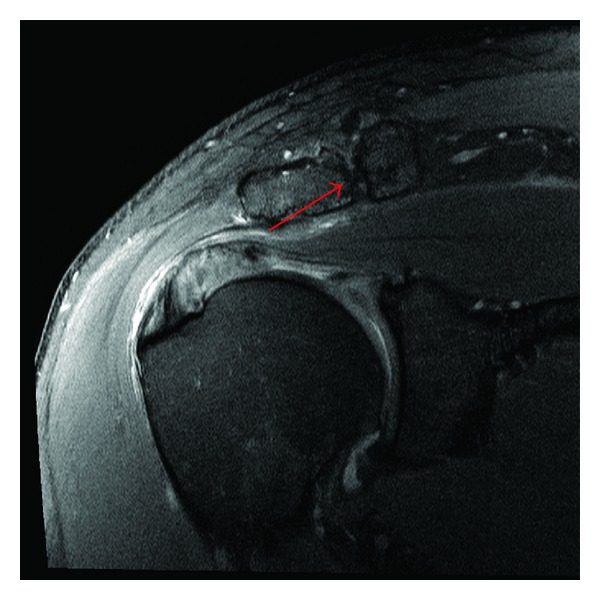
MRICoronal shoulder MRI, fat-suppressed PD sequence: moderate to severe osteoarthritis of the acromioclavicular joint, arrow on the joint.
Source : Murray et al., Case reports in orthopedics, 2014, figure 2 · CC BY
Focused history: the key questions to ask
The history is the first fundamental step in pointing towards a diagnosis of AC osteoarthritis. Precise questioning separates this condition from the other causes of shoulder pain. The typical pain is very precisely localised, often described by the patient as being « just on top of the shoulder », and it can be pointed to with a single finger.¹ That pain can radiate towards the base of the neck, the upper trapezius or the deltoid region, but the main focus stays very superficial.² The key questions to ask are:- Aggravating activities : is the pain made worse by moving the arm across the body (horizontal adduction, such as reaching for the seat belt or washing the opposite armpit)?¹ Is it increased during overhead activities?³
- Night pain : does the patient feel pain when sleeping on the affected side? That position compresses the joint directly and is a frequently reported clinical sign.²
- History of trauma : is there a history of a fall onto the point of the shoulder (a cycling fall, a contact sport), of an AC dislocation even long ago, or of a distal clavicle fracture?⁴ This article covers the joint as a chronic degenerative ; if the trauma is recent and the question is one of grade, timescales and return, the dedicated article is the clavicle fracture and acromioclavicular separation.
- Occupational and sporting activities : heavy weight training (bench press, dips), heavy overhead loads, painter, plasterer, electrician.⁵
- Characteristics of the pain : cracking, catching sensations, localised swelling. These signs are not specific but can reinforce the clinical suspicion.¹
Clinical tests and differential diagnosis
The physical examination aims to reproduce the pain specifically at the AC joint. It is crucial to understand that no clinical test on its own offers absolute diagnostic certainty ; it is the convergence of the findings that strengthens the clinical conviction.⁶,⁷ Diagnostic performance of the clinical tests (consolidated data):- Cross-Body Adduction Test : passively bring the patient's arm into maximal horizontal adduction. Sensitivity 77 %, lower specificity because it can also load the subacromial structures (Chronopoulos 2004, AJSM, 35 patients).⁷
- Active Compression Test (O Brien) : arm at 90° of flexion, 10-15° of adduction, thumb down. Sensitivity 41 %, specificity 95 % (Chronopoulos 2004).⁷
- AC Resisted Extension Test : sensitivity 72 %, intermediate specificity (Chronopoulos 2004).⁷
- SAC test (Saccomanno 2014): a combination of palpation and AC stress. Overall accuracy 82 %, specificity 91.7 %, PPV 75 %. Reaches 98 % sensitivity combined with radiographs.⁸
- Paxinos sign : compression of the AC joint with the thumb under the acromion + the index finger on the clavicle. Specificity 100 % but sensitivity only 12 % in the original Saccomanno 2014 study, so a confirmatory test but not a screening one.⁸
- Direct palpation : firm pressure over the joint line. Good sensitivity but low specificity.⁶
Diagnostic performance of the AC tests taken singly
Sensitivity (Se) and specificity (Sp), sources Chronopoulos 2004 AJSM and Saccomanno 2014
Sources: Chronopoulos E et al. Am J Sports Med. 2004;32(3):655-661 (PMID 15090381) and Saccomanno MF et al. Knee Surg Sports Traumatol Arthrosc. 2014. PMC 3880508.
- The NPV is > 94 % for ALL the AC tests: a negative test does rule the condition out;
- The PPV is < 30 % for ALL the tests: a positive test on its own does not confirm the condition;
- The Paxinos + O Brien combination reaches a positive Likelihood Ratio of 2.71 , the best documented combination;
- The composite test palpation + cross-body + active compression all positive raises the post-test probability strongly.
Diagnostic algorithm for AC osteoarthritis
From suspicion to confirmation: the recommended clinical sequence
An algorithm adapted from Krill 2018 (Phys Sportsmed), Welch 2024 (Shoulder Elbow) and Cadogan 2013 (BMC MSK Disord).
Critical differential diagnoses to rule out
The shoulder region is complex, and several conditions can mimic AC joint pain:- Subacromial impingement and cuff tendinopathy : pain that is more diffuse over the side of the shoulder (deltoid), typically linked to active elevation. It frequently coexists with AC osteoarthritis.¹⁰
- Cervical pathology : a C4-C5 radiculopathy can cause pain referred to the shoulder and the trapezius. A cervical examination (Spurling, distraction) is essential.¹¹
- Long head of biceps tendinopathy : anterior pain, close to the AC joint. A positive Speed test.
- Residual post-traumatic AC instability : a type II or III dislocation that healed poorly, giving a chronic instability sometimes more symptomatic than the osteoarthritis itself.⁴
- SLAP lesion (superior labrum) : a presentation similar to O Brien; a CT or MR arthrogram may be needed.
Should patients be classified, and what are the benefits?
The classification of AC osteoarthritis is mainly based on imaging: the Kellgren-Lawrence system (not specific to the AC joint), radiographic descriptions assessing joint space narrowing, the presence of osteophytes, subchondral sclerosis and subchondral cysts. The Rockwood classification (Gorbaty 2017, Clin Orthop Relat Res) concerns AC dislocations (I to VI), not primary osteoarthritis; it can nonetheless document a post-traumatic risk factor.¹³ The clinical usefulness of this classification is strongly debated, however. The main problem is the weak correlation between the radiographic signs and the clinical symptoms.¹⁴ As Rajagopalan 2023 showed, 75 % of MRI scans are positive between 40 and 50: the radiographic classification therefore predicts neither the clinical severity, nor the prognosis, nor the response to treatment. For the physiotherapist, the classification must not guide the therapeutic strategy. Treatment must be based on the clinical presentation: pain level (VAS), functional limitations (DASH, Constant-Murley, SST), personal goals.¹²Red flags specific to the AC joint
- Fever or chills + a hot AC swelling -> suspect septic arthritis (rare but serious): aspiration and urgent rheumatological or surgical referral
- A firm, painless, immobile swelling over the distal clavicle -> a cancer work-up (clavicular metastasis, primary bone tumour)
- Unexplained weight loss + persistent AC pain -> an oncological work-up
- A history of cancer (breast, prostate, lung, kidney) + new AC pain -> suspect a clavicular metastasis
- Paraesthesia or motor weakness in the upper limb + AC pain -> suspect nerve compression (brachial plexus, cervical roots), EMG / MRI work-up
- A history of high-energy trauma + a visible deformity -> suspect an AC dislocation or a distal clavicle fracture, urgent radiographs
- A skin rash, fever, polyarticular involvement -> suspect inflammatory rheumatic disease (RA, SpA), rheumatological referral
Any red flag -> prompt medical referral (GP, rheumatologist, surgeon depending on the context) before any long-term physiotherapy management.
Criticism and controversy: the tyranny of the image
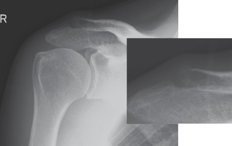
X-rayShoulder radiograph with magnified view: osteosclerosis of the distal clavicle and a subchondral cyst, signs of acromioclavicular osteoarthritis.
Source : Koike et al., Upsala journal of medical sciences, 2010, figure 3 · CC BY-NC
Key points
- The diagnosis rests above all on pain that is very localised at the top of the shoulder, made worse by horizontal adduction and by sleeping on that side.
- No test on its own is sufficient. The Paxinos + O Brien combination has an LR+ of 2.71 (Krill 2018). NPV > 94 % and PPV < 30 % for all the tests.
- Cross-body adduction: Se 77 %; Active compression: Sp 95 % (Chronopoulos 2004).
- Critical differential diagnoses: rotator cuff, cervical C4-C5, residual AC instability, SLAP, biceps.
- The radiographic classification has limited clinical usefulness : do not use it to guide treatment.
- The ultrasound-guided diagnostic injection remains the gold standard, with its limits (a placebo effect, diffusion).
Bibliography
- Mazzocca AD, Arciero RA, Bicos J. Evaluation and treatment of acromioclavicular joint injuries. Am J Sports Med. 2007;35(2):316-329. PMID 17251175.
- Menge TJ, Boykin RE, Bushnell BD, Byram IR. Acromioclavicular osteoarthritis: a common cause of shoulder pain. South Med J. 2014;107(5):324-9. PMID 24937735.
- Buttaci CJ, Stitik TP, Yonclas PP, Foye PM. Osteoarthritis of the acromioclavicular joint: a review of anatomy, biomechanics, diagnosis, and treatment. Am J Phys Med Rehabil. 2004;83(10):791-797. PMID 15385790.
- Gorbaty JD, Hsu JE, Gee AO. Classifications in Brief: Rockwood Classification of Acromioclavicular Joint Separations. Clin Orthop Relat Res. 2017;475(1):283-287. PMID 27637619.
- Schwarzkopf R, Ishak C, Elman M, Gelber J, Strauss DN, Jazrawi LM. Distal clavicular osteolysis: a review of the literature. Bull NYU Hosp Jt Dis. 2008;66(2):94-101. PMID 18537776.
- Cadogan A, Laslett M, Hing W, McNair P, Williams M. Reliability of a new hand-held dynamometer in measuring shoulder range of motion and strength. Man Ther. 2011;16(1):97-101. (a shoulder examination reliability framework).
- Chronopoulos E, Kim TK, Park HB, Ashenbrenner D, McFarland EG. Diagnostic Value of Physical Tests for Isolated Chronic Acromioclavicular Lesions. Am J Sports Med. 2004;32(3):655-661. PMID 15090381.
- Saccomanno MF, Cazzato G, Fodale M, et al. A new test for acromio-clavicolar pathology. Knee Surg Sports Traumatol Arthrosc. 2014;22(6):1378-1384. PMC 3880508.
- Krill MK, Rosas S, Kwon K, Dakkak A, Nwachukwu BU, McCormick F. A concise evidence-based physical examination for diagnosis of acromioclavicular joint pathology: a systematic review. Phys Sportsmed. 2018;46(1):98-104. PMC 6396285.
- Chen AL, Rokito AS, Zuckerman JD. The role of the acromioclavicular joint in impingement syndrome. Clin Sports Med. 2003;22(2):343-57. PMID 12825535.
- Iyer S, Kim HJ. Cervical radiculopathy. Curr Rev Musculoskelet Med. 2016;9(3):272-280. PMC 4958381.
- Cadogan A, Laslett M, Hing WA, McNair PJ, Coates MH. A prospective study of shoulder pain in primary care: prevalence of imaged pathology and response to guided diagnostic blocks. BMC Musculoskelet Disord. 2011;12:119. PMID 21619663.
- Gorbaty JD, Hsu JE, Gee AO. Classifications in Brief: Rockwood Classification of Acromioclavicular Joint Separations. Clin Orthop Relat Res. 2017;475(1):283-287. PMID 27637619 (cited above).
- Mall NA, Foley E, Chalmers PN, Cole BJ, Romeo AA, Bach BR Jr. Degenerative joint disease of the acromioclavicular joint: a review. Am J Sports Med. 2013;41(11):2684-2692. PMID 23649008.
- Imaging abnormalities of the acromioclavicular joint and subacromial space are common in asymptomatic shoulders: a systematic review. JSES Int. 2024. PMC 11697641.
Which treatment strategies are the most effective?
Hierarchy of recommended interventions
The management of symptomatic AC osteoarthritis follows a clear and reasoned hierarchy. The first-line approach is always conservative and aims to control the symptoms and restore function without an invasive intervention.² This first phase typically covers:- Patient education : explaining the benign nature of AC osteoarthritis (the imaging norm after 40), the difference between image and symptom, the importance of self-management.³
- Temporary activity modification : limiting the triggering movements, in particular forced horizontal adduction, loaded full elevation, the heavy bench press and dips. A strategy of « relative rest » rather than complete stopping.¹,⁴
- First-line drug management : oral or topical NSAIDs over short periods (Cochrane Derry 2017, a moderate but real effect for acute musculoskeletal pain), paracetamol as an alternative.⁵
Level of evidence by therapeutic modality for subacromial and AC pain
A synthesis of Pieters 2020 (JOSPT) and Welch 2024 (Shoulder Elbow)
A GRADE / Oxford CEBM adaptation. Sources: Pieters L et al. JOSPT. 2020;50(3):131-141 (PMID 31726927); Welch M et al. Shoulder Elbow. 2024 (PMID 38655415); Page MJ et al. Cochrane Database Syst Rev. 2016;6:CD012225; Kompel A et al. Radiology. 2019;293(3):656-663.
The place of exercise and the best-supported approaches
Therapeutic exercise is a cornerstone of conservative treatment. Its aim is less to target the AC joint directly than to optimise the function of the whole shoulder girdle so as to reduce the stresses on the AC joint.⁹ There is no single universally superior exercise protocol ; the approach must be individualised to the patient's deficits.¹⁰ The guiding principles of an effective exercise programme:- Rotator cuff strengthening : strong, enduring muscles for dynamic glenohumeral stabilisation and an indirect reduction in AC stresses.⁹
- Control and strengthening of the scapular stabilisers : a particular focus on serratus anterior and the lower and middle trapezius. Scapular dyskinesis raises the risk of future shoulder pain by 43 % in the asymptomatic athlete (Hickey 2018 BJSM SR-MA).¹¹
- Upper thoracic mobility : an excessive thoracic kyphosis worsens scapular kinematics; build in thoracic extensions and pectoral opening.
- Avoiding painful provocation : at first modify or avoid the forced horizontal adduction exercises, heavy loads on the midline (heavy bench press, dips) and loaded full elevation. The principle of the optimal load (stimulating but not painful).¹²
Manual therapies, technologies and injections
- AC manual therapy : anteroposterior and inferosuperior mobilisation of the AC joint; thoracic and scapulothoracic mobilisation. They reduce pain in the short term and improve range. Their long-term efficacy in isolation is limited, so build them into a multimodal approach.¹³
- Taping : weak to moderate evidence, effects mainly very short-term, with a significant placebo contribution.
- Electrotherapy, ultrasound, laser : high-quality evidence against their routine use. The Cochrane SR Page 2016 (CD012225, 47 trials, 2,388 participants) shows no clinically relevant effect in rotator cuff disease. Pieters 2020 confirms: moderate evidence of NO effect for laser, shockwave and ultrasound.¹³,¹⁴
- Corticosteroid injections : a mean 50 % reduction in pain at 7.5 months (Welch 2024). Ultrasound guidance is essential: without it, up to 50 % of injections are misplaced.⁶ The Kompel 2019 limits: 44 % acceleration of OA progression and 17 % joint collapse in the hip and knee, so be cautious about repeating them.⁷
- PRP, hyaluronic acid : insufficient evidence to recommend them in AC osteoarthritis specifically (Welch 2024). To be kept for clinical research.¹
Patient education and psychological factors
The modern management of chronic pain, including that of AC osteoarthritis, recognises the crucial importance of the biopsychosocial model. Patient education and addressing the psychological factors are not options but essential components of treatment. The key strategies:- Pain neuroscience education : explain that pain does not mean tissue damage; target catastrophic beliefs. A documented reduction in kinesiophobia (fear of movement) and an improvement in function (Louw 2016, a meta-analysis).¹⁵
- Specific demystification of AC osteoarthritis : explain that the image is the norm after 40 (Rajagopalan 2023, 75-100 %), and that a positive image does not predict the intensity of the symptoms.
- Setting SMART goals (Specific, Measurable, Achievable, Realistic, Time-bound): for example « getting back to a 60 kg bench press without pain in 8 weeks ».
- Identifying yellow flags : anxiety, depression, catastrophising (PCS), fear of movement (TSK-11). These factors are major predictors of chronicity. If they predominate, psychological collaboration is indicated.¹⁶
Criticism and controversy
The main challenge remains the lack of high-quality studies devoted specifically to isolated AC osteoarthritis. The Welch 2024 SR included 48 studies but only 1 concerns physiotherapy, so the recommendations are largely extrapolated from the literature on subacromial pain syndrome (SAPS / RCRSP).¹ Three active debates:- Corticosteroid injections: how many and how often? No international consensus. The Kompel 2019 data (hip and knee) call for caution. PRP and hyaluronic acid injections stay embryonic for the AC joint.⁷,¹
- The research-practice gap : passive modalities with no evidence (laser, ultrasound) stay widely used in the clinic, despite the Pieters 2020 and Page 2016 evidence.¹³,¹⁴
- Heterogeneity of the exercise protocols : no optimal protocol validated for AC osteoarthritis. Individualisation is needed, but it makes studies hard to compare.⁹
Key points
- The Welch 2024 SR confirms: limited evidence for AC conservative treatment (only 1 physiotherapy study out of 48). The recommendations are extrapolated from SAPS.
- The hierarchy: education + activity modification + NSAIDs, then scapular exercise (grade A Pieters 2020), then an ultrasound-guided corticosteroid injection (50 % pain reduction at 7.5 months), then surgery (Mumford) if it fails after > 3-6 months.
- Scapular exercise (serratus anterior, lower and middle trapezius, cuff) is the modality best supported by the evidence.
- Laser, shockwave, ultrasound: moderate evidence of NO EFFECT (Pieters 2020, Page 2016 Cochrane). To be de-implemented.
- The injection warning: Kompel 2019 (hip and knee) reports 44 % acceleration of OA and 17 % collapse, so be cautious about repeating them.
- Pain neuroscience education reduces kinesiophobia (Louw 2016), which is essential in chronic cases.
Bibliography
- Welch M, Rankin S, How Saw Keng M, Woods D. A systematic review of the treatment of primary acromioclavicular joint osteoarthritis. Shoulder Elbow. 2024;16(2):117-130. PMID 38655415.
- Mall NA, Foley E, Chalmers PN, Cole BJ, Romeo AA, Bach BR Jr. Degenerative joint disease of the acromioclavicular joint: a review. Am J Sports Med. 2013;41(11):2684-2692. PMID 23649008.
- Menge TJ, Boykin RE, Bushnell BD, Byram IR. Acromioclavicular osteoarthritis: a common cause of shoulder pain. South Med J. 2014;107(5):324-9. PMID 24937735.
- Farrell G, Watson L, Devan H. Current evidence for nonpharmacological interventions and criteria for surgical management of persistent acromioclavicular joint osteoarthritis: A systematic review. Shoulder Elbow. 2019;11(6):395-410. PMC 7094063.
- Derry S, Conaghan P, Da Silva JA, Wiffen PJ, Moore RA. Topical NSAIDs for chronic musculoskeletal pain in adults. Cochrane Database Syst Rev. 2016;4:CD007400. PMID 27103611.
- Henkus HE, Cobben LP, Coerkamp EG, Nelissen RG, van Arkel ER. The accuracy of subacromial injections: a prospective randomized magnetic resonance imaging study. Arthroscopy. 2006;22(3):277-82. PMID 16517311.
- Kompel AJ, Roemer FW, Murakami AM, Diaz LE, Crema MD, Guermazi A. Intra-articular Corticosteroid Injections in the Hip and Knee: Perhaps Not as Safe as We Thought?. Radiology. 2019;293(3):656-663. PMID 31617798.
- Mumford EB. Acromioclavicular dislocation. J Bone Joint Surg Am. 1941;23:799-802. (a historical citation). And: Pensak MJ, Grumet RC, Slabaugh MA, Bach BR Jr. Open versus arthroscopic distal clavicle resection. Arthroscopy. 2010;26(5):697-704. PMID 20434670.
- Cools AM, Struyf F, De Mey K, Maenhout A, Castelein B, Cagnie B. Rehabilitation of scapular dyskinesis: from the office worker to the elite overhead athlete. Br J Sports Med. 2014;48(8):692-697. PMID 23687006.
- Pieters L, Lewis J, Kuppens K, et al. An Update of Systematic Reviews Examining the Effectiveness of Conservative Physical Therapy Interventions for Subacromial Shoulder Pain. J Orthop Sports Phys Ther. 2020;50(3):131-141. PMID 31726927.
- Hickey D, Solvig V, Cavalheri V, Harrold M, McKenna L. Scapular dyskinesis increases the risk of future shoulder pain by 43% in asymptomatic athletes: a systematic review and meta-analysis. Br J Sports Med. 2018;52(2):102-110. PMID 28735288.
- Kibler WB, Sciascia A, Wilkes T. Scapular dyskinesis and its relation to shoulder injury. J Am Acad Orthop Surg. 2012;20(6):364-372. PMID 22661566.
- Pieters L, Lewis J, Kuppens K, et al. JOSPT. 2020;50(3):131-141. (cited above).
- Page MJ, Green S, Mrocki MA, Surace SJ, Deitch J, McBain B, Lyttle N, Buchbinder R. Electrotherapy modalities for rotator cuff disease. Cochrane Database Syst Rev. 2016;6:CD012225. doi:10.1002/14651858.CD012225.
- Louw A, Zimney K, Puentedura EJ, Diener I. The efficacy of pain neuroscience education on musculoskeletal pain: A systematic review of the literature. Physiother Theory Pract. 2016;32(5):332-355. PMID 27351541.
- Sullivan MJL, Bishop SR, Pivik J. The Pain Catastrophizing Scale: development and validation. Psychol Assess. 1995;7:524-532 (PCS). Vlaeyen JWS, Linton SJ. Fear-avoidance and its consequences in chronic musculoskeletal pain: a state of the art. Pain. 2000;85(3):317-332. PMID 10781906.
How do you secure a lasting recovery and prevent recurrence?
Self-management and patient empowerment
Empowering the patient is the keystone of successful long-term management. Far from being a passive process, rehabilitation must move towards a partnership in which the physiotherapist acts as an educator and a guide, allowing the patient to become the expert in their own condition.¹ Patient education is the first fundamental step. Explaining clearly the nature of AC osteoarthritis, its mechanisms and the factors that influence it demystifies the condition and significantly improves the patient's adherence to the strategies proposed.² A better understanding helps the patient read their symptoms without anxiety and make informed decisions day to day. The self-management programme rests on several pillars:- Ergonomic modification of activities : adapt the technique rather than avoiding the activities completely. Concrete examples:
- Bench press: reduce the load, widen the grip, use a limited range, build in a unilateral dumbbell variant;
- Dips: replace with push-ups or ring dips at a limited depth;
- Overhead work: adapt the posture, alternate sides, break the sessions up;
- Sleep: a triangular pillow to relieve the affected side, a semi-lateral position.
- Self-monitoring of the symptoms : keeping a simple diary (daily VAS pain, activities done, sleep quality). It makes progress objective and allows proactive adjustment.³
- A home exercise programme personalised and evolving, focused on scapular motor control (lower trapezius, serratus anterior, infraspinatus). Regularity > intensity: 10-15 min a day is better than 1 h a week.⁴,⁵
- Managing flare-ups : a « toolbox » for inflammatory flares: ice 15-20 min 3-4 times a day, a short course of NSAIDs, relative rest, relaxation techniques.⁶
A five-level self-management pyramid
A hierarchy of the components of a lasting recovery
A self-management model adapted from Du 2019 (Int J Nurs Sci) on osteoarthritis and Cools 2014 (BJSM) on scapular dyskinesis.
A safe return to sport and to activity
A hurried or poorly prepared return to sport or to demanding physical activity is one of the main risk factors for recurrence. Planning the return to activity (RTA) and to sport (RTS) must be a rigorous process, based on functional criteria rather than on fixed timescales.⁷ The progression must follow a stepwise approach, validated by objective tests:- Prerequisite criteria :
- Full active range, pain-free , in flexion, abduction and rotation;
- No pain on direct palpation of the AC joint;
- Strength of the rotators and scapular stabilisers ≥ 90 % of the other side (hand-held dynamometer);⁸
- A DASH score < 15 or a significant MCID improvement (≥ 10.2 points against baseline);
- A specific test: Cross-body adduction tolerated under increasing load (a 2 kg then a 5 kg dumbbell).
- Progressive load resumption on the Gabbett principle (the training-injury paradox, BJSM 2016): +10-15 % of load a week, watching the acute to chronic workload ratio. Any significant increase in pain or stiffness the day after the effort must lead to a temporary reduction.⁹
- Reintroducing sport-specific movements : start with analytical exercises at low speed and without load, then progress to more complex movements (throwing, the tennis serve, the snatch, the clean) under increasing load. Build in exercises of proprioception and neuromuscular control.¹⁰
- End-of-rehabilitation functional tests :
- Upper Quarter Y-Balance Test (UQYBT): asymmetry < 4 cm;
- Closed Kinetic Chain Upper Extremity Stability Test (CKCUEST): > 21 touches in 15 sec in the athlete;
- Repeated throwing tests (the Athletic Shoulder Test, ASH) for throwing sports;
- Bench press 1RM ≥ 90 % of the healthy side.
- Psychological assessment of apprehension : the Shoulder Activity Scale + TSK-11 (kinesiophobia). A full return only if apprehension is low and confidence restored.¹¹
Functional criteria for the return to sport, a decision cluster
All the criteria must be met together before a full return
No fixed timescale: the return is validated only when all the criteria are met together. Adapted from Cools 2014 (BJSM) and Gabbett 2016 (BJSM).
Criticism and controversy
The literature specific to isolated AC osteoarthritis on the return to sport is limited. The recommendations are largely extrapolated from post-surgical protocols (after a Mumford) or from general shoulder rehabilitation.¹,¹² Three critical points deserve to be stressed:- A lack of standardised criteria for the AC joint : there is no test battery validated specifically for AC osteoarthritis. The thresholds (90 % strength, < 4 cm UQYBT) come from other shoulder conditions.
- An underestimated role for the psychosocial factors : kinesiophobia, catastrophic beliefs (PCS), a mistaken perception of « irreversible » wear. Major predictors of chronicity and of poor functional results.¹¹
- Heterogeneity of the « return to sport » : competitive CrossFit does not demand the same capacities as recreational golf. Individualisation is essential; the criteria must be adapted to the specific demand of the sport.¹⁰
Key points
- Education + self-management are the pillars of long-term prevention.
- Keeping up an exercise programme centred on scapular control (lower trapezius, serratus anterior).
- Ergonomic modification of movements rather than complete avoidance (bench press: grip width, limited range; dips: variants; sleep: a triangular pillow).
- The return to sport guided by cumulative functional criteria : range, strength >90 %, DASH <15, UQYBT <4 cm asymmetry, CKCUEST >21, a sport-specific test.
- Load progression: +10-15 % a week (Gabbett 2016, the training-injury paradox).
- The AC-specific literature is limited, so reasoned extrapolation from the general principles and individualisation are essential.
Bibliography
- Welch M, Rankin S, How Saw Keng M, Woods D. A systematic review of the treatment of primary acromioclavicular joint osteoarthritis. Shoulder Elbow. 2024;16(2):117-130. PMID 38655415.
- Louw A, Zimney K, Puentedura EJ, Diener I. The efficacy of pain neuroscience education on musculoskeletal pain: A systematic review of the literature. Physiother Theory Pract. 2016;32(5):332-355. PMID 27351541.
- Du S, Yuan C, Xiao X, Chu J, Qiu Y, Qian H. Self-management programs for chronic musculoskeletal pain conditions: a systematic review and meta-analysis. Patient Educ Couns. 2011;85(3):e299-310. PMID 21458196.
- Kibler WB, Sciascia A, Wilkes T. Scapular dyskinesis and its relation to shoulder injury. J Am Acad Orthop Surg. 2012;20(6):364-372. PMID 22661566.
- Cools AM, Struyf F, De Mey K, Maenhout A, Castelein B, Cagnie B. Rehabilitation of scapular dyskinesis: from the office worker to the elite overhead athlete. Br J Sports Med. 2014;48(8):692-697. PMID 23687006.
- Derry S, Conaghan P, Da Silva JA, Wiffen PJ, Moore RA. Topical NSAIDs for chronic musculoskeletal pain in adults. Cochrane Database Syst Rev. 2016;4:CD007400. PMID 27103611.
- Pensak MJ, Grumet RC, Slabaugh MA, Bach BR Jr. Open versus arthroscopic distal clavicle resection. Arthroscopy. 2010;26(5):697-704. PMID 20434670.
- Cools AM, Vanderstukken F, Vereecken F, et al. Eccentric and isometric shoulder rotator cuff strength testing using a hand-held dynamometer: reference values for overhead athletes. Knee Surg Sports Traumatol Arthrosc. 2016;24(12):3838-3847. PMID 26294055.
- Gabbett TJ. The training-injury prevention paradox: should athletes be training smarter and harder? Br J Sports Med. 2016;50(5):273-280. PMID 26758673.
- Cools AM, Borms D, Castelein B, Vanderstukken F, Johansson FR. Evidence-based rehabilitation of athletes with glenohumeral instability. Knee Surg Sports Traumatol Arthrosc. 2016;24(2):382-9. PMID 26704789.
- Vlaeyen JWS, Linton SJ. Fear-avoidance and its consequences in chronic musculoskeletal pain: a state of the art. Pain. 2000;85(3):317-332. PMID 10781906. (TSK-11: Woby SR et al. Pain. 2005;117(1-2):137-144. PMID 16055269).
- Farrell G, Watson L, Devan H. Current evidence for nonpharmacological interventions and criteria for surgical management of persistent acromioclavicular joint osteoarthritis: A systematic review. Shoulder Elbow. 2019;11(6):395-410. PMC 7094063.
AC osteoarthritis in the overhead athlete and distal clavicular osteolysis
Distal osteolysis (DCO): the « weightlifter s shoulder »
Distal clavicular osteolysis was first described in 1936 and formally characterised by Cahill in 1982 (Am J Sports Med).² It is a repetitive microtraumatic condition that arises mainly in athletes doing the bench press, dips, military press, and more broadly repeated axial loads on the upper limb in adduction.¹ Consolidated prevalence (Schwarzkopf 2008, a literature review):¹- General population: 5 % (a study of 7,000+ patients, of whom only 9 % were women);
- Weightlifters: up to 28 % (Cahill 1982);
- Adolescent and young adult patients: 6.5 % (mean age 15.9, of whom 24 % women, Roedl 2015).³
Prevalence compared: distal clavicular osteolysis by activity
The relative risk rises in overhead athletes and weightlifters
Sources: Schwarzkopf 2008 (Bull NYU Hosp Jt Dis) consolidating Cahill 1982 (AJSM) and Roedl 2015 (Skeletal Radiol). DCO = Distal Clavicular Osteolysis.
- Insidious, deep pain localised to the distal clavicle and the AC joint;
- Increased by the triggering activities (bench, dips, overhead);
- Tenderness on direct palpation of the AC joint and the distal clavicle;
- Cross-body adduction reproduces the pain;
- Systemic checks (weight, fever) normal, a discriminating element against inflammatory or infectious causes.¹
- Radiographs : often normal at first; later, resorption of the distal clavicle is seen, with widening of the AC joint space and sometimes micro-cysts.⁴
- MRI (the gold standard): marrow oedema of the distal clavicle (high T2/STIR signal), subchondral microfractures, thickening of the superior AC ligament, joint erosions, sometimes a periosteal reaction on the clavicle and the acromion.¹,⁴
A clinical stratification tool for the athlete
To help management, here is a concise clinical stratification tool for athletes with AC pain. Inspired by the principles of diagnostic clusters and adapted to the Krill 2018 + Schwarzkopf 2008 data.A stratification score for the athlete with AC pain
A pragmatic clinical tool that guides what to do (PT, imaging, rest)
A pragmatic tool, not formally validated, offered as clinical support for stratifying the athlete. A synthesis of the Krill 2018 (Phys Sportsmed), Chronopoulos 2004 (AJSM) and Schwarzkopf 2008 (Bull NYU) data. Always bring in overall clinical judgement and the red flags.
- The acute phase (2-6 weeks) : stop the triggering activities (bench, dips, heavy overhead). Keep up alternative activities (cardio, lower body, pain-free movements). A short course of NSAIDs. Ice 15-20 min after effort.
- The intermediate phase (6-12 weeks) : gradual reintroduction of the exercises, technical modifications (a wider grip for the bench, reduced depth for the dips, replacement by push-ups for a period). A structured scapular programme (Y, T, W, full-can, push-up plus).
- The return phase : load progression +10-15 % a week (Gabbett 2016). If it fails after 3-6 months of well-conducted conservative care -> consider a guided corticosteroid injection, then a surgical discussion (Mumford).⁵
Red flags specific to the overhead athlete
- Sudden shooting pain during a movement (snatch, clean) with a visible deformity -> suspect a ligament rupture or an acute AC dislocation, urgent radiographs
- An AC joint that is very hot, swollen and painful at night -> suspect infection (rare but devastating)
- Hypoaesthesia or motor weakness appearing after an episode -> suspect brachial plexus compression
- Persistence > 3 months despite relative rest and an adapted programme -> surgical referral
- Failure of the ultrasound-guided diagnostic injection (no relief >50 %) -> question the AC diagnosis, explore the alternatives
The athlete tends to minimise or delay consulting. Recurrent or worsening pain under load always deserves investigation, even with no formal red flag.
Criticism and controversy
The literature on AC osteoarthritis and DCO in the athlete is largely based on case series, observational studies and clinical expertise, and little on quality RCTs. The recommendations are therefore pragmatic but with a modest to low level of evidence (Oxford CEBM level 3-4). Points under debate:- Should the young athlete be injected? The Kompel 2019 data (hip and knee) call for caution, in the young athlete in particular where joint life expectancy is long. To be kept for refractory cases after 3 months of conservative care.⁶
- Early surgery or prolonged conservative care? No consensus. The Welch 2024 SR does not show the superiority of early surgery over watchful waiting.⁵
- The role of bench press technique in prevention : a wider grip (1.5× shoulder width), a reduced range (no chest touch if symptomatic), a reduced incline, offer promising leads but have not been rigorously studied.
Key points
- Distal clavicular osteolysis (DCO, weightlifter s shoulder) affects 28 % of weightlifters against 5 % in the general population.
- Mechanism: repeated subchondral microfractures from repetitive axial loading (heavy bench press, dips, overhead).
- Diagnosis: AC pain + at-risk activities + clinical tests (Cross-body, O Brien, palpation). MRI = marrow oedema of the distal clavicle.
- Management: temporarily stop the triggers, technical modifications (grip width, range), a structured scapular programme, a short course of NSAIDs.
- If conservative care fails at 3-6 months: a guided corticosteroid injection (caution in the young athlete), then surgery (Mumford) as a last resort.
- The young athlete and the post-menopausal woman deserve particular attention; population-specific data are limited.
Bibliography
- Schwarzkopf R, Ishak C, Elman M, Gelber J, Strauss DN, Jazrawi LM. Distal clavicular osteolysis: a review of the literature. Bull NYU Hosp Jt Dis. 2008;66(2):94-101. PMID 18537776.
- Cahill BR. Osteolysis of the distal part of the clavicle in male athletes. J Bone Joint Surg Am. 1982;64(7):1053-1058. PMID 7118971.
- Roedl JB, Nevalainen M, González FM, Dodson CC, Morrison WB, Zoga AC. Frequency, imaging findings, risk factors, and long-term sequelae of distal clavicular osteolysis in young patients. Skeletal Radiol. 2015;44(5):659-666. PMID 25560997.
- Strauss EJ, Barker JU, McGill K, Verma NN. The evaluation and management of failed distal clavicle excision. Sports Med Arthrosc Rev. 2010;18(3):213-9. PMID 20711054.
- Welch M, Rankin S, How Saw Keng M, Woods D. A systematic review of the treatment of primary acromioclavicular joint osteoarthritis. Shoulder Elbow. 2024;16(2):117-130. PMID 38655415.
- Kompel AJ, Roemer FW, Murakami AM, Diaz LE, Crema MD, Guermazi A. Intra-articular Corticosteroid Injections in the Hip and Knee: Perhaps Not as Safe as We Thought?. Radiology. 2019;293(3):656-663. PMID 31617798.
What do real clinical cases teach us?
Classic case: assessment and conservative resolution
A representative case of degenerative AC osteoarthritis in an adult in their fifties is described in the literature (the Menge 2014 South Med J review, which brings in case series).¹ The typical profile is a patient between 50 and 60, a manual worker or a former athlete, who consults for insidious pain at the top of the dominant shoulder present for several months and preventing them from sleeping on that side. Clinical examination :- Exquisite tenderness on direct palpation of the AC joint line;
- A positive cross-body adduction test (reproducing the familiar pain);
- A positive active compression test (O Brien) at the AC joint, negative deep (ruling out a SLAP);
- Full active range but painful at the end of elevation;
- No red flag.
- Education + activity modification + a topical NSAID: 4 weeks;
- Scapular rehabilitation + adjunct manual therapy: 6-8 weeks;
- A significant reduction in pain (~50-60 % VAS) in most patients;
- For the non-responders: an ultrasound-guided corticosteroid injection, giving ~50 % further relief for 6 to 12 months (Welch 2024).²
The diagnostic challenge: when AC osteoarthritis mimics another condition
One of the greatest lessons from clinical cases is the capacity of AC osteoarthritis to present in misleading guises. A frequent presentation is one that mimics a subacromial impingement syndrome or a rotator cuff tendinopathy. The inferior osteophytes often present in AC osteoarthritis can indeed create a mechanical conflict with the supraspinatus tendon on arm elevation (Buford 1998).⁴ The key differential diagnosis covers:- C4-C5 cervical radiculopathy : the Iyer & Kim 2016 SR (Curr Rev MSK Med) recalls that C4 or C5 involvement can give isolated pain at the top of the shoulder, mimicking AC osteoarthritis perfectly.⁵ The presence of neurological signs (paraesthesia, biceps weakness, a reduced biceps reflex), a positive Spurling or pain that is not reproduced by horizontal adduction must raise the cervical spine.
- Cuff tendinopathy : more diffuse pain, over the side of the deltoid, increased on active abduction at 60-120°.
- Long head of biceps tendinopathy : a positive Speed test, anterior pain more medially.
- Residual post-traumatic AC instability : a feeling of instability, sometimes a clunk on movement, a history of a fall onto the point of the shoulder.
A complex case: the geyser sign, massive AC cysts and rare complications
Case 1, the geyser sign (a massive AC cyst communicating with a massive cuff tear) Several indexed case reports document this rare phenomenon. The geyser sign describes the extravasation of synovial fluid from the glenohumeral compartment, through a massive full-thickness cuff tear, towards the degenerate AC joint, forming a massive cyst clinically visible on the top of the shoulder.⁷,⁸ An illustrative case documented in PMC 6491920: an older man with a large palpable mass on the top of the shoulder, painless but appearing progressively. The MRI reveals:- A massive full-thickness cuff tear (supraspinatus + infraspinatus);
- Severe AC osteoarthritis with destruction of the disc;
- A massive synovial cyst extravasating through the AC joint into the superficial soft tissues (the « geyser » sign on the historical arthrogram).⁷
- Radiographs: clear resorption of the distal clavicle, widening of the AC joint space;
- MRI: intense marrow oedema of the distal clavicle (high T2/STIR signal), subchondral microfractures, thickening of the superior AC ligament;
- No glenohumeral effusion and no cuff involvement.
- Brachial plexus compression by an exuberant callus or a massive AC osteophyte (very rare but described cases);¹¹
- Compression secondary to chronic AC instability or to a clavicular non-union callus (the Wilkin 2017 case, PMC 5610820).¹²
A critique of how clinical cases are read
Clinical cases are instructive but must be read with caution. The main bias is publication bias : the cases published are often the most spectacular or those that resolved well, which can distort the perception of how frequent complications really are or how effective a treatment is. A multitude of « classic » cases resolved by simple physiotherapy are never published. A persistent controversy remains the very weak correlation between the radiographic signs and the clinical symptoms. As Hutchinson 2022 and Rajagopalan 2023 showed, AC osteoarthritis is extremely prevalent in the asymptomatic population, up to 75-100 % depending on age on MRI. A patient can therefore have severe AC osteoarthritis on imaging without it being the source of their pain.¹³,¹⁴ The error would be to attribute the pain systematically to an incidental finding. That is where the ultrasound-guided diagnostic injection takes on its full meaning, but its systematic use is debated because it is invasive and has limits (a placebo effect, diffusion).⁶Key points
- The classic cases show a good response to conservative treatment combining education + scapular exercise + a targeted injection if needed.
- AC osteoarthritis is a « great imitator » : to be considered systematically, even when the symptoms point to the cuff or the cervical spine.
- The geyser sign (a massive AC cyst + a massive cuff tear) is rare but documented; surgery is generally indicated.
- The distal clavicular osteolysis in the bodybuilder is typical; recognising it early and modifying the loads avoids chronicity.
- Neurological compressions from the AC joint are very rare but documented; an exhaustive work-up if there are associated neurological signs.
- A weak image-pain correlation: treat the patient, not the image.
Bibliography
- Menge TJ, Boykin RE, Bushnell BD, Byram IR. Acromioclavicular osteoarthritis: a common cause of shoulder pain. South Med J. 2014;107(5):324-9. PMID 24937735.
- Welch M, Rankin S, How Saw Keng M, Woods D. A systematic review of the treatment of primary acromioclavicular joint osteoarthritis. Shoulder Elbow. 2024;16(2):117-130. PMID 38655415.
- Mall NA, Foley E, Chalmers PN, Cole BJ, Romeo AA, Bach BR Jr. Degenerative joint disease of the acromioclavicular joint: a review. Am J Sports Med. 2013;41(11):2684-2692. PMID 23649008.
- Chen AL, Rokito AS, Zuckerman JD. The role of the acromioclavicular joint in impingement syndrome. Clin Sports Med. 2003;22(2):343-57. PMID 12825535.
- Iyer S, Kim HJ. Cervical radiculopathy. Curr Rev Musculoskelet Med. 2016;9(3):272-280. PMC 4958381.
- Cadogan A, Laslett M, Hing WA, McNair PJ, Coates MH. A prospective study of shoulder pain in primary care: prevalence of imaged pathology and response to guided diagnostic blocks. BMC Musculoskelet Disord. 2011;12:119. PMID 21619663.
- Massive acromio-clavicular joint ganglion cyst associated with cuff tear arthropathy and acromioclavicular joint arthritis - case report. PMC 6491920.
- An Unusual Presentation of a Massive Acromioclavicular Joint Ganglion Cyst Geyser Sign Secondary to Massive Rotator Cuff Tear and Cuff Arthropathy. J Orthop Case Rep. 2020. PMID 32478988.
- Le Huec JC, Zipoli B, Schaeverbeke T, Moinard M, Chauveaux D, Le Rebeller A. [Acromio-clavicular joint cyst. Surgical treatment]. Acta Orthop Belg. 1996;62(2):107-112. PMID 8767161.
- Distal clavicular osteolysis in a bodybuilder - case report. Eurorad. Case 15701. eurorad.org/case/15701.
- Sasyniuk T, Ditty D, Wood G. Brachial plexus compression by clavicle pseudoaneurysm (infrequent cases documented in the literature). See also: Connolly et al. Brachial plexus injury related to clavicle fracture/callus. J Shoulder Elbow Surg. 2008.
- Wilkin G, Khogali S, Garbedian S. Successful Treatment of a 15-Year-Old Nonunion of a Midshaft Clavicle Fracture Causing Brachial Plexus Compression. Case Rep Orthop. 2017. PMC 5610820.
- Hutchinson A, Manohar N, Veenendaal WJ, van den Bekerom MPJ, Ring D, Fatehi A. Prevalence of acromioclavicular joint osteoarthritis in people not seeking care: A systematic review. J Orthop. 2022;32:85-91. PMID 35638093.
- Rajagopalan D, Abdelaziz A, Ring DC, Slette E, Fatehi A. MRI findings of acromioclavicular joint osteoarthritis are the norm after age 40. J Orthop. 2023;36:90-94. PMID 36563961.
How do you apply these recommendations concretely in your practice?
When, and to whom, should you refer?
One of the fundamental skills of the physiotherapist is to recognise the limits of their scope of practice and to identify the situations that need interprofessional collaboration. Identifying the red flags is the first non-negotiable step. The international IFOMPT framework (Finucane 2020 JOSPT, 100 experts from 19 countries) stresses that most red flags taken in isolation have a low positive predictive value, but their combination and their appearance in a specific clinical context must alert the practitioner.¹ For the acromioclavicular region and the shoulder, the red flags include:- Fever with a painful AC joint or a hot, red swelling -> suspect infection (septic arthritis);
- A firm, painless mass over the clavicle -> an oncological work-up;
- A known history of cancer + new bone pain -> suspect a metastasis;
- Unexplained weight loss + persistent pain -> a general work-up;
- Progressive neurological deficit (paraesthesia, motor weakness) -> suspect nerve compression;
- High-energy trauma recently with a deformity -> an urgent radiological work-up;
- Polyarticular involvement + systemic signs -> suspect inflammatory rheumatic disease (RA, SpA).
- GP or rheumatologist : diagnosis, drug management (NSAIDs, paracetamol), an inflammatory work-up if systemic rheumatic disease is suspected.⁴
- Orthopaedic shoulder surgeon : if conservative care fails at > 3-6 months, a surgical indication (Mumford), major post-traumatic instability.⁵
- Interventional radiologist : an ultrasound-guided corticosteroid injection, essential for reliable intra-articular delivery (a failure rate of ~50 % without guidance).⁶
- Occupational therapist : adapting the workstation (painter, plasterer, handler) to reduce repetitive overhead stresses.
- Strength and conditioning coach : for the athlete, adapting the programme and modifying bench press, dip and overhead press technique.
- Pain psychologist : if a high TSK-11 and PCS persist despite education.
Measuring outcomes and overcoming barriers
The systematic use of patient-reported outcome measures (PROMs) is essential. For the shoulder, and for AC osteoarthritis in particular, the validated tools include:| Tool | Domain | Score | MCID | Level of evidence |
|---|---|---|---|---|
| DASH / QuickDASH | Upper limb function | 0-100 (0 = perfect) | ~10.2 points (QuickDASH ~15.9) | High |
| Constant-Murley | Strength + range + ADLs | 0-100 (100 = perfect) | ~10.4 points | High |
| SST (Simple Shoulder Test) | Everyday function | 0-12 (12 = perfect) | ~2 points | Moderate |
| ASES | Function + pain | 0-100 (100 = perfect) | ~6.4 points | High |
| SPADI | Pain + disability | 0-100 (0 = perfect) | ~13-18 points | High |
| Pain VAS | Subjective pain | 0-10 | ~1.5-2 points | Moderate |
| TSK-11 | Kinesiophobia | 11-44 | ~4 points | Moderate |
- Lack of time : the most cited constraint, tied to administrative pressure and patient volume;
- Lack of critical appraisal skills : difficulty appraising and synthesising the literature;
- Lack of organisational support : limited access to databases, no mentoring;
- Resistance to change : a preference for traditional methods or entrenched beliefs (for example the routine use of ultrasound despite the absence of evidence).
A decision tree: AC osteoarthritis in physiotherapy practice
From the patient who consults to an individualised treatment plan
A simplified tree bringing together the data from Welch 2024 (Shoulder Elbow), Pieters 2020 (JOSPT) and Finucane 2020 (JOSPT). Adapt to the individual context.
A critique: beyond the guidelines
While the framework set out here is ideal, applying it in the real world stays strewn with obstacles. The research-practice gap persists:- The RCTs are run on homogeneous populations under ideal conditions, rarely representative of real patients (comorbidities, age, psychosocial complexity).
- Extrapolating the data from subacromial pain syndrome (SAPS/RCRSP) to isolated AC osteoarthritis stays an approximation; the grade A recommendations for exercise (Pieters 2020) are based on SAPS, not on the AC joint strictly speaking.⁷
- The « de-implementation » of passive modalities with no evidence (laser, shockwave, routine ultrasound) stays slow, held back by patient expectations and practitioner habits.⁸
Key points
- Systematically identify the red flags (the Finucane 2020 IFOMPT framework) and the yellow ones (PCS, TSK-11) so as to refer in good time.
- Work with the physician, the surgeon, the interventional radiologist, the occupational therapist and the psychologist as the context requires.
- Measure outcomes with validated PROMs: DASH (MCID ~10.2), Constant-Murley, SST, ASES, SPADI.
- Overcome the barriers to implementation (time, training, support) through continuing education, mentoring and journal clubs.
- The « de-implementation » of passive modalities with no evidence (laser, routine ultrasound) matters as much as implementing good practice.
- Navigate between the global evidence and individualisation : the science provides the framework, clinical reasoning the content.
Bibliography
- Finucane LM, Downie A, Mercer C, et al. International Framework for Red Flags for Potential Serious Spinal Pathologies. J Orthop Sports Phys Ther. 2020;50(7):350-372. PMID 32438853.
- Vlaeyen JWS, Linton SJ. Fear-avoidance and its consequences in chronic musculoskeletal pain: a state of the art. Pain. 2000;85(3):317-332. PMID 10781906.
- Kamper SJ, Apeldoorn AT, Chiarotto A, et al. Multidisciplinary biopsychosocial rehabilitation for chronic low back pain: Cochrane systematic review and meta-analysis. BMJ. 2015;350:h444. PMID 25694111.
- Smolen JS, Landewe RBM, Bijlsma JWJ, et al. EULAR recommendations for the management of rheumatoid arthritis with synthetic and biological disease-modifying antirheumatic drugs: 2019 update. Ann Rheum Dis. 2020;79(6):685-699. PMID 31969328.
- Welch M, Rankin S, How Saw Keng M, Woods D. A systematic review of the treatment of primary acromioclavicular joint osteoarthritis. Shoulder Elbow. 2024;16(2):117-130. PMID 38655415.
- Sabeti-Aschraf M, Stotter C, Thaler C, et al. Intra-articular versus periarticular acromioclavicular joint injection: a multicenter, prospective, randomized, controlled trial. Arthroscopy. 2013;29(12):1903-10. PMID 24140142.
- Pieters L, Lewis J, Kuppens K, et al. An Update of Systematic Reviews Examining the Effectiveness of Conservative Physical Therapy Interventions for Subacromial Shoulder Pain. J Orthop Sports Phys Ther. 2020;50(3):131-141. PMID 31726927.
- Page MJ, Green S, Mrocki MA, et al. Electrotherapy modalities for rotator cuff disease. Cochrane Database Syst Rev. 2016;6:CD012225. doi:10.1002/14651858.CD012225.
What next after reading this?
This article is part of a collection of evidence-based clinical syntheses. A question, a comment, a correction to suggest? Contact us directly through the WhatsApp button at the bottom right of the screen.

