


















In brief
The same fall onto the point of the shoulder, the same patient, two neighbouring bones and two decisions that obey the same logic: it is the classification that decides, not the look of the shoulder. This article deals with acute injuries of the shoulder girdle. For the same joint in chronic degenerative disease, with no recent trauma, the dedicated article is acromioclavicular osteoarthritis ; for its recognition as an occupational disease, acromioclavicular osteoarthritis and occupational disease.
Clinical synthesis based on the Cochrane review by Lenza (2019) on surgery versus conservative treatment of the clavicle, the meta-analysis by Woltz (JBJS Am 2017), the Edinburgh cohort of Murray (JBJS Am 2013) on non-union risk factors, the randomised trial by Boström Windhamre (JSES 2022) comparing hook plate with physiotherapy, and the ISAKOS consensus of Beitzel (Arthroscopy 2014): 58 references checked one by one on PubMed.
Clinical synthesis
We should start with one figure, because it shifts everything else. In the Edinburgh cohort, clavicle non-union affects 6.2 % of fractures at all sites combined at 24 weeks, and 4.5 % of midshaft fractures alone (PMID 15252081). The same department, ten years later, in 941 midshaft fractures that were displaced in adults, finds 13.3 % (PMID 23824382). These are not two contradictory figures: they are two different populations, and the gap between them is exactly what the classification exists to spot.
This is where the reasoning is decided. A clavicle fracture that is not displaced almost always heals on its own; a displaced comminuted fracture in a smoker fails to heal in one case out of three. A grade I or II separation is rehabilitated; a grade IV or VI is operated on. Between the two sits a grade III that has divided the literature for forty years. In both conditions, it is neither the pain, nor the deformity, nor the patient's worry that decides: it is what the film shows, and which category it belongs to.
The corollary is uncomfortable, and it is solid. Operating on every displaced fracture means performing 7.5 fixations to prevent one non-union ; operating only on those whose predicted risk exceeds 40 % brings that number down to 1.7 (PMID 23824382). And the meta-analysis of the six randomised trials confirms it from another angle: the plate does divide the risk of non-union (RR 0.14 ; 95 % CI 0.06 to 0.32), but as soon as implant removals are counted, 17.6 % of operated patients go back to theatre against 16.6 % of those never operated on (PMID 28632595). Reoperation is not removed, its nature is changed.
The thresholds in this article are the ones that decide. They are also the ones most often quoted from memory, and most often quoted wrong.
Shortening is the clearest example of this. The 2 cm threshold circulates everywhere; it comes from a series of 39 patients already in delayed union or malunion, with no control group (PMID 11317682). The 20 mm threshold comes from a series of 52 patients in whom none had lost range or strength (PMID 9250733). And the only prospective cohort that has looked for a correlation between shortening and function does not find it, even beyond 2 cm (PMID 26080806). The French recommendation, for its part, sets 1.5 cm in the young active patient (PMID 28043849). Four sources, four values, none of them from a trial that randomised on a threshold.
On the acromioclavicular side, the news is simpler and more reassuring. The only recent randomised trial comparing surgery with physiotherapy in grades III and V finds the non-operated patients significantly better at three months, then strictly equivalent at 6, 12 and 24 months: Constant 88 against 91 for grade III, 90 against 91 for grade V (PMID 35007749). With, in the same trial, the caveat that has to be told to the patient: 18 % of the non-operated patients ended up having surgery within 19 months.
And for rehabilitation, an honesty is needed that the published protocols do not have. One review screened 1,742 studies and kept only four that described a detailed protocol, then concluded in black and white: « no randomised trial or comparative study was identified or cited as the basis for these rehabilitation protocols » (PMID 34093044). The timeframes we give are expert landmarks, not results. What stays solid is what can be measured: union, range of motion, strength symmetry, and the six-week reassessment model that predicts union with an area under the curve of 87.3 % (PMID 31977816).
- Clavicle non-union moves from 4.5 %–13.3 % depending on whether all midshaft fractures or only the displaced ones are counted. Displacement is the first sort.
- The strongest factor is smoking (OR 3.76), ahead of comminution (1.75) and displacement (1.17).
- No shortening threshold has been validated prospectively. The four values in circulation come from four different study designs.
- Grades I and II : rehabilitation. Grades IV and VI : surgery. Grades III and V : a time-limited trial of rehabilitation, then reassessment.
- The residual deformity worries the patient far more than it hinders them, and this has been checked: of 28 patients bothered by the look of it, only 11 were considering surgical correction.
Why are these two injuries covered in the same article?
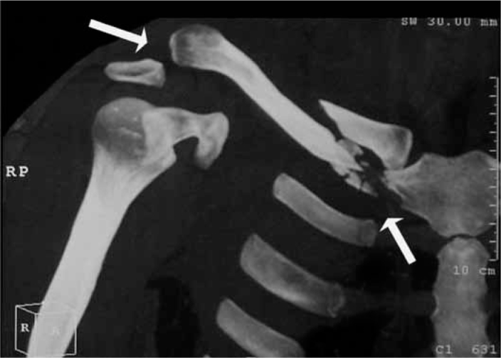
CT scan3D CT reconstruction of the shoulder, anterior view, showing together the clavicular fracture and the acromioclavicular dislocation (arrows).
Source : Correa et al., Revista Brasileira de Ortopedia, 2011, figure 3a · CC BY-NC-ND
Because they come through the same door of the clinic. The patient has fallen onto the point of the shoulder or onto an outstretched hand, they can no longer lift the arm, and the question they ask is identical in both cases. This chapter sets out what brings them together, what separates them, and how often they are met.
The clavicle is the only bony strut between the axial skeleton and the upper limb. Everything else in the shoulder girdle hangs from muscles. This peculiarity explains two things at once: the clavicle breaks often, and the joint that links it to the acromion separates often, because these are the two points through which a force applied to the shoulder is obliged to pass.
The mechanism is the same in both cases. The evidence-based review from the American Academy of Orthopaedic Surgeons describes, for separation, « a direct fall onto the superior aspect of the shoulder with the arm adducted » (PMID 31008872). Video analysis of shoulder injuries in Super Rugby, over 397 matches, finds exactly two patterns: contact with the ground, arm in horizontal adduction, flexion and internal rotation, and impact on the lateral aspect of the shoulder, elbow flexed, arm by the side (PMID 25156881). Depending on the angle and the energy, it is the bone that gives way or the ligaments.
What mechanics say about the coracoclavicular complex
The coracoclavicular ligaments, conoid and trapezoid, are what suspends the clavicle from the coracoid. A cadaveric study on 19 specimens measures their failure in uniaxial tension at 500 ± 134 N, for a stiffness of 103 ± 30 N/mm and an elongation at failure of 7.7 ± 1.9 mm (PMID 10653552). Two lessons for the clinic. First, this complex barely lengthens before it tears: there is no comfortable stretch zone, either it is intact or it is not. Second, in this loading configuration, the authors find no significant difference between the contributions of the conoid and the trapezoid. The division of roles taught in the textbooks cannot be read in that particular measurement.
Where the clavicle breaks, and where it refuses to unite
868 consecutive fractures treated without surgery, Edinburgh prospective cohort. The commonest site is not the one that causes the most trouble.
Source: Robinson CM, Court-Brown CM, McQueen MM, Wakefield AE. J Bone Joint Surg Am 2004;86(7):1359-1365 (PMID 15252081). Prospective observational cohort over 51 months, 868 patients, median age 29.5 years. The bars on the right are on a 0 to 30% scale.
This chart carries a counter-intuitive message that is useful in the consulting room: the lateral fifth, two and a half times less common than the shaft, refuses to unite two and a half times more often. It is the site that gets underestimated, because it hurts less at the start and the frontal radiograph shows it poorly.
A clarification is needed about the 66.9 % in this chart, because 80 % is what one reads everywhere. Both figures are correct and they do not measure the same thing. The classic 80 % divides the clavicle into thirds and counts the middle third (PMID 30666620). The Edinburgh classification divides it into fifths at the ends, and therefore assigns to the lateral fifth fractures that the division into thirds placed in the middle third (PMID 9619941). This is not a disagreement between authors, it is a shifted anatomical boundary, and it explains a good part of the differences in figures from one publication to another.
How many patients, and which ones
The clavicle accounts for 2.6 to 4 % of all fractures, and 80 % of them sit in the middle third (Cochrane review, PMID 30666620). The Chinese national survey, across 512,187 questionnaires, measures an incidence of 17.4 per 100,000 person-years (95 % CI 13.8 to 21.0), with road traffic accidents and falls accounting for 91 % of causes (PMID 29404668). The Edinburgh series fills in the demographics: in men, incidence peaks before the age of 20 and falls thereafter ; in women, it is steadier, with two peaks, adolescence and old age (PMID 9619941).
The risk of clavicle fracture in high school, sport by sport
567 fractures across 31.5 million athlete exposures, United States national surveillance over nine seasons. Rate per 100,000 exposures.
Source: McCarthy MM, Bihl JH, Frank RM, Salem HS, McCarty EC, Comstock RD. Orthop J Sports Med 2019;7(7):2325967119861812 (PMID 31384623). In sports that are comparable between the sexes, the rate remains 2.91 times higher in boys (95 % CI 1.97 to 4.30). In competition 4.58 against 0.87 in training, a ratio of 5.27.
On the acromioclavicular side, the only recent prospective population cohort comes from the Copenhagen region. Across 549,225 inhabitants and one full year, 106 acute separations: incidence 19.3 per 100,000 person-years, male to female ratio 8.6 to 1, bimodal distribution with peaks at 20-24 years and 55-59 years. Sport is responsible in 80 cases out of 106, and cycling alone in 51 of them (PMID 37811384). Separation is thought to account for about 9 % of shoulder injuries (PMID 41761265), and in elite rugby it is the shoulder injury most frequently reported (PMID 25156881).
This last point is worth keeping in mind in the clinic: the cyclist who has fallen onto the shoulder is statistically the typical patient with an acromioclavicular separation, ahead of the rugby player.
Four epidemiological landmarks worth knowing
Each comes from a prospective cohort or a systematic review, not from an author's estimate.
Sources: PMID 30666620, PMID 37811384, PMID 31384623.
Red flags at first contact, before any mobilisation
- Sensory or motor deficit of the upper limb, even partial, even of delayed onset : a brachial plexopathy can appear up to a month after a displaced fracture treated without surgery. Surgical opinion and imaging of the plexus before any rehabilitation (PMID 31784383).
- Breathlessness, chest pain, desaturation : in the United States national inpatient database for clavicle fracture, haemopneumothorax is the commonest associated non-vascular, non-neural injury, after rib fractures (PMID 33717875). An emergency.
- Cold limb, reduced or asymmetric radial pulse, expanding supraclavicular haematoma : subclavian vascular injury. An absolute emergency, no physiotherapy of any kind.
- Skin stretched, blanched or threatened over the tip of the fragment : a fracture threatening to become open. Surgical opinion the same day.
- High-energy trauma : in the same database, clavicle fracture is significantly associated with concussion and with splenic and thoracic injuries. The shoulder is not necessarily the patient's most serious injury.
- One single mechanism, two injuries: a fall onto the point of the shoulder or onto an outstretched hand. Depending on the angle and the energy, it is the bone or the ligaments that give way.
- The shaft carries 80 % of fractures, but it is the lateral fifth that unites worst (11.5 % non-unions against 4.5 %).
- The typical patient with a separation is a man (8.6 to 1), a cyclist before being a rugby player, and he is 20-24 or 55-59 years old.
- The coracoclavicular complex fails at 500 N after 7.7 mm of elongation : there is no mechanical middle ground.
How do you tell a clavicle fracture from an acromioclavicular separation?
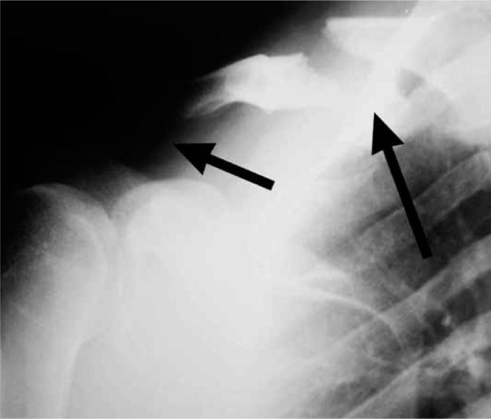
X-rayShoulder X-ray: extra-articular fracture of the medial end of the clavicle (arrow) and marked widening of the acromioclavicular joint (arrow).
Source : Correa et al., Revista Brasileira de Ortopedia, 2011, figure 2 · CC BY-NC-ND
The honest answer fits in one sentence: by radiography, and by radiography alone. This chapter explains why the acromioclavicular clinical tests do not serve the purpose they are believed to serve, what they are really worth, and which views to request so that the classification is reliable.
Clinically, the two presentations resemble one another closely enough to be confused on a regular basis. Pain at the top of the shoulder, inability to raise the arm, a deformity visible under the skin, a patient supporting the elbow with the opposite hand. Palpation points the way: the exquisite tenderness sits over the acromioclavicular joint line in one case, at the fracture site in the other. But that distinction, reliable in a relaxed patient, is far less so in a 25-year-old who reaches the emergency department two hours after a rugby collision, with muscle guarding that rules out any fine examination.
What the acromioclavicular tests are really worth
This is the point where teaching and measurement diverge most. The three classic tests were assessed by Chronopoulos et al. in 35 patients operated on for resection of the distal clavicle, that is to say chronic and isolated acromioclavicular lesions (PMID 15090381). Their values are in the table below, and the row that matters is not the sensitivity row.
| Test | Sensitivity | Specificity | PPV | NPV | Overall accuracy |
|---|---|---|---|---|---|
| Forced horizontal adduction | 77 % | Not reported separately | < 30 % | > 94 % | 79 % |
| Acromioclavicular resisted extension | 72 % | Not reported separately | < 30 % | > 94 % | 84 % |
| Active compression test (O'Brien) | 41 % | 95 % | < 30 % | > 94 % | 92 % |
This table says one precise thing, and it is the opposite of common practice. A negative predictive value above 94 % together with a positive predictive value below 30 % means that these tests serve to rule out, not to confirm. Three negative tests make acromioclavicular pain very unlikely ; three positive tests do not make it likely. The authors do conclude that a combination of tests does better than a single test, which is one way of saying that none of them is enough on its own.
And outside the context for which they were validated, they fall apart. Cadogan et al. recruited 153 primary care patients consulting for a painful shoulder, then gave them a fluoroscopically guided acromioclavicular anaesthetic block, with an 80 % response as the reference standard. The result: « none of the traditional tests was associated with an 80 % anaesthetic response », and their combinations discriminated no better than chance (area under the curve 0.507 ; 95 % CI 0.366 to 0.647) (PMID 23634871). The reference meta-analysis on shoulder tests is even more categorical: « no impingement test or acromioclavicular pathology test demonstrated significant diagnostic accuracy » (PMID 17720798).
The acromioclavicular tests have never been validated in acute trauma. Using them to decide between a fracture and a separation means using them outside their measured domain.
The practical consequence is simple. Faced with an acute shoulder injury, the physiotherapist does not do the clinical sorting: they check that the radiograph has been taken, that it has been taken properly, and they read the report knowing what should be in it.
The views that make the classification reliable
This point is too often neglected, and it tips decisions. The Rockwood classification rests on a measurement relative to the uninjured side, which assumes that both shoulders appear on the same film. Without that, its reliability is poor: earlier work reported kappa values of the order of 0.20–0.50, that is to say an agreement between observers barely better than mediocre (PMID 33582181).
Lau et al. standardised the protocol: bilateral Zanca views on the same radiographic plate. Interobserver reliability then rises to a Fleiss kappa of 0.624 and intraobserver agreement to a Cohen kappa of 0.696, on the grade III versus grade V distinction. The authors identify two residual causes of error: the muscle guarding that masks the displacement, and the under-grading of a high-grade lesion that follows from it.
To this are added two views that are only requested if one knows why. The true axillary view looks for posterior displacement of the distal clavicle, which defines grade IV and on its own changes management. The cross-body adduction view looks for overlap of the clavicle on the acromion, which separates subtypes IIIA and IIIB in the ISAKOS consensus (PMID 24485119). The most recent Delphi consensus from the American Shoulder and Elbow Surgeons, which brought together 18 surgeons over three rounds with a 100 % response rate, makes the use of multiple radiographic views one of its 76 consensus statements (PMID 39842657).
What the report must contain, condition by condition
For a clavicle fracture, four pieces of information govern what follows, and they are not always written down: the site (middle third, lateral fifth, medial fifth), the displacement and in particular whether or not there is cortical apposition, the comminution, and the shortening as measured. These are the four variables retained as independently predictive in the Edinburgh models (PMID 15252081, PMID 23824382). A report that says « midshaft clavicle fracture » and no more cannot be used to decide anything.
For a separation, what is needed is the coracoclavicular distance on both sides, expressed as a percentage increase, the search for a posterior displacement, and if possible a note on any overlap in cross-body adduction. The measurement of the distance itself is excellent between observers, with an intraclass correlation coefficient of 0.982 (PMID 36570359): it is the visual interpretation that varies, not the ruler.
Red flags at the diagnostic stage
- Shoulder trauma without a radiograph : the question is not whether one can do without it, it is that the classification, and therefore management, cannot be settled without it. Do not start rehabilitation on a clinical diagnosis in this setting.
- A unilateral radiograph when separation is suspected : the classification becomes unreliable and under-grading is the documented direction of error. Request bilateral views before concluding that the grade is low.
- Medial pain, dysphagia, breathlessness or difficulty swallowing : consider a posterior sternoclavicular injury, which fits neither of the two classifications in this article and which is a surgical emergency.
- A painful shoulder in a patient over 60 after minimal trauma : think of a fragility fracture and an associated cuff tear. See osteoporosis and the prevention of fragility fractures and symptomatic rotator cuff tear.
- In acute trauma, the diagnosis is radiographic. The acromioclavicular tests were validated on isolated chronic lesions, and they fail outside that setting.
- Their profile is that of a test of exclusion : NPV above 94 %, PPV below 30 %.
- The Rockwood classification is only reliable with bilateral Zanca views on the same plate : kappa 0.624 against 0.20 to 0.50 without.
- Four words to look for in the report of a fracture: site, displacement, comminution, shortening.
What exactly does the Rockwood classification say, and what does it decide?
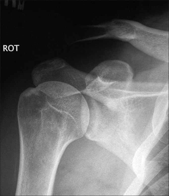
X-rayShoulder X-ray showing a stage III acromioclavicular dislocation, with marked elevation of the clavicle relative to the acromion.
Source : Marchie et al., International Journal of Shoulder Surgery, 2009, figure 2 · CC BY
Six grades, two ligaments, one measurement expressed as a percentage of the uninjured side. The Rockwood classification is simple to state and regularly misquoted, particularly on its numerical limits. This chapter gives them with their source, then says what each grade calls for.
The logic is ligamentous before it is radiographic. Two systems hold the distal clavicle: the acromioclavicular ligaments, which mainly control horizontal translation, and the coracoclavicular ligaments, conoid and trapezoid, which suspend the clavicle and control vertical translation. The grade says which ones have given way, and by how much the clavicle has ridden up.
The six Rockwood grades, frontal view
The percentage is the increase in coracoclavicular distance compared with the uninjured side. Solid line: intact ligament. Broken line: torn ligament.
Numerical limits: He X, Kong D, Lu X, et al. J Orthop Surg Res 2026;21(1):237 (PMID 41761265), full text. Management by grade: Frank RM, Cotter EJ, Leroux TS, Romeo AA. J Am Acad Orthop Surg 2019;27(17):e775-e788 (PMID 31008872). Schematic diagram, not to anatomical scale.
Two limits are worth keeping exactly as they are, because they are often quoted the wrong way round. Grade III corresponds to an increase in coracoclavicular distance of 25–100 % compared with the uninjured side. Grade V starts beyond 100 %, with a range classically described as 100 to 300 %. So it is neither the pain nor the look of the deformity that separates III from V, it is a measurement taken with callipers on a bilateral film.
| Grade | Ligaments | Radiograph | Reference management |
|---|---|---|---|
| I | Acromioclavicular sprain, coracoclavicular ligaments intact | Normal, less than 10% widening | Non-surgical |
| II | Acromioclavicular ligaments torn, coracoclavicular ligaments intact | Widening of 10 to 25% | Non-surgical |
| III | Both systems torn | Widening of 25 to 100% | Controversial : a trial of rehabilitation as first line |
| IV | Both systems torn | Displacement posteriorly, clavicle in or through the trapezius fascia; seen on the axillary view | Surgical |
| V | Both systems torn, deltotrapezial fascia stripped | Widening of 100 to 300% | Controversial |
| VI | Both systems torn | Displacement inferiorly, clavicle in a subacromial or subcoracoid position | Surgical |
The ISAKOS subclassification, and why it concerns the physiotherapist directly
In 2014, the ISAKOS upper extremity committee proposed splitting grade III in two, noting that no consensus existed on its treatment. The definition is remarkable, because it is the only one in the whole classification to bring rehabilitation into the criterion itself :
- Grade IIIA : stable joint, no overlap of the clavicle on the cross-body adduction view and no significant scapular dysfunction.
- Grade IIIB : unstable, defined by scapular dysfunction resistant to treatment and by overlap of the clavicle on the same view (PMID 24485119).
In other words: a grade III only becomes a IIIB after it has resisted rehabilitation. The physiotherapist is not merely the person who delivers conservative treatment, they produce the information that tips the classification.
With a caveat that has to be given at the same time. The reliability study of this modification, on 40 radiographs read by six specialist surgeons, finds acceptable overall agreement (kappa 0.637) but agreement that is poor for IIIA (kappa 0.215) and only moderate for IIIB (0.473). The authors conclude that « visual inspection was not reliable in distinguishing a IIIA from a IIIB » (PMID 36570359). The subclassification guides the reasoning, it does not decide mechanically.
- Grade III : 25 to 100 % increase in coracoclavicular distance. Grade V : beyond 100 %. It is a measurement, not an impression.
- Grades IV (posterior) and VI (inferior) cannot be seen on a frontal view alone.
- The IIIA / IIIB subclassification makes the response to rehabilitation a criterion for grading. Its visual reliability remains poor for IIIA.
- Consensus management: I and II without surgery, IV and VI with, III and V under debate.
Which thresholds really put clavicle fixation up for discussion?
Displacement, shortening, comminution: three words that everyone quotes, with figures that almost nobody traces back to the original study. This chapter goes back to each source, gives what it actually measured, and offers the only decision tool validated on a prospective cohort.
Before the thresholds, a point of method. The non-union rate of a clavicle fracture means nothing without the population it describes. This is the leading source of fruitless arguments between professionals, and the chart below is enough to clear it up.
The non-union rate depends first of all on what is being measured
Six populations, six figures, all of them correct. Each describes a different fracture.
Sources, in order: PMID 35984091, PMID 15252081 (two values), PMID 23824382, PMID 31977816, PMID 34779668. Scale 0 to 35%. Definitions of non-union differ from one study to another, something a systematic review notes explicitly (PMID 25027978).
Two readings stand out. The first: in the adolescent, even when completely displaced, a clavicle fracture practically never goes to non-union (0.4 % in 282 patients followed for two years). The second: it is the Neer type II lateral clavicle that holds the record, with 31 % radiological non-union, while keeping, according to the authors of the meta-analysis themselves, a « good functional outcome despite this rate ».
The three factors that really predict non-union
The Edinburgh multivariate model, built on 941 midshaft fractures followed to union or non-union, retains only three variables (PMID 23824382) :
- Smoking, odds ratio 3.76. It is by far the first of them, and the authors write that « smoking cessation should be an integral part of treatment ». It is the only factor on which the patient and the clinician can act immediately.
- Comminution, odds ratio 1.75.
- Displacement, odds ratio 1.17 per unit.
The earlier model from the same team, on 868 patients, added for the shaft the absence of cortical apposition, the female sex and increasing age (PMID 15252081). Female sex reappears independently in a population cohort of 1,350 fixations, as a factor for revision for non-union (OR 2.2) (PMID 24990977).
Operating on every displaced fracture means performing 7.5 fixations to prevent one non-union. Operating only on those whose predicted risk exceeds 40% means performing 1.7.
That sentence is the surgical translation of the model, and it comes straight from the article (PMID 23824382). It explains why the indications swung back and forth for twenty years: both camps were right about different populations.
The tool that was missing: reassess at six weeks rather than decide on day one
This is the most useful work of the last decade for the physiotherapist, and it is little known. Nicholson et al. prospectively followed 200 patients with a midshaft fracture that was completely displaced, with non-union defined on CT at six months. They compared a model built on data from the day of injury with a model built on a reassessment at six weeks (PMID 31977816).
The six-week reassessment model
Three simple criteria, two of which are available in the practice. Area under the curve 87.3% against 64.8% for the day-of-fracture model.
Source: Nicholson JA, Clement ND, Clelland AD, MacDonald D, Simpson AHRW, Robinson CM. J Bone Joint Surg Am 2020;102(7):557-566 (PMID 31977816). Level I prognostic study. The 40-point QuickDASH threshold comes from a ROC curve analysis, with an area under the curve of 76.8% for that criterion alone.
What this model changes for practice: the question « should we operate? » does not have to be settled on day one. It is much better asked at six weeks, with a patient already in rehabilitation, a measured QuickDASH and a follow-up radiograph. And the physiotherapist holds two of the three criteria.
Shortening: four thresholds, four study designs, no trial
Here is the point where the literature is most often quoted from memory. The table below lists the four values in circulation, with what each study actually did.
| Threshold quoted | Source | Actual design | What the study establishes | What it does not establish |
|---|---|---|---|---|
| 20 mm | Hill 1997, JBJS Br (PMID 9250733) | Retrospective series, 52 of 66 patients reviewed | Initial shortening of at least 20 mm is strongly associated with non-union (p < 0.0001) and with an unsatisfactory result | No patient had any significant loss of range or strength. And of the 28 bothered by the appearance, only 11 were considering surgery |
| 2 cm | Wick 2001, Arch Orthop Trauma Surg (PMID 11317682) | 39 patients already in delayed union or malunion, all of them operated on, with no control group | That 30 of the 33 Allman I fractures in this selected series were shortened by at least 2 cm | Nothing about the risk carried by a fracture shortened by 2 cm at the outset : the population is built on failure |
| 18 mm (men) 14 mm (women) | Lazarides 2006, JSES (PMID 16517363) | Retrospective, 132 united fractures, satisfaction questionnaire | Beyond these values, a significant association with a result judged unsatisfactory. Mean Constant 84, 25.8% dissatisfied | The threshold is different for each sex, which always disappears when it is quoted |
| 15 mm | Ropars 2017, OTSR (PMID 28043849) | Narrative review, expert recommendation | Surgery is recommended beyond 1.5 cm of shortening in the young active patient | This is not a measurement, it is a recommendation. It is lower than the English-language thresholds |
| None | Figueiredo 2015, BMC Musculoskelet Disord (PMID 26080806) | Prospective cohort, 59 patients, DASH at 6 weeks and 1 year | No correlation between shortening and function (p = 0.073 and p = 0.706), including when only shortenings above 2 cm are analysed | Modest sample, a single centre. But it is the only prospective design that has asked the question |
The overall reading fits in one sentence. No trial has randomised patients on either side of a shortening threshold. The four values in circulation come from two retrospective series, from a series with no controls built on failure, and from an expert recommendation. The only prospective cohort that has looked for the correlation does not find it. That does not mean shortening is unimportant: it means that it does not decide on its own, and that a figure announced to the patient as a boundary is a boundary the literature has never drawn.
The indications that are not up for discussion
They are rare and they are clear-cut. The French review from the Société française de chirurgie orthopédique lists them: depressed shoulder, floating shoulder, open fracture, fracture with a neurovascular complication (PMID 28043849). To which is added, in practice, the fracture that threatens the skin. Faced with any of these situations, the question of a threshold does not arise.
Red flags during follow-up of a fracture treated without surgery
- Mobility felt at the fracture site at six weeks : this is one of the three criteria in the predictive model. Combined with a high QuickDASH, the risk of non-union rises to 60%. Refer back.
- Pain that increases after the sixth week instead of easing : the normal trajectory is downward. A reversal should prompt a search for delayed union.
- Later onset of paraesthesia or of a deficit : post-fracture plexopathy can appear at day 8, day 14 or day 30, on granulation tissue or exuberant callus (PMID 31784383).
- Current smoker : this is not a red flag in the strict sense, but the odds ratio of 3.76 makes it the first modifiable lever for union. Raising it is an act of care, not a digression.
- The non-union rate ranges from 0.4 %–31 % depending on the population. Always ask which fracture is being talked about.
- Three independent factors: smoking (OR 3.76), comminution (1.75), displacement (1.17).
- The best decision tool is not on day one but at six weeks, and two of its three criteria are available in the practice.
- No shortening threshold has been validated prospectively. 20 mm, 2 cm, 18/14 mm and 15 mm come from four different study designs.
- Indications not open to discussion: open skin, threatened skin, neurovascular injury, floating shoulder.
Should a grade III separation be operated on?
This is the oldest and the most poorly settled question in shoulder trauma. It deserves better than an opinion: four randomised trials pooled in a meta-analysis, a recent trial with a physiotherapy arm, and a twenty-year follow-up. This chapter sets them end to end.
A reminder of what is at stake in figures. Grade III accounts for 55.7 % of all separations (PMID 37811384). So this is not a special case: it is the commonest case, and it is also the most debated. And the debate is not about radiographic reduction, which surgery achieves better, but about what that reduction brings the patient.
What the meta-analysis of the trials devoted to grade III alone says
Xie et al. restricted their meta-analysis to randomised trials dealing exclusively with grade III, which is both rare and important: most studies mix grades III and V, whose prognoses are not comparable. Four trials, 244 patients (PMID 39587562).
The main result is a non-result, and that is information in itself. No difference in Constant score in the long term: mean difference 4.82 points, 95 % confidence interval −6.42 to 16.06, p = 0.400. The interval crosses zero by a wide margin. Surgery, on the other hand, gives better early pain relief and a better coracoclavicular distance at every time point. And it comes with a higher incidence of post-traumatic osteoarthritis and of implant problems.
The trial that finally compares surgery with physiotherapy
Most trials set two surgical techniques against each other, or surgery against « doing nothing ». The Swedish trial by Boström Windhamre et al. does something else: it randomises 124 patients aged 18 to 65 with an acute grade III or V separation between hook plate and physiotherapy, with the same standardised outpatient rehabilitation protocol in both arms and 24 months of follow-up (PMID 35007749).
Hook plate against physiotherapy, what the trial measures at each time point
124 patients randomised, grades III and V stratified. The direction of the difference reverses: at three months, it is the non-operated patients who do better.
Source: Boström Windhamre H, von Heideken J, Une-Larsson V, Ekström W, Ekelund A. J Shoulder Elbow Surg 2022;31(6):1122-1136 (PMID 35007749). Single-centre randomised trial, complete follow-up in 118 of the 121 patients at 24 months. At the end, 97% of the Constant score of the uninjured shoulder had been recovered and 86% of patients rated the result as excellent or good.
Three things to take from this trial, and they point in different directions.
- At three months, it is the non-operated patients who do better, significantly so, on the Constant score, the subjective shoulder value, the QuickDASH and pain at rest as well as on movement. The hook plate costs a quarter of a year of recovery. The same signal appears in the Canadian multicentre trial on general health: physical score 52.13 against 45.75 at three months in favour of the non-operated group (p < 0.001), with a return above the population norm at six months without surgery and at two years with it (PMID 28832388).
- From six months onwards there is no longer any difference, neither for grade III nor for grade V.
- But eleven patients, that is 18 % of the non-operated group, ended up in theatre within 19 months: six grade III and five grade V. This is the sentence to say to the patient who asks for a guarantee: conservative treatment succeeds in more than four patients out of five, not in all of them.
Twenty years on, the deformity stays, the loss of function does not
The Finnish trial by Joukainen et al. recalled 25 of its 35 randomised patients 18 to 20 years after the injury (PMID 26535287). The result is twofold, and it is exactly what a patient needs to hear.
On one side, the anatomical difference persists: the joint is clinically more prominent or unstable without surgery, and radiologically wider (8.3 against 3.4 mm ; p = 0.004). On the other, the functional difference does not exist: Constant 83 in the operated group against 85 in the non-operated group, UCLA 25 against 27, simple shoulder test 11 against 12, with no significant difference, neither for grade III nor for grade V. And the authors add that « both groups showed radiological signs of acromioclavicular osteoarthritis and calcification of the coracoclavicular ligaments in equal proportions ».
At twenty years, the bump is still there and the shoulder works just as well. This is the most useful result to know for a consultation that opens with « is it going to stay like that? ».
And what about the reduction brace?
The idea is appealing: since the problem is a displacement, why not reduce it without operating? One published case report answered yes in a young and highly motivated patient, with healing in a grade II position (see the chapter of clinical cases). But the matched prospective cohort that measured this strategy in 25 patients, compared with 23 patients in early functional rehabilitation and 23 operated on, reaches a blunt conclusion (PMID 39442863) :
- No functional or cosmetic difference at twelve months between brace, surgery and early functional rehabilitation.
- The radiological indices do not improve over time with a brace, which leads the authors to write that its usefulness is « questionable ».
- Surgery, for its part, does improve the coracoclavicular index, but with no functional benefit over early rehabilitation.
A more modest randomised trial, comparing a simple sling with a brace in 35 grade III patients, likewise finds no difference in coracoclavicular index, neither at injury, nor at six weeks, nor at twelve (PMID 37426936). The comfort sling therefore remains the default choice, and there is no argument for imposing a constraining device.
So where does the line fall?
It falls on time and on function, not on the grade alone. Three elements draw it.
First, the trial of rehabilitation has a duration. The published protocols set 6 to 12 weeks for grade III (PMID 34093044). This is not a validated figure, it is an expert framework, but it has the advantage of existing and of being bounded.
Second, that delay is not neutral. A systematic review of eight comparative studies finds better Constant scores and subjective values after early surgery, with 26.0 % subluxation or recurrence in the early group against 38.1 % in the delayed group (PMID 25119054). A more recent matched series, on grades IIIb and V, defines « chronic » as surgery beyond 21 days, and finds in the acute group better functional scores and above all a scapular dysfunction score twice as good (1.4 ± 1.6 against 3.4 ± 2.5 ; p = 0.0004) (PMID 36584871).
Third, these two facts contradict one another in appearance and reconcile in practice. If you want to operate early, you have to decide early ; if you want to try rehabilitation, you accept operating later and a little less well. The way out of this tension is the one ISAKOS proposes: do not decide on the grade, but identify quickly the patients whose horizontal instability and scapular dysfunction herald the failure of conservative care, and refer those patients without waiting for the twelfth week.
Signs that should prompt referral of a grade III during rehabilitation
- Scapular dysfunction that does not yield to scapular control work : this is literally the ISAKOS definition of grade IIIB, and the criterion that tips things towards a surgical opinion (PMID 24485119).
- Overlap of the clavicle on the acromion in cross-body adduction, if that view has been taken. Horizontal instability.
- Persistent mechanical pain beyond the twelfth week, with functional limitation, in a high-demand patient.
- Worsening displacement on a follow-up film : this is not the expected course.
- Do not let more than three weeks slip by for a patient whose surgical indication is already clear at the initial assessment: the result of delayed surgery is poorer.
- Across the four randomised trials devoted to grade III alone, no long-term functional difference (mean difference 4.82 Constant points, 95 % CI −6.42 to 16.06).
- In the trial with a physiotherapy arm, the non-operated patients do better at three months, and after that there is no longer any difference.
- 18 % of the non-operated patients end up having surgery within 19 months: that is the honest information to give.
- At twenty years, the prominence persists, function is identical, and radiological osteoarthritis is just as common in both arms.
- The reduction brace does not durably improve the radiograph and adds nothing beyond early rehabilitation.
Conservative or surgery: what do the trials really show?
Two comparative tables, one condition each, with the level of evidence carried on every row. The rule applied here: a conclusion is not ranked by its fame, but by the quality the assessors gave it.
First it is worth saying where the debate comes from. In 2007, the multicentre trial of the Canadian Orthopaedic Trauma Society randomised 132 patients with a displaced midshaft fracture between plate and sling. The results are clear-cut: radiographic union at 16.4 weeks after plating against 28.4 without, 2 non-unions in 62 operated patients against 7 in 49 non-operated, 9 symptomatic malunions against 0, and better functional scores at every time point (PMID 17200303). That trial tipped a generation of practice.
Fifteen years and five trials later, the synthesis is more nuanced, and it is that nuance which matters to the physiotherapist who sees the patient after the decision has been made.
What the evidence is worth, modality by modality
The level is the one assigned by the Cochrane review's assessors or by the review itself, not an editorial judgement.
Sources: PMID 30666620 (rows 1 to 3), PMID 24879341 (rows 4 and 5), PMID 39587562 (row 6). The downgrades are justified in the review by risk of bias, statistical heterogeneity and imprecision.
Displaced midshaft fracture: the table row by row
| Outcome | Conservative treatment | Fixation | Verdict and level of evidence |
|---|---|---|---|
| Function at 1 year or more | Reference | + 2.3 Constant points | No clinically important difference. Low quality evidence (I² = 83 %) |
| Pain | Reference | −0.60 mm on a 0 to 100 VAS | No difference. Low quality evidence |
| Non-union | 11.6% symptomatic non-unions | 3.4% mechanical failures | Advantage to surgery. RR 0.14 (0.06 to 0.32) in the dedicated meta-analysis. Level I |
| Symptomatic malunion | 11.3 % | 1.2 % | Advantage to surgery |
| Infection | 0 % | 3.2 % | Advantage to conservative care, by construction |
| Implant removal for discomfort | Not applicable | 10.2 % | A cost specific to surgery |
| Reoperation, removals included | 16.6 % | 17.6 % | A tie : RR 1.01 (0.64 to 1.59). Level I |
| Quality of life at 1 year | Reference | + 0.30 point | No difference. Low quality evidence |
| Functional recovery if the fracture unites | Identical in both arms: 1 patient in 2 at 3 months, 9 in 10 at 6 months | Cohort drawn from a randomised trial | |
The last row is the one least often quoted and the one that helps most in rehabilitation. When the fracture unites, the return to normal shoulder function follows exactly the same curve with or without a plate : 26.7 % against 25.0 % at six weeks, 52.3 % against 44.2 % at three months, 86.0 % against 90.8 % at six months (PMID 34254832). The authors found no predictive factor for faster recovery. In other words: what decides the speed of recovery is not the treatment, it is the union.
The special case of the adolescent, and that of the lateral clavicle
In the adolescent, the FACTS multicentre prospective cohort followed 282 patients aged 10 to 18 with a fracture that was completely displaced, for two years. No difference in patient-reported score, including after matching on age and severity. The operated patients, on the other hand, had 10.4 % unplanned reoperations against 1.4 % and 20.8 % clinically significant complications against 5.2 %. And across the whole cohort, non-union 0.4 %, symptomatic malunion 0.4 %, refracture 2.6 % (PMID 35984091). In the adolescent, the balance leans clearly towards conservative care.
For the Neer type II lateral clavicle, the situation is the reverse, and paradoxical. Surgery significantly increases the union rate, but the meta-analysis of 2,284 patients writes that « patients treated without surgery had a good functional outcome despite a non-union rate of 31 % » (PMID 34779668). A radiological non-union is therefore not synonymous with clinical failure here. The authors also point out that hook plates give poorer Constant scores than coracoclavicular fixation, with more complications and more revisions, and no gain in union.
Acromioclavicular separation: the table row by row
| Outcome | Conservative treatment | Surgery | Verdict and level of evidence |
|---|---|---|---|
| Function at 3 months | Better (Constant, subjective value, QuickDASH, pain) | Poorer | Advantage to conservative care. Randomised trial |
| Function at 6, 12 and 24 months | Constant 88 (grade III) and 90 (grade V) | Constant 91 in both grades | No difference, p = 0.477. Randomised trial |
| Long-term function, grade III alone | Reference | + 4.82 points | No difference. Meta-analysis of 4 RCTs |
| Function at 18 to 20 years | Constant 85, UCLA 27 | Constant 83, UCLA 25 | No difference. Randomised trial, very long follow-up |
| Radiographic reduction | Not achieved, wider joint (8.3 mm) | Achieved and maintained (3.4 mm) | Advantage to surgery, p = 0.004 |
| General health at 3 months | 52.13 (SF-36 physical) | 45.75 | Advantage to conservative care, p < 0.001. Multicentre RCT |
| Return above the population norm | 6 months | 2 years | Advantage to conservative care |
| Later acromioclavicular osteoarthritis | 29.3% (pooled prevalence) | 6.7 to 25% depending on the technique | No difference between the injured shoulder and the uninjured one (p = 0.120). Should not steer the decision |
| Later crossover to surgery | 18% within 19 months | Not applicable | The honest caveat of conservative treatment |
The second-to-last row is the one that links this article to the one on osteoarthritis. A systematic review of 94 articles and 3,812 patients measured the prevalence of acromioclavicular osteoarthritis after treatment of a separation, and concludes unambiguously: « the choice of treatment for an acromioclavicular separation should not be influenced by the potential development of acromioclavicular osteoarthritis » (PMID 39226400). The reason is striking: the prevalence was no different between the injured shoulder and the contralateral one (p = 0.120). What is seen on the follow-up radiograph at ten years is largely what time would have produced anyway. For what to do with an established acromioclavicular osteoarthritis, with no recent trauma, the dedicated article is acromioclavicular osteoarthritis.
Finally, when surgery is chosen, the network meta-analysis of 26 trials and 1,581 patients shows that not all techniques are equal: adding acromioclavicular fixation or a graft improves functional scores and reduces recurrence, at the price of a longer operating time, while the hook plate, the coracoclavicular screw and K-wires rank at the bottom (PMID 36871607). This is useful information for understanding the operation note of the patient one receives.
- On the clavicle, surgery divides the risk of non-union, but the overall reoperation rate is identical once implant removals are counted (17.6 % against 16.6 %).
- The quality of the evidence is low to very low for almost every outcome in the Cochrane review.
- If the fracture unites, recovery is the same with or without a plate. It is the union that decides, not the treatment.
- In the adolescent, surgery multiplies unplanned reoperations sevenfold with no functional gain.
- On the acromioclavicular side, later osteoarthritis should not enter into the decision : it is no more frequent on the injured side.
What rehabilitation, phase by phase, and on what criteria does one move from one phase to the next?
This chapter opens with an admission, because keeping quiet about it would give expert timeframes the authority of a result. It then gives the only published schedule with numerical criteria, and what, in the fracture, really drives progression.
The admission that has to come first
A 2021 review screened 1,742 studies to identify rehabilitation protocols for acromioclavicular injuries that were detailed and grounded in physiological reasoning. It kept four. And it writes, without hedging: « no randomised trial or comparative study was identified or cited as the basis for these rehabilitation protocols » (PMID 34093044). The authors criticise these protocols for their standardisation and their « arbitrary timeframes », and propose an approach by goals rather than by dates.
They name in passing three recurring obstacles, under the acronym PASS : pain (Pain), apprehension (Apprehension), and stiffness of the anterior chest wall (Stiffness) which prevents scapular control from being regained (Scapular control). It is a useful framework for working out why a rehabilitation programme has stalled.
Rehabilitation timeframes are expert landmarks, not trial results. What can be measured is union, range of motion, strength symmetry. That is what should govern the move from one phase to the next.
The published schedule, with its progression criteria
The most recent narrative review on acromioclavicular separation publishes a five-phase schedule, with, for each one, its goals, its means and above all its criteria for moving on, several of which are numerical (PMID 41761265). It is the only one at this level of detail. It applies to conservative treatment as well as after surgery, with a shift of one to four weeks.
Five phases, and the criterion that moves you to the next one
Acromioclavicular separation. Left column: timeframes for conservative treatment. In brackets: timeframes after surgery.
Source: He X, Kong D, Lu X, et al. J Orthop Surg Res 2026;21(1):237 (PMID 41761265), table 4 of the full text. These timeframes are expert landmarks: no randomised trial compares two rehabilitation schedules in this condition (PMID 34093044).
What changes with the grade
The historical protocol of Cote et al., modelled on Gladstone's and taken up by the 2021 review, modulates these phases by grade (PMID 34093044) :
| Grade | Immobilisation | What is specific to the first week | Horizon |
|---|---|---|---|
| I | None, or for comfort | Early mobility from the first week, while avoiding internal rotation, cross-body adduction and end range | Return to sport at 2 weeks |
| II | Sling | Scapular retraction, then closed chain and open chain | Return to full activity when the specific tasks can be performed |
| III | Sling | Same protocol as grade II, with early lower limb and trunk work | A trial of rehabilitation of 6 to 12 weeks, then reassessment |
| IV to VI | Same protocol as grade III, but the surgical indication is in principle already settled for IV and VI | According to the surgical decision | |
| After reconstruction | Brace for 6 to 8 weeks | Active assisted, closed chain at 8 weeks, isotonic and open chain from 12 to 18 weeks | Progression set by bone-tendon healing |
One detail is worth underlining, because it is counter-intuitive and because it recurs in several protocols: cross-body adduction is the movement to avoid, in the early phase as well as after surgery. It is also the one used as a provocation test and as a stress radiographic view. The same movement, depending on the moment, is either an assessment tool or an assault.
For the clavicle fracture, it is union that drives things
Here there is no published protocol at this level of detail, and that has to be said rather than copying across the one for separation. What the literature gives is more modest and more solid:
- Immobilisation in a sling is preferable to the figure-of-eight bandage. A randomised trial in 60 patients finds significantly higher pain on day one with the bandage (VAS 6.8 against 5.6 ; p = 0.034), with no difference in shortening (9 against 7.5 mm ; p = 0.30) (PMID 26530661). The Cochrane review of conservative interventions reaches the same conclusion on very low quality evidence (PMID 24879341), and the French review keeps the sling with the elbow to the body as « the best tolerated » (PMID 28043849).
- For the lateral clavicle, the only published sequence is simple : a sling for two weeks, then progressive mobility and strengthening (PMID 37716731).
- Low-intensity pulsed ultrasound does not speed up union : a difference of −0.32 days, confidence interval −5.85 to 5.21, from a trial at low risk of bias with moderate quality evidence (PMID 24879341). It is one of the few passive modalities for which a clear answer is available, and that answer is negative.
The landmark that really structures follow-up is the sixth week, set out in chapter 4. Two of its three criteria are within reach of the practice: a QuickDASH below 40 points and the absence of felt mobility at the fracture site. A patient who meets both conditions at six weeks has a 3 % risk of non-union, and rehabilitation can be stepped up with confidence. A patient who fails two of the three criteria has 60 %, and the surgical question should be asked again rather than postponed.
And the piece of data that allows reassurance without lying: when the fracture unites, one patient in two has regained normal shoulder function at three months and nine in ten at six months, regardless of the initial treatment (PMID 34254832).
The scapula, the thread running through both conditions
Scapular dysfunction is not a refinement of rehabilitation: it has entered the classification. The ISAKOS grade IIIB is defined by scapular dysfunction resistant to treatment (PMID 24485119). It is also described after surgical treatment of grade III, in the form of the painful and malpositioned scapula syndrome (PMID 22461014), and the scapular dysfunction score is twice as poor when surgery has been delayed beyond three weeks (PMID 36584871).
In other words, scapular control is not only a means of rehabilitation: it is also what is observed in order to classify, to decide on referral, and to judge the result. That is why it occupies phases 2 and 3 of the schedule, before any strength work.
Red flags during rehabilitation
- Persistent night pain beyond phase 2 : it appears explicitly as a criterion for not moving on. Do not progress the load without having resolved it.
- Onset or worsening of a neurological deficit at any moment: post-fracture plexopathy can be late, and it is surgical in the forms caused by granulation tissue (PMID 31784383).
- Mobility felt at the fracture site after the sixth week : a criterion of the predictive model, to be passed on to the referring doctor.
- Scapular dysfunction that does not yield after several weeks of specific work in a grade III separation: this is the criterion that tips it into IIIB.
- A shoulder that stiffens globally instead of recovering, especially in a diabetic patient: consider a frozen shoulder and change strategy. See adhesive capsulitis.
- No randomised trial underpins the published protocols. The timeframes are landmarks, the criteria are what decide.
- Five phases, and five criteria for moving on, three of them numerical: pain of 3 or less, flexion 90 degrees and external rotation 30 degrees, testing 4 out of 5, then 80 % and 90 % symmetry in closed chain.
- Cross-body adduction is the movement to protect in the early phase and after surgery.
- For the fracture: a sling rather than a figure-of-eight bandage, pulsed ultrasound useless, and a structured reassessment at six weeks.
- The control of the scapula is at once a means, a criterion for grading and a criterion of outcome.
When can contact sport be resumed?
This is the athlete's real question, and the one on which the literature is most disappointing. Return rates are excellent, timeframes are known, and safety criteria do not exist. This chapter gives all three and says which one is missing.
Let us start with the good news, which is solid. Across 120 studies and 4,327 cases of operated separation, the return to sport rate is 91.5 %, including 85.6 % at the same level. In collision athletes, it rises to 97.3 %, including 97.2 % at the same level (PMID 37345238). An independent review of 462 athletes finds 94 % returning and 84 % at the same level (PMID 31089792).
And on the clavicle side, the most telling piece of data concerns conservative treatment in professionals in an extreme contact sport: across 30 American football players in the North American league, 96.9 % returned, at 245 days on average, including 27.6 % within the same season, with career length and performance identical to those of matched controls (PMID 28776634).
Return to sport rates and timeframes, series by series
The first four rows concern operated separations, the last one clavicle fractures treated without surgery.
Sources: Cleary BP, Hurley ET, et al. Am J Sports Med 2024;52(5):1350-1356 (PMID 37345238); Verstift DE, Welsink CL, et al. Knee Surg Sports Traumatol Arthrosc 2019;27(12):3803-3812 (PMID 31089792); Jack RA, Sochacki KR, et al. Orthopedics 2017;40(5):e836-e843 (PMID 28776634). Bars on a 0 to 100% scale. Levels of evidence: IV for both systematic reviews.
The problem: the only widely used criterion is the calendar
This is the most important sentence in this chapter, and it is written in black and white by the authors of the largest review: « there is a lack of consensus in the literature on what constitutes a safe return to sport ». Return to play criteria are reported in 83.3 % of studies, and the item most frequently reported is elapsed time (PMID 37345238).
This finding matches the one about rehabilitation: the protocols are set by dates, not by tests. The only source that offers numerical discharge thresholds is the five-phase schedule in the previous chapter, with a full pain-free range, a closed chain stability test at 90 % of the uninjured side and isokinetic rotation and scapular strength at 85 % of the uninjured side (PMID 41761265). These are expert thresholds, but they have the advantage of being measurable and of not depending on a calendar.
A timeframe is not a criterion. It says how much time has passed, not whether the shoulder is ready.
Return to sport after a clavicle fracture
The data from American professional sport show that a return to contact is possible without surgery, and at a high level. But two caveats must go with it.
The first is the time it takes. 245 days on average, with a standard deviation of 120 days: that is long, and the spread is considerable. Slightly more than one player in four comes back in the same season, which means that three in four do not.
The second comes from the American high school surveillance, across 567 fractures. The proportion of medical disqualifications was 40.0 % in the operated group against 22.6 % in the non-operated group (proportion ratio 1.77 ; 95 % CI 1.31 to 2.39). The authors themselves qualify this result: the initial severity of the fracture and the point in the season at which it occurs may explain part of the gap (PMID 31384623). This is an association, not a demonstration that surgery delays the return.
The safety rule, on the other hand, is not up for discussion: a contact sport is not resumed on a fracture that has not united. It is union, not the disappearance of pain, that permits contact. And 86.5 % of displaced fractures unite by six months (PMID 34254832), which tallies fairly well with the time observed in professionals.
What can honestly be said to the patient
- « You will get back to it » : this is true in more than nine cases out of ten, across all sports, and in more than 97 % of cases in collision sport after surgery for a separation.
- « At the same level » : true in 84 to 86 % of cases on average, and in 97 % of cases in collision athletes. In throwing athletes, by contrast, only 79.2 % regain their level while 97.1 % return: the throwing movement is more demanding than contact.
- « How long will it take » : four to six months after surgery for a separation, rather six to eight months after a clavicle fracture in a contact athlete, and the spread should be announced rather than a date.
- « How will we know » : this is where one has to be frank. There is no validated test. One relies on union, full pain-free range, strength symmetry and tolerance of progressive exposure to contact.
What should postpone a return to contact
- A fracture that has not united radiologically, however complete the absence of pain. The risk is refracture and non-union.
- Pain at the fracture site on palpation or on loading after specific work.
- Strength asymmetry above 15 % in rotation or in the scapular stabilisers, according to the available expert thresholds.
- Apprehension about contact that has not been resolved: it appears explicitly among the three barriers identified in acromioclavicular rehabilitation.
- Persistent scapular dysfunction : it predicts the failure of conservative care in separation and signals a poorly rebuilt chain.
- Return to sport is the rule: 91.5 % after surgery for a separation, 96.9 % after a clavicle fracture treated without surgery in contact sport professionals.
- Mean timeframes: 4 to 6 months for the operated separation, 245 days for the fracture in the professional.
- There is no validated return to play criterion, and the only widely used one is elapsed time.
- The throwing athlete returns just as often but regains their level less often (79.2 % against 97.2 % in collision).
- On a fracture, it is union that permits contact, not the absence of pain.
Which complications must be recognised?
Four families, very unequal in frequency and in severity: non-union, which is common and benign; malunion, which worries more than it hinders; neurological injury, which is rare and insidious; and the complications of surgery, which are commonplace and underestimated.
Non-union, common and often silent
The figures are in chapter 4, but one detail deserves to be singled out, because it changes the way it is talked about. In the meta-analysis of the six randomised trials, the authors note that one third of the patients who developed a non-union received no further treatment (PMID 28632595). In other words, a radiological non-union is not automatically a symptomatic non-union.
The clearest demonstration comes from the lateral clavicle: the meta-analysis of 2,284 patients finds 31 % non-union after conservative treatment, while writing that these patients « had a good functional outcome despite this rate » (PMID 34779668). One patient in three does not unite, and the group as a whole does well. This apparent paradox rests on the relative stability conferred by the coracoclavicular ligaments when they are intact.
What should raise the alarm is therefore not the absence of a healing line on a film, it is persistent mechanical pain at the site, felt mobility, and a functional trajectory that reverses.
Malunion, or the gap between what worries and what hinders
This is the subject where the physiotherapist has most to contribute, because the data exist and are rarely passed on. The Cochrane review measures 11.3 % symptomatic malunions after conservative treatment against 1.2 % after surgery (PMID 30666620). The gap is real and it must not be played down.
But the series that founded the 20 mm threshold says something else, and its raw data speak best. Of 52 patients reviewed at 38 months on average, none had any significant limitation of range or loss of strength attributable to the fracture. Twenty-eight complained about the appearance, and among them, only eleven would have considered accepting a surgical correction (PMID 9250733).
Twenty-eight patients bothered by the look of their clavicle, eleven ready to be operated on for it. The figure the consultation lacks is not the one about the deformity, it is this one.
The sentence to say is therefore precise: the bump will not disappear, it will soften a little with remodelling, it will very probably limit neither range nor strength, and most of the people who find it unsightly do not for all that wish to undergo an operation. That does not invalidate the distress, it puts it back in its place.
Neurological injury: rare, but with two faces
Brachial plexopathy after a clavicle fracture is « extremely rare » according to the authors who report it (PMID 31784383). Its rarity is precisely what makes it dangerous: nobody looks for it. It takes two forms that have to be told apart, because their prognosis and their treatment differ.
- Acute compression by the displaced fragments. The signs appear in the first few days. MRI shows compression with no loss of continuity of the plexus. One published case reports a 70 % recovery at six months on simple conservative treatment, and the author concludes that this form can be considered without surgery in the young patient, provided the MRI has been done before deciding (PMID 35780083).
- Late compression by granulation tissue or exuberant callus. Three published cases in patients aged 70, 62 and 68, all with a displaced midshaft fracture treated with a figure-of-eight bandage, in whom the palsy appeared at day 8, day 30 and day 14, with no initial neurological sign at all. Surgery found in all three cases a rupture of the subclavius muscle and compressive granulation tissue. Complete recovery of strength 11, 6 and 6 months after the operation (PMID 31784383).
The practical consequence is direct for rehabilitation follow-up: a patient who had no neurological sign on day one can develop one a month later. Sensorimotor testing is not done once and for all at the initial assessment.
Thoracic and vascular complications
They are rare but they exist, and they are not distributed in the same way depending on whether one is talking about the fracture or about its treatment. The distinction matters and is often blurred.
At the time of injury, the United States national inpatient database, across 411,612 patients, shows that a clavicle fracture is most often accompanied by a rib fracture, then by a spinal fracture, and that the commonest non-vascular, non-neural injury is haemopneumothorax, ahead of pulmonary, bronchial or diaphragmatic injuries. The association with concussion and with splenic injuries is significant (PMID 33717875). These figures concern admitted patients, that is a severely injured population: they do not apply to the isolated fracture of a cyclist who has fallen at a standstill.
After fixation, the Ontario population cohort of 1,350 patients gives the figures specific to surgery: 16 pneumothoraces, that is 1.2 %, and injuries to the brachial plexus as well as to the subclavian vessels found in five patients or fewer for each (PMID 24990977). The authors conclude that these complications « should continue to be considered rare ».
The complications of surgery, commonplace and underestimated
This is probably the point least well conveyed to the patient at the time of the decision.
What happens after clavicle fixation, at two years
Population cohort, 1,350 patients aged 16 to 60 operated on for a closed midshaft fracture.
Source: Leroux T, Wasserstein D, et al. J Bone Joint Surg Am 2014;96(13):1119-1125 (PMID 24990977). Prognostic level of evidence IV. The authors note that these rates, although low, are higher than those previously reported.
A level I trauma centre series completes the picture with a result that bears directly on follow-up: overall complications affect 14.5 % of operated patients, but they are three times more frequent after intramedullary nailing than after plating (32 % against 10 %, p = 0.003). Conversely, symptomatic hardware requiring removal is more frequent after plating (26 % against 7 %). And 35 % of the complications are judged to be technique-related, and therefore potentially avoidable (PMID 26994518).
On the acromioclavicular side, two complications recur. The scapular dysfunction, described even after surgical treatment of grade III (PMID 22461014) and twice as marked when surgery has been delayed beyond three weeks (PMID 36584871). And the loss of reduction, which reaches 26.0 % with early surgery and 38.1 % with delayed surgery (PMID 25119054). Finally, the hook plate gives lower Constant scores than coracoclavicular fixation, with more complications and more revisions (PMID 34779668), and it is always removed at a second operation.
The five situations that call for a medical opinion without waiting for the next session
- A sensory or motor deficit of later onset, even a mild one, even in a patient who was doing well: late plexopathy from granulation tissue. Typical interval 8 to 30 days.
- Breathlessness, chest pain or desaturation, including a long way from surgery: pneumothorax, including a delayed one.
- Redness, discharge, fever over a fixation scar : deep infection accounts for 2.6 % of reoperations, at a median of five months.
- Increasing mechanical pain at the fracture site after the sixth week with felt mobility: suspected evolving non-union.
- Painful and progressive prominence of the hardware under the skin, or sudden loss of the acromioclavicular reduction: implant failure.
- In itself, a radiological non-union is not a clinical failure : one third receive no further treatment, and 31 % non-union at the lateral clavicle coexists with a good functional result.
- Symptomatic malunion affects 11.3 % of conservatively treated patients, but the cosmetic concern rarely leads to a wish for surgery.
- The late plexopathy appears between day 8 and day 30 in a patient who was initially unaffected: neurological testing is repeated.
- After fixation, one patient in four goes back to theatre, most often for implant removal.
- Intramedullary nailing has three times as many complications as the plate, which in turn is removed more often.
What do real clinical cases teach us?
Five published and indexed case reports, chosen because each one throws light on a decision that the series do not settle. None is a reconstructed story: each carries its PubMed identifier.
Case 1: an acute plexopathy treated without surgery
Man of 51, midshaft clavicle fracture after a fall on a staircase. Paraesthesia appears on day one, then a motor deficit of the elbow, wrist and hand on day three. MRI shows compression of the plexus by the displaced fragments, with no loss of continuity. Electromyography confirms the plexopathy. The patient is treated with a simple sling: he reports an improvement of 70 % at six months (PMID 35780083).
What this case adds. The author sets out a useful distinction: an acute compression by bone fragments can be considered without surgery in a young patient, unlike a compression by exuberant callus or granulation tissue. And he sets an explicit condition: the MRI must come before the decision, in order to check that there is no rupture or kinking of the plexus. A neurological deficit is therefore not automatically a surgical indication, but it is always an indication for imaging and a specialist opinion.
Case 2: three late plexopathies, and what they had in common
Three patients aged 70, 62 and 68, all with a displaced midshaft fracture treated with a figure-of-eight bandage, all with no neurovascular sign at all at the time of injury. The plexus palsy appeared at day 8, day 30 and day 14. The interval between symptoms and surgery was 27, 75 and 28 days. In all three cases, the operation found the same thing: a rupture of the subclavius muscle and abnormal growth of granulation tissue around the fracture site, compressing the plexus. Complete recovery of strength 11, 6 and 6 months after the operation (PMID 31784383).
What this case adds. The trio of common factors identified by the authors transfers directly to screening: advanced age, displaced midshaft fracture, granulation tissue. And above all, a lesson about the follow-up schedule: a normal neurological assessment on day one does not excuse you from repeating it. The median interval between injury and deficit in this series falls precisely in the window when the patient is in rehabilitation.
Case 3: a grade V reduced with a brace, in a very particular patient
Man of 31, an orthopaedic trainee, a Rockwood V separation in a snowboarding accident. Rather than operate, the team reduces and stabilises the joint with an acromioclavicular brace worn for six weeks, which simultaneously depresses the clavicle and elevates the humerus. After removal, the reduction is almost anatomical. Weight-bearing films at six months show a joint healed in a Rockwood II position, and the patient has regained his previous function with a satisfactory cosmetic appearance (PMID 33111203).
What this case adds, and above all how to read it. It shows that a non-surgical reduction of a high grade is possible. But it has to be set against the matched prospective cohort published five years later by the same Berlin team, which measured this strategy in 25 patients: no lasting improvement in the radiological indices, and no functional or cosmetic benefit over early functional rehabilitation at twelve months (PMID 39442863). It is a fine example of the hierarchy of evidence at work: the isolated case opens a line of enquiry, the cohort closes it. The patient was, moreover, an orthopaedic trainee, that is a brace wearer whose compliance is hardly representative.
Case 4: a postoperative plexopathy resolved by watchful waiting
Man of 54, comminuted midshaft fracture that progressed to a symptomatic non-union after conservative treatment. Plate fixation is carried out. Forty-eight hours later, a left brachial plexopathy appears. Imaging finds no cause justifying urgent revision. The team opts for watchful waiting: complete recovery, with occasional neuralgia and a slight residual limitation of forward elevation (PMID 32649110).
What this case adds. It is a reminder that clavicle surgery is not neurologically trivial, and that the patient's position in theatre as well as intraoperative traction are possible causes. The authors themselves write that further work is needed to know whether this waiting approach can be generalised: on one case, it cannot be yet.
Case 5: the only published case of physiotherapy management
Woman of 30, displaced midshaft clavicle fracture and brachial plexus injury after a road traffic accident. Imaging finds an epidural collection and pseudomeningoceles, that is signs of root avulsion. After fixation, a structured programme is delivered: pain management, multimodal sensory retraining, graded motor imagery, then progressive restoration of range, strength and endurance of the upper limb, with neuromuscular control work (PMID 39246957).
What this case adds. It is the only published report that describes in detail the physiotherapy side of this combination. And it illustrates a point that the orthopaedic protocols do not cover: when a nerve injury is added to the fracture, rehabilitation changes in kind. It is no longer a progression of range and load, it is a work of sensorimotor reintegration that borrows its tools from neurological rehabilitation.
- The neurological deficit after a clavicle fracture is rare, late in half of the published cases, and of variable prognosis depending on its mechanism.
- Compression by fragments : can be considered without surgery, after MRI. Compression by granulation tissue or callus : operated on in every published case.
- An isolated case favourable to one strategy (the reduction brace) was contradicted by the prospective cohort that assessed it afterwards.
- When a nerve injury is associated with the fracture, rehabilitation becomes sensorimotor before it is articular.
How is this applied in practice?
This chapter condenses everything above into a triage algorithm, a list of what to record at the first assessment, and the sentences that answer the questions the patient actually asks.
Triage algorithm for a shoulder girdle injury
The two conditions share the first three steps. It is the radiograph that separates them, and the classification that decides thereafter.
Summary of the reference management pathways: PMID 31008872 (grades), PMID 31977816 (reassessment at 6 weeks), PMID 34779668 (Neer II), PMID 28043849 (indications not open to discussion), PMID 33582181 (bilateral views).
The eight lines to write at the first assessment
They take no longer than an ordinary assessment, and they are exactly the ones that will be useful at six weeks.
- The precise mechanism : a fall onto the point of the shoulder, onto an outstretched hand, the energy of the injury, whether a helmet and protective gear were worn.
- The complete radiographic diagnosis : site and displacement for the fracture, grade and percentage increase in coracoclavicular distance for the separation. If the report does not give them, record that as missing information.
- Smoking status. It is the first modifiable factor for union, with an odds ratio of 3.76 (PMID 23824382).
- A complete and dated neurological examination of the upper limb, to be repeated, not done once.
- A baseline QuickDASH, because the 40-point threshold at six weeks is one of the three criteria in the predictive model.
- The sporting or occupational demand : level, contact or throwing sport, deadline, load-carrying demands.
- Scapular observation in elevation and in resisted depression, which will serve as a comparator.
- What worries the patient, put in their own words. In half of cases it is the appearance, and there is an answer for that.
Four useful sentences in the consulting room
| What the patient asks | What can be answered, and its source |
|---|---|
| « Will the bump go away? » | No, it will soften a little. But in the series that founded the 20 mm threshold, no patient had lost range or strength, and of 28 bothered by the appearance, only 11 were considering surgery (PMID 9250733) |
| « Should I have had surgery? » | If the fracture unites, recovery is identical : one patient in two at three months, nine in ten at six months, with or without a plate (PMID 34254832) |
| « And if it does not unite? » | It is possible, and it is not necessarily a problem: one third of non-unions received no further treatment in the meta-analysis of the randomised trials (PMID 28632595) |
| « Will I be able to play rugby again? » | Yes, in more than nine cases out of ten, and 97.3 % in collision athletes after surgery for a separation. But there is no validated test to say when: it is union and strength symmetry that decide (PMID 37345238) |
What should trigger a call to the referring doctor
- At six weeks, if the QuickDASH stays above 40 and mobility is felt at the fracture site: two of the three criteria in the predictive model, that is a 60 % risk of non-union.
- Between the sixth and the twelfth week, in a grade III separation, if the scapular dysfunction does not settle: this is the criterion that tips it into ISAKOS grade IIIB, and the result of surgery degrades with delay.
- At any time, if a neurological, respiratory, vascular or infectious sign appears.
- Before any return to contact on a fracture, if union has not been confirmed radiologically.
Where these two injuries meet the rest of the shoulder
Acute trauma does not exhaust the question. Three extensions are common in practice.
The acromioclavicular joint becomes osteoarthritic, with time and not because of the injury: the systematic review of 3,812 patients finds no difference in prevalence between the injured shoulder and the uninjured one (PMID 39226400). When pain at the top of the shoulder sets in with no recent trauma, that is a different picture and different management: see acromioclavicular osteoarthritis, and acromioclavicular osteoarthritis and occupational disease for the occupational recognition side.
The injured shoulder can stiffen rather than recover, especially in the diabetic patient: this is the diagnosis to consider when there is a global loss of passive range, and it changes the strategy completely (adhesive capsulitis).
And the differential diagnosis of shoulder trauma is not limited to these two injuries: glenohumeral dislocation and the instability that follows it make up the other great traumatic condition of the region (shoulder instability and recurrent dislocation), and in patients over 60, a cuff tear frequently accompanies the injury (symptomatic rotator cuff tear, subacromial pain). Finally, when a fracture follows minimal trauma, the bone question arises: osteoporosis and the prevention of fragility fractures.
- Three steps common to both conditions: red flags, bilateral imaging, classification. Only then, management.
- Eight lines at the first assessment, including the baseline QuickDASH and the smoking status, both of which will be useful at six weeks.
- Four sourced answers to the four questions the patient really asks.
- Two alert moments: the sixth week for the fracture, the twelfth for grade III.
Frequently asked questions
Do you really need 2 cm of shortening to operate on a clavicle?
This is the most quoted threshold and the least solid. It comes from a series of 39 patients who already had delayed union or malunion, all of them operated on, with no control group (PMID 11317682). A population built on failure cannot measure the risk attached to a threshold. The only prospective cohort that has looked for a correlation between shortening and function does not find one, including beyond 2 cm (PMID 26080806). The French recommendation sets 1.5 cm instead, in the young active patient (PMID 28043849). Shortening matters, but it has no validated boundary.
Figure-of-eight bandage or sling?
A sling. A randomised trial in 60 patients finds significantly higher pain on day one with the bandage (6.8 against 5.6 out of 10 ; p = 0.034), with no difference in final shortening (PMID 26530661). The Cochrane review reaches the same conclusion, on very low quality evidence from two underpowered trials (PMID 24879341). The French review keeps the sling with the elbow to the body as the best tolerated option (PMID 28043849). No data show that a constraining device improves reduction.
When can a clavicle fracture be mobilised?
There is no randomised trial comparing two mobilisation schedules in this fracture. What we know is indirect but useful: the only published sequence for the lateral clavicle is a sling for two weeks, then progressive mobility and strengthening (PMID 37716731); and when the fracture unites, the speed of recovery is the same with or without a plate (PMID 34254832). The logic that follows is to mobilise early within pain-free ranges, not to load until union is under way, and to structure a reassessment at six weeks rather than to look for a universal date.
Should a grade III be operated on?
The meta-analysis of the four randomised trials devoted to grade III alone finds no long-term functional difference (mean difference 4.82 Constant points, confidence interval −6.42 to 16.06 ; p = 0.400) (PMID 39587562). The trial comparing surgery with physiotherapy even finds the non-operated patients better at three months (PMID 35007749). The reasonable position is a trial of rehabilitation limited to 6 to 12 weeks, with immediate referral in case of horizontal instability and resistant scapular dysfunction, which together define grade IIIB (PMID 24485119).
Will the deformity go away?
No, and it is better to say so at once. After a separation treated without surgery, the bump is still there eighteen to twenty years later, with a radiologically wider joint (8.3 against 3.4 mm), but functional scores identical to those of operated patients (PMID 26535287). After a fracture, the lump fades with remodelling without disappearing. The figure that helps most: of 28 patients bothered by the look of their clavicle, only 11 were considering surgical correction (PMID 9250733).
Does an acromioclavicular separation lead to osteoarthritis?
No more than the passing of time does. A systematic review of 94 articles and 3,812 patients finds a prevalence of acromioclavicular osteoarthritis of 6.7 % to 29.3 % depending on treatment, but above all no difference between the injured shoulder and the contralateral one (p = 0.120). The authors conclude that the choice of treatment should not be influenced by this risk (PMID 39226400). The trial with twenty years of follow-up points the same way: as many radiological signs of osteoarthritis in both arms (PMID 26535287). For established acromioclavicular osteoarthritis and its management, the dedicated article is acromioclavicular osteoarthritis.
Can clinical tests diagnose an acromioclavicular injury?
Above all they can rule one out. In isolated chronic lesions, forced horizontal adduction has a sensitivity of 77 % and the active compression test a specificity of 95 %, but all of them have a negative predictive value above 94 % and a positive predictive value below 30 % (PMID 15090381). Outside that context they collapse: in primary care, with an anaesthetic block as the reference standard, none was associated with a positive response and their combinations discriminated no better than chance (PMID 23634871). In acute trauma, the diagnosis is radiographic.
Is a radiograph of the uninjured side needed?
Yes, and it is not an administrative detail. The Rockwood classification is a measurement that is relative to the opposite side. Without bilateral views, its reliability falls to kappa values of 0.20 to 0.50 ; with bilateral Zanca views on the same plate, it reaches 0.624 between observers (PMID 33582181). The documented direction of error is under-grading, that is, taking a high grade for a low one.
Does smoking really change anything?
It is the strongest factor in the Edinburgh multivariate model, with an odds ratio of 3.76, ahead of comminution (1.75) and displacement (1.17). The authors write explicitly that stopping smoking « should be an integral part of treatment » (PMID 23824382). It is also the only non-union risk factor the patient can act on: raising it is not a moral digression, it is the most profitable lever in the whole of management.
Should a clavicle non-union always be operated on?
No. In the meta-analysis of the six randomised trials, one third of the patients who developed a non-union received no further treatment (PMID 28632595). And the clearest demonstration comes from the Neer type II lateral clavicle, where 31 % radiological non-union coexists with a good overall functional result (PMID 34779668). What decides is persistent mechanical pain and functional limitation, not the look of the film.
In an adolescent, should a severely displaced fracture be operated on?
Recent data lean clearly towards conservative care. The FACTS multicentre prospective cohort followed 282 adolescents aged 10 to 18 with a fracture that was completely displaced for two years: no difference in patient-reported score, but 10.4 % unplanned reoperations against 1.4 % and 20.8 % significant complications against 5.2 % in the operated group. Across the whole cohort, non-union affected only 0.4 % of patients (PMID 35984091).
Can ultrasound still be offered to speed up union?
No. It is one of the few passive modalities assessed by a trial at low risk of bias, and the answer is clear: no difference in time to union between low-intensity pulsed ultrasound and placebo, with a difference of −0.32 days and a confidence interval of −5.85 to 5.21. The Cochrane review rates this evidence as moderate quality, that is, the highest level in the whole file (PMID 24879341).
References
58 references, resolved one by one through the PubMed E-utilities API on 16 August 2026. For each of them, the abstract was read and the figures quoted in the article checked at source. The metadata below (authors, journal, year, pagination, DOI) are extracted from the PubMed XML by script, not copied by hand.
Clavicle: epidemiology, classification and current concepts (7)
- Robinson CM. Fractures of the clavicle in the adult. Epidemiology and classification. J Bone Joint Surg Br. 1998;80(3):476-84. PMID 9619941. DOI 10.1302/0301-620x.80b3.8079.
- Chen W, Zhu Y, Liu S, et al. Demographic and socioeconomic factors influencing the incidence of clavicle fractures, a national population-based survey of five hundred and twelve thousand, one hundred and eighty seven individuals. Int Orthop. 2018;42(3):651-658. PMID 29404668. DOI 10.1007/s00264-018-3815-0.
- McCarthy MM, Bihl JH, Frank RM, et al. Epidemiology of Clavicle Fractures Among US High School Athletes, 2008-2009 Through 2016-2017. Orthop J Sports Med. 2019;7(7):2325967119861812. PMID 31384623. DOI 10.1177/2325967119861812.
- Amer KM, Congiusta DV, Suri P, et al. Clavicle fractures: Associated trauma and morbidity. J Clin Orthop Trauma. 2021;13:53-56. PMID 33717875. DOI 10.1016/j.jcot.2020.08.020.
- van der Meijden OA, Gaskill TR, Millett PJ. Treatment of clavicle fractures: current concepts review. J Shoulder Elbow Surg. 2012;21(3):423-9. PMID 22063756. DOI 10.1016/j.jse.2011.08.053.
- Ropars M, Thomazeau H, Huten D. Clavicle fractures. Orthop Traumatol Surg Res. 2017;103(1S):S53-S59. PMID 28043849. DOI 10.1016/j.otsr.2016.11.007.
- Hoogervorst P, van Schie P, van den Bekerom MP. Midshaft clavicle fractures: Current concepts. EFORT Open Rev. 2018;3(6):374-380. PMID 30034818. DOI 10.1302/2058-5241.3.170033.
Clavicle: non-union and predictive factors (5)
- Robinson CM, Court-Brown CM, McQueen MM, et al. Estimating the risk of nonunion following nonoperative treatment of a clavicular fracture. J Bone Joint Surg Am. 2004;86(7):1359-65. PMID 15252081. DOI 10.2106/00004623-200407000-00002.
- Murray IR, Foster CJ, Eros A, et al. Risk factors for nonunion after nonoperative treatment of displaced midshaft fractures of the clavicle. J Bone Joint Surg Am. 2013;95(13):1153-8. PMID 23824382. DOI 10.2106/JBJS.K.01275.
- Nicholson JA, Clement ND, Clelland AD, et al. Displaced Midshaft Clavicle Fracture Union Can Be Accurately Predicted with a Delayed Assessment at 6 Weeks Following Injury: A Prospective Cohort Study. J Bone Joint Surg Am. 2020;102(7):557-566. PMID 31977816. DOI 10.2106/JBJS.19.00955.
- Jørgensen A, Troelsen A, Ban I. Predictors associated with nonunion and symptomatic malunion following non-operative treatment of displaced midshaft clavicle fractures--a systematic review of the literature. Int Orthop. 2014;38(12):2543-9. PMID 25027978. DOI 10.1007/s00264-014-2450-7.
- Leroux T, Wasserstein D, Henry P, et al. Rate of and Risk Factors for Reoperations After Open Reduction and Internal Fixation of Midshaft Clavicle Fractures: A Population-Based Study in Ontario, Canada. J Bone Joint Surg Am. 2014;96(13):1119-1125. PMID 24990977. DOI 10.2106/JBJS.M.00607.
Clavicle: conservative treatment against fixation (6)
- Canadian Orthopaedic Trauma Society. Nonoperative treatment compared with plate fixation of displaced midshaft clavicular fractures. A multicenter, randomized clinical trial. J Bone Joint Surg Am. 2007;89(1):1-10. PMID 17200303. DOI 10.2106/JBJS.F.00020.
- Woltz S, Krijnen P, Schipper IB. Plate Fixation Versus Nonoperative Treatment for Displaced Midshaft Clavicular Fractures: A Meta-Analysis of Randomized Controlled Trials. J Bone Joint Surg Am. 2017;99(12):1051-1057. PMID 28632595. DOI 10.2106/JBJS.16.01068.
- Lenza M, Buchbinder R, Johnston RV, et al. Surgical versus conservative interventions for treating fractures of the middle third of the clavicle. Cochrane Database Syst Rev. 2019;1(1):CD009363. PMID 30666620. DOI 10.1002/14651858.CD009363.pub3.
- Nicholson JA, Clement ND, Clelland AD, et al. Acute plate fixation of displaced midshaft clavicular fractures is not associated with earlier return of normal shoulder function when union is achieved. Bone Jt Open. 2021;2(7):522-529. PMID 34254832. DOI 10.1302/2633-1462.27.BJO-2021-0049.R1.
- Heyworth BE, Pennock AT, Li Y, et al. Two-Year Functional Outcomes of Operative vs Nonoperative Treatment of Completely Displaced Midshaft Clavicle Fractures in Adolescents: Results From the Prospective Multicenter FACTS Study Group. Am J Sports Med. 2022;50(11):3045-3055. PMID 35984091. DOI 10.1177/03635465221114420.
- Asadollahi S, Hau RC, Page RS, et al. Complications associated with operative fixation of acute midshaft clavicle fractures. Injury. 2016;47(6):1248-52. PMID 26994518. DOI 10.1016/j.injury.2016.02.005.
Clavicle: shortening and malunion (4)
- Hill JM, McGuire MH, Crosby LA. Closed treatment of displaced middle-third fractures of the clavicle gives poor results. J Bone Joint Surg Br. 1997;79(4):537-9. PMID 9250733. DOI 10.1302/0301-620x.79b4.7529.
- Wick M, Müller EJ, Kollig E, et al. Midshaft fractures of the clavicle with a shortening of more than 2 cm predispose to nonunion. Arch Orthop Trauma Surg. 2001;121(4):207-11. PMID 11317682. DOI 10.1007/s004020000202.
- Lazarides S, Zafiropoulos G. Conservative treatment of fractures at the middle third of the clavicle: the relevance of shortening and clinical outcome. J Shoulder Elbow Surg. 2006;15(2):191-4. PMID 16517363. DOI 10.1016/j.jse.2005.08.007.
- Figueiredo GS, Tamaoki MJ, Dragone B, et al. Correlation of the degree of clavicle shortening after non-surgical treatment of midshaft fractures with upper limb function. BMC Musculoskelet Disord. 2015;16:151. PMID 26080806. DOI 10.1186/s12891-015-0585-3.
Clavicle: immobilisation, lateral clavicle and return to sport (5)
- Lenza M, Belloti JC, Andriolo RB, et al. Conservative interventions for treating middle third clavicle fractures in adolescents and adults. Cochrane Database Syst Rev. 2014;(5):CD007121. PMID 24879341. DOI 10.1002/14651858.CD007121.pub3.
- Ersen A, Atalar AC, Birisik F, et al. Comparison of simple arm sling and figure of eight clavicular bandage for midshaft clavicular fractures: a randomised controlled study. Bone Joint J. 2015;97-B(11):1562-5. PMID 26530661. DOI 10.1302/0301-620X.97B11.35588.
- Uittenbogaard SJ, van Es LJM, den Haan C, et al. Outcomes, Union Rate, and Complications After Operative and Nonoperative Treatments of Neer Type II Distal Clavicle Fractures: A Systematic Review and Meta-analysis of 2284 Patients. Am J Sports Med. 2023;51(2):534-544. PMID 34779668. DOI 10.1177/03635465211053336.
- Lian J, Chan FJ, Levy BJ. Classification of Distal Clavicle Fractures and Indications for Conservative Treatment. Clin Sports Med. 2023;42(4):685-693. PMID 37716731. DOI 10.1016/j.csm.2023.05.007.
- Jack RA, Sochacki KR, Navarro SM, et al. Performance and Return to Sport After Nonoperative Treatment of Clavicle Fractures in National Football League Players. Orthopedics. 2017;40(5):e836-e843. PMID 28776634. DOI 10.3928/01477447-20170719-03.
Acromioclavicular separation: epidemiology and classification (7)
- Haugaard KB, Bak K, Seem K, et al. Rockwood type III is the most common type of acromioclavicular joint dislocation: A prospective cohort study investigating the incidence and epidemiology of acute acromioclavicular joint dislocations in an urban population. Shoulder Elbow. 2023;15(5):505-512. PMID 37811384. DOI 10.1177/17585732221123314.
- Beitzel K, Mazzocca AD, Bak K, et al. ISAKOS upper extremity committee consensus statement on the need for diversification of the Rockwood classification for acromioclavicular joint injuries. Arthroscopy. 2014;30(2):271-8. PMID 24485119. DOI 10.1016/j.arthro.2013.11.005.
- Lau ETC, Hong CC, Poh KS, et al. A relook at the reliability of Rockwood classification for acromioclavicular joint injuries. J Shoulder Elbow Surg. 2021;30(9):2191-2196. PMID 33582181. DOI 10.1016/j.jse.2021.01.016.
- Velasquez Garcia A, Abdo G. Reliability of the ISAKOS Modification to Subclassify Rockwood Type III Acromioclavicular Joint Injuries. Orthop J Sports Med. 2022;10(12):23259671221133379. PMID 36570359. DOI 10.1177/23259671221133379.
- Frank RM, Cotter EJ, Leroux TS, et al. Acromioclavicular Joint Injuries: Evidence-based Treatment. J Am Acad Orthop Surg. 2019;27(17):e775-e788. PMID 31008872. DOI 10.5435/JAAOS-D-17-00105.
- He X, Kong D, Lu X, et al. Comprehensive review of acromioclavicular joint dislocation: anatomy, mechanism, imaging, treatment and rehabilitation-narrative review. J Orthop Surg Res. 2026;21(1). PMID 41761265. DOI 10.1186/s13018-026-06753-z.
- Usman J, McIntosh AS, Quarrie K, et al. Shoulder injuries in elite rugby union football matches: Epidemiology and mechanisms. J Sci Med Sport. 2015;18(5):529-33. PMID 25156881. DOI 10.1016/j.jsams.2014.07.020.
Acromioclavicular separation: treatment trials and meta-analyses (8)
- Boström Windhamre H, von Heideken J, Une-Larsson V, et al. No difference in clinical outcome at 2-year follow-up in patients with type III and V acromioclavicular joint dislocation treated with hook plate or physiotherapy: a randomized controlled trial. J Shoulder Elbow Surg. 2022;31(6):1122-1136. PMID 35007749. DOI 10.1016/j.jse.2021.12.003.
- Xie C, Fan S, Chen L, et al. Comparative efficacy of operative versus conservative treatment for Rockwood type III acromioclavicular joint dislocation: a systematic review and meta-analysis of randomized controlled trials. BMC Musculoskelet Disord. 2024;25(1):960. PMID 39587562. DOI 10.1186/s12891-024-08100-x.
- Joukainen A, Kröger H, Niemitukia L, et al. Results of Operative and Nonoperative Treatment of Rockwood Types III and V Acromioclavicular Joint Dislocation: A Prospective, Randomized Trial With an 18- to 20-Year Follow-up. Orthop J Sports Med. 2014;2(12):2325967114560130. PMID 26535287. DOI 10.1177/2325967114560130.
- Bi AS, Robinson J, Anil U, et al. Treatment options for acute Rockwood type III-V acromioclavicular dislocations: a network meta-analysis of randomized controlled trials. J Shoulder Elbow Surg. 2023;32(6):1146-1158. PMID 36871607. DOI 10.1016/j.jse.2023.01.039.
- Mah JM, Canadian Orthopaedic Trauma Society (COTS). General Health Status After Nonoperative Versus Operative Treatment for Acute, Complete Acromioclavicular Joint Dislocation: Results of a Multicenter Randomized Clinical Trial. J Orthop Trauma. 2017;31(9):485-490. PMID 28832388. DOI 10.1097/BOT.0000000000000881.
- Maleitzke T, Barthod-Tonnot N, Maziak N, et al. Noninvasive bracing of acromioclavicular joint dislocations is not superior to early functional rehabilitation and not inferior to surgical stabilization in Rockwood type III and V injuries. J Shoulder Elbow Surg. 2025;34(5):1236-1244. PMID 39442863. DOI 10.1016/j.jse.2024.08.040.
- Finsterwald M, Dao Trong ML, Hollo D, et al. Conservative treatment of Rockwood type III acromioclavicular joint separation: a randomized controlled trial sling vs. brace. JSES Int. 2023;7(4):527-531. PMID 37426936. DOI 10.1016/j.jseint.2023.02.017.
- Vossen RJM, Verstift D, Van Etten-Jamaludin FS, et al. Choice of Acromioclavicular Dislocation Treatment Should Not Be Influenced by Risk of Development of Acromioclavicular Osteoarthritis: A Systematic Review With Meta-Analysis. JBJS Rev. 2024;12(9). PMID 39226400. DOI 10.2106/JBJS.RVW.24.00085.
Acromioclavicular separation: timing of surgery and return to sport (4)
- Song T, Yan X, Ye T. Comparison of the outcome of early and delayed surgical treatment of complete acromioclavicular joint dislocation. Knee Surg Sports Traumatol Arthrosc. 2016;24(6):1943-50. PMID 25119054. DOI 10.1007/s00167-014-3225-9.
- Dey Hazra RO, Hanhoff M, Kühnapfel A, et al. Superior clinical results for early arthroscopic treatment of grade IIIb and V acromioclavicular joint instability compared to delayed operative treatment. J Shoulder Elbow Surg. 2023;32(6):1185-1195. PMID 36584871. DOI 10.1016/j.jse.2022.11.023.
- Cleary BP, Hurley ET, Kilkenny CJ, et al. Return to Play After Surgical Treatment for Acromioclavicular Joint Dislocation: A Systematic Review. Am J Sports Med. 2024;52(5):1350-1356. PMID 37345238. DOI 10.1177/03635465231178784.
- Verstift DE, Welsink CL, Spaans AJ, et al. Return to sport after surgical treatment for high-grade (Rockwood III-VI) acromioclavicular dislocation. Knee Surg Sports Traumatol Arthrosc. 2019;27(12):3803-3812. PMID 31089792. DOI 10.1007/s00167-019-05528-w.
Clinical examination of the shoulder and diagnostic values (3)
- Chronopoulos E, Kim TK, Park HB, et al. Diagnostic value of physical tests for isolated chronic acromioclavicular lesions. Am J Sports Med. 2004;32(3):655-61. PMID 15090381. DOI 10.1177/0363546503261723.
- Cadogan A, McNair P, Laslett M, et al. Shoulder pain in primary care: diagnostic accuracy of clinical examination tests for non-traumatic acromioclavicular joint pain. BMC Musculoskelet Disord. 2013;14:156. PMID 23634871. DOI 10.1186/1471-2474-14-156.
- Hegedus EJ, Goode A, Campbell S, et al. Physical examination tests of the shoulder: a systematic review with meta-analysis of individual tests. Br J Sports Med. 2008;42(2):80-92; discussion 92. PMID 17720798. DOI 10.1136/bjsm.2007.038406.
Rehabilitation, consensus and anatomy (4)
- LeVasseur MR, Mancini MR, Berthold DP, et al. Acromioclavicular Joint Injuries: Effective Rehabilitation. Open Access J Sports Med. 2021;12:73-85. PMID 34093044. DOI 10.2147/OAJSM.S244283.
- Barnes LA, Shi LL, Walters T, et al. Acromioclavicular joint injuries: multicenter expert consensus study using the Delphi method. J Shoulder Elbow Surg. 2025;34(8):1989-1999. PMID 39842657. DOI 10.1016/j.jse.2024.11.028.
- Murena L, Canton G, Vulcano E, et al. Scapular dyskinesis and SICK scapula syndrome following surgical treatment of type III acute acromioclavicular dislocations. Knee Surg Sports Traumatol Arthrosc. 2013;21(5):1146-50. PMID 22461014. DOI 10.1007/s00167-012-1959-9.
- Harris RI, Wallace AL, Harper GD, et al. Structural properties of the intact and the reconstructed coracoclavicular ligament complex. Am J Sports Med. 2000;28(1):103-8. PMID 10653552. DOI 10.1177/03635465000280010201.
Indexed clinical cases (5)
- Kim MS. Conservative treatment for brachial plexus injury after a displaced clavicle fracture: a case report and literature review. BMC Musculoskelet Disord. 2022;23(1):632. PMID 35780083. DOI 10.1186/s12891-022-05601-5.
- Saito T, Matusmura T, Takeshita K. Brachial plexus palsy after clavicle fracture: 3 cases. J Shoulder Elbow Surg. 2020;29(2):e60-e65. PMID 31784383. DOI 10.1016/j.jse.2019.09.027.
- Maleitzke T, Maziak N, Plachel F, et al. Can an acute high-grade acromioclavicular joint separation be reduced and stabilized without surgery? A surgeon's experience. Arch Orthop Trauma Surg. 2020;140(12):2021-2027. PMID 33111203. DOI 10.1007/s00402-020-03630-0.
- Johnson CS, Acebo JB, Aya K, et al. Transient Brachial Plexopathy After Fixation of a Clavicle Fracture Nonunion: A Case Report. JBJS Case Connect. 2020;10(2):e0434. PMID 32649110. DOI 10.2106/JBJS.CC.19.00434.
- Rai A, Sharath HV, Raghuveer R, et al. The Physiotherapy Management of Postoperative Mid-shaft Clavicular Fracture With Brachial Plexus Injury: A Case Report. Cureus. 2024;16(8):e66461. PMID 39246957. DOI 10.7759/cureus.66461.
Taking the shoulder further
Knowing how to classify an injury is the beginning. Rehabilitating a painful shoulder, acute or chronic, has to be worked through in full.

