

















In brief
Rotator cuff-related shoulder pain (RCRSP), or subacromial pain syndrome, is an umbrella term covering tendinopathy, bursitis and non-traumatic partial tears of the cuff; the concept of subacromial impingement has been abandoned. It is the most frequent cause of shoulder pain. The diagnosis is above all clinical, imaging not being recommended as a first line because it correlates poorly with symptoms (96 % of ultrasound abnormalities in asymptomatic people aged 40-70). Progressive therapeutic exercise and education are the first-line treatment, the progression of load counting more than the type of exercise, and subacromial decompression brings no additional benefit. RCRSP accounts for 44 to 65 % of consultations for shoulder pain.
A clinical synthesis based on the most recent meta-analyses and international consensus statements: the 2025 JOSPT CPG (Desmeules), Dutch SAPS (Diercks 2014), Pieters JOSPT 2020, CSAW Lancet 2018, Paavola BJSM 2021 FIMPACT 5 years, Cochrane Karjalainen 2019.
Clinical synthesis
- The terminology is changing: favour "rotator cuff-related shoulder pain" (RCRSP) or "subacromial pain syndrome" (SAPS) over the simplistic mechanical concept of "subacromial impingement" (Lewis 2016, Cuff & Littlewood 2018, Salamh & Lewis 2020).
- It is the most frequent cause of shoulder pain (44-65 % of consultations), affecting around 1 person in 5 over a lifetime, with a peak after 50.
- The risk factors are biopsychosocial: mechanical overload (overhead work/sport, repetitive throwing - Yamamoto 2010), diabetes, hypertension, hypercholesterolaemia (Giri 2023 Ann Phys Rehabil Med - an SR/MA of 25 studies), smoking, age.
- The pathophysiology is a failed healing response of the tendon to overload, and not simple inflammation. The continuum model (Cook & Purdam 2009) remains the reference teaching framework.
- Pain correlates poorly with imaging: 96 % of ultrasound abnormalities in asymptomatic people aged 40-70 (Girish 2011), 20 % of tears from the age of 60 (Yamamoto 2010). Psychosocial factors play a major role (Chester 2018, Mallows 2017).
- The diagnosis is above all clinical. Imaging is not recommended as a first line except in the presence of red flags (2025 JOSPT CPG Desmeules).
- No single test is enough. The 2025 JOSPT CPG recommends the Painful Arc to rule in and Hawkins-Kennedy to rule out. Salamh & Lewis 2020 argue for retiring the historical special tests.
- Examination of the cervical spine is a compulsory step to rule out referred pain, the major differential diagnosis.
- The modern approach stratifies into subgroups: high vs low irritability, a psychosocial-dominant profile (Chester 2018), nociceptive/nociplastic mechanisms, for precision physiotherapy.
- The progressive therapeutic exercise and education are the first-line treatment (2025 JOSPT CPG - a strong recommendation, Pieters 2020 JOSPT umbrella SR).
- No exercise protocol has been shown to beat another, but no trial has looked for that: Naunton 2020 (Clin Rehabil SR/MA) compares exercise with placebo, not the protocols with each other, and finds a benefit, an uncertain one, only for progressive resisted exercise. The key is progressive loading matched to tolerance.
- The subacromial decompression surgery brings no additional benefit vs placebo or vs exercise (CSAW Lancet 2018 Beard, FIMPACT 5y Paavola 2021 BJSM, Cochrane Karjalainen 2019).
- The corticosteroid injections give transient relief (a small effect, ≤ 8 weeks - Mohamadi 2017 CORR meta-analysis). No superiority over exercise in the medium to long term.
- The shockwave therapy (ESWT) is useful for calcific tendinopathy (2024 SR/MA reviews). PRP is not superior to placebo.
- The manual therapy brings a short-term benefit only when combined with exercise (Desjardins-Charbonneau 2015 SR/MA).
- The overhead athlete (thrower, swimmer, volleyball player) shows a specific profile: GIRD, scapular dyskinesis, SLAP involvement - return to sport on criteria (strength > 90 %, symmetry of the ER/IR ratio, a throwing progression).
- Management must be biopsychosocial : kinesiophobia, catastrophising and low self-efficacy are major predictors (Chester 2018 BJSM cohort n>1000).
- The course is generally favourable (50-60 % improvement at 1 year), but intense initial pain and an adverse psychosocial profile predict persistence.
- The red flags (a history of cancer, weight loss, non-mechanical night pain, fever, major trauma) call for immediate medical referral.
- Return to sport / to work must be based on functional criteria (pain < 2/10, full range, strength ≥ 90 % of the sound side) and not on a preset timetable (2022 JOSPT CPG Return to Work, Lafrance).
- Measure outcomes with validated PROMs: SPADI, DASH, Constant-Murley, PSFS makes shared decision-making with the patient easier (Hoffmann 2014).
- Drug history : look for recent exposure to fluoroquinolones (ciprofloxacin, levofloxacin, ofloxacin), and to corticosteroid therapy given systemically or locally. Both raise the risk of tendinopathy and rupture, especially after the age of 60, in weight-bearing tendons and when the two are combined.
Contents
- What are the fundamentals to know about rotator cuff-related shoulder pain (RCRSP)?
- How can RCRSP be assessed and diagnosed with certainty?
- What are the most effective treatment strategies for RCRSP?
- What is specific to the overhead athlete: thrower, swimmer, volleyball player
- How do you ensure durable recovery and prevent recurrence?
- What do concrete clinical cases teach us about RCRSP?
- How do you apply these recommendations concretely in your practice?
What are the fundamentals to know about RCRSP?
How is RCRSP defined, who does it affect and what are the risk factors?
The contemporary definition, validated by the main guidelines (2025 JOSPT CPG Desmeules, 2022 JOSPT CPG Lafrance for return to work, Dutch SAPS Diercks 2014), rests on three pillars:⁴,⁵- Reproducible pain over the shoulder region (middle deltoid, anterolateral acromial area), triggered by active movements in elevation and rotation, without major trauma.
- Functional limitation (dressing, reaching a shelf, sleeping on the side) with no neurological deficit and no frank instability.
- Exclusion of the red flags and of the main differential diagnoses (cervical radiculopathy, frozen shoulder, glenohumeral instability, dominant acromioclavicular osteoarthritis).
📊 Prevalence of cuff tears by age (Yamamoto 2010)
A Japanese general population cohort, n = 683, systematic bilateral ultrasound
Source: Yamamoto A, Takagishi K, Osawa T, et al. J Shoulder Elbow Surg. 2010;19(1):116-120. PMID 19540777. And Girish G, Lobo LG, Jacobson JA, et al. AJR. 2011;197(4):W713-9. PMID 21940544.
- Age > 50 : progressive deterioration of the tendon matrix, falling cellularity and healing capacity. The prevalence of tears rises sharply after 60 (Yamamoto 2010).⁶
- Repeated mechanical overload : manual work with the arm elevated (painter, plumber, electrician), overhead sports (throwing, swimming, volleyball, javelin), see chapter 4, devoted to the overhead athlete.
- Diabetes : the meta-analysis Giri 2023 finds a moderate association with cuff disease (OR 1.49; 95 % CI 1.43 to 1.55), possibly through non-enzymatic glycation of collagen and chronic microangiopathy.⁸
- Hypertension and dyslipidaemia : associations of the same order (OR 1.40 for hypertension, OR 1.48 for dyslipidaemia), the authors judging the risk of bias highest for hypertension.⁸
- Smoking : reduced tendon oxygenation, documented delay in healing after surgery.
- Psychosocial factors : kinesiophobia, catastrophising and low self-efficacy, independent predictors of the prognosis at 6-12 months (Chester 2018, a multicentre cohort of 1030 patients).⁹,¹⁰
⚖️ Metabolic risk factors for cuff disease
A qualitative synthesis: meta-analysis Giri 2023 (Ann Phys Rehabil Med)
Sources: Giri A, O'Hanlon D, Jain NB. Ann Phys Rehabil Med. 2023 (PMC9974529) for the metabolic factors, Yamamoto 2010 for age, Chester 2018 (PMID 27445360) for the psychosocial factors. A qualitative visualisation (narrative synthesis), not weighted by a pooled OR.
What happens in the cuff and how does it evolve naturally?
The conceptual framework most used to understand tendon pathology of the cuff remains the Cook & Purdam 2009 continuum, taken up and refined by the successive international consensus statements on tendinopathy. The Nature Reviews Disease Primers 2021 review (Millar et al.) places rotator cuff tendinopathy in a three-phase model that is non-inflammatory in the classic sense of the term:¹¹,¹²- Reactive : a proliferative and non-inflammatory response to an acute overload (painting a ceiling, an unusual day's work, a sudden increase in sporting load). The tendon thickens transiently through an increase in water and proteoglycan content in order to resist the stress. A reversible state with a reduction in load and analgesic isometrics.
- Dysrepair : if the overload persists, the matrix disorganises, cellularity rises, angiogenic factors appear. A partially reversible state: progressive loading stimulates remodelling.
- Degenerative : areas of cell necrosis, severe matrix disorganisation, dense neovascularisation. Often localised and surrounded by still-healthy tissue. The structural changes are barely reversible; the therapeutic objective is to optimise the function of the remaining healthy tissue, not to "heal" the degenerate area.¹¹
🔄 The cuff continuum: 3 tendon states
The Cook-Purdam framework transposed to cuff pathology (Millar 2021 Nat Rev Dis Primers)
Source: Cook JL, Purdam CR. Br J Sports Med. 2009;43(6):409-416. PMID 18812414. Framework integrated by Millar NL et al. Nat Rev Dis Primers. 2021;7(1):1. doi:10.1038/s41572-020-00234-1.
Key points
- The RCRSP (Lewis 2016) is an umbrella term covering tendinopathy, bursitis and non-traumatic partial tears of the cuff. The historical term "subacromial impingement" has been abandoned by the international guidelines.¹,²,³
- Prevalence : 44-65 % of shoulder pain in consultation, a lifetime prevalence of 1 in 5, up to 20 % of tears after 60 (Yamamoto 2010), 96 % of ultrasound abnormalities in the asymptomatic (Girish 2011).⁶,⁷
- Risk factors : age, overhead load, diabetes and metabolic comorbidity (Giri 2023), smoking. The psychosocial (kinesiophobia, catastrophising, Chester 2018) ones are independent predictors of the prognosis.⁸,⁹,¹⁰
- The Cook-Purdam continuum (reactive → dysrepair → degenerative) remains the reference teaching framework for guiding load modulation.¹¹,¹²
- Natural course : 50-60 % spontaneous improvement at 1 year, but 40-50 % with residual pain or disability without structured intervention (Kuijpers 2004).¹³imaging is not a prognostic predictor once the psychosocial factors are taken into account.
Bibliography
- Lewis J. Rotator cuff related shoulder pain: Assessment, management and uncertainties. Man Ther. 2016;23:57-68. PMID 26882892.
- Cuff A, Littlewood C. Subacromial impingement syndrome — What does this mean to and for the patient? A qualitative study. Musculoskelet Sci Pract. 2018;33:24-28. PMID 29065348.
- Lewis J. The end of an era? J Orthop Sports Phys Ther. 2018;48(3):127-129. doi:10.2519/jospt.2018.0102.
- Desmeules F, Roy JS, Lafrance S, et al. Rotator Cuff Tendinopathy Diagnosis, Nonsurgical Medical Care, and Rehabilitation: A Clinical Practice Guideline. J Orthop Sports Phys Ther. 2025;55(4):235-274. doi:10.2519/jospt.2025.13182.
- Diercks R, Bron C, Dorrestijn O, et al. Guideline for diagnosis and treatment of subacromial pain syndrome: Dutch Orthopaedic Association. Acta Orthop. 2014;85(3):314-322. PMID 24847788.
- Yamamoto A, Takagishi K, Osawa T, et al. Prévalence and risk factors of a rotator cuff tear in the général population. J Shoulder Elbow Surg. 2010;19(1):116-120. PMID 19540777.
- Girish G, Lobo LG, Jacobson JA, et al. Ultrasound of the shoulder: asymptomatic findings in men. AJR Am J Roentgenol. 2011;197(4):W713-9. PMID 21940544.
- Giri A, O'Hanlon D, Jain NB. Risk factors for rotator cuff disease: A systematic review and meta-analysis of diabetes, hypertension, and hyperlipidemia. Ann Phys Rehabil Med. 2023;66(1):101631. PMC9974529.
- Chester R, Jerosch-Herold C, Lewis J, Shepstone L. Psychological factors are associated with the outcome of physiotherapy for people with shoulder pain: a multicentre longitudinal cohort study. Br J Sports Med. 2018;52(4):269-275. PMID 27445360.
- Mallows A, Debenham J, Walker T, Littlewood C. Association of psychological variables and outcome in tendinopathy: a systematic review. Br J Sports Med. 2017;51(9):743-748. PMID 27852585.
- Cook JL, Purdam CR. Is tendon pathology a continuum? A pathology model to explain the clinical presentation of load-induced tendinopathy. Br J Sports Med. 2009;43(6):409-416. PMID 18812414.
- Millar NL, Silbernagel KG, Thorborg K, et al. Tendinopathy. Nat Rev Dis Primers. 2021;7(1):1. doi:10.1038/s41572-020-00234-1.
- Kuijpers T, van der Windt DAWM, van der Heijden GJMG, Bouter LM. Systematic review of prognostic cohort studies on shoulder disorders. Pain. 2004;109(3):420-431. PMID 15157703.
How can RCRSP be assessed and diagnosed with certainty?
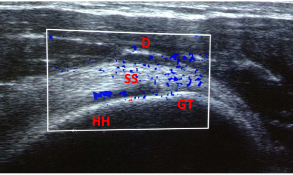
UltrasoundPower Doppler shoulder ultrasound in an athlete: intratendinous neovascularisation of the supraspinatus tendon, a characteristic ultrasound sign of tendinopathy.
Source : Notarnicola et al., BMC Research Notes, 2012, figure 2 · CC BY
Which questions should you ask to understand the patient and their history?
The history opens the whole line of reasoning. 📝 It must characterise the pain, identify the actionable risk factors and, above all, stratify irritability and the psychosocial profile, which will dictate the pace of loading.⁴- Precise site : ask the patient to point. Pain over the shoulder region, middle deltoid, anterolateral acromial area → consistent with RCRSP. Cervicoscapular pain, radiation into the upper limb with paraesthesiae → suspicion of cervical radiculopathy. Focal acromioclavicular pain → isolated AC involvement.
- Mode of onset : insidious over several weeks to months (typical of RCRSP) vs acute after clear trauma (a fall on the shoulder, traction on the arm) → suspicion of a traumatic tear or a glenohumeral lesion.
- Time course :
- Waking at night when rolling onto the shoulder → typical of RCRSP, frequent and disabling.
- Pain on starting to move, improving after warming up → consistent with RCRSP.
- Constant pain, at night without movement, deterioration in general condition → reconsider (a tumour, an infection, a systemic rheumatological process).
- Triggering movements : elevation above the head, rotations (putting on a belt, doing up a bra behind the back), hand behind the back, carrying a load with the arm elevated. These "patterns" point to RCRSP rather than to frozen shoulder (where the loss of passive mobility dominates).
- Recent changes in load : an intensification of manual work, overhead sport (thrower, swimmer, volleyball player, see chapter 4), moving house, painting, DIY, carrying a child.
- Medical history : age > 50, diabetes, hypertension, dyslipidaemia, smoking (Giri 2023).⁵
- Psychosocial profile : fear of movement, catastrophising, beliefs about pain ("my cuff is torn, so I am going to make it worse"), level of self-efficacy, major predictors of the prognosis (Chester 2018, Mallows 2017).⁶,⁷
- Baseline PROMs : SPADI (Shoulder Pain and Disability Index, 13 items, 0-100, MCID ≈ 8-10 points), DASH or QuickDASH, PSFS for the patient's target activities. They make it possible to quantify severity and to follow progress.
🚩 Red flags to screen for from the history onwards
- Clear trauma + complete functional impairment + the epaulette sign → suspicion of glenohumeral dislocation. An orthopaedic emergency.
- Trauma + marked weakness + complete loss of active elevation in a young person → suspicion of an acute massive tear of the cuff → prompt surgical work-up (Desmeules CPG 2025).¹
- Fever + hot swelling + exquisite tenderness → septic arthritis, an emergency.
- A progressive palpable mass, night sweats, unexplained weight loss, deterioration in general condition → oncological work-up (bone metastases, primary tumour, rare but to be considered).
- Paraesthesiae, motor deficit, absent reflexes in a root territory → cervical radiculopathy, thoracic outlet syndrome, plexopathy → neurological examination + cervical MRI.
- Atypical referred pain (the left shoulder in a cardiac patient, the right shoulder in a biliary or pulmonary one) → rule out projected visceral pain.
Which clinical tests should you run and which other conditions must be ruled out?
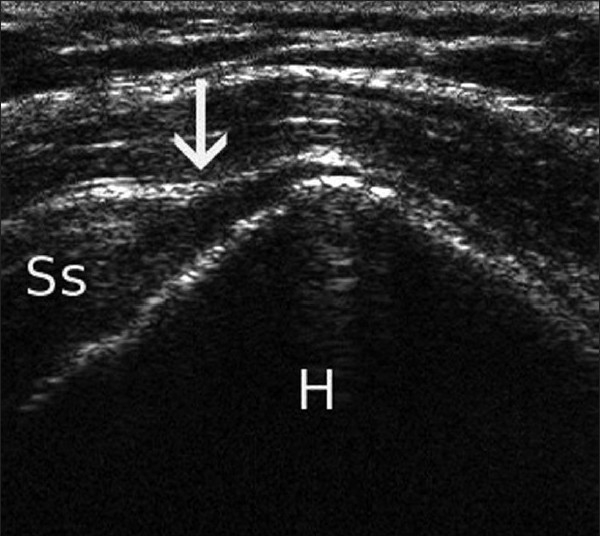
UltrasoundShoulder ultrasound: full-thickness tear of the supraspinatus tendon, arrow on the discontinuity above the humeral head.
Source : de Beer et al., International Journal of Shoulder Surgery, 2010, figure 1 · CC BY
- Painful Arc Sign (a painful arc between 60° and 120° of active abduction in the scapular plane) → a reasonable rule-in for RCRSP (the highest specificity among the usual tests).
- Hawkins-Kennedy test (90° flexion + passive internal rotation) → a reasonable rule-out (the highest sensitivity); a negative test lowers the probability of RCRSP.
| Clinical test | What it assesses | Performance (Hegedus 2012 / Alqunaee 2012) | 2025 JOSPT CPG recommendation |
|---|---|---|---|
| Painful Arc Sign | Reproduction of pain at 60-120° of active abduction | Sp ~ 80 %, Se ~ 53 % | Recommended (rule-in) |
| Hawkins-Kennedy | 90° flexion + passive internal rotation | Se ~ 80 %, Sp ~ 56 % | Recommended (rule-out) |
| Neer test | Maximal passive flexion | Se ~ 79 %, Sp ~ 53 % | Limited diagnostic value |
| Jobe / empty can | Supraspinatus test | Special tests are not discriminating | Not recommended for discrimination |
| Drop arm test | Suspicion of a major tear | Good specificity, low sensitivity | An adjunct if a tear is strongly suspected |
| Overall passive mobility | Telling RCRSP from a frozen shoulder | Preserved passive mobility = the diagnostic pivot | Essential |
| SPADI / DASH / PSFS (PROMs) | Overall severity, follow-up | Documented validity and responsiveness | Recommended for follow-up |
| Imaging (ultrasound / MRI) | Anatomical work-up as a second-line investigation | Weak symptom / image correlation (Girish 2011) | Not as a first line |
- Suspicion of an acute traumatic tear in a younger person with marked functional impairment.
- Failure of at least 6-12 weeks of structured conservative treatment with reassessment of the diagnosis.
- Suspicion of an alternative diagnosis (a tumour, a systemic rheumatological disorder, an acutely painful calcification).
- Preoperative work-up if surgery is being considered.
- Cervical radiculopathy (C5-C6) : the most frequent "mimic". Cervicoscapular pain radiating into the arm, sometimes paraesthesiae in the C5 territory (lateral aspect of the arm) or C6 (thumb, index finger). Spurling test, Lhermitte sign, examination of the reflexes (biceps, brachioradialis).
- Frozen shoulder (adhesive capsulitis) : loss of passive mobility (lateral rotation at 0° of abduction and passive elevation in particular) is the central criterion. Often associated with diabetes. It runs in 3 phases over 12-24 months.
- Glenohumeral instability : suspect it in a young person with a history of dislocation/subluxation. Apprehension tests, relocation test.
- Isolated acromioclavicular osteoarthritis : focal pain over the AC joint, pain on cross-body adduction, tenderness on direct palpation.
- Acute calcific tendinopathy : abrupt pain, sometimes extremely intense, without trauma. An anteroposterior radiograph in external rotation = an immediate diagnosis (dense calcium).
- Rheumatoid arthritis / spondyloarthritis : multi-joint involvement, prolonged morning stiffness, inflammatory markers.
- Projected visceral pathology : cardiac (left shoulder), biliary or diaphragmatic (right shoulder), apical pulmonary (Pancoast sign).
Should patients be classified, and along which axes?
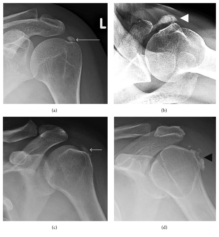
X-rayShoulder radiographs in four views: calcific deposits within the rotator cuff tendons, arrows on the calcifications of the supraspinatus, subscapularis, infraspinatus and teres minor, characteristic of calcific tendinopathy.
Source : Beckmann, Radiology Research and Practice, 2016, figure 8 · CC BY
- Irritability :
- High irritability : pain ≥ 7/10, frequent night waking, pain at rest, pain already on small movements. → First approach: education + analgesic modalities + sub-painful isometrics + low-load exercises. Short-term objective: modulate the irritability before progressing the load.
- Moderate irritability : pain 4-6/10, functional, triggered by moderate effort. → Progress to cuff strengthening and scapular stabilisation.
- Low irritability : pain ≤ 3/10, functional, triggered by high load only. → Progress to functional loading, eccentric exercises, return to the target activities.
- Psychosocial profile :
- A dominant psychosocial profile (high kinesiophobia, catastrophising, low self-efficacy, painful comorbidity, depression): pain education, a cognitive behavioural approach, possibly shared care with a psychologist alongside the motor programme (Chester 2018, Mallows 2017).⁶,⁷
- A dominant motor profile (a strength deficit, poor scapular control, intolerance of load with no yellow flags): a progressive strengthening programme centred on the cuff and the scapula.
Key points
- The diagnosis of RCRSP is essentially clinical. Imaging is not recommended as a first line (Desmeules 2025 JOSPT CPG, Dutch SAPS 2014).¹,²
- The 2025 JOSPT CPG retains only two useful tests : Painful Arc Sign (rule-in) and Hawkins-Kennedy (rule-out). The other historical tests (Neer, Jobe, drop arm taken in isolation) are not discriminating (Hegedus 2012, Hanchard 2013, Alqunaee 2012, Salamh & Lewis 2020).⁸,⁹,¹⁰,¹¹
- PROMs recommended: SPADI, DASH/QuickDASH, PSFS, at baseline and at every reassessment.
- Major differential diagnoses : cervical radiculopathy, frozen shoulder, glenohumeral instability, isolated AC osteoarthritis, acute calcific tendinopathy, projected visceral pathology.
- Red flags to screen for: clear trauma + impairment (dislocation, massive tear), fever + swelling (septic arthritis), a mass + deterioration + night sweats (oncological), neurological signs (radiculopathy, plexopathy).
- Stratify along 2 axes (an editorial synthesis, absent from the 2025 JOSPT CPG): irritability (high / moderate / low) and psychosocial profile. The content and the pace of the programme follow from them.
Bibliography
- Desmeules F, Roy JS, Lafrance S, et al. Rotator Cuff Tendinopathy Diagnosis, Nonsurgical Medical Care, and Rehabilitation: A Clinical Practice Guideline. J Orthop Sports Phys Ther. 2025;55(4):235-274. doi:10.2519/jospt.2025.13182.
- Diercks R, Bron C, Dorrestijn O, et al. Guideline for diagnosis and treatment of subacromial pain syndrome: Dutch Orthopaedic Association. Acta Orthop. 2014;85(3):314-322. PMID 24847788.
- Girish G, Lobo LG, Jacobson JA, et al. Ultrasound of the shoulder: asymptomatic findings in men. AJR Am J Roentgenol. 2011;197(4):W713-9. PMID 21940544.
- Lewis J. Rotator cuff related shoulder pain: Assessment, management and uncertainties. Man Ther. 2016;23:57-68. PMID 26882892.
- Giri A, O'Hanlon D, Jain NB. Risk factors for rotator cuff disease: A systematic review and meta-analysis of diabetes, hypertension, and hyperlipidemia. Ann Phys Rehabil Med. 2023;66(1):101631. PMC9974529.
- Chester R, Jerosch-Herold C, Lewis J, Shepstone L. Psychological factors are associated with the outcome of physiotherapy for people with shoulder pain: a multicentre longitudinal cohort study. Br J Sports Med. 2018;52(4):269-275. PMID 27445360.
- Mallows A, Debenham J, Walker T, Littlewood C. Association of psychological variables and outcome in tendinopathy: a systematic review. Br J Sports Med. 2017;51(9):743-748. PMID 27852585.
- Hegedus EJ, Goode AP, Cook CE, et al. Which physical examination tests provide clinicians with the most value when examining the shoulder? Br J Sports Med. 2012;46(14):964-978. PMID 22773322.
- Hanchard NCA, Lenza M, Handoll HHG, Takwoingi Y. Physical tests for shoulder impingements and local lésions of bursa, tendon or labrum that may accompany impingement. Cochrane Database Syst Rev. 2013;(4):CD007427. PMID 23633343.
- Alqunaee M, Galvin R, Fahey T. Diagnostic accuracy of clinical tests for subacromial impingement syndrome: a systematic review and meta-analysis. Arch Phys Med Rehabil. 2012;93(2):229-36. PMID 22289231.
- Salamh P, Lewis J. It Is Time to Put Special Tests for Rotator Cuff-Related Shoulder Pain out to Pasture. J Orthop Sports Phys Ther. 2020;50(5):222-225. PMID 32272031.
- Lewis J. The end of an era? J Orthop Sports Phys Ther. 2018;48(3):127-129. doi:10.2519/jospt.2018.0102.
What are the most effective treatment strategies for RCRSP?
Where do you start? What is the recommended hierarchy of interventions?
The 2025 JOSPT CPG Desmeules and the Dutch SAPS 2014 are aligned: two unavoidable pillars before any other intervention.⁶,⁷- Patient education and reassurance : deconstruct the "impingement" model (Cuff & Littlewood 2018), explain the not purely structural nature of the pain, set realistic expectations (recovery = 3-6 months for most, sometimes longer where there are psychosocial factors), normalise the presence of ordinary imaging abnormalities with age (Girish 2011, Yamamoto 2010).⁸
- A progressive exercise programme centred on the cuff and on scapular stabilisation: the cornerstone of treatment, strongly recommended as a first line (Pieters 2020), even though the placebo-controlled trials remain few and of low certainty (Naunton 2020).⁴,⁵
🔺 Therapeutic pyramid for RCRSP
From the most invasive (top) to the most universal (base): a hierarchy of intent
Sources: Beard DJ et al. CSAW Lancet 2018 (PMID 29169668); Paavola M et al. FIMPACT 5 years BJSM 2021 (PMID 33020137); Karjalainen Cochrane 2019 (PMID 30707445); Mohamadi 2017 (PMID 27469590); Naunton 2020 SR/MA; Pieters 2020 JOSPT update.
Where does exercise fit in, and is there a superior approach?
🏋️ Exercise is the best-documented active treatment in RCRSP, with a high level of evidence (Desmeules 2025 JOSPT CPG).⁶ The debate of the past decades has been about the best protocol : eccentric vs concentric, progressive vs non-progressive, high vs low load. The meta-analysis Naunton 2020 (Clin Rehabil, 7 trials, 468 participants) does not settle that debate, because it does not compare the protocols with each other: it sets progressive resisted exercise, and then non-progressive non-resisted exercise, separately against placebo or no treatment. The first achieves an uncertain clinical benefit on pain and function (15 points out of 100, 95 % CI 9 to 21, between 6 weeks and 6 months), the second no significant benefit (4 points, 95 % CI -2 to 9), with low certainty in both cases.⁴ The review Pieters 2020 (JOSPT update) strongly recommends exercise as a first line but concludes that the type, dose and duration of exercise remain to be established.⁵ Several practical principles emerge:- Cuff strengthening : abduction, external rotation (ER) and internal rotation (IR) in closed as well as open chain. Progression from sub-painful to a significant load.Sub-painful isometrics are useful in a phase of high irritability to modulate the pain without provoking a flare-up.
- Scapular stabilisation and motor control : activation of serratus anterior, of the scapular fixators (middle and lower trapezius, rhomboids), correction of dyskinesis, particularly in the overhead athlete (see chapter 4).
- Functional reintegration : exercises that integrate the shoulder into the whole kinetic chain, movements specific to the work or the sport, a gradual return to the target load.
- Dosage : 2 to 3 sessions a week for at least 8 to 12 weeks, movements within a tolerable zone (acceptable pain, modulated by irritability).Adherence is more decisive than the exact technique: a patient who keeps up a simple programme for 12 weeks will do better than a patient who abandons a complex one at 4 weeks.
⚖️ Effect of the active interventions in RCRSP
A qualitative synthesis: the effect against placebo or no treatment (Naunton 2020, Pieters 2020)
Sources: Naunton J et al. Clin Rehabil. 2020 (doi:10.1177/0269215520934147); Pieters L et al. JOSPT 2020 (doi:10.2519/jospt.2020.8498); Desjardins-Charbonneau A et al. JOSPT 2015 for manual therapy (doi:10.2519/jospt.2015.5455). A qualitative visualisation.
Adjunct therapies and surgery: what should be expected of them?
💡 The adjunct modalities must all be considered as a complement to exercise, never as a substitute. Manual therapy (glenohumeral and thoracic mobilisation, manipulation of the cervicothoracic region): the meta-analysis Desjardins-Charbonneau 2015 (JOSPT): an adjunct short-term effect on pain when combined with exercise. No superiority in the long term. It can make it easier to get a highly irritable patient moving.¹⁰ Subacromial corticosteroid injections : the meta-analysis Mohamadi 2017 (Clin Orthop Relat Res) quantified their effect precisely: a small relief, perceptible in the short term (4-8 weeks), which dies away by 6 months.⁹ A potential indication: a patient with high irritability in whom an analgesic window would allow active work to start. The risk: if the injection is presented as a "treatment", it can on the contrary delay active management and keep alive the purely biomedical model. NSAIDs and analgesics : reasonable relief in the short term, without changing the long-term prognosis. The 2025 JOSPT CPG mentions NSAIDs as an occasional option for managing a flare.⁶ ESWT (shockwave therapy) : the best-documented indication is in calcific tendinopathy of the cuff. For RCRSP without calcification, the additive effect on exercise is more debated. To be reserved for refractory chronic cases. Subacromial decompression surgery : this is the pivotal point of the decade. Three RCTs converge:- CSAW Beard 2018 (Lancet, n = 313, 32 UK hospitals, 3 arms: arthroscopic decompression vs diagnostic arthroscopy vs active monitoring): no clinically significant superiority of decompression over diagnostic arthroscopy, and a clinically marginal difference vs monitoring.¹
- FIMPACT Paavola 2018/2021 (BMJ then BJSM 5 years): sham-controlled, decompression vs diagnostic arthroscopy vs supervised exercise. At 5 years, none of the three interventions is superior to the others.²
- Cochrane Karjalainen 2019 (CD005619): a systematic review pooling the RCTs: it confirms the absence of a clinically important benefit of subacromial decompression vs control interventions.³
| Modality | Main indication | Level of evidence | Expected effect |
|---|---|---|---|
| Education + load modulation | All patients | High | ↗ adherence, ↘ recurrence, ↘ fear of movement |
| Progressive exercise (cuff + scapula) | All patients, 8-12+ weeks | High | The central pillar, Naunton 2020 and Pieters 2020 |
| Education + reassurance + load adaptation | All patients | High | The foundation. Deconstructing the "impingement" model (Lewis 2018) |
| Manual therapy (adjunct) | Short term, high irritability | Moderate | A small, short-term effect (Desjardins-Charbonneau 2015) |
| NSAIDs | An acute painful episode | Moderate in the short term | No effect on the long-term prognosis |
| Subacromial corticosteroid injection | A patient with high irritability | A small, transient effect | Dies away by 6 months (Mohamadi 2017) |
| ESWT | Calcific tendinopathy above all | Moderate if there are calcifications | More debated without calcification |
| Subacromial decompression (surgery) | — | Not superior to placebo | CSAW 2018, FIMPACT 2021, Cochrane 2019¹,²,³ |
| Surgical tendon repair | An acute traumatic tear in a young person | Selected cases | No clear superiority vs exercise (Cochrane Karjalainen 2019)¹¹ |
| Ultrasound / laser / TENS alone | A symptomatic adjunct | Low | Insufficient evidence |
Beyond the physical: how do you act on the psychosocial factors?
🧠 Psychosocial factors are a major determinant of the prognosis in RCRSP, and are frequently under-treated. The multicentre cohort Chester 2018 (n = 1030, BJSM) established that kinesiophobia, catastrophising and low self-efficacy are independent predictors of the outcome of physiotherapy at 6 and 12 months.¹² The review Mallows 2017 confirms these results for tendinopathy in general.¹³ Patient education must be active and proactive on four points:- Reframe the pain : the presence of an imaging abnormality is not "proof" that the pain will be permanent or that the tissue is going to "break". 96 % of asymptomatic men have ultrasound abnormalities (Girish 2011)¹⁴: a key message to deliver very early.
- Deconstruct the myths : "my cuff is catching and wearing out", "it needs an operation", "if I move I will make it worse", "rest heals the cuff", so many structural beliefs that keep kinesiophobia alive (Cuff & Littlewood 2018, a qualitative study).⁸
- Set realistic expectations : recovery = 3 to 6 months for most, sometimes longer where there are psychosocial factors, non-linear, with fluctuations to be expected.
- Self-efficacy : give the patient concrete tools (a written programme, PROM follow-up, simple criteria for adjusting the load) so that they become an actor in their recovery.
Critique and controversy
The "exercise protocol war" (pure eccentric vs progressive vs concentric-eccentric) has in fact never been settled for RCRSP: neither Naunton 2020 nor Pieters 2020 compares the protocols with each other, and Pieters concludes that the type, dose and duration of exercise remain to be documented.⁴,⁵ The commercial pressure around specific techniques (a "single protocol") owes more to marketing than to evidence. More problematic: despite three pivotal RCTs (CSAW, FIMPACT, Cochrane) showing the absence of superiority of subacromial decompression, the procedure is still widely performed in many health systems.¹,²,³ This "knowing-doing gap" is a major challenge of the coming decade, with far from negligible economic and iatrogenic-exposure consequences. The clinician must pass these data on to the patient to inform a shared decision (chapter 7). Finally, the "symptomatic patient vs abnormal imaging" dichotomy is still poorly accepted by some: a typical clinical presentation of RCRSP with a partial tear on ultrasound is not an indication for surgery in the short term: a fact that still sometimes contradicts common discourse.Key points
- ✅ First line : education + reassurance + a progressive exercise programme (cuff + scapula) over at least 8-12 weeks. A high level of evidence (2025 JOSPT CPG Desmeules).⁶
- ✅ No exercise protocol has been shown to beat another in RCRSP, for want of trials comparing them (Naunton 2020, Pieters 2020): what is documented is the benefit of progression and of resisted loading.⁴,⁵
- ✅ Manual therapy : a short-term adjunct only (Desjardins-Charbonneau 2015).¹⁰
- ⚠️ Corticosteroid injections : a small and transient effect (Mohamadi 2017). To be used sparingly, never as a standalone "treatment".⁹
- ❌ Subacromial decompression is not superior to placebo in non-traumatic RCRSP (CSAW Beard 2018 Lancet, FIMPACT Paavola 2021 BJSM 5 years, Cochrane Karjalainen 2019).¹,²,³
- ✅ Systematically address psychosocial factors : Chester 2018 (n = 1030), kinesiophobia and catastrophising are independent predictors of the prognosis.¹²
Bibliography
- Beard DJ, Rees JL, Cook JA, et al. Arthroscopic subacromial décompression for subacromial shoulder pain (CSAW): a multicentre, pragmatic, parallel group, placebo-controlled, three-group, randomised surgical trial. Lancet. 2018;391(10118):329-338. PMID 29169668.
- Paavola M, Malmivaara A, Taimela S, et al. Subacromial décompression versus diagnostic arthroscopy for shoulder impingement: a 5-year follow-up of a randomised, placebo surgery controlled clinical trial. Br J Sports Med. 2021;55(2):99-107. PMID 33020137.
- Karjalainen TV, Jain NB, Page CM, et al. Subacromial décompression surgery for rotator cuff disease. Cochrane Database Syst Rev. 2019;1(1):CD005619. PMID 30707445.
- Naunton J, Street G, Littlewood C, Haines T, Malliaras P. Effectiveness of progressive and resisted and non-progressive or non-resisted exercise in rotator cuff related shoulder pain: a systematic review and meta-analysis of randomized controlled trials. Clin Rehabil. 2020;34(9):1198-1216. doi:10.1177/0269215520934147.
- Pieters L, Lewis J, Kuppens K, et al. An Update of Systematic Reviews Examining the Effectiveness of Conservative Physical Therapy Interventions for Subacromial Shoulder Pain. J Orthop Sports Phys Ther. 2020;50(3):131-141. doi:10.2519/jospt.2020.8498.
- Desmeules F, Roy JS, Lafrance S, et al. Rotator Cuff Tendinopathy Diagnosis, Nonsurgical Medical Care, and Rehabilitation: A Clinical Practice Guideline. J Orthop Sports Phys Ther. 2025;55(4):235-274. doi:10.2519/jospt.2025.13182.
- Diercks R, Bron C, Dorrestijn O, et al. Guideline for diagnosis and treatment of subacromial pain syndrome: Dutch Orthopaedic Association. Acta Orthop. 2014;85(3):314-322. PMID 24847788.
- Cuff A, Littlewood C. Subacromial impingement syndrome — What does this mean to and for the patient? A qualitative study. Musculoskelet Sci Pract. 2018;33:24-28. PMID 29065348.
- Mohamadi A, Chan JJ, Claessen FM, et al. Corticosteroid Injections Give Small and Transient Pain Relief in Rotator Cuff Tendinosis: A Meta-analysis. Clin Orthop Relat Res. 2017;475(1):232-243. PMID 27469590.
- Desjardins-Charbonneau A, Roy JS, Dionne CE, et al. The Efficacy of Manual Therapy for Rotator Cuff Tendinopathy: A Systematic Review and Meta-Analysis. J Orthop Sports Phys Ther. 2015;45(5):330-350. doi:10.2519/jospt.2015.5455.
- Karjalainen TV, Jain NB, Heikkinen J, et al. Surgery for rotator cuff tears. Cochrane Database Syst Rev. 2019;12(12):CD013502. PMID 31813166.
- Chester R, Jerosch-Herold C, Lewis J, Shepstone L. Psychological factors are associated with the outcome of physiotherapy for people with shoulder pain: a multicentre longitudinal cohort study. Br J Sports Med. 2018;52(4):269-275. PMID 27445360.
- Mallows A, Debenham J, Walker T, Littlewood C. Association of psychological variables and outcome in tendinopathy: a systematic review. Br J Sports Med. 2017;51(9):743-748. PMID 27852585.
- Girish G, Lobo LG, Jacobson JA, et al. Ultrasound of the shoulder: asymptomatic findings in men. AJR Am J Roentgenol. 2011;197(4):W713-9. PMID 21940544.
- Lewis J. The end of an era? J Orthop Sports Phys Ther. 2018;48(3):127-129. doi:10.2519/jospt.2018.0102.
What is specific to the overhead athlete (thrower, swimmer, volleyball player)?
GIRD, scapular dyskinesis and the "thrower's shoulder" profile
The review by Wilk et al. 2002 (Am J Sports Med) and then 2013 (Int J Sports Phys Ther) describes precisely the chronic adaptations of the thrower's dominant shoulder:¹,²- An increase in external rotation at 90° of abduction (up to +15 to +20° vs the non-dominant side), allowing the cocking phase of the throw.
- A decrease in internal rotation at 90° of abduction: the well-known GIRD (Glenohumeral Internal Rotation Deficit). A GIRD is defined clinically as a difference > 20° between the dominant and the non-dominant side. Part of it is adaptive (humeral retroversion acquired during skeletal maturation), another part pathological (a stiff posterior capsule, accentuation of the posterosuperior internal impingement).
- Total ROM (TROM) : the sum of ER + IR must be comparable on the dominant and the non-dominant side (the rule of a < 5° difference). A loss of TROM is a warning sign: independently of the GIRD.
- Scapular dyskinesis : an altered scapulohumeral rhythm, reduced activation of serratus anterior, dominance of upper trapezius. The scapular dyskinesis test allows a reproducible visual assessment.³
- Strength deficits of the external rotators : a lowered ER/IR ratio (ER force/IR force), the classic target is > 0.66, or even > 0.75 for high-level throwing sports.
🎯 The typical GIRD of the thrower
Dominant vs non-dominant side, rotation at 90° of abduction
⚠️ Interpretation: GIRD > 20° = a warning sign. The loss of Total ROM (TROM > 5° between the two sides) is a more powerful risk factor than an isolated GIRD. Sources: Wilk KE et al. Am J Sports Med. 2002 (PMID 11799012); Manske R, Wilk KE et al. Int J Sports Phys Ther. 2013 (PMID 24175137).
Differential diagnoses specific to the overhead athlete
Shoulder pain in the overhead athlete is not limited to RCRSP. Several entities must be actively looked for:- SLAP lesion (Superior Labrum Anterior to Posterior): detachment of the superior labrum. In the thrower, a sensation of an internal "pop", pain in late cocking, a loss of throwing velocity. The O'Brien, Speed and Yergason tests have limited diagnostic value (chapter 2, Salamh & Lewis 2020). MR arthrography remains the reference examination if surgery is being considered.⁴
- Posteroinferior (subclinical multidirectional) instability : capsular laxity with chronic microsubluxation. Sulcus, posteroinferior apprehension and drawer tests. Often responsible in the competitive swimmer.
- Posterosuperior internal impingement (described by Walch): pathological contact of the supraspinatus tendon and the posterosuperior labrum in maximal abduction-external rotation (the cocking phase). It can be associated with a SLAP lesion and with a GIRD.
- Isolated tendinopathy of the long head of biceps : reproducible by palpating the bicipital groove, the Speed and Yergason tests.
- Anterosuperior subscapularis impingement : anterior pain, weakness in internal rotation against resistance, the belly-press sign.
Prevention in the overhead athlete: what do we know?
The review by Cools et al. 2015 (Braz J Phys Ther) brings together the main determinants of prevention:³- Specific strengthening of the scapular fixators : serratus anterior, middle and lower trapezius, rhomboids. Typical exercises: Y-T-W, push-up plus, dynamic hug, robbery (low rows).
- Strengthening of the external rotators : restore an ER/IR ratio > 0.66, isometric and concentric-eccentric exercises with a band or a pulley.
- Posterior capsular stretching : sleeper stretch, cross-body stretch, the structural effect is debated but they are widely used to correct a pathological GIRD.
- Monitoring of the throwing load : pitch count in the baseball pitcher (rules published by USA Baseball/MLB Pitch Smart by age), serve volume in tennis, weekly distance in the swimmer.
- Periodisation : development, competition and recovery phases, avoiding abrupt jumps in volume / intensity.
Specific criteria for return to overhead sport
🏈 The return to overhead sport after an episode of RCRSP must never be based on elapsed time alone. The function-based criteria derive from the Wilk 2013 and Cools 2015 recommendations:²,³- No pain at rest and during everyday movements.
- Isometric strength symmetry > 90 % vs the sound side (ER at 0° and 90° of abduction, IR, abduction).
- ER/IR ratio > 0.66 (preferably > 0.75 for the high-level thrower).
- Symmetrical TROM (a < 5° difference between the sides at 90° of abduction).
- Progressive tolerance of the target load : a progressive throwing/serving programme (interval throwing programme, return-to-serve programme), low volumes and controlled intensity to begin with, a gradual increase respecting a rule of acceptable pain and manageable fatigue.
- Restoration of the whole kinetic chain : a poor trunk-to-upper-limb transfer overloads the shoulder. The return to sport goes through assessment and work on the lower limbs, the core and energy transfer.
⭐ Key points
- The thrower's shoulder is an adapted shoulder: increased ER, decreased IR (GIRD), with a warning threshold of > 20° for the GIRD and > 5° for the loss of TROM (Wilk 2002, Wilk 2013).¹,²
- Critical differential diagnoses: SLAP, posteroinferior instability, posterosuperior internal impingement, biceps tendinopathy, anterosuperior impingement.⁴
- A prevention programme (Cools 2015): scapular fixators + external rotators + posterior stretching + load monitoring. Widely recommended in the young thrower.³
- Return to sport = functional criteria: strength > 90 %, ER/IR ratio > 0.66, symmetrical TROM, a progressive throwing programme. No "magic timetable".
- The imaging keeps its role in confirming or ruling out a clinically suspected SLAP, but must not replace a thorough clinical examination.
Bibliography
- Wilk KE, Meister K, Andrews JR. Current concepts in the rehabilitation of the overhead throwing athlete. Am J Sports Med. 2002;30(1):136-151. PMID 11799012.
- Wilk KE, Macrina LC, Cain EL, Dugas JR, Andrews JR. The recognition and treatment of superior labral (slap) lesions in the overhead athlete. Int J Sports Phys Ther. 2013;8(5):579-600. PMID 24175139.
- Cools AM, Johansson FR, Borms D, Maenhout A. Prévention of shoulder injuries in overhead athletes: a science-based approach. Braz J Phys Ther. 2015;19(5):331-339. PMID 26537804.
- Salamh P, Lewis J. It Is Time to Put Special Tests for Rotator Cuff-Related Shoulder Pain out to Pasture. J Orthop Sports Phys Ther. 2020;50(5):222-225. PMID 32272031.
- Desmeules F, Roy JS, Lafrance S, et al. Rotator Cuff Tendinopathy Diagnosis, Nonsurgical Medical Care, and Rehabilitation: A Clinical Practice Guideline. J Orthop Sports Phys Ther. 2025;55(4):235-274. doi:10.2519/jospt.2025.13182.
- Lewis J. Rotator cuff related shoulder pain: Assessment, management and uncertainties. Man Ther. 2016;23:57-68. PMID 26882892.
How do you ensure durable recovery and prevent recurrence?
How do you make the patient an actor in their recovery?
🎯 Structured self-management is one of the key principles defended by the 2025 JOSPT CPG Desmeules.² It rests on four concrete pillars:- Education about pain and about the not purely structural nature of RCRSP: imaging abnormalities are ordinary with age (96 % in asymptomatic men, Girish 2011; up to 20 % of tears after 60, Yamamoto 2010).³,⁴ This information alone reduces fear of movement in many patients (Cuff & Littlewood 2018, a qualitative study).⁵
- A written home exercise programme : 2 to 3 sessions a week, cuff and scapular stabilisation exercises, movements within a tolerable zone. Simplicity and repeatability come before complexity.
- Progressive load management : a return to everyday and work activities, the principle of graded exposure to the avoided movements. Applying the principles of the 2022 Lafrance CPG (Return to Work).⁶
- PROM follow-up : SPADI, DASH or PSFS every 4-6 weeks to objectify progress and disconnect the clinical decision from emotionally "bad days".
When and how do you plan a safe return to sport and to work?
🏋️ The return to activities (work, everyday life, sport) must be based on functional criteria and not on a preset timetable. The 2022 Lafrance CPG (JOSPT) offers specific recommendations for return to work in cuff disorders: a gradual return, adaptation of the workstation where possible, joint management with the employer and the occupational physician, support for occupational self-efficacy.⁶ General criteria to aim for before a full return to activity (a synthesis of the Desmeules 2025 CPG, Wilk 2002 for overhead):- Pain ≤ 2/10 at rest and during everyday activities (dressing, reaching a high shelf, sleeping on the side).
- Active range preserved or symmetrical (elevation in particular, rotations at 90° of abduction).
- Isometric strength symmetry > 90 % vs the sound side (abduction, ER, IR).
- Progressive tolerance of the target load : sustain the critical work or sporting movement for the usual duration without clear reproduction of the pain.
- A clinically significant improvement in the PROM (SPADI: a reduction of ≥ 8-10 points; DASH: a reduction of around ≥ 10 points).
- For the overhead athlete: additional specific criteria (ER/IR ratio > 0.66, symmetrical TROM, a throwing progression: chapter 4).
🚦 Flow chart: a function-based return to activity
A sequenced, criterion-based decision (vs calendar-based)
Adapted from the 2025 JOSPT CPG Desmeules recommendations (RCRSP)² and the 2022 JOSPT CPG Lafrance (Return to Work).⁶
Critique and controversy
The thresholds for return to activity (strength > 90 %, pain ≤ 2/10) are logical and derive from good practice, but their predictive value for non-recurrence at 1-2 years remains only moderately documented for RCRSP, less solidly than for the ACL, for example. Most studies measure the rate of return, not the rate of recurrence. In everyday practice, applying these criteria strictly takes time, tools (a dynamometer, PROMs) and training. The risk is that these criteria remain theoretical, and that the clinician allows the return on looser criteria (no pain at rest alone, which is very insufficient). Finally, the tension between self-management and support is subtle: too much autonomy without follow-up can lead to dropping out; too much support keeps dependence alive. The right balance depends on the patient's psychosocial profile (chapter 2).Key points
- Structured self-management is a key principle of the 2025 JOSPT CPG Desmeules: education + a written programme + progressive load management + PROM follow-up.²
- The psychosocial factors (kinesiophobia, catastrophising, self-efficacy) are independent determinants of the prognosis (Chester 2018, n = 1030).⁷
- The return to activity must be based on functional criteria : pain ≤ 2/10, strength > 90 %, PROM improvement ≥ MCID, progressive tolerance of the target load.
- The 2022 Lafrance CPG (JOSPT) offers specific recommendations for return to work in cuff disorders.⁶
- A realistic trajectory: improvement perceived at 4-6 weeks, a peak at 3-6 months, a return to everyday activities at ≈ 3 months, a return to competitive sport at 4-9 months. 40-50 % of patients remain symptomatic at 1 year without structured intervention (Kuijpers 2004).¹
Bibliography
- Kuijpers T, van der Windt DAWM, van der Heijden GJMG, Bouter LM. Systematic review of prognostic cohort studies on shoulder disorders. Pain. 2004;109(3):420-431. PMID 15157703.
- Desmeules F, Roy JS, Lafrance S, et al. Rotator Cuff Tendinopathy Diagnosis, Nonsurgical Medical Care, and Rehabilitation: A Clinical Practice Guideline. J Orthop Sports Phys Ther. 2025;55(4):235-274. doi:10.2519/jospt.2025.13182.
- Girish G, Lobo LG, Jacobson JA, et al. Ultrasound of the shoulder: asymptomatic findings in men. AJR Am J Roentgenol. 2011;197(4):W713-9. PMID 21940544.
- Yamamoto A, Takagishi K, Osawa T, et al. Prévalence and risk factors of a rotator cuff tear in the général population. J Shoulder Elbow Surg. 2010;19(1):116-120. PMID 19540777.
- Cuff A, Littlewood C. Subacromial impingement syndrome — What does this mean to and for the patient? A qualitative study. Musculoskelet Sci Pract. 2018;33:24-28. PMID 29065348.
- Lafrance S, Charron M, Roy JS, et al. Diagnosing, Managing, and Supporting Return to Work of Adults With Rotator Cuff Disorders: A Clinical Practice Guideline. J Orthop Sports Phys Ther. 2022;52(10):647-664. PMID 35881707.
- Chester R, Jerosch-Herold C, Lewis J, Shepstone L. Psychological factors are associated with the outcome of physiotherapy for people with shoulder pain: a multicentre longitudinal cohort study. Br J Sports Med. 2018;52(4):269-275. PMID 27445360.
- Mallows A, Debenham J, Walker T, Littlewood C. Association of psychological variables and outcome in tendinopathy: a systematic review. Br J Sports Med. 2017;51(9):743-748. PMID 27852585.
- Lewis J. Rotator cuff related shoulder pain: Assessment, management and uncertainties. Man Ther. 2016;23:57-68. PMID 26882892.
What do concrete clinical profiles teach us about RCRSP?
Profile 1: RCRSP with a classic presentation
The most frequent profile in practice is the adult patient (typically 45-65) with insidious pain over the shoulder region, triggered by movements in elevation and worse at night when rolling onto the shoulder, without initial trauma. Irritability is moderate : pain 4-6/10 on effort, functional at rest, sleep sometimes disturbed. Passive mobility is preserved, the clinical examination reproduces the pain (Painful Arc and Hawkins-Kennedy often positive, 2025 JOSPT CPG).¹Recommended management (aligned with Naunton 2020, Pieters 2020, Desmeules 2025):³,⁴,¹
- Education about the not purely structural nature of the pain, deconstructing the "impingement" model.
- A progressive exercise programme (cuff + scapula), 2-3 sessions a week, movements within a tolerable zone.
- PROM follow-up (SPADI or DASH) at 4-6 weeks to objectify progress.
- Adaptation of the occupational load if there is overhead exposure (2022 Lafrance CPG).⁵
Expected prognosis : a clinically perceptible improvement at 4-6 weeks, a return to everyday activities at 3 months for most. This profile corresponds to the target indication of standard evidence-based treatment, and to the epidemiological base of the SRs/MAs cited.
Profile 2: The cervical "mimic"
The most frequent diagnostic trap is C5-C6 cervical radiculopathy presenting as a "false" RCRSP. The patient describes pain over the shoulder region, but a more careful history reveals associated cervicoscapular pain, with paraesthesiae in the C5 territory (lateral aspect of the arm) or C6 (thumb, index finger), sometimes a subtle motor deficit. Shoulder movements can reproduce the pain atypically (pain on taking up a cervical position more than on pure shoulder movement). ⚠️ Salamh & Lewis 2020 (JOSPT) stress the importance of a systematic cervical examination before making the diagnosis of RCRSP.⁶Diagnostic keys :
- Spurling test, cervical distraction.
- Examination of the reflexes (biceps, brachioradialis), of dermatomal sensation, of segmental motor strength.
- Cervical imaging (MRI) if the suspicion is confirmed: neurosurgical or rheumatological referral according to context.
Not recognising this profile means treating a cuff that is not the source of the problem. The pain persists and the patient loses confidence in physiotherapy.
Profile 3: Acute calcific tendinopathy
The calcific tendinopathy of the cuff is a particular subtype. The typical presentation: shoulder pain of abrupt onset, sometimes with no trigger, of extreme intensity (pain "10/10", the shoulder impossible to move, an antalgic posture). The diagnosis is radiographic: an anteroposterior film in external and internal rotation shows a dense calcification in the area of the cuff tendons.Specific management :
- Acute phase: NSAIDs, analgesics, sometimes a local corticosteroid injection to manage the crisis (Mohamadi 2017, a small/transient effect but useful in these hyperalgesic presentations).⁷
- Second phase: an exercise programme similar to standard RCRSP.
- ESWT (shockwave therapy): this is l'the best-documented indication for ESWT in cuff pathology: several recent SRs/MAs show a superior effect on calcific resorption and on pain (moderate to strong evidence).
- Possibly ultrasound-guided needling or percutaneous lavage for refractory residual calcifications.
Profile 4: A dominant psychosocial profile
The most complex profile is that of a patient whose RCRSP sits within a dominant psychosocial context : high kinesiophobia, catastrophising, low self-efficacy, sometimes painful comorbidity (chronic low back pain, fibromyalgia), depression, anxiety. The multicentre cohort Chester 2018 (n = 1030 shoulder patients, 6-month follow-up) established that these variables explain a large part of the variance in outcomes: independently of the initial severity and of imaging.²The exercise programme alone is insufficient for this profile. Management requires:
- An in-depth pain education (the concepts of pain "modulated" by the nervous system, hypersensitivity vs tissue damage).
- An approach informed by cognitive behavioural therapy, as far as the physiotherapist's skills allow, or in shared care with a psychologist specialising in chronic pain.
- A graded exposure to the avoided movements, with explicit work on kinesiophobia (graded exposure).
- Realistic management of expectations: the trajectory is slower, fluctuations are expected, the prognosis depends more on how the psychosocial factors evolve than on structural parameters.⁸
🧩 The 4 typical RCRSP profiles
A simplified stratification derived from the 2025 JOSPT CPG Desmeules¹
An illustrative diagram: an editorial synthesis based on the 2025 JOSPT CPG¹ and on the data of Chester 2018² and Mallows 2017⁸. These profiles are not exclusive: the same patient can combine several traits (for example profile 1 + psychosocial traits).
Critique and controversy: where do case reports sit in the hierarchy of evidence?
🧠 The individual clinical cases are fundamental for generating hypotheses and flagging rare presentations, but their level of evidence is the lowest of the whole scientific hierarchy. A single case illustrates a possibility, not a generality. It cannot control for placebo, regression to the mean or spontaneous evolution. Publication bias is massive (failures are rarely published). That is why we have preferred here to work with typical profiles anchored in the aggregated literature, rather than presenting fictional or anecdotal individual cases.📐 Hierarchy of scientific evidence: where does each type of study belong?
Strength of evidence decreasing from the top (meta-analyses) towards the bottom (isolated cases)
A simplified GRADE / Oxford CEBM hierarchy. The length of the coloured bar on the right illustrates the relative strength of evidence. Practical implication: where a seductive case report and a meta-analysis diverge, the decision must follow the meta-analysis. Case reports remain valuable for generate hypotheses, flagging rare presentations, or illustrating a line of clinical reasoning.
⭐ Key points
- Four typical RCRSP profiles met in practice: (1) a classic presentation, (2) a C5-C6 cervical mimic, (3) acute calcific tendinopathy, (4) a dominant psychosocial profile.
- Profile 1 (the most frequent) responds well to education + progressive exercises (Naunton 2020, Pieters 2020, Desmeules 2025).³,⁴,¹
- Profile 2 (the cervical mimic) must be ruled out by a systematic cervical examination before making the diagnosis of RCRSP (Salamh & Lewis 2020 JOSPT).⁶
- Profile 3 (acute calcific) benefits from acute-phase analgesia (possibly a corticosteroid injection, Mohamadi 2017) and from ESWT (recent SR/MA evidence).⁷
- Profile 4 (dominant psychosocial) requires a biopsychosocial approach and CBT: an independent predictor of the prognosis (Chester 2018 n = 1030).²,⁸
- ⚠️ Level of evidence : an individual case report = level 5 (the weakest). When they diverge, follow the meta-analyses (level 1a), not the isolated case. The typical profiles illustrate, they do not demonstrate anything.
Bibliography
- Desmeules F, Roy JS, Lafrance S, et al. Rotator Cuff Tendinopathy Diagnosis, Nonsurgical Medical Care, and Rehabilitation: A Clinical Practice Guideline. J Orthop Sports Phys Ther. 2025;55(4):235-274. doi:10.2519/jospt.2025.13182.
- Chester R, Jerosch-Herold C, Lewis J, Shepstone L. Psychological factors are associated with the outcome of physiotherapy for people with shoulder pain: a multicentre longitudinal cohort study. Br J Sports Med. 2018;52(4):269-275. PMID 27445360.
- Naunton J, Street G, Littlewood C, Haines T, Malliaras P. Effectiveness of progressive and resisted and non-progressive or non-resisted exercise in rotator cuff related shoulder pain: a systematic review and meta-analysis of randomized controlled trials. Clin Rehabil. 2020;34(9):1198-1216. doi:10.1177/0269215520934147.
- Pieters L, Lewis J, Kuppens K, et al. An Update of Systematic Reviews Examining the Effectiveness of Conservative Physical Therapy Interventions for Subacromial Shoulder Pain. J Orthop Sports Phys Ther. 2020;50(3):131-141. doi:10.2519/jospt.2020.8498.
- Lafrance S, Charron M, Roy JS, et al. Diagnosing, Managing, and Supporting Return to Work of Adults With Rotator Cuff Disorders: A Clinical Practice Guideline. J Orthop Sports Phys Ther. 2022;52(10):647-664. PMID 35881707.
- Salamh P, Lewis J. It Is Time to Put Special Tests for Rotator Cuff-Related Shoulder Pain out to Pasture. J Orthop Sports Phys Ther. 2020;50(5):222-225. PMID 32272031.
- Mohamadi A, Chan JJ, Claessen FM, et al. Corticosteroid Injections Give Small and Transient Pain Relief in Rotator Cuff Tendinosis: A Meta-analysis. Clin Orthop Relat Res. 2017;475(1):232-243. PMID 27469590.
- Mallows A, Debenham J, Walker T, Littlewood C. Association of psychological variables and outcome in tendinopathy: a systematic review. Br J Sports Med. 2017;51(9):743-748. PMID 27852585.
- Girish G, Lobo LG, Jacobson JA, et al. Ultrasound of the shoulder: asymptomatic findings in men. AJR Am J Roentgenol. 2011;197(4):W713-9. PMID 21940544.
- Yamamoto A, Takagishi K, Osawa T, et al. Prévalence and risk factors of a rotator cuff tear in the général population. J Shoulder Elbow Surg. 2010;19(1):116-120. PMID 19540777.
- Lewis J. Rotator cuff related shoulder pain: Assessment, management and uncertainties. Man Ther. 2016;23:57-68. PMID 26882892.
- Beard DJ, Rees JL, Cook JA, et al. Arthroscopic subacromial décompression for subacromial shoulder pain (CSAW). Lancet. 2018;391(10118):329-338. PMID 29169668.
How do you apply these recommendations concretely in your practice?
When, and to which other health professionals, should you refer?
The first non-negotiable step in triage is screening for red flags. The clinician must have a low threshold for referral: sensitivity takes precedence over specificity when serious conditions are at stake.🚩 Red flags specific to the shoulder
- Glenohumeral dislocation : clear trauma + the epaulette sign + complete functional impairment + intense pain. An orthopaedic emergency.
- An acute traumatic massive tear in a relatively young person: trauma + marked weakness + complete loss of active elevation + pain. A prompt surgical work-up (2025 JOSPT CPG).¹
- Infection (septic arthritis) : fever + hot swelling + exquisite tenderness + erythema. An emergency.
- A tumour (rare but to be considered): a progressive palpable mass, night pain without movement, night sweats, unexplained weight loss, deterioration in general condition. An oncological work-up.
- Cervical radiculopathy / plexopathy : radicular pain in a territory, paraesthesiae, a motor or sensory deficit, an absent reflex → neurological examination + cervical MRI.
- Projected visceral pain : cardiac (the left shoulder above all), biliary or diaphragmatic (the right shoulder), apical pulmonary (Pancoast). Adapt the general examination if the presentation is atypical.
- Systemic rheumatological disease : rheumatoid arthritis, spondyloarthritis, polymyalgia rheumatica (an older patient, shoulder + pelvic girdle pain, raised ESR/CRP).
⚠️ Any red flag → prompt medical referral (general practitioner, emergency department, sports physician, orthopaedic surgeon) before, or alongside, physiotherapy management.
- General practitioner / sports physician : an overall work-up, management of metabolic comorbidity (diabetes, hypertension, dyslipidaemia, Giri 2023), blood tests if a rheumatological disorder is suspected.
- Rheumatologist : suspicion of a systemic inflammatory disorder, multi-joint involvement.
- Orthopaedic shoulder surgeon : an acute traumatic massive tear, documented failure of 6-12 weeks of structured conservative treatment, a surgical request from the patient after being informed of the evidence-based data.
- Psychologist specialising in chronic pain : a dominant psychosocial profile (chapter 6, profile 4), high kinesiophobia, psychiatric comorbidity.
- Occupational physician / ergonomist : adaptation of the workstation where there is overhead exposure (2022 Lafrance CPG).³
- Strength and conditioning coach for the overhead athlete: reviewing the throwing load, periodisation, the progression of the return to sport (chapter 4).
How do you measure outcomes and overcome barriers to implementation?
📊 Measuring outcomes is the sine qua non of quality practice. Several validated PROMs exist for RCRSP, each with its strengths and its limits:⁴📋 PROMs for RCRSP: a functional comparison
Choosing according to the clinical context (community physiotherapy, research, follow-up)
⚠️ A practical tip: in community physiotherapy, SPADI + PSFS are amply enough to follow RCRSP. The Constant-Murley is reserved for specialist settings (shoulder surgery, research). DASH/QuickDASH = the standard for whole upper limb disorders.
- Digital tools : a tablet or smartphone app that the patient fills in in the waiting room, automatic scoring, graphical display of progress.
- Continuing education : practical workshops on using PROMs and on shared decision-making.
- Clinical champions : opinion leaders in the practice or the clinic who spread the use of EBP.
- Building it into the routine : SPADI / PSFS at every follow-up consultation (every 4-6 weeks), not only at the start and the end of care.
- Shared decision-making (shared decision-making, Hoffmann 2014 JAMA)⁵: present the patient with the options and their level of evidence (evidence-based progressive exercise, surgical decompression not superior to placebo, a corticosteroid injection with a small and transient effect), and discuss them in the light of their preferences and their timetable. It significantly increases adherence and satisfaction. ✅
Critique and controversies: beyond the guidelines
The red flag paradox still holds: no sign taken in isolation has a high positive predictive value. It is the convergence of evidence plus probabilistic clinical reasoning that guides referral, not a checklist. The risk is twofold: over-diagnosing (sending every painful tendon for MRI) or under-diagnosing (missing a reduced dislocation or an acute traumatic tear by putting the pain down to a simple RCRSP). The "knowing-doing" gap is particularly striking in RCRSP: the SR/MA Naunton 2020 and the SR Pieters 2020 have placed progressive exercise in the first line for 5 years, the rejection of subacromial decompression as superior to placebo has been confirmed by 3 pivotal RCTs since 2018-2021, and yet practice resists.⁶,⁷,⁸,⁹ Moving to genuine EBP requires more than the diffusion of information: it requires an overhaul of the economic incentives and of continuing education. Finally, the standardisation versus personalisation tension is central. PROMs and CPGs are essential for comparability and safety, but the clinician must be able to navigate between standardised tools and individualisation (the patient's real needs). Stratification by irritability and psychosocial profile proposed in chapter 2, which is an editorial synthesis and not a recommendation, is one way of reconciling these two demands.⭐ Key points
- Referral is crucial for safety : red flags (dislocation, an acute massive tear, infection, oncological, radiculopathy, projected pain) → prompt medical referral.
- Psychosocial yellow flags (kinesiophobia, catastrophising, Chester 2018 n = 1030)² → collaboration with a psychologist, a biopsychosocial approach.
- Measure outcomes with validated PROMs: SPADI (short, shoulder-specific, MCID 8-10 pts), DASH/QuickDASH (whole upper limb), Constant-Murley (orthopaedic), PSFS (patient-centred).⁴
- The shared decision-making (Hoffmann 2014 JAMA)⁵, informing the patient of the evidence-based data (exercise = the first line; decompression surgery not superior to placebo; corticosteroids a small transient effect), increases adherence and satisfaction.
- For refractory cases after 3-6 months: reconsider the diagnosis (targeted imaging, cervical examination), discuss with a sports physician or a surgeon according to context.
- The "knowing-doing gap" is the major challenge of the coming decade: bringing into everyday practice the evidence accumulated since 2017-2025 (CSAW Beard 2018, FIMPACT Paavola 2021, 2025 JOSPT CPG Desmeules).⁶,⁷,¹
Bibliography
- Desmeules F, Roy JS, Lafrance S, et al. Rotator Cuff Tendinopathy Diagnosis, Nonsurgical Medical Care, and Rehabilitation: A Clinical Practice Guideline. J Orthop Sports Phys Ther. 2025;55(4):235-274. doi:10.2519/jospt.2025.13182.
- Chester R, Jerosch-Herold C, Lewis J, Shepstone L. Psychological factors are associated with the outcome of physiotherapy for people with shoulder pain: a multicentre longitudinal cohort study. Br J Sports Med. 2018;52(4):269-275. PMID 27445360.
- Lafrance S, Charron M, Roy JS, et al. Diagnosing, Managing, and Supporting Return to Work of Adults With Rotator Cuff Disorders: A Clinical Practice Guideline. J Orthop Sports Phys Ther. 2022;52(10):647-664. PMID 35881707.
- Diercks R, Bron C, Dorrestijn O, et al. Guideline for diagnosis and treatment of subacromial pain syndrome: Dutch Orthopaedic Association. Acta Orthop. 2014;85(3):314-322. PMID 24847788.
- Hoffmann TC, Montori VM, Del Mar C. The connection between evidence-based medicine and shared décision making. JAMA. 2014;312(13):1295-1296. PMID 25268434.
- Beard DJ, Rees JL, Cook JA, et al. Arthroscopic subacromial décompression for subacromial shoulder pain (CSAW). Lancet. 2018;391(10118):329-338. PMID 29169668.
- Paavola M, Malmivaara A, Taimela S, et al. Subacromial décompression versus diagnostic arthroscopy for shoulder impingement: a 5-year follow-up. Br J Sports Med. 2021;55(2):99-107. PMID 33020137.
- Naunton J, Street G, Littlewood C, Haines T, Malliaras P. Effectiveness of progressive and resisted and non-progressive or non-resisted exercise in rotator cuff related shoulder pain: a systematic review and meta-analysis of randomized controlled trials. Clin Rehabil. 2020;34(9):1198-1216. doi:10.1177/0269215520934147.
- Pieters L, Lewis J, Kuppens K, et al. An Update of Systematic Reviews Examining the Effectiveness of Conservative Physical Therapy Interventions for Subacromial Shoulder Pain. J Orthop Sports Phys Ther. 2020;50(3):131-141. doi:10.2519/jospt.2020.8498.
- Giri A, O'Hanlon D, Jain NB. Risk factors for rotator cuff disease: A systematic review and meta-analysis of diabetes, hypertension, and hyperlipidemia. Ann Phys Rehabil Med. 2023;66(1):101631. PMC9974529.
- Karjalainen TV, Jain NB, Page CM, et al. Subacromial décompression surgery for rotator cuff disease. Cochrane Database Syst Rev. 2019;1(1):CD005619. PMID 30707445.
- Lewis J. Rotator cuff related shoulder pain: Assessment, management and uncertainties. Man Ther. 2016;23:57-68. PMID 26882892.
What next after reading this?
This article is part of a collection of evidence-based clinical syntheses. A question, a comment, a correction to suggest? Contact us directly through the WhatsApp button at the bottom right of the screen.

