


















In brief
The question that occupied twenty years of literature was not the right one. For a long time “immobilise” was set against “mobilise early”, whereas the dividing line that matters separates passive movement from active movement. For early passive motion, twenty trials and 1,841 patients find no increase in the risk of retear, with an odds ratio of 1.05 (Mazuquin 2021): passive mobilisation gains a few degrees of range without costing healing. For early active movement, the same team measures a relative risk of structural failure of 1.52 beyond 3 cm of tear and of 2.74 for suture-bridge repairs (Kluczynski 2016). A second shift, less well known: the risk window is not the sling window. Retears occur on average at 19.2 weeks, mainly between the sixth and the twenty-sixth (Iannotti 2013), that is to say during the rehabilitation sessions, not during immobilisation. Third: there is no clinical practice guideline on the subject. The only framework available is a 2016 expert consensus, and the protocols actually applied all depart from it, in the direction of greater aggressiveness (Galetta 2021).
This synthesis is addressed to the physiotherapist who receives a shoulder operated on for a cuff repair and must decide, session after session, what may be mobilised, what must be protected, and what can honestly be announced. It deals neither with the surgical indication nor with the unoperated tendon: it begins once the decision has been made and the construct is in place.
That is also what sets it apart from the two pages the site already devotes to the cuff, and it should be said straight away so that everyone goes to the right place. Rotator cuff tendinopathy and subacromial pain deals with the painful but unruptured tendon, the one that is not repaired. Symptomatic (degenerative) rotator cuff tear deals with the torn tendon and the decision whether or not to operate, with what first-line rehabilitation achieves without surgery. This page begins where the second one stops: afterwards. Neither of the two contains the word “immobilisation”, or “retear”, or “passive mobilisation”, and that is normal: these are postoperative questions, and they have their own body of literature.
Three figures that frame the subject
French operative volume, the real timing of retear risk, and the horizon for return to work.
Sources: Villatte 2020 (PMID 32863170), Iannotti 2013 (PMID 23780533), Haunschild 2021 (PMID 33395315). The French figure comes from the PMSI hospital database and not from a registry: it counts coded procedures, not patients followed up.
Clinical synthesis
- Passive and active do not raise the same question. Early passive mobilisation does not increase the risk of retear: odds ratio 1.05 (95 % CI 0.64 to 1.75) across twenty trials and 1,841 patients (Mazuquin 2021). Early active movement, by contrast, is associated with a relative risk of structural failure of 1.52 beyond 3 cm, and of 2.74 for suture-bridge repairs (Kluczynski 2016). The shorthand “mobilise early” erases exactly the distinction that matters.
- What early motion gains is real but small. Fifteen degrees of flexion at three months, about five at six and twelve months (Riboh 2014). In the most recent trials the differences are of the order of one to three degrees at one year (Mazuquin 2021), and the most cautious analysis concludes that these differences do not reach the threshold of clinical relevance (Silveira 2021). That is not a reason to do nothing: it is a reason not to sell early motion as an acceleration of recovery.
- Biology imposes a longer deadline than the sling does. In the only animal model with a true rotator cuff, Sharpey's fibres do not appear in appreciable numbers before twelve weeks and the tendon-to-bone junction is still not mature at fifteen (Sonnabend 2010). In humans, retears occur on average at 19.2 weeks, with a linear increase up to the twenty-sixth (Iannotti 2013), and a series of 638 arthroscopies places 46.3 % of them between the sixth week and the third month (Yamaura 2023).
- Three weeks in a simple sling are no worse than six weeks in a brace. Non-inferiority trial in 120 patients with a small to medium tear: WORC of 83 % against 87 % at one year, and 89 % healing on MRI in both groups (Jenssen 2018). The abduction brace adds nothing to the simple sling across four trials and 302 patients (Chen 2023).
- The available schedule is a consensus, not a guideline. Two weeks of strict immobilisation, protected passive range from the second to the sixth week, active range thereafter, progressive strengthening from the twelfth (Thigpen 2016). No clinical practice guideline validates it, and the 66 protocols published online all depart from it in the direction of greater aggressiveness (Galetta 2021).
- Stiffness is the symmetrical risk, and it is more common than people think. From 4.9 % of stiffness leading to a capsular release (Huberty 2009) to 14.2 % of patients outside the mobility thresholds at three months (Cho 2022). The factors that recur are diabetes, female sex, partial tear, and, in a French series, preoperative anxiety (Aïm 2022). It is stiffness, not retear, that moved practice towards early motion.
- A retorn cuff is not failed rehabilitation. The weighted mean rate is 26.6 % at 23.7 months across more than 8,000 shoulders, and patient-reported outcomes improve whether or not integrity is restored (McElvany 2015). The score gap between a healed cuff and a retorn cuff reaches the threshold of clinical relevance for the ASES, not for the Constant (Haque 2018).
- What is announced has to be counted in months, not in weeks. The minimal clinically important benefit is reached around the sixth month (Manderle 2020), return to the previous level of work at 8.15 months for 62.3 % of patients (Haunschild 2021), and the second year adds no more than two to three score points, below the relevance threshold (Sahoo 2022).
What exactly was repaired, and why the operative report governs what follows
A cuff repair is not a single procedure: it is a family of procedures, each of which constrains the first session differently. The tendon repaired, its size, the technique and the associated procedures are not technicians' details, they decide what is allowed.
A common procedure, and one that is becoming more so
Cuff surgery is the leading reason for shoulder surgery, and the shoulder is the third area of orthopaedic activity after the hip and the knee. Analysis of the French PMSI hospital database counts 234,612 primary shoulder surgery procedures in 2018, of which 173,799 for the rotator cuff, that is 74 % of the total; among them, 61,055 were tendon repairs proper. Activity rose by 24.5 % between 2012 and 2018, and the projection scenarios give an increase of between 13.6 % and more than 300 % by 2050 depending on the assumption chosenPMID 32863170. The phenomenon is not a French one: the Finnish national registry had already documented a clear rise in the incidence of cuff repairsPMID 26265152, and an American projection on Medicare beneficiaries aged 65 years and over predicts a doubling of arthroscopic repairs by 2050PMID 42024215.
This volume has a practical consequence: the private-practice physiotherapist sees these shoulders regularly, and sees them without any shared protocol. That is the subject of chapter 5.
Shoulder surgery in France, and the share taken by the cuff
Primary shoulder surgery procedures coded in the PMSI in 2018, and the change measured over 2012-2018.
Source: Villatte 2020, Orthopaedics & Traumatology: Surgery & Research, analysis of the PMSI 2012-2018 with projection to 2070 (PMID 32863170).
Five lines of the operative report that change the session
The first assessment of an operated shoulder begins with a reading, not with a test. Five items concretely change what is allowed, and none of them can be guessed from the examination.
Which tendons were repaired. This is the most often neglected point, and the one with the heaviest consequences. A repair of the subscapularis is not rehabilitated like an isolated supraspinatus repair: forced passive external rotation puts the suture under tension, and active internal rotation loads it directly. The instruction therefore differs in both directions, in range as in activation, and the patient does not know it.
The size and retraction of the tear. This is the factor that decides how cautious to be, and the literature shows it very reproducibly: it is beyond 3 cm, and still more beyond 5, that early motion ceases to be neutral (chapter 3). A massive tear, marked retraction or advanced fatty infiltration change the conversation.
The repair technique. Single row, double row, suture bridge: the meta-analysis of structural failures shows that the risk associated with early motion is not the same for every constructPMID 25943112. The physiotherapist does not have to judge the technique: they have to know which one they are protecting.
The associated procedures. Tenodesis or tenotomy of the long head of biceps, acromioclavicular resection, labral repair, acromioplasty. They are not neutral for the timetable or for the prognosis: biceps tenodesis is associated with reaching the clinically important benefit threshold later, whereas resection of the distal end of the clavicle is associated with reaching it earlierPMID 33079576. The site also covers long head of biceps tendinopathy and labral tears in their own right.
The surgeon's named instructions. They take precedence over any general rule, including those in this article. Between-surgeon variability is such that it has become a subject of study in its own rightPMID 32386779: if there is a disagreement, you call, you do not decide alone.
| What the operative report says | What that changes in the session | What to ask for if the information is missing |
|---|---|---|
| Tendons repaired, subscapularis included | A subscapularis suture contraindicates forced passive external rotation and early active internal rotation. An isolated posterosuperior repair does not impose that limit. | The exact tendon, and the permitted passive external rotation limit in degrees. |
| Size and retraction | Beyond 3 cm, early active motion is no longer neutral. Beyond 5 cm, even early passive motion is no longer documented as safe. | The size in centimetres, or at least the category small / medium / large / massive. |
| Repair technique | The excess risk of early active motion differs between transosseous, single row and suture bridge. | The type of construct, and the number of anchors. |
| Associated procedures | A biceps tenodesis lengthens the time to clinically important benefit; an acromioclavicular resection shortens it. | The complete list of procedures, and the precautions specific to each. |
| Tissue quality and fatty infiltration | It does not change today's session, but it changes the prognosis and therefore what is announced. | The preoperative fatty infiltration stage if it was graded. |
| The surgeon's explicit instructions | They take precedence over any general rule, including consensus statements. | The prescribed sling duration, the permitted ranges, the start date for active work. |
Key points from this chapter
- The cuff accounts for 74 % of primary shoulder surgery in France, with 61,055 tendon repairs coded in 2018 and shoulder activity up 24.5 % over six years.
- The operative report is the first item of the assessment, before any test.
- The tendon repaired changes the instructions in both directions: subscapularis limits passive external rotation and active internal rotation.
- Tear size is the factor that decides how cautious to be, far more than the time since surgery.
- The surgeon's named instruction takes precedence over any general rule.
How long does the repaired tendon take to hold on its own?
All postoperative rehabilitation follows from a single fact: the speed at which the tendon-to-bone junction becomes solid again. That fact runs slower than the sling, and the gap between the two is the source of most scheduling errors.
What biology says, and what it does not say
The rotator cuff, in the strict anatomical sense, exists in practice only in higher primates, which limits the animal models available. The reference study repaired lesions in eight middle-aged baboons, sacrificed in pairs at four, eight, twelve and fifteen weeks. At four weeks, bone-to-tendon healing was immature. At eight weeks, the repair looked healed macroscopically, but the Sharpey's fibres that actually hold the assembly together did not appear in appreciable numbers before twelve weeks. At fifteen weeks, the bone-to-tendon junction was almost, but not quite, mature. The authors draw an explicit conclusion for humans: protect the repair for at least twelve to fifteen weeksPMID 20357340.
This result is solid and it is limited, and both have to be held together. Solid, because it is histological and because it concerns the only relevant model. Limited, because eight animals do not give a distribution, and because a baboon has neither diabetes, nor smoking, nor fatty infiltration. It therefore does not prescribe a timetable: it gives the order of magnitude, and that order of magnitude is three months, not six weeks.
The duration of immobilisation and the duration of healing do not coincide. The sling comes off at six weeks; the tendon-to-bone junction is not mature before fifteen. Most of the risk window therefore takes place in the clinic, not at home.
In humans: when do repairs actually fail?
The question received a direct answer in a prospective multicentre study: 113 patients operated on for a 1 to 4 cm full-thickness tear, with sequential MRI at six intervals, from the second to the fifty-second week. Nineteen retears were diagnosed within the year, that is 17 %. The mean time was 19.2 weeks, with a linear increase in the number of retears over the first twenty-six weeks, then a single further retear between the twenty-sixth and the fifty-secondPMID 23780533. The authors summarise it as follows: retears occur mainly between six and twenty-six weeks, and a substantial number between twelve and twenty-six.
A Japanese series of 638 consecutive arthroscopies, with MRI at six weeks, three months, six months and one year, sets out the distribution of the 41 retears observed: 22.0 % within the first six weeks, 46.3 % between six weeks and three months, 26.8 % between three and six months, and only 4.9 % between six months and one year. Early retears were larger than late onesPMID 36842463. The meta-analysis of 31 level 1 and 2 studies gives consistent orders of magnitude by follow-up window: 15 % at three months, 21 % between three and six months, 16 % between six and twelvePMID 34465332.
One nuance that binary imaging hides has to be added. A repair can fail without a hole: the tendon lengthens and moves relative to its insertion while remaining in continuity. A study using radio-opaque markers documented this “failure with continuity”, which MRI readily classifies as healedPMID 23019253. That does not change management, but it explains why a patient with reassuring imaging can remain weak.
Two timelines that do not coincide
Histological maturation of the tendon-to-bone junction in the primate model, and the real distribution of retears in humans.
Sources: Sonnabend 2010 (PMID 20357340) for histological maturation, Yamaura 2023 (PMID 36842463) for the distribution of retears. The two scales are not superimposable: one describes tissue in eight animals, the other a clinical series of 638 shoulders. They are placed side by side to show that the risk window extends beyond the immobilisation period, not to establish causality.
Key points from this chapter
- In the only animal model with a true cuff, the tendon-to-bone junction is not mature before twelve to fifteen weeks, even though it looks healed to the eye from eight.
- In humans, retears occur on average at 19.2 weeks, with a linear progression up to the twenty-sixth.
- Nearly half of them are concentrated between the sixth week and the third month, that is to say during active rehabilitation.
- A repair can fail while remaining in continuity: the tendon lengthens without a hole appearing, and imaging then classifies it as healed.
- Practical consequence: vigilance does not stop when the sling comes off, it is only just beginning.
Strict immobilisation or early passive mobilisation: what has the literature actually shown?
This is the question that occupied the field for twenty years, and the answer has changed. Not in the way people think: it is not “early motion won”, it is “that was not the right question”.
The starting point: caution inherited, not measured
The historical position was to immobilise strictly for four to six weeks, for fear of making the suture fail. That caution was not absurd: it followed directly from what the histology in the previous chapter shows. It had simply never been compared with anything else.
The first series of meta-analyses compared early passive motion, started in the first week, with strict immobilisation. The result, remarkably stable from one synthesis to the next, fits into two sentences: early passive motion gains range in the short term, and it does not cost healing. The meta-analysis of five randomised trials finds 14.70 degrees more forward flexion at three months (95 % CI 5.52 to 23.87; p = 0.002), 4.31 degrees at six months and 4.18 at twelve, external rotation greater by 10.43 degrees at the third month only, and retear rates that do not differ at one year: 16.3 % after immobilisation against 21.1 % after early passive motion (relative risk 0.82; 95 % CI 0.57 to 1.20; p = 0.31)PMID 24813324. Two other meta-analyses from the same period conclude in the same directionPMID 25127908PMID 25143489.
The most recent and largest synthesis confirms and refines this. Twenty randomised trials, 1,841 patients: no significant difference in pain or in function, except on the SANE self-assessment scale at six months (mean difference 6.54; 95 % CI 2.24 to 10.84). On range of motion, significant differences but ones that melt away over time: 7.36 degrees of flexion at six weeks, 8.45 at three months, 3.57 at six months, 1.42 at one year. And on repair integrity, an odds ratio of 1.05 (95 % CI 0.64 to 1.75), that is to say nothingPMID 34048450.
The shift that a quick reading misses: passive is not active
The same group of researchers asked the question twice, changing a single word, and the two answers are not the same.
First study, on passive movement: 28 studies and 1,729 repairs. Pooling only the level 1 studies that directly compared early and delayed motion, there was no significant difference in retear, 13.7 % against 10.5 % (relative risk 1.30; 95 % CI 0.74 to 2.30; p = 0.36). But stratification by size reveals two opposite things. For tears of 3 cm or less repaired transosseously or with a single row, early motion did better: 18.7 % retears against 28.2 % (relative risk 0.66; 95 % CI 0.47 to 0.95; p = 0.02). For tears larger than 5 cm, it did clearly worse: 52.2 % against 22.6 % across all techniques (relative risk 2.31; 95 % CI 1.16 to 4.61; p = 0.01), and 56.4 % against 20 % for double-row repairsPMID 25296646.
Second study, on active movement: 37 studies and 2,251 repairs, early active motion being defined as started before the sixth week. This time the excess risk appears in small tears as well. For tears of 3 cm or less repaired transosseously or with a single row, 39.7 % structural failures against 24.3 % (relative risk 1.63; 95 % CI 1.28 to 2.08). Beyond 3 cm, 40.5 % against 26.7 % across all techniques (relative risk 1.52; 95 % CI 1.17 to 1.97), and 48 % against 17.5 % for suture-bridge repairs (relative risk 2.74; 95 % CI 1.59 to 4.73). The authors conclude that early active range of motion “may not be advisable” after cuff repairPMID 25943112.
Two meta-analyses, the same team, the same year, the same subject, one word of difference: passive or active. It is that word that decides, and it is the one the shorthand “mobilise early” erases.
The risk of structural failure with early motion, by tear size
Relative risks measured against delayed motion. Above 1, early motion exposes more; below, it protects.
Sources: Kluczynski 2015 for passive motion, 28 studies and 1,729 repairs (PMID 25296646); Kluczynski 2016 for active motion, 37 studies and 2,251 repairs (PMID 25943112). These stratifications rest on indirect comparisons between level 1 to 4 studies, and not on head-to-head randomised trials: they indicate a direction, not a reliable magnitude.
What the trials themselves say
Four randomised trials deserve to be known in detail, because each addresses a different variant of the question and because they illuminate one another.
Free active motion against six weeks in a sling. The Canadian multicentre trial randomised 206 patients with a full-thickness tear: in one group, patient-led weaning from the sling and painless active range for six weeks; in the other, six weeks in a sling with no active motion. At six weeks, the early group had significantly better flexion and abduction. After that, no difference at all, in range, in pain, in strength or in quality of life, over twenty-four months. On twelve-month ultrasound, 25 % of subjects had a full-thickness tear, with no difference between groups, 30 % in the early group against 33 % in the standard group. The authors conclude that painless active motion may be considered in the first six weeksPMID 30827428.
Sling against no sling at all. In 80 patients with a small or medium superior tear, with passive mobilisation in both groups for four weeks, going without a sling gave better external rotation at six weeks (23.5 degrees against 15.3; p = 0.017) and better active elevation at six weeks and then at three months (139.0 degrees against 125.8; p = 0.015). Ultrasound at six months showed no difference in integrity (p = 0.902). On multivariate analysis, immobilisation was associated with a lower SANE and with higher painPMID 30893230.
Three weeks against six weeks. Non-inferiority trial in 120 patients with a small to medium tear of supraspinatus and of the upper part of infraspinatus. Three weeks in a simple sling against six weeks in a brace with an abduction pillow, active motion as soon as it came off in both cases. At one year, WORC of 83 % against 87 %, non-inferiority demonstrated; age-adjusted Constant of 86 against 90 (p = 0.37); MRI showing 50 patients healed in each group, that is 89 %. Four complications in the short group, none in the long groupPMID 30195953.
Patient-led rehabilitation. The British RaCeR pilot trial randomised 73 adults before surgery: removal of the sling as soon as possible and movement guided by symptoms, against four weeks of immobilisation. The difference in time out of the sling between the groups was 50 percentage points, which validates the feasibility of the protocol. Eighteen full-thickness retears were reported, seven in the early group and eleven in the standard groupPMID 33305619. This is not an efficacy trial and it must not be read as one: it is a feasibility study, whose merit is to have shown that a large trial was possible, by randomising after surgery rather than before.
A Korean trial followed for five years in 75 patients completes the long-term picture: six retears in all, two in the early group and four in the delayed group, with no statistical difference, and functional scores that plateau from the twelfth month in both groupsPMID 38469925. An Iranian trial in small and medium tears points the same wayPMID 37608119.
What the most cautious synthesis concludes
The systematic review in the Journal of Orthopaedic & Sports Physical Therapy, which graded the certainty of the evidence, deserves to be quoted as it stands because it is the least enthusiastic. Across eight studies and 756 participants, it finds high-certainty evidence in favour of early active motion for flexion and abduction at six weeks and for external rotation at six weeks, three months and six months. But it also finds moderate-certainty evidence of a WORC quality-of-life score that is worse at six weeks in the early group, and no difference in cuff integrity. Its conclusion is explicit: the differences between groups do not appear clinically importantPMID 33998264.
The overview of meta-analyses, which ranked seven meta-analyses covering 5,896 patients, reaches what looks like the opposite conclusion: none found immobilisation superior to early motion, but the best available evidence suggests that early motion improves range while increasing the risk of retear, and that tear size might provide the best decision strategyPMID 28288280. The two conclusions do not contradict each other if the passive-active distinction is held: the gain in range is real and modest, the excess risk exists but is concentrated on active motion and on large tears.
Two more recent meta-analyses do not change this framework. The first, thirteen trials and 1,082 patients, finds gains in range of the order of one to three degrees, in favour of early motion, with no difference in retear, either for passive or for active motion, in small to large tearsPMID 38049792. The second, eleven trials comparing early exercise with immobilisation in a brace, finds the same gains in range and, above all, a clear reduction in the risk of postoperative stiffness: relative risk 0.34 (95 % CI 0.19 to 0.60), with no benefit on pain or on functional scoresPMID 40082920.
This last figure is the most useful in the whole chapter, because it names the real benefit of early motion. Early motion does not make recovery faster: it makes the shoulder stiffen less. And stiffness is a common problem, costly in sessions, whose scale chapter 6 measures.
Key points from this chapter
- Early passive mobilisation does not increase the risk of retear: odds ratio 1.05 across twenty trials and 1,841 patients.
- Early active movement is associated with an excess risk of structural failure that grows with tear size: relative risk 1.52 beyond 3 cm, 2.74 with a suture bridge.
- Beyond 5 cm, even early passive motion leaves the zone documented as safe: 52.2 % retears against 22.6 %.
- The gain in range from early motion is real but small and transient: about 15 degrees of flexion at three months, one to four degrees at one year.
- The best-established benefit of early motion is not the speed of recovery, it is the reduction in the risk of stiffness: relative risk 0.34.
- The right question is therefore not “early or late” but “what, on what size of tear”.
How long the sling, and which one?
Three distinct questions hide behind immobilisation: how long, with which device, and how many hours a day. All three have answers, and the third is the only one where the evidence clearly favours strict wear.
How long: the range really is a range
The only trial to have compared two durations head-on is the Norwegian non-inferiority trial described above: three weeks in a simple sling are no worse than six weeks in an abduction brace, on the WORC at one year as on healing at MRIPMID 30195953. Its scope stops, however, at its sample: small to medium tears of supraspinatus and of the upper part of infraspinatus. Nothing in this trial licenses shortening immobilisation for a massive tear, for a subscapularis repair or for poor-quality tissue.
The detail that does not make the headline also has to be read: four complications occurred in the short group, one acute postoperative infection, two frozen shoulders treated by injection and one revision for a loose anchor, against none in the long group. With groups of sixty, that proves nothing; it is enough not to present shortening as a gain without a downside.
Which device: the abduction brace has not proved itself
The theoretical argument for the abduction brace is appealing: by holding the arm 30 degrees away from the body, it should reduce tension on the repaired tendon and improve healing. The meta-analysis of four randomised trials and 302 patients does not find this. Neither on the Constant score (mean difference 3.06; 95 % CI −0.42 to 6.53; p = 0.08), nor on the WORC, nor on the visual analogue scale for pain, nor on range of motion, nor on healing failures (odds ratio 0.86; 95 % CI 0.32 to 2.37; p = 0.78). The authors conclude that the simple sling is probably the better choice, if only for its cost-effectivenessPMID 37085009.
This result has a direct practical consequence in the clinic: when a patient arrives with a bulky abduction brace they can barely tolerate, it is not for the physiotherapist to replace it, but it is for them to report it to the surgeon rather than let the patient abandon it of their own accord. Weaning and giving up are not the same thing.
How many hours a day: the one point where the evidence is clear-cut
A prospective study of 65 patients embedded temperature sensors in the slings to measure actual wear, and compared that measurement with what patients reported. Self-report proved unreliable: 82.8 % sensitive but only 28.6 % specific, accurate in 53.1 % of cases, and weakly correlated with actual wear (r = 0.32; p = 0.009). Two profiles wore it markedly less: men, 91 % less likely to be compliant than women (odds ratio 0.09; 95 % CI 0.02 to 0.42; p = 0.002), and obese patients, 88 % less, or morbidly obese patients, 98 % less. Above all, actual wear was associated with outcomes: at six weeks, a threshold of 13.6 hours a day separated 0 % imaging failures from 16 % (p = 0.01), and at one year, a threshold of 15.4 hours separated 3 % from 28 % (p = 0.008)PMID 36948483.
This is an observational study and association is not causation: a patient who wears the sling little is probably also a patient who does more with the arm. But the order of magnitude is instructive, and it gives a concrete message to carry into the session: the question “how many hours a day do you really keep it on?” is more useful than “are you wearing your sling properly?”, to which everyone answers yes.
| Question | What the evidence shows | Level and limitation | What to do with it in the session |
|---|---|---|---|
| Three or six weeks of immobilisation? | Non-inferiority of three weeks in a sling against six weeks in a brace, WORC 83 % against 87 % at one year, 89 % MRI healing on both sides. | One randomised trial, 120 patients, small to medium tears only. | You do not shorten it on your own initiative; you know the range exists and you discuss it with the surgeon. |
| Abduction brace or simple sling? | No difference on Constant, WORC, pain, range or healing failure. | Meta-analysis of four trials, 302 patients, high risk of bias for two of them. | You substitute nothing; you report the intolerance to the prescriber rather than let the patient give up. |
| How many hours a day? | Thresholds of 13.6 h/day at six weeks and 15.4 h/day at one year associated with fewer imaging failures. | Observational study of 65 patients, association and not causation. | You ask about the actual wear, in hours, and you target men and obese patients, who are less compliant. |
| Can the sling be dispensed with? | In small and medium tears, going without a sling gives better early mobility with no difference in integrity at six months. | One randomised trial, 80 patients, passive mobilisation in both arms. | A surgical decision, never a rehabilitation one. The fact that it is possible does not make it indicated. |
| What is done during immobilisation? | Protected passive motion, the elbow, the wrist, the hand, cervicoscapular control and education are not immobilisation. | Expert consensus, no dedicated trial. | You fill the period rather than endure it: that is where adherence over the next three months is decided. |
Key points from this chapter
- Three weeks in a simple sling are worth six weeks in a brace for small to medium tears, MRI healing included.
- The abduction brace brings nothing measurable compared with the simple sling, and costs more.
- Actual wear counts: beyond 13.6 hours a day at six weeks, imaging failures fall from 16 % to zero in the only series that measured it objectively.
- What the patient reports about compliance is not reliable: specificity of 28.6 %, correlation of 0.32 with the measurement.
- None of these data apply to massive tears or to subscapularis repairs, which are absent from these trials.
On what criteria should you move to active motion, then to strengthening?
There is no clinical practice guideline on rehabilitation after cuff repair. The only shared framework is an expert consensus, which the protocols actually applied all contradict in the same direction. Saying so honestly is better than inventing a timetable.
The only framework available, and its exact status
In 2016 the American Society of Shoulder and Elbow Therapists published the first international multidisciplinary consensus statement devoted to rehabilitation after arthroscopic cuff repair. Its philosophy fits into one sentence: gradually apply controlled loads to the healing repair, taking into account tear size, tissue quality and patient-specific variables. The framework it describes comprises two weeks of strict immobilisation, the staged introduction of protected passive range from the second to the sixth week, restoration of active range thereafter, and progressive strengthening from the twelfth week, followed where appropriate by a functional progression towards sport or demanding workPMID 26995456.
What this document is and what it is not has to be named. It is an expert consensus, not a guideline built on a graded systematic review. It has not been validated by a trial, and its authors do not present it as such. Its value is that of a common landmark, and that value is real in a field that had none.
What the protocols actually do
One study collected 66 rehabilitation protocols published online by American academic orthopaedic institutions and by general search, then compared them with the consensus. Two findings. First, their rarity: only 16 of the 187 accredited institutions, that is 8.5 %, published an accessible protocol. Second, the direction of the gap: the protocols recommended more aggressive rehabilitation than the consensus on the duration of immobilisation, the initiation of passive range, that of self-assisted range and that of strengthening, with significance below 0.001 on all four points. Protocols published after 2016 tended towards more caution, without meeting the consensusPMID 33418088.
Variability does not stop at documents: it runs through the care team itself. One study compared the opinions of surgeons and therapists within the same teams and found disagreements on the key timingsPMID 32386779. And the author of the largest meta-analysis notes it in conclusion: the programmes of the twenty trials included varied substantially in time spent in a sling and in the pace of exercise progressionPMID 34048450.
When a whole field has nothing but an expert consensus, the right stance is not to manufacture a better one on your own. It is to know on what criterion you are deciding, and to be able to say so to the patient and to the surgeon alike.
Progressing on criteria rather than on dates
No set of progression criteria has been validated after cuff repair: that is a fact, and the table that follows is therefore a proposed structure, not a piece of data. What it does is align each decision with what is documented elsewhere in this article: the healing window from chapter 2, the passive-active distinction from chapter 3, and the risk factors from chapter 7. The timings given are those of the consensus; it is the criteria, not the dates, that authorise progression.
| Phase | Consensus landmark | Proposed entry criteria | What remains out of reach |
|---|---|---|---|
| 1. Protection | Weeks 0 to 2, strict immobilisation | None: this is the starting point. Assessment from the operative report, education, management of pain and sleep. | Any glenohumeral range that has not been prescribed. Any contraction of the repaired cuff. |
| 2. Protected passive | Weeks 2 to 6 | Surgeon's agreement, clean wound, night pain under control. Ranges limited to the prescribed values, particularly in external rotation if subscapularis has been repaired. | Active motion, including “just to see”. Self-assisted work that has not been taught. Carrying loads, even light ones. |
| 3. Assisted active, then active | Weeks 6 to 12 | Functional and painless passive range, no return of night pain, acceptable scapular control in partial elevation, no pain that persists beyond the session. | Resisted strengthening of the repaired cuff. Elevation against gravity at end range if control gives way. |
| 4. Progressive strengthening | From week 12 | Full or near-full active range, movement without gross compensation, tolerance of a light load with no delayed pain lasting more than twenty-four hours. | Explosive work, throwing, overhead loading as long as strength is not symmetrical at inner range. |
| 5. Return to work and to the task | Variable, often beyond the sixth month | Strength and endurance compatible with the real demands of the job or the sport, tested in the loaded position, not only at assessment. | Return to a demanding job without a graded trial. See chapter 8 for the real timings. |
What authorises moving to the next phase
Proposed decision tree, to be applied at the end of each phase. The timings remain those of the consensus; it is the answers that decide.
Structure proposed by this article, aligned with the timings of the American Society of Shoulder and Elbow Therapists consensus (PMID 26995456). No set of progression criteria has been validated by a trial after cuff repair: this tree organises a decision, it does not ground it in direct evidence.
What rehabilitation brings beyond the timetable
A Chilean randomised trial added a session of pain neuroscience education to the standard postoperative programme and measured its effect on pain, function and kinesiophobiaPMID 35412432. The question is not trivial: fear of movement is measurable in a substantial share of these patients, it is associated with worse functional scores in the early postoperative period, and it decreases during rehabilitation without disappearingPMID 35883122PMID 41310540. An observational study of the psychological factors associated with shoulder scores after cuff surgery points the same wayPMID 30179945.
This is a useful foothold in a phase where the physiotherapist has few mechanical levers: during the first six weeks, most of what they can do is explain, reassure without lying, and get the sling worn. The site also devotes a whole dossier to frozen shoulder, whose painful and cognitive component overlaps closely with this subject.
Key points from this chapter
- There is no clinical practice guideline on rehabilitation after cuff repair: the only shared framework is a 2016 expert consensus.
- That consensus puts strict immobilisation at two weeks, protected passive motion from two to six, active motion thereafter and strengthening at twelve weeks.
- The 66 protocols published online are all more aggressive than it is, with significance below 0.001 on all four key timings.
- No set of progression criteria has been validated: reasoning by criteria remains preferable to reasoning by dates, but it has to be presented as a structure, not as evidence.
- During the first six weeks, education and adherence are the main levers available, and they are not incidental.
How to prevent and recognise postoperative stiffness?
Stiffness is the risk that mirrors retear, and it is stiffness, far more than the fear of failure, that moved practice towards early motion. It is common, it has risk factors that can be identified before surgery, and it almost always resolves.
How common, and by what definition
Three figures circulate, and they do not measure the same thing. The historical series of 489 consecutive arthroscopies by a single operator uses a demanding definition: patients who were dissatisfied because of their stiffness, to the point of accepting a secondary capsular release. There were 24 of them, that is 4.9 %PMID 19664508. A Korean study uses a mobility threshold measured at three months, passive flexion below 120 degrees or external rotation with the elbow at the side below 30 degrees: of 274 patients with no preoperative stiffness, 39 were affected, that is 14.2 %PMID 35210518. A Swiss registry of 1,330 primary arthroscopies finds 112 stiff shoulders within six months, that is 8.4 %PMID 34310220. A Colombian series comparing open and arthroscopic surgery documents the same order of magnitudePMID 38630250.
Remembering “about one shoulder in ten” is reasonable, bearing in mind that the proportion going as far as revision surgery is five times lower.
Who stiffens, and what that changes even before the first session
The risk factors overlap from one series to another, and several are known before surgery, which makes it possible to adjust what is said from the very first consultation.
In the series of 489 arthroscopies, stiffness was more common in patients with a work-related injury (8.6 %), those under 50 years of age (8.6 %), in cases of associated calcific tendinopathy (16.7 %), associated adhesive capsulitis (15.0 %), PASTA-type partial tear (13.5 %) or concomitant labral repair (11.0 %). It was, on the contrary, less common when the tear was large or involved several tendonsPMID 19664508. This last point is counter-intuitive and important: the large tear, the one that most frightens the therapist, is also the one that stiffens least.
The Swiss predictive model finds a higher risk in women, in cases of partial tear, low preoperative passive abduction and absence of tendon degeneration, with a predicted risk ranging from 2.3 % to 38.9 % depending on the profile. Its discriminative capacity remains modest, with an area under the curve of 0.67PMID 34310220. The Korean study retains two independent factors on multivariate analysis: diabetes and the time to starting rehabilitationPMID 35210518. It is the only factor on the list that the physiotherapist acts on directly.
A French series adds the psychological angle. Of 77 consecutively operated patients, eight developed a frozen shoulder at six months. The rate of preoperative anxiety among them was 50 % against 17 % in the others (p = 0.04), with more women (p = 0.028) and more occupational disease claims (75 % against 18 %; p = 0.027). Their Constant score at six months was 55 against 72 (p = 0.004). Neither depression nor kinesiophobia was predictive in this seriesPMID 35077897. A recent prognostic study adds that the majority of patients who are stiff at six weeks show pain sensitisationPMID 40064421.
The paradox to hold on to in the session
The meta-analysis of eleven trials comparing early exercise with immobilisation in a brace finds a clear reduction in the risk of stiffness with early motion, relative risk 0.34 (95 % CI 0.19 to 0.60)PMID 40082920. But chapter 3 showed that early active motion increases the risk of structural failure in large tears. The two results do not contradict each other: they point to two different populations.
The young patient, with a work-related injury, operated on for a partial tear or a calcification, is at risk of stiffness. The older patient, operated on for a large retracted tear, is at risk of retear. The same instruction cannot serve both.
This is probably the most useful practical conclusion in the whole article. Stratification is done neither on timing nor on departmental habit, but on the dominant risk profile, which can be read in the operative report and in two or three items of history. When the profile is ambiguous, early passive motion is the only modality that costs nothing in either direction, apart from tears larger than 5 cm.
When stiffness sets in all the same, the news is better than people think. In the series of 489 arthroscopies, the 24 patients who underwent a secondary capsular release, performed 4 to 19 months after the repair, were all satisfied with the final result, and control arthroscopy found complete healing of the initial repair in 23 of them, that is 95.8 %PMID 19664508. Postoperative stiffness therefore does not herald a failed repair, and the question of whether it is a matter for rehabilitation or for revision surgery remains open in the literaturePMID 33554171.
Key points from this chapter
- About one shoulder in ten stiffens after repair; only one in twenty will go as far as capsular release.
- The at-risk profiles can be identified before surgery: young patient, woman, work-related injury or occupational disease, partial tear, associated calcification, diabetes, preoperative anxiety.
- Counter-intuitive but consistent: the large tear stiffens less than the small one.
- The only risk factor directly modifiable by the physiotherapist is the time to starting rehabilitation.
- Secondary capsular release, when it is needed, finds a healed repair in 95.8 % of cases and satisfied every patient in the reference series.
How to recognise delayed healing or a retear?
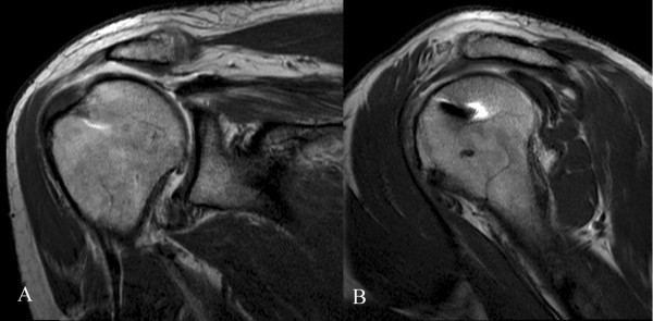
MR arthrographyMR arthrography of the shoulder at 38 months: recurrent tear of the thinned supraspinatus tendon, with leakage of contrast medium and superior migration of the humeral head.
Source : Tudisco et al., BMC Musculoskeletal Disorders, 2013, figure 4 · CC BY
Retear is common, often silent, and rarely dramatic. What matters is not to fear it but to know what it changes, when it occurs, and which signs require the session to be suspended and the surgeon called.
The figures, and what they allow us to say
The largest synthesis available examined 108 articles reporting both imaging and clinical data, that is more than 8,000 shoulders. The weighted mean retear rate there is 26.6 % at 23.7 months. Retears were associated with more marked fatty infiltration, a larger initial tear, older age and double-row repairs. Clinical improvement reached on average 72 % of the maximum possible improvement, and patient-reported outcomes improved whether or not cuff integrity was restored. The authors also note, in conclusion, that the number of publications has exploded without outcomes improvingPMID 24753240.
The meta-analysis of risk factors, fourteen studies and 5,693 patients, draws up a long and coherent list: age, body mass index, diabetes, fatty infiltration of subscapularis and infraspinatus, symptom duration, bone mineral density, length, width and area of the tear, retraction, critical shoulder angle, acromiohumeral space, distance between the myotendinous junction and the glenoid, operative time and biceps procedurePMID 34089878. Two factors deserve to be singled out because they are modifiable or negotiable. Smoking: across fourteen articles and 73,817 participants, it multiplies the risk of retear by 2.06 (95 % CI 1.30 to 3.28; p = 0.002) and that of revision surgery by 1.29PMID 34813890. Osteoporosis: it emerges as an independent factor for non-healing in a dedicated seriesPMID 21813440.
How many repaired cuffs retear, and at what point
Proportion of retears documented on imaging, by follow-up window, and weighted mean across all durations.
Sources: Longo 2021 for the distribution by follow-up window (PMID 34465332) and McElvany 2015 for the weighted mean (PMID 24753240). These percentages describe heterogeneous populations and protocols: they frame a conversation, they do not predict an individual patient.
What a retear really changes for the patient
Less than is feared, and knowing that changes the way an imaging result is announced. The meta-analysis of twelve studies and 800 patients, with a mean retear rate of 22 % at 27.5 months, finds in patients with a healed cuff a Constant score higher by 8.61 points, a UCLA score higher by 2.96, an ASES score higher by 9.49 and pain lower by 0.62 point. The authors take care to relate these gaps to the published thresholds of clinical relevance: the difference reaches that threshold for the ASES, but not for the ConstantPMID 29796103.
In other words: a retorn cuff does on average less well than a healed cuff, the gap is real, it is modest, and a great many patients with a residual defect remain clearly improved compared with their preoperative state. Announcing a retear as a failure makes a patient lose the benefit they have actually gained.
The signs that call for suspending progression and contacting the surgeon
- A sudden loss of strength, occurring during an identifiable movement. The patient often describes a precise action, a door pulled, a stumble caught, a child lifted. This is the most suggestive presentation, and it typically occurs in the risk window, between the sixth week and the sixth monthPMID 36842463.
- Active range that regresses while passive range is preserved. This is the classic dissociation. It does not prove a retear, but it requires that no load be added and that an opinion be sought.
- Night pain that comes back after having settled. Sleep normally improves after repair, and it improves earlyPMID 37167606: a clear return is a change of trajectory, not a fluctuation.
- Fever, discharge, redness or wound dehiscence. These do not belong to the retear register but they require the session to be stopped immediately and an opinion the same day. You do not massage, you do not mobilise the area.
- Stiffness that sets in between two sessions, with loss of passive range. It belongs to chapter 6 and not to this one, but it justifies adjusting the programme without waiting for the next surgical consultation: it is at three months that the stiffness threshold is measured, and at that stage the prognosis for recovery is still being decided.
- A new sensory or motor deficit in a nerve territory. It does not belong to the cuff. The site covers suprascapular nerve syndrome and neuralgic amyotrophy, two presentations that can arise afterwards and be mistaken for a failed repair.
The rule that simplifies things: faced with one of these signs, you do not adjust the load, you suspend progression and you contact the surgical team. A confirmed retear cannot be salvaged in a session, and an infection even less so; conversely, progression continued on a repair that is failing turns a partial defect into a complete tear.
| Factor | What the evidence shows | Modifiable? | What to do with it |
|---|---|---|---|
| Tear size and retraction | The most consistent factor, found in both large syntheses. | No | It governs the caution of chapter 3 and the message of chapter 8. |
| Fatty infiltration | Associated with retear across more than 8,000 shoulders. | No | To be asked of the surgeon; it explains slow strength recovery without anyone being at fault. |
| Age | Consistently associated. | No | Does not justify giving up strengthening, only reviewing its pace. |
| Smoking | Relative risk of retear 2.06; of revision 1.29, across 73,817 participants. | Yes | The only factor on this list where a brief intervention by the therapist makes sense. |
| Diabetes and low bone density | Retained as independent factors in several series. | Partly | They strengthen the case for caution, and for diabetes they intersect with the risk of stiffness. |
| Early active motion in a large tear | Relative risk of structural failure 1.52 beyond 3 cm, 2.74 with a suture bridge. | Yes | This is the only factor entirely in the therapist's hands. |
Key points from this chapter
- The weighted mean retear rate is 26.6 % at 23.7 months across more than 8,000 shoulders.
- Patient-reported scores improve whether or not integrity is restored; the gap between a healed cuff and a retorn cuff reaches the threshold of clinical relevance for the ASES, not for the Constant.
- Smoking doubles the risk of retear: it is the best-documented modifiable factor.
- The most suggestive presentation is a sudden loss of strength during an identifiable action, between the sixth week and the sixth month.
- Faced with a warning sign, you suspend progression: you do not salvage a failing repair by adjusting the load.
What can honestly be announced: pain, sleep, work, sport?
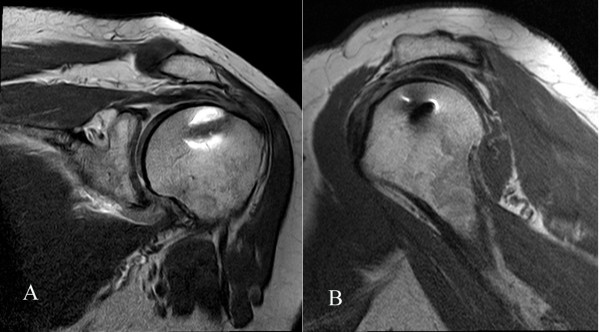
MR arthrographyMR arthrography of the shoulder, coronal and oblique sagittal views, 49 months after rotator cuff repair: continuous, healed supraspinatus tendon, without retraction or superior migration of the humeral head.
Source : Tudisco et al., BMC Musculoskeletal Disorders, 2013, figure 1 · CC BY
This is the question the patient asks at the first session, and the one where the gap between expectation and reality costs the most. The data exist, they are precise, and they are counted in months.
The time to improvement as actually measured
A series of 203 patients dated the achievement of thresholds of clinical relevance by survival analysis. The minimal clinically important difference was reached on average at 5.77 months on the ASES score, substantial clinical benefit at 6.22 months, and the patient acceptable symptom state at 7.23 months. The corresponding values on the SANE were 6.25, 7.05 and 9.26 months, and on the Constant 6.94, 7.13 and 8.66. Reaching these thresholds later: patients with a work-related injury, current smokers, those who had an associated biceps tenodesis, those operated on for a partial tear, and those whose preoperative score was already highPMID 33079576. The thresholds themselves were established in 288 patients: for the ASES, 11.1 points for minimal benefit, 17.5 for substantial benefit and 86.7 as the acceptable statePMID 30685283.
A meta-analysis of fifteen studies and 1,371 patients completes the picture by showing where the curve stops. The first year brings 41.1 ASES points, 34.2 Constant points and 42.9 WORC points. The second year brings no more than 2.3, 3.2 and 2.0 points respectively, all below the threshold of clinical relevancePMID 34473586. What the patient will gain, they will therefore gain for the most part in the first twelve months.
Sleep, the milestone the patient can read most easily
Of 293 patients enrolled prospectively, 262, that is 89.8 %, reported sleep disturbance before surgery. Among them, 221, that is 84.4 %, were free of it at two years, and those whose disturbance resolved had better functional scores than those in whom it persistedPMID 37167606. A series from the same centre places the return to normal sleep around the sixth month for the majority of patientsPMID 35219845, and a European multicentre study confirms the clear improvement in sleep after repairPMID 39189149.
It is a useful milestone because it is concrete, because it comes before functional recovery, and because the patient can check it themselves without needing a score.
Work, and the gap according to the job
The meta-analysis of thirteen series and 1,224 patients finds a weighted mean of 62.3 % returning to the previous level of work, at 8.15 months on average, with a standard deviation of 2.7 months. The rate fell significantly with the physical intensity of the job (p < 0.001), while it differed neither between open and arthroscopic surgery, nor between patients with a work-related injury and the rest. More than 35 % of patients did not regain their previous level of workPMID 33395315.
A second meta-analysis, covering all activities across fifteen studies and 1,065 patients, gives more favourable figures because it measures something else: 88.5 % return to activity across all categories, that is 83.8 % for work, 88.2 % for sport and 97.3 % for daily activities, with a mean duration of 6.59 monthsPMID 36792854. The difference between 62.3 % and 83.8 % is not a contradiction: the first measures return to the same level, the second return to work, modified duties included.
The milestones that can be announced, and when
Mean times measured in the literature, from sleep to resuming the previous job.
Sources: Manderle 2020 for the clinical thresholds (PMID 33079576), Dolan 2022 for sleep (PMID 35219845), Kholinne 2023 for return to activity (PMID 36792854), Haunschild 2021 for work (PMID 33395315). These are means from heterogeneous series: they serve to frame what is announced, not to promise a date.
What also has to be said, and which comes before this article
A well-informed patient knows that the question of surgery itself remains debated. The 2019 Cochrane review, nine trials and 1,007 participants, concludes that repair probably provides little or no improvement in pain compared with non-operative treatment at one year, with moderate certainty for pain and low certainty for function, and that these trials mostly concerned small degenerative supraspinatus tearsPMID 31813166. This page does not deal with the indication: the article on degenerative tears does, with the criteria for surgical referral.
Saying this to a patient who has already been operated on is obviously of no use and can do harm. It does, however, remind the therapist that the success of management is not measured by cuff integrity on a follow-up MRI, but by what the patient can do with their arm.
Key points from this chapter
- The threshold of clinically important benefit is reached on average around the sixth month, the acceptable symptom state around the seventh.
- The second year brings no more than two to three score points, below the relevance threshold: the essentials are settled in the first twelve months.
- Sleep normalises around the sixth month in the majority, and it is the milestone the patient can read most easily.
- 62.3 % of patients regain their previous level of work, at 8.15 months; the rate falls when the job is physically demanding.
- Telling a patient who works with their arms overhead that it will take six weeks is preparing a disappointment that will cost more than initial frankness.
What do published case reports teach us?
Four published case reports, chosen because each documents a situation that randomised trials exclude: the older patient, the adolescent, neurological comorbidity and the irreparable tear.
A medium tear in a 73-year-old woman, with pool work from the second week
A 73-year-old woman operated on for a 2.5 cm non-retracted full-thickness supraspinatus tear followed a programme combining land-based and water-based work, started two weeks after surgery and run over six weeks for eighteen sessions. The five patient-reported outcome scales improved, exceeding the minimal detectable change for the SPADI and the Penn Shoulder Score. Pain at rest went from 4 to 2 out of 10, and from 9 to 6 on activity, at eight weeks after surgery. No adverse event occurredPMID 26206216.
What this case illustrates. It establishes nothing about the effectiveness of water, and the authors say so. What it does document is the feasibility of low-load work started early on a medium tear, in a patient of an age that trials rarely recruit. The start date, two weeks, corresponds exactly to the end of the consensus phase of strict immobilisation.
A 13-year-old boy, subscapularis and long head of biceps, thirty weeks of rehabilitation
Cuff pathology in adolescents is rare, about 1 % of upper limb injuries before the age of 20 years, and subscapularis involvement is rarer still. A 13-year-old boy, a throwing athlete, with a complete subscapularis tear and a partial tear of the long head of biceps, underwent open subscapularis repair with biceps tenodesis. The postoperative programme comprised 22 sessions spread over 30 weeks, including a graded return-to-throwing programme. Range, strength and reported scores, including the Penn Shoulder Score and the QuickDASH, improved to a clinically significant degree, with return to sportPMID 40322514.
What this case illustrates. Two things. First the duration: thirty weeks for a healthy adolescent, whose healing is theoretically the best possible. Then the tendon: a subscapularis repair imposes its own rotation limits, and adding a biceps tenodesis lengthens the time to clinically important benefit, which the series of 203 patients in chapter 8 had measuredPMID 33079576.
A 48-year-old woman with post-polio syndrome
The sequelae of poliomyelitis expose patients to overuse injuries of the upper limb, in people who use their arms to compensate for a lower limb deficit. A 48-year-old woman with post-polio syndrome underwent a cuff repair and was then rehabilitated with Maitland mobilisations and low-intensity functional exercises, chosen explicitly so as not to trigger the fatigue characteristic of this syndrome. Measurements taken before surgery, before and after rehabilitation, and then two years later, show a return to independence that was maintained over time, with no overuse fatigue or weaknessPMID 17244697.
What this case illustrates. The dominant constraint was not protecting the suture but managing fatigue: in this profile, dosage takes precedence over the timetable. It is also a useful reminder for all patients whose upper limb carries an unusual functional load, wheelchair or crutch users in particular.
A massive irreparable tear treated by lower trapezius transfer
A 57-year-old woman with a massive irreparable tear underwent a lower trapezius transfer with an Achilles tendon allograft, a procedure for which the recommended immobilisation is strict, at 90 degrees of abduction and maximal external rotation, for six to eight weeks. Pool rehabilitation was nonetheless started at fifteen days and conventional rehabilitation at twenty-five, with twelve months of follow-up. The functional result was good and was maintained, with no suture dehiscence and no slackening of the allograftPMID 35803750.
What this case illustrates, and what it does not license. A single case report does not overturn a surgical instruction, and certainly not for a tendon transfer, where failure is hard to retrieve. This case belongs here because it recalls that the real load of an exercise depends on its modality as much as on its date: water-based work at fifteen days is not active work at fifteen days. The reasoning that matters is never “early or late”, it is “how much load, on what tissue, at what stage of its healing”.
Key points from this chapter
- The four case reports cover profiles that trials exclude: older patient, adolescent, neurological comorbidity, irreparable tear.
- Thirty weeks of rehabilitation in a fully healthy adolescent give the honest order of magnitude to announce to everyone else.
- A subscapularis repair and a biceps tenodesis change both the range limits and the expected timescale.
- In a patient whose arms carry a functional load, dosing fatigue takes precedence over the timetable.
- A single case does not overturn a surgical instruction; it recalls that modality counts as much as date.
How to apply all this from the first session?
This chapter condenses the article into concrete decisions. It replaces neither the operative report nor the surgeon's instruction: it gives the order in which to look at them.
What is established, what is probable, what remains a working hypothesis
Modalities of rehabilitation after cuff repair, ranked by the strength of the available evidence.
Ranking established by this article from the sources cited in chapters 3 to 7: Mazuquin 2021 (PMID 34048450), Chen 2023 (PMID 37085009), Jenssen 2018 (PMID 30195953), Hao 2025 (PMID 40082920), Kluczynski 2016 (PMID 25943112), Thigpen 2016 (PMID 26995456). This is not a formal GRADE rating: no graded guideline exists on this subject.
The five minutes that decide the rest
First, read before you touch. Tendons repaired, subscapularis included. Size and retraction. Technique. Associated procedures. The surgeon's numerical instructions. If one of these five items is missing, you ask for it, you do not estimate it.
Second, place the patient on the two axes of risk. Risk of stiffness: young patient, woman, work-related injury or occupational disease, partial tear, associated calcification, diabetes, preoperative anxiety. Risk of retear: age, large tear, retraction, fatty infiltration, smoking, low bone density. The two lists overlap little, and that is what makes a decision possible.
Third, build the programme on the modality, not on the date. Protected passive motion is the neutral modality: it costs nothing in either direction, except beyond a 5 cm tear. Active motion is the modality that commits: you bring it forward only with a reason, and never on a large tear before the prescribed date.
Fourth, measure the actual wear of the sling, in hours. Not “are you wearing it?” but “how many hours a day have you kept it on this week, nights included?”. It is men and overweight patients who wear it least.
Fifth, announce in months. Sleep normalised around the sixth month, clinically perceptible benefit around the sixth, previous work around the eighth for two patients in three. A patient who hears six weeks at the first session will come back disappointed at the twelfth.
| Period | What you do | What you do not do | What you watch |
|---|---|---|---|
| Weeks 0 to 2 | Education, management of sleep and pain, elbow, wrist and hand, cervicoscapular control, checking sling wear. | Any glenohumeral range that has not been prescribed; any contraction of the repaired cuff. | The wound, fever, unusual pain, the patient's ability to sleep. |
| Weeks 2 to 6 | Protected passive motion within the prescribed ranges, strictly respecting the external rotation limit if subscapularis has been repaired. | Active motion, even briefly. Self-passive work not taught in the session. Carrying loads. | Night pain, gain in passive range, actual adherence in hours. |
| Weeks 6 to 12 | Assisted active then active motion, in the order described in chapter 5, with scapular control. | Resisted strengthening of the repaired cuff before the twelfth week in a large tear. | This is the densest retear window: sudden loss of strength, active-passive dissociation. |
| Weeks 12 to 24 | Progressive strengthening, endurance, increasing load below shoulder height then above it. | Throwing, explosive work and overhead work as long as strength is not symmetrical. | Pain delayed more than twenty-four hours after a session: a dose signal, not an injury signal. |
| Beyond the 6th month | Progression towards the real demands of the job or the sport, tested in the loaded position. | Declaring management finished on an isolated score, with no functional test. | What the patient still avoids and does not mention spontaneously. |
Key points from this chapter
- Read the operative report before touching the shoulder: five items, none of which can be estimated.
- Place the patient on the two axes of risk, stiffness and retear: the two lists overlap little.
- Protected passive motion is the neutral modality; active motion is the one that commits.
- Measure sling wear in hours, not in yes or no.
- Announce the timescales in months, from the first session.
Frequently asked questions
How long should the shoulder be immobilised after a rotator cuff repair?
Three to six weeks for a small to medium tear, and the range really is a range: a non-inferiority trial in 120 patients showed that three weeks in a simple sling was no worse than six weeks in a brace, with 89 % healing on MRI in both groupsPMID 30195953. Beyond small and medium tears the evidence is lacking and the surgeon's instruction takes precedence. The question is not only how long, but what is done during that time: passive mobilisation inside the brace is not immobilisation.
Does early passive mobilisation increase the risk of retear?
No, on average across trials: the largest meta-analysis, twenty trials and 1,841 patients, finds an odds ratio of 1.05 (95 % CI 0.64 to 1.75)PMID 34048450. The nuance concerns size: beyond 5 cm, early passive mobilisation was associated with 52.2 % retears against 22.6 % with delayed motionPMID 25296646. Early passive motion is safe in small and medium tears; it has not been shown to be safe in large ones.
What is the difference between early passive motion and early active motion?
The whole difference lies in what the repaired tendon undergoes. Passive motion does not make the cuff work, active motion does. The two meta-analyses from the same group show it: with early passive motion, no difference in retear, 13.7 % against 10.5 %PMID 25296646; with early active motion, a relative risk of structural failure of 1.52 beyond 3 cm and of 2.74 for suture-bridge repairsPMID 25943112. Confusing the two turns a safe instruction into a risky one.
When does a retear occur after rotator cuff repair?
Between the sixth and the twenty-sixth week, not in the first few days. In 113 patients followed by MRI at six intervals, the mean time was 19.2 weeks, with only one further retear between the sixth and the twelfth monthPMID 23780533. A series of 638 arthroscopies places the peak between six weeks and three months, where 46.3 % of retears are concentratedPMID 36842463. The risk window therefore extends well beyond the sling period.
When can strengthening begin after a rotator cuff repair?
The American Society of Shoulder and Elbow Therapists consensus places progressive strengthening at the twelfth week, after two weeks of strict immobilisation, protected passive range from the second to the sixth week, then restoration of active rangePMID 26995456. This framework is an expert consensus, not a trial result, and the protocols published online are more aggressive than it isPMID 33418088. In practice, the date matters less than the criterion: full and painless passive range, scapular control, and no return of night pain.
Is an abduction brace or a simple sling needed?
A meta-analysis of four randomised trials and 302 patients finds no difference between the two, either on the Constant score, or on the WORC, or on pain, or on range of motion, or on healing failures (odds ratio 0.86; 95 % CI 0.32 to 2.37). The authors conclude that the simple sling is preferable for its cost-effectivenessPMID 37085009.
How common is stiffness after a rotator cuff repair?
It depends on the definition. In 489 consecutive arthroscopies, 4.9 % of patients were dissatisfied because of stiffness that led to a capsular releasePMID 19664508. With a mobility threshold measured at three months, the incidence rises to 14.2 % in 274 patients with no preoperative stiffnessPMID 35210518, and a registry of 1,330 arthroscopies finds 8.4 % stiffness within six monthsPMID 34310220. The factors that recur are diabetes, female sex, partial tear and preoperative anxietyPMID 35077897.
When will my patient be able to return to work?
On average at eight months, and not everyone. The meta-analysis of thirteen series and 1,224 patients finds 62.3 % return to the previous level of work, at 8.15 months on average, with a rate that falls the more physically demanding the job isPMID 33395315. Another meta-analysis, covering all activities, finds 83.8 % return to work and a mean duration of 6.59 monthsPMID 36792854. Telling a patient who lifts loads overhead that it will take six weeks is preparing a disappointment.
Does a retear mean that rehabilitation has failed?
No, and this is important to know before announcing an imaging result. The weighted mean retear rate is 26.6 % at 23.7 months across more than 8,000 shoulders, and the associated factors are fatty infiltration, tear size and agePMID 24753240. A meta-analysis of twelve studies and 800 patients shows that the score difference between a healed cuff and a retorn cuff reaches the threshold of clinical relevance for the ASES but not for the ConstantPMID 29796103: many patients with a residual defect remain improved.
Does the patient sleep better after a rotator cuff repair?
Yes, and it is often the clearest improvement. In 293 operated patients, 89.8 % reported sleep disturbance before surgery, and 84.4 % of those saw it resolve by two yearsPMID 37167606. A series from the same centre places the return to normal sleep around the sixth month for the majority of patientsPMID 35219845. Sleep is therefore a useful milestone to announce, and a follow-up indicator that the patient understands better than a score.
How can I know exactly what the surgeon repaired?
By reading the operative report. Five items change the session: the tendons repaired, in particular subscapularis involvement, which contraindicates forced passive external rotation and early active internal rotation; the size and retraction of the tear; the repair technique; associated procedures, long head of biceps tenodesis in particular; and the surgeon's explicit instructions, which take precedence over any general rule.
Should a degenerative rotator cuff tear be operated on?
That question comes before this article, and the site gives it a page of its own. The 2019 Cochrane review, nine trials and 1,007 participants, concludes that repair probably provides little or no improvement in pain compared with non-operative treatment at one year, with moderate certainty for pain and low certainty for functionPMID 31813166. This article does not deal with the indication: it begins once the decision has been made and the construct is in place.
References and verification
The 58 references in this article were resolved one by one against PubMed metadata, through the NCBI E-utilities API, on 16 August 2026: full author list, journal, year, volume, issue, pagination and DOI, without truncation. The figures quoted come from structured abstracts, never from a reconstruction. The two ICD-11 codes in the structured data were checked against the simple tabulation list published by the World Health Organization. The only French operative volume figure comes from the PMSI hospital database and not from a registry: it counts coded procedures, not patients followed up, and no French rotator cuff surgery registry exists to date.
For what comes before surgery, the site covers rotator cuff tendinopathy and subacromial pain, symptomatic degenerative rotator cuff tear and calcific tendinopathy. For the diagnoses to consider when an operated shoulder is not progressing as expected: frozen shoulder, suprascapular nerve syndrome and neuralgic amyotrophy. For the other major shoulder operation and its aftermath, see total shoulder arthroplasty.

