














On-siteDPCFIFPL

Upper limb
The painful shoulder with Adam Meakins
Adam Meakinsadammeakins
Go further
Neuralgic amyotrophy: recognising the sequence of sudden pain then weakness, telling it from a frozen shoulder or a cuff tear, and rehabilitating it.
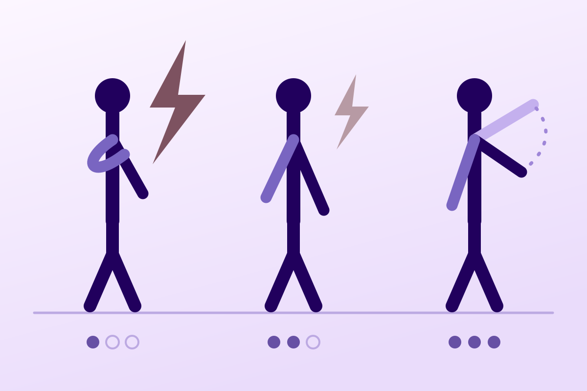
A sudden shoulder pain, then a motor deficit appearing as the pain eases: this sequence points to neuralgic amyotrophy, not a tendinopathy.
65%recovery at ten months according to a 2026 review
What follows covers each of these points in detail, with sources. It is there if you need it.
This topic is taught in a course: The painful shoulder with Adam Meakins, on-site with Adam Meakins.
1 / 12
11 article chapters · 57 min in total
Comprendre◔ 6 min
Neuralgic amyotrophy is an acute inflammatory neuropathy affecting several nerves of the brachial plexus.
Chronologie◔ 5 min
Pain and deficit are offset in time: this is the only sign available without any investigation.
Examiner◔ 6 min
The examination is neurological, not orthopaedic: patient undressed to the waist, viewed from behind and from the front.
Différencier◔ 6 min
This syndrome is missed because its initial phase is indistinguishable from the three most common shoulder diagnoses.
Examens◔ 4 min
Neuralgic amyotrophy remains a clinical diagnosis, ahead of any investigation.
Déclencheurs◔ 5 min
The most frequent trigger is an upper respiratory tract infection in the preceding weeks.
Rééduquer◔ 7 min
No rehabilitation reinnervates a nerve: recovery follows its own timetable.
Pronostic◔ 5 min
Long considered benign, this syndrome in fact leaves frequent sequelae long term.
Cas cliniques◔ 5 min
These case reports prove nothing about efficacy; they show how a diagnosis is missed.
En pratique◔ 4 min
Reassessing a painful shoulder at 3-4 weeks is the move that catches the diagnosis, and it costs nothing.
Questions◔ 4 min
A prospective cohort finds an incidence 30 to 50 times higher than the historical estimate.
Train on this
Upper limb
Adam Meakinsadammeakins
Next comes practice: the course that teaches this topic, with Adam Meakins.
Our courses on this topic may be covered by DPC or FIFPL, the French continuing-education funding schemes, depending on your profession and situation.
A sudden, very intense shoulder pain, often at night. Then it eases, and that is when something is wrong: the arm is weak. This sequence, pain first and deficit afterwards, belongs to no tendinopathy and to no adhesive capsulitis. It is the signature of neuralgic amyotrophy, and it is almost always at the moment the pain gives way that the diagnosis is decided.
In brief
Three figures that change what you do
Real frequency, pattern of the lesion, long-term outcome.
Sources: incidence, prospective cohort in primary care covering 14,118 inhabitants (PMID 26016482); distribution of the lesion and long-term outcome, series of 246 cases (PMID 16371410).
In this chapter: the definition and the three names of the disease, the history of its incidence, which has been revised by a factor of thirty to fifty, the current pathophysiological model, and the two reasons why a physiotherapist can see cases without ever recognising one.
Parsonage-Turner syndrome is an acute, multifocal inflammatory neuropathy that strikes the nerves of the brachial plexus territory. It carries the name of the two authors who described it in the Lancet in 1948 under the title of “shoulder-girdle syndrome”.36 In French it is called névralgie amyotrophiante or amyotrophie névralgique, and in English neuralgic amyotrophy, and sometimes still acute brachial neuritis or idiopathic brachial plexopathy. These four names designate the same thing, and this terminological scatter does not help in recognising it.
The definition fits in one sentence: a neuropathic pain of sudden onset in the shoulder region, followed by patchy paresis and muscle wasting of the upper limb, with or without sensory disturbance.11 The word that counts in that sentence is “followed”. This is not pain with weakness, it is pain then weakness, and the interval between the two is what distinguishes this syndrome from everything it resembles.
For thirty years, the reference for frequency was a population study conducted in Rochester, Minnesota, over the period 1970-1981: 579 records reviewed, 11 cases retained, 1.64 cases per 100,000 inhabitants per year.7 At that level, a community physiotherapist could reasonably practise a whole career without meeting a single one, and that conviction took hold.
It was refuted in 2015 by a study of a different design. Rather than reviewing records, the authors counted prospectively for one year every new presentation for neck, shoulder or arm in two primary care centres serving 14,118 inhabitants, after training the general practitioners to recognise the picture. Of 492 new patients, 34 were suspected and 14 confirmed after neurological assessment, an annual incidence of 1 in 1000 for the classic phenotype alone.1 The authors conclude that the syndrome is 30 to 50 times more common than was believed.
The same disease, five incidence figures
Logarithmic scale. The gap does not measure a variation in frequency, it measures a difference in method.
Positions calculated in decimal logarithm, not estimated by eye. Sources: PMID 2996415 (Rochester 1970-1981), PMID 41606804 (2026 review), PMID 41235535 (German claims data 2013-2022), PMID 26016482 (prospective primary care cohort). The four measurements do not bear on the same object: retrospective register, secondary literature, reimbursement coding and prospective counting after training the doctors.
A study published in 2026 settles the debate from the other end. The authors used the claims data of a German insurer covering about 26 million insured people over ten years: they find an incidence of 7.7 to 12.8 per 100,000, which is nearly 90 % less than the prospective cohorts.8 Their conclusion is not that the prospective estimate was wrong; it is that the majority of cases are not correctly diagnosed and therefore never appear in reimbursement data. The same work notes that diaphragmatic paresis, a marker of phrenic nerve involvement, is coded in only 0.45 % of cases, a frequency far below what the clinical literature describes.
The gap between 1.64 and 100 per 100,000 does not measure a disease that changes. It measures what a clinician sees according to whether or not they know what they are looking for.
The accepted hypothesis is that of a lesion with three components, none of which is sufficient on its own: a genetic predisposition, an immunological trigger, and mechanical strain on the nerve segment or segments concerned.4 This model explains two observations that used to puzzle: why the disease strikes precise nerves and not the whole plexus, and why it recurs in some patients and never in others.
The genetic component has had a name since 2005: three mutations of the SEPT9 gene, coding for septin 9, have been identified in six families with the hereditary form, linked to chromosome 17q25 and autosomal dominant in transmission.29 In those families, the attacks are triggered by environmental factors, chief among them an infection or a childbirth.
The mechanical component is the one whose reading imaging has changed most over the past ten years. We now know that the affected nerves often show hourglass-like constrictions, that is, a focal narrowing of the calibre of the nerve or of some of its fascicles, sometimes accompanied by torsion phenomena.12 These lesions are not anecdotal: in 123 patients whose diagnosis was confirmed by electromyography, 3-tesla magnetic resonance neurography found them in 90.2 %.19
Four background markers
What the literature establishes about the form, sensory involvement, recurrence and phrenic nerve involvement.
Sources: PMID 26662794 (review, classic phenotype); PMID 16371410 (series of 246 cases, sensory involvement and recurrences); PMID 41235535 (German claims data, coding of diaphragmatic paresis).
The first is statistical. An acute shoulder pain in an adult has default diagnoses, and they are far more common: rotator cuff tendinopathy, adhesive capsulitis, subacromial impingement. Faced with a banal presentation, the clinical mind goes to the probable. That is a good reflex most of the time, and it is exactly what causes a picture whose real frequency has just been revised by a factor of thirty to be missed.
The second is chronological, and it is the most costly. The patient consults during the painful phase, when there is still nothing to see but a shoulder that hurts and cannot be moved. The motor deficit arrives later, often after the diagnosis has been made and rehabilitation started. It is then interpreted as a consequence of pain and inactivity, not as a new neurological sign. The reviews written for orthopaedic surgeons say it plainly: knowledge of the clinical signs of this syndrome is not widespread, and it is the distinction from spinal and shoulder conditions that matters in practice.14
Key points
In this chapter: the three-stage chronology, what is particular about the pain in its suddenness and its timing, why the appearance of the deficit at the moment the pain gives way is the most discriminating clinical fact in the picture, and what that chronology implies for the physiotherapist who sees the patient between the two.
Almost every acute painful shoulder that a physiotherapy practice receives has a history in which pain and limitation go together: they appear together, they settle together. Neuralgic amyotrophy does exactly the opposite. Its pain and its deficit are offset in time, and that offset is the only sign that can be obtained without any investigation.
The onset is sudden. Patients date it to the hour, which is already rare in shoulder conditions. The pain is intense, continuous, often nocturnal or revealed at night, and it is not relieved by the usual antalgic positions. It has a neuropathic character, and it is described in the literature as one of the most severe symptoms these patients have known.11
Its duration is a quantified figure: in the series of 246 cases, the severe, continuous initial pain lasts about four weeks on average, and it evolves in three successive phases.3 In the untreated patients of the prednisolone series, the median time to first relief was 20.5 days.22 There is a difference between the sexes worth knowing, because it changes what is said to the patient: the initial pain lasted on average 45 days in men against 23 in women.3
This is the tipping point, and it is counter-intuitive. Just as the patient is at last beginning to feel better on the pain side, they discover that they can no longer perform certain movements. The reviews put it consistently: a weakness that occurs some time after the pain.412 The patient does not present it that way. They say that their pain is better but that their arm “is not keeping up”, and they readily attribute that weakness to the weeks spent without moving.
The timeline that makes the diagnosis
Two offset curves. It is the offset, not each curve taken in isolation, that distinguishes this syndrome from everything it resembles.
Qualitative diagram: the vertical axis is not graduated and the plotting of the curves has no measurement value. The time markers, on the other hand, are sourced. Mean duration of the initial pain of about 4 weeks, in three successive phases, and sequelae in about two thirds of patients followed for 3 years or more: PMID 16371410. Median time to first relief of 20.5 days in untreated patients: PMID 19321467. Recovery spread over months to years: PMID 19588414.
Two practical consequences follow from this timeline, and they are of different kinds.
The first concerns the timing of the consultation. The patient generally arrives in the red zone, when there is only pain. At that stage nothing allows a clinical decision, and it is not abnormal to start with the probable diagnoses. What is avoidable, on the other hand, is failing to reassess. A shoulder referred for rotator cuff tendinopathy which, three weeks later, hurts less but moves less well actively, is not a tendinopathy doing badly. It is a picture that has changed in nature.
The second concerns the interpretation of the weakness. A pain-related weakness is an inhibition weakness: it gives way when you reassure, it varies with the pain of the day, it follows no territory. A denervation weakness does not give way, does not vary, and follows a nerve territory. The rule that condenses this chapter is the one the orientation articles retain: a motor weakness out of proportion to what the pain and the loss of passive range explain must prompt the diagnosis to be reconsidered.13
A pain that improves while a deficit sets in is not good news followed by a complication. It is one and the same diagnosis revealing itself in two stages.
Muscle wasting appears next, and it is often early and rapid compared with what simple immobility would produce. It has a value that is not only diagnostic but chronological: its presence indicates that the denervation dates back several weeks. It is visible to the eye over the supraspinous and infraspinous fossae when the suprascapular nerve is involved, and it can be read on the contour of the deltoid or of serratus anterior according to the nerve affected.
Red flags of this chapter
Key points
In this chapter: the three things to look at before touching the patient, scapular winging and what it is really worth as an orienting sign, the logic of patchy territories that fit no root, and the nerve-by-nerve table of muscles and of the movements that reveal them.
The examination of a suspected neuralgic amyotrophy is not a shoulder examination. It is a neurological examination of the upper limb, carried out on a patient undressed to the waist, from behind as much as from the front. Three elements are obtained before anything is even moved: the muscle contours, the position of the scapula at rest, and the behaviour of that scapula when the arm goes up.
The contours. The supraspinous and infraspinous fossae are read from behind, comparing sides. Asymmetrical hollowing of the infraspinous fossa is the most visible sign of suprascapular nerve involvement. The point of the shoulder, the contour of the deltoid, the curve of the biceps and the thenar eminence deserve the same comparative examination: since the lesion is patchy, so is the wasting.
The scapula at rest, then in movement. It is the sign most often missed, for a simple reason: it cannot be seen from the front, and it cannot always be seen at rest. You have to ask for forward elevation of the arm, or a push against a wall, and look at the medial border of the scapula.
Scapular winging is taken to be a rare and non-specific sign. A French series of 128 unilateral winged scapulae investigated clinically and electrically over twenty-five years corrects that impression sharply.15 Long thoracic nerve palsy is the leading cause, with 70 cases, which an independent review of the winged scapula confirms.16 And among those long thoracic palsies, 61 were due to neuralgic amyotrophy. In other words: faced with a scapula that wings without trauma, the most likely diagnosis is not orthopaedic, it is neurological, and it is the one in this article.
From a winging scapula to the diagnosis
Series of 128 unilateral winged scapulae investigated clinically and by electrodiagnosis over twenty-five years.
Source: PMID 29314072, 128 patients over 25 years. The other causes reported in this series are the spinal accessory nerve (39), orthopaedic causes (11), involvement of both nerves (5), facioscapulohumeral dystrophy (5), voluntary winging (6) and no identified cause (2); the numbers published by category do not partition exactly into 128, so they are reproduced as they stand and without any derived percentage. The 61/70 ratio, on the other hand, is the one the authors write. The right side was involved in 62 of the 128 patients.
Two forms must be distinguished. The authors of the series of 128 cases note that the clinical data allow the identification of two distinct profiles, one for the long thoracic nerve and the other for the spinal accessory nerve.15 Their clinical semiology is classic and needs no equipment. Winging from involvement of the long thoracic nerve, which paralyses serratus anterior, increases on forward elevation of the arm and on pushing against a wall, with a medial border of the scapula that lifts. Winging from the spinal accessory nerve, which paralyses trapezius, comes with a drooping shoulder and increases in abduction. Look for both, not only the first: the spinal accessory was involved in 39 of the winged scapulae in that same series.15
This is the second great principle of the examination, and it is decisive for the differential diagnosis in the next chapter. A cervical radiculopathy affects one root, and a root innervates a coherent set of muscles, with a dermatome and a reflex. Neuralgic amyotrophy affects nerves, sometimes even fascicles inside a nerve, and it affects several of them with no metameric logic.
The most elegant demonstration of this mechanism comes from imaging. In twenty patients with a so-called spontaneous anterior interosseous syndrome, 3-tesla magnetic resonance neurography finds lesions of individual fascicles inside the trunk of the median nerve, in the arm, 14.6 centimetres on average above the humeroradial joint line, strictly respecting the internal topography of the nerve: only those fascicles that will later form the anterior interosseous nerve are affected.20 The authors conclude that this syndrome is, in the majority of cases, not an operable compression at the elbow but a multifocal mononeuropathy. The same logic holds for the other distal nerves affected: the lesion almost always sits upstream, in the fascicles of the parent nerve.17
A deficit that fits no root is not a badly performed examination. It is the expected result when the lesion is fascicular.
The most frequently affected nerves are, in the order in which the standardised ultrasound approach describes them: suprascapular, long thoracic, median and its anterior interosseous branch, radial and its posterior interosseous branch, axillary, spinal accessory, musculocutaneous.17 Across the whole series of 246 cases, involvement of the upper and middle trunk, with the long thoracic and the suprascapular alone or combined, accounts for 71.1 % of cases.3
| Nerve | Muscles | What is seen | The movement that reveals it |
|---|---|---|---|
| Long thoracic | Serratus anterior | Winging of the medial border of the scapula | Forward elevation of the arm, push against a wall |
| Suprascapular | Supraspinatus, infraspinatus | Hollowing of the supraspinous and infraspinous fossae | Resisted external rotation with the elbow at the side, comparing sides |
| Spinal accessory | Trapezius | Drooping shoulder, lateral winging of the scapula | Abduction, resisted shoulder shrug |
| Axillary | Deltoid | Flattening of the point of the shoulder | Resisted abduction at 90 degrees |
| Musculocutaneous | Biceps brachii, brachialis | Loss of the curve of the biceps | Resisted elbow flexion, biceps reflex |
| Anterior interosseous | Flexor pollicis longus, flexor digitorum profundus (II) | Thumb-index pinch that flattens, with no sensory disturbance at all | Ask for a circle to be made with thumb and index finger: the patient forms a triangle |
| Posterior interosseous | Finger extensors | Dropped fingers, wrist extension preserved | Resisted extension of the metacarpophalangeal joints |
| Phrenic | Diaphragm | Breathlessness when lying flat, orthopnoea | Ask about sleeping flat. Medical advice if positive |
Table to be scrolled horizontally on a small screen.
Two entries in this table deserve a further word. Involvement of the anterior interosseous is purely motor and without any sensory disturbance, which makes it easy to confuse with a tendon problem of the hand; it is the nerve most frequently found in an ultrasound series of 53 patients, where it accounted for 23 % of the affected nerves.18 This figure comes from a series referred for investigation, therefore enriched in distal forms, and it does not contradict the proximal predominance of the series of 246 cases: the two measure different populations. Involvement of the posterior interosseous, for its part, gives dropped fingers with preserved wrist extension, and a published case shows how readily it can be taken for an entrapment syndrome or a compressive neuropathy.34
Key points
In this chapter: the differential table for the acute painful shoulder, the sentence that separates neuralgic amyotrophy from each of its three great imitators, a decision tree usable at the end of the assessment, and the rarer entities to which this syndrome is sometimes wrongly attributed.
This syndrome is not missed because it is odd. It is missed because its initial phase is indistinguishable from the three most common diagnoses of the painful shoulder, and because each of those three diagnoses has a dedicated article on this site: rotator cuff tendinopathy, adhesive capsulitis and symptomatic degenerative rotator cuff tear. This chapter says in one sentence what separates each of them from neuralgic amyotrophy.
| Entity | Onset of the pain | Motor deficit | Passive range | What decides |
|---|---|---|---|---|
| Neuralgic amyotrophy | Sudden, datable to the hour, very intense, nocturnal. About 4 weeks3 | Appears AFTER, as the pain decreases. Patchy, in the nerve trunks | Preserved at first, then limited by secondary stiffness | The pain then deficit sequence, the early muscle wasting and a territory that follows no root |
| Cervical radiculopathy | Often gradual, associated neck pain, radiation into the limb | Present from the outset, in a single root, with concordant dermatome and reflex | Preserved | A coherent metameric territory, and neck movements that reproduce the pain |
| Rotator cuff tear | Gradual or after an effort; night pain common | Present from the outset and stable, from tendon discontinuity, not from denervation | Preserved | The deficit is there from day one and does not worsen when the pain gives way |
| Adhesive capsulitis | Gradual over several weeks | No true deficit; strength is limited by pain and by range | LOST, globally and above all in external rotation | The loss of PASSIVE range, in particular external rotation with the elbow at the side |
| Rotator cuff tendinopathy | Gradual, linked to load and to overhead movements | None. Inhibition weakness that varies with the pain | Preserved | The pain follows the load, and strength becomes normal again once the pain is removed |
| Calcific tendinopathy | Hyperacute in the resorptive phase, sometimes more intense still | None | Very limited by pain in the acute phase, but recoverable | The calcification on the radiograph, and the total absence of any neurological sign |
Table to be scrolled horizontally on a small screen.
Against cervical radiculopathy. Radiculopathy affects one root, therefore a territory coherent with its dermatome and its reflex; neuralgic amyotrophy affects nerves and fascicles, therefore a patchy territory that fits into no root.2017 Let us add two elements of history: neck pain is usual in one, unusual in the other, and pain reproduced by neck movements does not belong to the picture of neuralgic amyotrophy. The subject is dealt with in detail in cervicobrachial neuralgia, whose name alone is enough, incidentally, to explain part of the confusion: the two entities share the word “neuralgia” without sharing a mechanism.
Against rotator cuff tear. In a tear, the deficit is present from day one and it is mechanical: the tendon no longer transmits. In neuralgic amyotrophy, the deficit arrives afterwards and it is neurological: the muscle no longer receives. The practical consequence is this: a deficit that sets in during treatment is not a tear getting worse, it is another disease. Two published case reports show exactly this trap, one in a patient operated on for a cuff tear whose palsy was setting in at the time of surgery,31 the other in a patient whose picture appeared ten weeks after a repair.32
Against adhesive capsulitis. It is the simplest and most reliable distinction, and it lies in one movement: measuring passive external rotation with the elbow at the side. Adhesive capsulitis loses it, neuralgic amyotrophy keeps it, at least as long as secondary stiffness has not set in. Conversely, adhesive capsulitis does not produce a true motor deficit and does not come with early muscle wasting. One must nevertheless guard against too rigid a reading: a painful shoulder kept immobile for several weeks stiffens, and a patient seen late can combine the two pictures.
Decision tree for an acute non-traumatic painful shoulder
To be worked through at the end of the assessment, and repeated at three weeks if the picture changes.
An editorial reading intended for clinical reasoning, and not a validated algorithm: no decision rule has been derived or tested on this presentation. The elements that make it up are sourced in the body of the article. The three-week delay before electromyography corresponds to the time needed for denervation signs to appear on needle examination.21
Long head of biceps tendinopathy causes anterior pain reproduced on palpation of the groove and on resisted flexion tests, with no deficit and no wasting: it shares only its location with neuralgic amyotrophy. Instability and recurrent dislocations have a traumatic history and apprehension in the cocked position, both absent here. Thoracic outlet syndrome is the closest on the neurological front, but its involvement bears on the lower trunk of the plexus, it is positional and it sets in gradually, where neuralgic amyotrophy is sudden and predominates on the upper trunk.
Finally, mention must be made of the diagnoses that must not be missed behind a suspected neuralgic amyotrophy. A plexopathy can be tumoural, infiltrative or post-radiation, and both electromyography and imaging serve first of all to rule out those causes.221 A palpable supraclavicular mass, a history of cancer, a deterioration in general condition or a gradual involvement over several months fall outside the scope of this article and call for medical referral without delay.
What calls for urgent medical advice
Key points
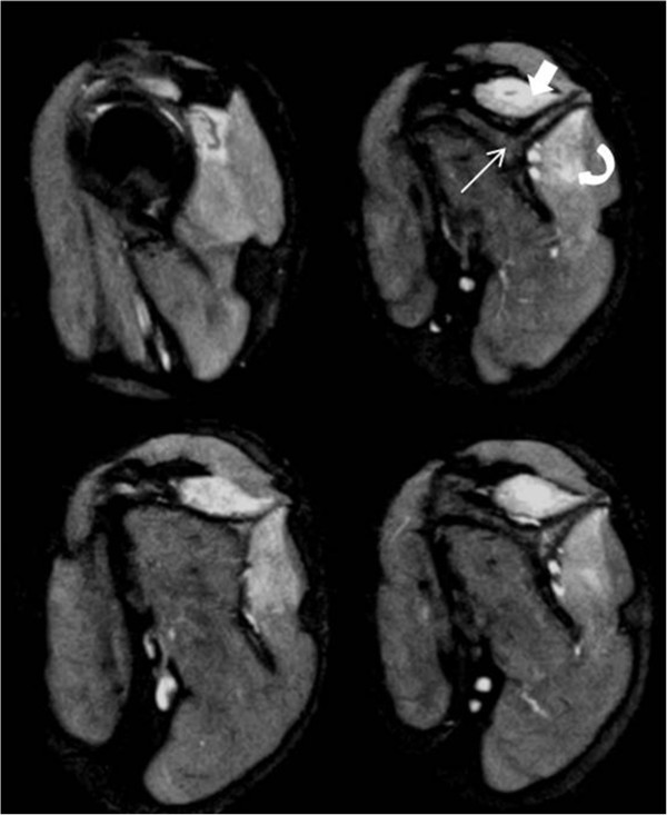
MRISagittal T2 fat-suppressed shoulder MRI: denervation oedema of the supraspinatus and infraspinatus, typical of Parsonage-Turner syndrome.
Source : Kumar et al., Journal of Medical Case Reports, 2013, figure 1 · CC BY
In this chapter: why the diagnosis remains clinical, what electromyography contributes and why it should not be requested too early, what ultrasound and MR neurography have changed over ten years, and the only serology worth requesting systematically.
We must start with the point that structures everything else: neuralgic amyotrophy is a clinical diagnosis. Investigations serve to rule out an infectious or malignant cause and to document the differential diagnosis, not to make the positive diagnosis.2 A patient whose history is typical and whose examination is concordant has neuralgic amyotrophy before any investigation, and a normal investigation does not refute it.
Electromyography confirms the plexopathy, specifies its location, its mechanism, its age, its severity and its prognosis.21 It is the investigation that objectifies the patchy character of the lesion and that formally separates it from a radiculopathy. The anatomy of the plexus, however, imposes an extensive examination, with multiple conduction studies and needle examination of many muscles: it is not a short investigation, and it gains from being requested with a precise question.
The timing is the most important practical fact in this chapter. The examination must be carried out at least three weeks after the lesion, the time needed for the electrical abnormalities to appear, in particular fibrillation potentials and positive sharp waves.21 An electromyography requested in the first few days of a deficit can be normal while the lesion is very much there, and that false negative then delays the diagnosis instead of speeding it up. Another useful marker for interpreting a report: after about three months, reinnervation can raise the amplitude of the motor responses and overestimate the proportion of preserved axons.21
Until the 2010s, this syndrome was a clinical and electrophysiological diagnosis, with no visible substrate. High-resolution ultrasound and magnetic resonance neurography have changed that by showing structural lesions: swelling of the nerve, incomplete then complete constrictions, and rotational phenomena of torsion or fascicular entanglement, which the standardised approach sorts into four categories.17
What each imaging technique finds
Two different populations, two different techniques. This is not a direct comparison.
Sources: PMID 28556181 (ultrasound, overall sensitivity of 74 % in 53 patients and 70 affected nerves); PMID 37610034 (3-tesla MR neurography, hourglass-like constrictions in 90.2 % of 123 patients whose diagnosis was confirmed by electromyography, sensitivity of 91.9 % in the severe forms and of 91.0 % on nerve-by-nerve analysis, agreement between two radiologists of 94.3 % per subject). Both series come from referral centres, where the patients referred are more severe than average: these figures cannot be transposed as they stand to a practice population.
One result from this imaging has a directly therapeutic reach. In the ultrasound series, the category of the abnormality predicted the outcome: where there was a complete constriction and rotational phenomena, reinnervation was absent or negligible, a significant difference between categories.18 These are the patients whom the surgical literature considers potential candidates for a neurolysis, and it is the only element that currently allows them to be identified other than by waiting for nothing to happen.
Imaging that shows a complete constriction does not change the diagnosis. It changes what can reasonably be hoped for from rehabilitation alone.
One operational recommendation comes out of a French series of six prospective cases: systematically look for hepatitis E virus infection in neuralgic amyotrophy, whatever the severity, if the onset dates back less than four months, that is, before the IgM antibodies disappear.30 In that series, the six patients totalled 26 inflammatory mononeuropathies, from one to eight per patient, affecting the suprascapular nerve in all six, the long thoracic in five, the anterior interosseous in three and the phrenic nerve in one. After two to eight years of follow-up, all the nerves had recovered clinically except in one patient. The numbers are small and that must be said, but the test is simple and carries no risk.
What the physiotherapist can do with all this
None of these investigations is requested by the physiotherapist. This chapter is therefore not about requesting, it is about writing a useful letter. A letter that says “painful shoulder, please investigate” triggers nothing. A letter that says “sudden pain on 12 March, easing for three weeks, secondary appearance of a deficit of forward elevation with winging of the medial border of the scapula, passive external rotation preserved, suspected neuralgic amyotrophy, electromyography would be useful” triggers the right consultation and the right investigation, at the right time.
Key points
In this chapter: the triggers to look for in the preceding four weeks, the hereditary form and what distinguishes it in practice, the differences between men and women that change the prognosis given, the postoperative form, which is the most costly trap, and phrenic nerve involvement.
The history must go back one month before the pain began. The most frequently reported trigger is an upper respiratory tract infection occurring a few weeks earlier.9 The other documented contexts are postoperative, post-traumatic and post-vaccination.13 An indirect argument in favour of the immunological mechanism comes from the German claims data: the diagnosis is made significantly more often in the first quarter of the year than in any other, which the authors link to the seasonality of infections.8
The case of hepatitis E deserves a place of its own, because it is common, little known and detectable. It is covered in the previous chapter as regards what to do.30 Let us note here that a neuralgic amyotrophy is not always idiopathic and that an identifiable cause exists in a proportion of patients.
The hereditary form accounted for 47 of the 246 cases in the large reference series, about one in five.3 That proportion does not transpose as it stands: the series comes from a referral centre specialising in the brachial plexus, which attracts familial and recurrent forms. It is autosomal dominant in transmission and linked, in the families studied, to mutations of the SEPT9 gene on chromosome 17q25.29 Clinically it closely resembles the common form, which explains why it is often distinguished only by asking about the family history. Its quantitative differences, on the other hand, are clear.
Hereditary form against idiopathic form
Same disease, same picture, four parameters that differ clearly.
Bars normalised within each card, the largest value taking the maximum width: the lengths compare from one line to another within a card, never from one card to another. Sources: PMID 16371410 for age at onset, number of attacks and involvement outside the plexus (series of 246 cases, 199 idiopathic and 47 hereditary); PMID 41606804 for the recurrence rates. The series of 246 cases reports for its part 26.1 % of recurrences in the idiopathic form over 6 years of mean follow-up, a value consistent with the 26 % of the review.
The hereditary form is also the one that gives the most severe maximum paresis and the poorest functional outcome.3 Two consequences follow. The first is that a question about a family history of painful shoulder with paralysis has its place in the history taking, which is not intuitive. The second is that what is said about recurrence is not the same: announcing a risk of relapse of about a quarter or of about three quarters does not call for the same vigilance or the same preparation.
The large series finds three differences.3 The initial pain lasts longer in men, 45 days against 23. The middle and lower parts of the plexus are more often affected in women, 23.1 % against 10.5 %. And the functional outcome of women is poorer. These three facts do not change the treatment, but they change what is said, and accurate information about the expected duration of the pain is worth a great deal in a disease where waiting is the main treatment.
It is the variant on which the orthopaedic literature insists most, and for a precise reason. In a patient who has just been operated on, intense shoulder pain and then a deficit are naturally put down to surgical positioning, to the postoperative course or to an anaesthetic block.13 The attribution is plausible, it is immediate, and it closes the reasoning. The result is a delayed diagnosis in a patient who is, moreover, following a postoperative rehabilitation that cannot work on a denervated muscle. The two case reports in the corresponding chapter illustrate the two sides of this trap: the one where the surgery preceded a palsy that was setting in,31 and the one where the picture appears some time after the operation.32
Phrenic nerve involvement gives a diaphragmatic paralysis, whose clinical expression is breathlessness that appears or worsens on lying flat, and difficulty sleeping flat. It is coded in only 0.45 % of the German reimbursement records, a frequency that the authors explicitly read as reflecting under-recognition rather than genuine rarity.8 It was present in one of the six patients of the French hepatitis E series.30 The question to ask fits in one sentence, and there is no reason not to ask it.
Key points
In this chapter: the most important result in the whole rehabilitation literature on this syndrome, and it is negative; what the only available randomised trial demonstrated and under what conditions; the table of modalities with their level of evidence; and what is done during the painful phase, when there is nothing to do but wait properly.
The limit must be set before describing the means: no rehabilitation reinnervates a nerve. Motor recovery, in this syndrome, depends on axonal regeneration and reinnervation, which follow their own timetable. What rehabilitation can do sits around that process: preventing the waiting from doing harm, organising compensations, treating the pain and fatigue that persist, and above all relearning a scapular control which, for its part, does not come back on its own.
A survey of 248 patients at least six months from their last attack reports a result that must be read slowly: standard physiotherapy management was ineffective or made symptoms worse in more than half of them.5 This is not a methodological detail, it is the finding that motivated all the later research on this subject.
The same work explains why. It shows a close correlation between four things: persistent pain, scapular instability, difficulties in overhead activities, and increased fatigability.5 In other words, the residual pain of these patients is not a nerve pain that lingers: it is a mechanical pain, produced by a shoulder that works badly because its scapula no longer positions itself correctly. Loading a shoulder in those conditions means loading a disorganised system, and that reproduces the symptom instead of treating it.
Strengthening a denervated muscle does not strengthen it. Strengthening around a scapula that no longer positions itself reproduces the very pain you claim to be treating.
A single randomised controlled trial has evaluated a rehabilitation programme in this syndrome, and its result is positive.6 Forty-seven patients with scapular dyskinesis, more than eight weeks from onset, were allocated between multidisciplinary rehabilitation and usual care. The programme comprised a multidisciplinary diagnostic consultation then eight sessions of physiotherapy and occupational therapy, centred on the motor relearning intended to correct the scapular dyskinesis and on self-management strategies for pain and fatigue.
The only randomised rehabilitation trial, in two measures
47 patients randomised, 22 against 15 analysed after dropouts. Primary outcome: functional capacity of the shoulder, arm and hand.
Source: PMID 36697215. Score used: Shoulder Rating Questionnaire, Dutch version. Group difference adjusted for sex, age and baseline score: 8.60 (95 % confidence interval 0.26 to 16.94; p = 0.044). The lower bound of the interval brushes zero: the effect is real but its size remains imprecise, which is expected with 37 patients analysed. The trial was not blinded, which probably inflates the effect measured on a patient-reported outcome.
Three points of precision about this trial, because a single trial of 47 patients cannot be cited without its limits. It was not blinded, and its primary outcome is a self-report questionnaire: on that type of outcome, the absence of blinding inflates the effects. Dropouts brought the analysis down to 37 patients out of 47. And the lower bound of the confidence interval, at 0.26, shows that the effect is significant by very little. It remains, by far, the best evidence available on this subject.
A second publication from the same trial adds an interesting nuance. The authors were seeking to find out whether the rehabilitation normalised the abnormal cerebral sensorimotor representations of these patients: on that outcome, no difference between the groups, whereas the clinical improvement was indeed significantly greater in the rehabilitation group.27 The benefit therefore exists without our being able to attach it to the mechanism it was supposed to have.
The rationale has been filled out since 2022. Compared with matched controls, 39 patients with residual symptoms were slower on a hand laterality judgement task, with reduced activity, specific to the affected limb, in two high-level visual regions.26 A notable fact: that activity decreased as the persistent pain increased. The authors draw from it the hypothesis that maladaptive cerebral plasticity, in the visuomotor integration areas, contributes to the residual motor dysfunction and to the pain that follows from it, and suggest using visuomotor strategies in rehabilitation.
That is not proof of efficacy, and it must be said in those terms. But it shifts the reasoning: the residual problem of these patients is not first of all a problem of strength, it is a problem of drive and coordination. That is consistent with what the review articles recommend, describing a multidisciplinary rehabilitation centred on scapular coordination, on energy distribution strategies and self-management.2
Modalities and level of evidence
Stacked cards, from the best supported to the least supported.
An editorial reading of heterogeneous sources, and not a formal GRADE assessment: none exists on this subject. Sources: PMID 36697215, 19321467, 19588414, 26662794, 41484261, 22850488. The “moderate” level of the first line is capped by the existence of a single, unblinded trial of 47 patients.
This is the period when the physiotherapist sees the patient and when there is no evidence of efficacy for anything at all. That does not mean there is nothing to do, but that what is done comes from clinical common sense and must be presented as such.
Frankness about the level of evidence
The Cochrane review devoted to the treatment of this syndrome identified no randomised or quasi-randomised trial, across every form of treatment. Of thirty articles of anecdotal data, only three counted more than ten treated cases.23 The 2024 German review counts six controlled cohort studies and no randomised trial on drug treatment.4 Since then, a randomised rehabilitation trial has been published.6 That is all. An article presenting detailed protocols with precise durations and loads would invent a certainty that does not exist.
Key points
In this chapter: why this syndrome's reputation for a good prognosis has been revised, what “recovering” means according to the studies and why two apparently contradictory figures are both correct, what really persists in the long term, and the elements that allow one duration rather than another to be given.
Parsonage-Turner syndrome was long presented as a painful but benign disease, from which one recovered. That reputation comes from the older series, of which the natural history study of 99 patients published in 1972 is the most cited.10 It was revised from the 2000s onwards, when larger cohorts and above all longer follow-ups showed something else.
A 2026 review written for shoulder surgeons states that the condition generally resolves, with a recovery rate of 65 % at ten months and a favourable prognosis in most cases.9 The series of 246 cases states, for its part, that recovery is less favourable than is usually supposed, with pain and paresis persisting in about two thirds of patients followed for three years or more.3 Both statements are published, both are verifiable, and they appear opposed.
They are not, and understanding why is useful in the consultation. They do not measure the same thing or at the same moment. “Recovering” in the sense of a surgical review most often means regaining a usable strength and an acceptable function. “Persisting” in the sense of the Dutch series means keeping one pain and one paresis, both detectable, which includes modest sequelae. The patient who has regained 90 % of their strength and manages daily life well is counted as recovered on one side and as carrying sequelae on the other.
What remains, according to what is measured
Four figures, four different definitions of the same question. Reading them together avoids picking one out of convenience.
Sources: PMID 41606804 (recovery at 10 months); PMID 16371410 (pain and paresis persisting at 3 years or more, series of 246 cases); PMID 38835178 (unable to work at 3 years in the absence of treatment); PMID 19254608 (severe fatigue in more than a third of 89 patients, on average 2 years after the last attack). These four studies have different populations, definitions and follow-up durations: they complement each other, they do not compare.
The most precise study on this point surveyed 89 patients, on average two years after their last attack, with validated scales of pain, fatigue, psychological distress and quality of life.25 Its results deserve to be known in detail, because they describe exactly the patients seen in rehabilitation:
The fifth point is the most important for practice, and it joins the previous chapter. If the residual pain does not follow the residual strength, then it is not by regaining strength that it will be treated. It is the mechanics of the shoulder and the distribution of effort that must be addressed. The second study, on 248 patients, names the culprits: scapular instability, overhead activities and fatigability.5
The pain that remains is not the trace of the nerve lesion. It is the price of a shoulder that works badly around a nerve that has recovered.
Three elements modulate the expected timescale, and two of them are accessible to the clinician.
The topography. A proximal lesion, of the suprascapular nerve for example, recovers faster than a distal one such as that of the anterior interosseous nerve.21 This is explained by the distance the axons have to travel again, and it allows greater precision with a patient whose affected nerve is known.
Imaging, when it has been done. A complete constriction accompanied by rotational phenomena came with an absent or negligible reinnervation in the ultrasound series.18 It is the only strong predictive element available, and it is the one that prompts discussion of surgery rather than of continued waiting.
The form. The hereditary form gives a more severe maximum paresis and a poorer functional outcome.3 In women, the functional outcome is also poorer, with more frequent involvement of the middle and lower parts of the plexus.3
How to put it without lying in either direction
There are two ways of getting this prognosis wrong. The first is to reassure too quickly, taking up the historical reputation for benignity: the patient will not understand why, eighteen months later, they still hurt when working with their arms in the air, and they will think they were badly treated. The second is to announce the two thirds with sequelae without saying what those sequelae are made of: the patient will hear that they will keep a paralysed arm. A wording that holds both ends: most people regain a usable arm, many keep some discomfort or fatigability, and it is counted in months, sometimes in years.
Key points
In this chapter: five genuinely published and verifiable cases, chosen because each illustrates a different mechanism of error. Two cases where the rotator cuff masked the diagnosis, one case where the territory affected was distal and misleading, one case where the diagnosis was made without technical facilities, and one case where the rehabilitation was documented in detail.
Case reports demonstrate nothing about efficacy. They serve another purpose: showing by which concrete routes a diagnosis is missed. On this syndrome, the case literature is abundant and of good quality, and the mechanisms of error it describes are remarkably consistent.
The Greenhill 2017 case is the more instructive of the two, because the chronology there is reversed relative to the classic picture.31 The severe neuropathic pain, the one that should have raised the alarm, had been preceded by a moderate shoulder pain. That moderate pain coincided with a genuine rotator cuff lesion, which on its own justified treatment. The patient was therefore operated on. But the surgery preceded a palsy that was in the process of setting in, and that palsy then hampered the postoperative rehabilitation. The authors conclude that atypical presentations expose the patient to two symmetrical risks: an unnecessary operation or a delayed diagnosis.
This case contains a lesson that goes beyond neuralgic amyotrophy: the presence of a genuine lesion on imaging does not exclude that a second condition is producing the symptoms. It is the same reasoning as the one developed in the article on symptomatic rotator cuff tear : a visible tear does not prove that it is the cause of the complaint.
The Ibrahim 2020 case describes the reverse situation, and one just as common: a picture that appeared ten weeks after a surgical repair of the cuff.32 At that stage, pain and weakness are naturally put down to the postoperative course or to a rehabilitation that is dragging. The diagnosis was retained after the other causes had been ruled out by cervical and shoulder MRI and by electromyography. It is exactly the postoperative form that the orthopaedic literature flags as the hardest to recognise, because the context supplies a ready-made explanation.13
The Yang 2015 case illustrates the forms that do not belong to the classic phenotype, which covers only 70 % of patients.234 The patient had a posterior interosseous nerve palsy, that is, dropped fingers, with no deficit of wrist extension. The authors stress that this picture is regularly taken for an entrapment syndrome or a compressive neuropathy, and that only knowledge of the phenotypes of the disease makes it possible to think of it. That is consistent with the fascicular mechanism demonstrated on imaging: the lesion sits upstream, in the fascicles of the parent nerve, even when the clinical expression is distal.2017
The Ohta 2017 case was published in a rural medicine journal, and that is what makes it interesting.33 A 28-year-old woman presented with acute bilateral shoulder pain and difficulty moving the right arm. The diagnosis was suspected on three elements only, all of them clinical: the progression of the symptoms, the severity of the pain, and the absence of musculoskeletal inflammation. It was then confirmed by neurologists, and the patient was treated with methylprednisolone with gradual improvement.
The authors' conclusion is one this site can adopt as its own: the diagnosis of an acute shoulder pain with no central neurological sign can be made by a careful clinical examination, including where access to investigations is limited. This case is also a reminder that the bilateral form exists, whereas the presentation is usually unilateral.13
The Roberts 2024 case is the only one in this selection to describe a rehabilitation programme precisely, in this instance an occupational therapy one.35 It concerns a 44-year-old woman admitted to intensive care for COVID-19, intubated and placed prone for sixteen hours a day, in whom the diagnosis was confirmed. The outcome measures show an improvement of 80 to 100 % in the affected left upper limb. Manual muscle testing returned to within normal limits, except for internal and external rotation of the shoulder, and grip, pinch and fine coordination remained improved but limited.
Two elements deserve to be noted. First the residue: even in a case presented as a success, it is the shoulder rotations that remain deficient, which matches the predilection of the suprascapular nerve. Second, the authors insist on the importance of having taken into account the psychosocial and sociocultural dimension of the patient, which they consider to have contributed to the result. That is an authors' judgement on a single case, not a datum, but it joins what the randomised trial formalised under the name of self-management.6
What these five cases do not prove
No case report demonstrates the efficacy of a treatment. The Ohta case received methylprednisolone and improved, but the disease also improves spontaneously: temporal sequence does not make causality. The Roberts case describes a good course under occupational therapy with no comparison group. What these cases do establish, on the other hand, is the existence of these presentations and the plausibility of the mechanisms of error described, which is precisely what is asked of them here.
Key points
In this chapter: what rehabilitation can and cannot do, stated frankly in two columns; what to do phase by phase; the measures to use to follow these patients; and the four decisions that really change their trajectory.
| What rehabilitation can do | What it cannot do |
|---|---|
| Correct the scapular dyskinesis by motor relearning. It is the only modality supported by a randomised trial.6 | Speed up reinnervation. No data suggest it, and the timetable of axonal regeneration does not depend on us. |
| Reduce the residual pain by acting on what it is correlated with: scapular instability, overhead movements, fatigability.5 | Strengthen a denervated muscle. The muscle does not receive the drive; the exercise loads the neighbouring structures. |
| Prevent the secondary stiffness, the avoidable complication of a shoulder immobilised by pain. | Shorten the painful phase. It lasts as long as it lasts; only early corticosteroids have a datum in that direction, and it is a weak one.22 |
| Teach energy distribution strategies and self-management of fatigue.2 | Guarantee a complete recovery. About two thirds keep something at three years.3 |
| Organise the useful compensations and adapt occupational and domestic activities, in liaison with occupational therapy.2435 | Substitute for the diagnosis. An undiagnosed patient receives the wrong treatment, whatever the skill of the therapist. |
| Explain the disease, its chronology and its prognosis. In a condition where waiting is the main treatment, that is an intervention in its own right. | Decide alone. Pain relief, corticosteroids, imaging and the indication for surgery are medical decisions. |
Table to be scrolled horizontally on a small screen.
| Phase | Objective | What is done | What is avoided |
|---|---|---|---|
| Acute pain 0 to 4 weeks |
Do not make it worse, refer, explain | Gentle passive maintenance, antalgic positioning, a detailed medical letter, information about the expected chronology | Any loading, any strengthening, any painful technique |
| Appearance of the deficit 2 to 8 weeks |
Recognise and document | Analytical muscle testing, examination of the scapula, mapping of the territories, referral for electromyography beyond 3 weeks of deficit21 | Attributing the weakness to inactivity, continuing a shoulder protocol without reassessing |
| Subacute 2 to 6 months |
Scapular motor relearning | Programme centred on the drive and the placement of the scapula, self-management of pain and fatigue, adaptation of activities624 | Analytical strengthening as the main focus5 |
| Reinnervation 6 months to 3 years |
Rebuild the function on what comes back | Progression guided by the drive regained, gradual return to overhead activities, management of fatigability | Treating a plateau at 6 months as a definitive state |
| Sequelae beyond |
Live with what remains | Energy conservation strategies, adjustments, spaced follow-up, information about the risk of recurrence9 | Promising a complete recovery, or conversely stopping all follow-up |
Table to be scrolled horizontally on a small screen.
The time limits in this table are indicative: they take up the mean duration of the initial pain of about four weeks,3 the minimum three-week delay before electromyography,21 the inclusion criterion of more than eight weeks in the randomised trial,6 and the order of magnitude of a recovery counted in months to years.23 No sequence of phases has been validated as such.
The question is not trivial: most of the usual shoulder scores are designed for tendon conditions. The authors of the study on residual complaints explicitly recommend two tools for assessing the natural course and the effects of a treatment in this syndrome: the Shoulder Pain and Disability Index and the Shoulder Disability Questionnaire.5 The randomised trial, for its part, used the Shoulder Rating Questionnaire as its primary outcome, with a threshold of clinically relevant improvement set at 12 points.6 To that are added, in a condition where fatigue is a major symptom and one not correlated with the paresis, a measure of fatigue and a measure of participation.25
Refer back without delay
Less than was believed for thirty years. The historical estimate was 1.64 per 100,000 inhabitants per year.7 A prospective cohort conducted in primary care, with doctors previously trained to recognise it, finds an incidence of 1 in 1000 per year for the classic form alone, which is 30 to 50 times more.1 The claims data of a German insurer covering 26 million people give an intermediate figure, of 7.7 to 12.8 per 100,000, which their authors read as the measure of an underdiagnosis rather than of a real frequency.8
About four weeks on average for the severe, continuous initial phase, with a marked difference between the sexes: 45 days in men against 23 in women.3 In untreated patients, the median time to first relief was 20.5 days.22 A residual pain, different from the initial pain and mechanical in mechanism, can persist well beyond.
Often, but not always, and rarely quickly. A recent review reports 65 % recovery at ten months.9 The large follow-up series, on the other hand, finds pain and paresis still detectable in about two thirds of patients followed for three years or more.3 These two figures do not contradict each other: they measure neither the same thing nor at the same moment. The accurate wording is that a majority of patients regain a usable arm, that many keep some discomfort or fatigability, and that this is counted in months, even in years.
It is very useful, but not straight away. The examination must be carried out at least three weeks after the deficit sets in, the time needed for the electrical abnormalities of denervation to appear.21 Requested earlier, it can be normal while the lesion exists, which delays the diagnosis instead of speeding it up. It confirms the plexopathy, locates it, dates it and gives prognostic elements.
The question is not settled, and that must be said. No randomised trial has ever evaluated a drug treatment for this syndrome.23 The available data come from an open-label series of 50 patients treated with prednisolone in the acute phase, compared with 203 untreated historical controls: median time to first relief of 12.5 days against 20.5, recovery of strength as early as the first month in 18 % against 6.3 %, and complete recovery at one year in 12 % against 1 %.22 On that basis the reviews recommend a short high-dose course if the patient is seen early and is still in pain.24 It is a medical decision.
Neither exactly. Classic analytical strengthening is the modality for which there is a frankly unfavourable datum: in 248 patients surveyed, standard physiotherapy was ineffective or made symptoms worse in more than half.5 Strict rest, for its part, allows a stiffness to set in. What is supported by a randomised trial is a programme of motor relearning of scapular control combined with self-management strategies for pain and fatigue, with a number needed to treat of 4.6
Yes. In the common form, recurrences affected 26.1 % of patients over six years of mean follow-up.3 In the hereditary form, the reported rate rises to about 75 %.9 It is information worth giving, because a patient who recognises the start of a new attack can be seen early, at the moment when the question of corticosteroids still arises.
There is a hereditary form, autosomal dominant, linked to mutations of the SEPT9 gene on chromosome 17q25.29 It accounted for 47 of the 246 cases in the reference series, which comes from a referral centre and therefore probably over-represents this form.3 It starts earlier, around 28 years against 41, gives more attacks and more often affects nerves situated outside the brachial plexus. A question about a family history of painful shoulder with paralysis therefore has its place in the history taking.
It is the most classic confusion, and it is resolved on the geometry of the deficit. A cervical radiculopathy affects one root, therefore a coherent set of muscles with a concordant dermatome and reflex; neuralgic amyotrophy affects nerves and sometimes fascicles inside a nerve, therefore a patchy territory that fits into no root.20 Added to that are the usual absence of neck pain and the fact that neck movements do not reproduce the pain. The subject is developed in the article devoted to cervicobrachial neuralgia.
Rarely, and on precise criteria. It concerns patients in whom imaging documents a complete constriction of the nerve or a torsion, situations in which spontaneous reinnervation was absent or negligible.18 A 2026 scoping review retained only six studies, all retrospective and heterogeneous, with numbers from 1 to 59 patients and a mean time to surgery of about eleven months after the onset.28 The results are better in young subjects and with a short duration of illness, but the level of evidence remains very low.
Yes, and that is exactly the trap described in a published case: a genuine cuff lesion coexisted with a palsy in the process of setting in, and the surgery preceded that palsy without preventing it.31 The rule that follows from it holds beyond this syndrome: an abnormality visible on imaging does not prove that it is the cause of the current symptoms.
Thirty-six references, each checked against two independent databases : Europe PMC for the complete author list, the pagination and the abstract, then CrossRef for a second opinion on the title, the journal and the year. The abstract of each source was read to check that it does establish what is attributed to it. Two older references have no indexed abstract and are cited as historical markers only, without any figure being attributed to them. The PMID links open the PubMed record, the DOIs the publisher's page.
How this article was built
Searches in PubMed and Europe PMC on the terms neuralgic amyotrophy, Parsonage-Turner syndrome, brachial plexitis and idiopathic brachial plexopathy, supplemented by targeted queries on incidence, the clinical spectrum, electrodiagnosis, ultrasound and MR neurography, rehabilitation, prognosis, genetics and triggering factors. The bibliography is generated from the verified metadata and not retyped by hand, so that no citation line can diverge from the source that validated it.
Four limits are accepted and flagged in the text. The treatment literature is poor: the Cochrane review devoted to the subject identified no randomised trial, and a single randomised rehabilitation trial exists to date, on 47 patients and without blinding. The imaging series come from referral centres, therefore from populations more severe than those of a practice. The incidence estimates vary by a factor of sixty according to the method used, and the article presents that gap rather than choosing a figure. Finally, the grading of the modalities presented here is an editorial reading of heterogeneous sources: no formal GRADE assessment exists on this subject, and no French or European practice guideline is devoted to it.
Disclaimer. This article is addressed to healthcare professionals. It does not replace a clinical examination or individual medical advice, and must not be used for self-diagnosis. Any acute, intense shoulder pain, all the more so when followed by a motor deficit, warrants a medical consultation.
Behind this article
An author who explains, a reviewer who checks.
How we write and check our content

Physiotherapist · co-founder of Physio Learning
Marked for life by his first four-hour lecture without a single image, he took a master’s in instructional design so that it would never happen to anyone again. He hunts down publication bias and unreadable slides with the same intransigence.

Head of scientific content
Physiotherapist specialising in neuro-musculoskeletal practice and holder of a master’s in public health. He checks the methodological rigour of every article: primary sources, levels of evidence, no exceptions.
