















In brief
Cervicobrachial neuralgia, more formally called cervical radiculopathy, results from compression or irritation of a cervical nerve root, causing pain that radiates from the neck into the upper limb in the territory of the affected root. Its annual incidence is 83 cases per 100,000, peaking between the ages of 50 and 54, with the C7 root involved in 45 to 60% of cases; the causes are mostly degenerative. The course is very favourable, with 75 to 90% of patients improving within 4 to 6 months on multimodal conservative treatment (education, exercise, manual therapy), the Wainner cluster being the most reliable diagnostic approach.
Clinical synthesis based on the most recent meta-analyses and guidelines: Blanpied 2017 JOSPT, the OPTIMa 2022 update, the Thoomes 2026 update and the 2025 NMA.
Clinical synthesis
- The cervical radiculopathy (the modern term for cervicobrachial neuralgia) is a compression or irritation of a cervical nerve root causing pain that radiates into the upper limb, with or without neurological signs.1,2
- The annual incidence is 83 cases per 100,000 (Rochester cohort 1976-1990, Radhakrishnan), with a peak between 50 and 54 years of age. The C7 root is affected in 45-60% of cases, followed by C6 (20-25%) and C8 (~10%).3,4,5
- The causes are mainly degenerative (osteoarthritis, spondylosis). In younger patients, disc herniation is more common.2,6
- The natural course is very favourable : 75 to 90% of patients see their symptoms improve within 4 to 6 months on conservative treatment.7,8
- Pain that clearly radiates below the elbow is a finding strongly associated with nerve compression confirmed on MRI.9
- No single test is sufficient. The ULNT1 is useful for ruling out (high sensitivity), whereas Spurling's test helps to confirm (high specificity).10,11
- The Wainner cluster (4 tests: Spurling A + distraction + ULNT1 + rotation < 60°) is the most reliable approach. If all four are positive, the probability of radiculopathy is very high.12
- Essential differential diagnoses: shoulder disorders, carpal tunnel syndrome (the concept of double crush), thoracic outlet syndrome, Pancoast syndrome (apical tumour).2,5
- The most effective approach is multimodal, combining education, exercise and manual therapy. The NMA by Núñez de Arenas-Arroyo 2025 (36 RCTs, 25 interventions) confirms the additive effect of its components.13
- Education is a key intervention: reassure the patient about the favourable prognosis, take the drama out of imaging (high prevalence of asymptomatic herniations after 50) and explain pain in order to reduce kinesiophobia.14,15
- Exercise is a cornerstone, but no approach has proved superior: individualise. Strengthening the deep neck flexors and scapular stabilisers is essential.16,17
- The cervical traction added to physiotherapy reduces pain and disability in the short and medium term (Romeo 2018 meta-analysis, 7 RCTs).18
- The manual therapy and neurodynamics are useful adjuncts. Boyles 2011 and Nee 2012 show a clinically relevant immediate benefit.19,20
- Return to sport or to daily activities must be progressive and based on functional criteria (full range of motion and strength, no neurological symptoms), not on a fixed timeline.21
- The correlation between MRI and symptoms is weak : 87.6% of asymptomatic 60-69 year-olds show disc degeneration (Brinjikji 2015). Treat a patient, not an image.22
- An urgent medical referral is essential in the presence of red flags (signs of myelopathy, progressive deficit, major trauma, fever, history of cancer) or after 4-6 weeks of failed conservative care.23
- Measuring outcomes with validated PROMs (NDI, DASH, PSFS) makes progress objective, improves communication and guides shared decision-making.24
Contents
- What are the essentials to know about cervical radiculopathy?
- How do you assess and diagnose cervical radiculopathy with confidence?
- Which treatment strategies are the most effective?
- How do you secure lasting recovery and prevent flares?
- The athlete and return to play: protocol and progression criteria
- What do real clinical cases teach us?
- How do you apply these recommendations concretely in your practice?
What are the essentials to know about cervical radiculopathy?
How is this condition defined, who does it affect and what are the risk factors?
Cervicobrachial neuralgia, more formally called cervical radiculopathy, is a neurological condition resulting from compression or inflammation of one or more nerve roots at the cervical spine.1,2 Patients typically describe pain radiating from the neck into the shoulder, arm, forearm and sometimes the hand, following the sensory territory (dermatome) of the affected root.1,3 This pain may be accompanied by paraesthesia (tingling, numbness), reduced sensation or muscle weakness (myotome).1,2
🧠 Epidemiologically, cervical radiculopathy is a common disorder. The landmark study by Radhakrishnan et al. (Rochester cohort, Minnesota, 1976-1990, n=561 patients) reports a mean annual incidence of 83.2 cases per 100,000 population, with a peak in individuals aged 50 to 54 years.3 Men are slightly more affected than women (ratio 1.4:1, Kelsey 1984 on 205 cases of acute cervical disc prolapse).4
Distribution of the affected nerve roots in cervical radiculopathy
From Radhakrishnan 1994 (Rochester cohort) and Caridi 2011 (HSS J review)
The C7 root is by far the most frequently affected (45-60% of cases), which explains why pain typically radiates towards the dorsum of the hand and the 3rd and 4th fingers. This distribution guides the clinical examination: systematically test the triceps brachii, wrist flexion and C7 sensation.
The causes of this nerve compression are mostly degenerative. Cervical spondylosis (vertebral osteoarthritis) and degenerative disc disease account for about 70-75% of cases, notably through the formation of osteophytes narrowing the intervertebral foramen.2,5 In younger patients, disc herniation is a more common cause than degenerative bone disease.6
Several risk factors have been identified. The landmark case-control study by Kelsey 1984 found significant associations with:
- The heavy manual work (frequent handling of heavy objects at work at the time symptoms appeared)
- The active smoking (accelerates disc degeneration through tissue hypoxia)
- The frequent driving of vehicles (vibration transmitted to the spine)
- The repeated diving from a diving board (repeated cervical microtrauma)4
What happens in the body and how does it evolve naturally?
The pathophysiology of cervical radiculopathy is twofold : mechanical and biochemical.5,7 The mechanical compression acting directly on the nerve root and its feeding vessels can cause ischaemia, intraneural oedema and focal demyelination, altering nerve conduction.5
Compression alone, however, does not explain the whole symptom picture. Inflammatory processes play a crucial role, especially with a disc herniation. When the nucleus pulposus of the intervertebral disc extrudes, it releases pro-inflammatory mediators (TNF-α, interleukins, prostaglandins) that create an “inflammatory soup” around the root, causing chemical irritation and sensitisation that contribute significantly to the neuropathic pain experienced.5
📈 One of the most important points for management is to know the natural history, which is generally very favourable. Several systematic reviews confirm that most patients improve with conservative treatment alone, without needing surgery.8,9 Eubanks (Am Fam Physician 2010) reports that 75 to 90% of patients achieve significant improvement or complete resolution with non-operative care, generally within 4 to 6 months, sometimes within a few weeks.10 The prospective Kuijper 2009 cohort (BMJ, n=205) even showed that simple interventions (a semi-rigid collar or physiotherapy) substantially reduce pain from the 6th week onwards compared with a wait-and-see policy.11
🚩 Red flags specific to the cervical spine (Finucane 2020)
- Signs of cervical myelopathy : clumsy hands, loss of balance, hyperreflexia, Hoffmann's sign, clonus, Babinski → urgent MRI
- Progressive neurological deficit (rapidly established muscle weakness, > 1 root) → neurosurgical referral
- Sphincter disturbance or gait disturbance → suspect spinal cord involvement
- Recent major cervical trauma (road traffic accident, fall from height) → rule out fracture, vertebral artery dissection
- Fever + neck pain ± deterioration in general condition → rule out infection (discitis, spondylodiscitis, epidural abscess)
- History of cancer + non-mechanical night pain + unexplained weight loss → oncological work-up (metastasis, Pancoast tumour)
- “Thunderclap” headache + neck pain → rule out arterial dissection, subarachnoid haemorrhage
- Age < 20 years or > 55 years with an atypical presentation → heightened vigilance
⚠️ Any confirmed red flag → urgent medical referral before any physiotherapy management. Full reference: Finucane LM et al., JOSPT 2020 (PMID 32438853).
Key points
- Definition: compression or irritation of a cervical root, radicular pain in the dermatomal territory (ICD-11 8B93.0).
- Epidemiology: 83 per 100,000 population per year, peak at 50-54 years, C7 root in 45-60% of cases.
- Aetiologies: 70-75% degenerative (spondylosis + disc disease), disc herniation more common in younger patients.
- Risk factors: heavy manual work, smoking, vibration, repeated diving (Kelsey 1984).
- Pathophysiology: dual mechanism: mechanical compression and biochemical irritation by inflammatory mediators.
- Prognosis 🌟: very favourable , with 75-90% improvement within 4-6 months on conservative treatment (Eubanks 2010, Kuijper 2009).
Bibliography, chapter 1
- Iyer S, Kim HJ. Cervical radiculopathy. Curr Rev Musculoskelet Med. 2016;9(3):272-280. PMID 27250042.
- Childress MA, Becker BA. Nonoperative Management of Cervical Radiculopathy. Am Fam Physician. 2016;93(9):746-754. PMID 27175952.
- Radhakrishnan K, Litchy WJ, O Fallon WM, Kurland LT. Epidemiology of cervical radiculopathy. A population-based study from Rochester, Minnesota, 1976 through 1990. Brain. 1994;117(Pt 2):325-335. PMID 8186959.
- Kelsey JL, Githens PB, Walter SD, et al. An epidemiological study of acute prolapsed cervical intervertebral disc. J Bone Joint Surg Am. 1984;66(6):907-914. PMID 6736091.
- Caridi JM, Pumberger M, Hughes AP. Cervical radiculopathy: a review. HSS J. 2011;7(3):265-272. PMID 23024624.
- Corey DL, Comeau D. Cervical radiculopathy. Med Clin North Am. 2014;98(4):791-799. PMID 24994052.
- Bono CM, Ghiselli G, Gilbert TJ, et al. An evidence-based clinical guideline for the diagnosis and treatment of cervical radiculopathy from degenerative disorders. Spine J. 2011;11(1):64-72. PMID 21168100.
- Huo L, Yang X, Feng T, et al. Management of Cervical Spondylotic Radiculopathy: A Systematic Review. Global Spine J. 2022. PMC 9609507.
- Wong JJ, Cote P, Sutton DA, et al. Effectiveness of Multimodal Rehabilitation Interventions for Management of Cervical Radiculopathy in Adults: An Updated Systematic Review from the Ontario Protocol for Traffic Injury Management (OPTIMa) Collaboration. J Rehabil Med. 2022. PMID 35797062.
- Eubanks JD. Cervical radiculopathy: nonoperative management of neck pain and radicular symptoms. Am Fam Physician. 2010;81(1):33-40. PMID 20052961.
- Kuijper B, Tans JT, Beelen A, Nollet F, de Visser M. Cervical collar or physiotherapy versus wait and see policy for recent onset cervical radiculopathy: randomised trial. BMJ. 2009;339:b3883. PMID 19812130.
- Finucane LM, Downie A, Mercer C, et al. International Framework for Red Flags for Potential Serious Spinal Pathologies. J Orthop Sports Phys Ther. 2020;50(7):350-372. PMID 32438853.
How do you assess and diagnose cervical radiculopathy with confidence?
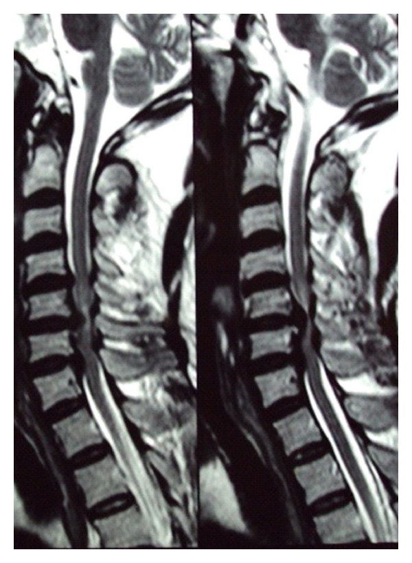
MRISagittal cervical MRI, two sequences: multilevel disc herniations from C4 to C7 with caudal migration and spinal cord compression.
Source : Rustagi et al., Case reports in orthopedics, 2011, figure 1 · CC BY
The diagnosis of cervical radiculopathy is a demanding clinical process that rests not on a single test or symptom, but on the convergence of findings from the history, the physical examination and, if necessary, imaging.1 Cervical radiculopathy is defined as a set of neurological symptoms and signs resulting from compression or irritation of a cervical root.1,2
Which questions should you ask to understand the patient and their history?
History-taking is the foundation of the diagnosis. Particular attention should be paid to the following elements:
- 🗺️ Mapping and quality of the pain : it is essential to ask the patient to describe the topography of their pain precisely. Pain that radiates below the elbow is strongly associated with nerve compression confirmed on MRI (Rainville 2017).3 The description (shooting pain, burning, electric shock) and the paraesthesia (tingling, pins and needles) are more reliable than assessing motor strength during the examination.
- 🔄 Influencing factors : identify the positions that aggravate or relieve the symptoms. Aggravation on coughing or sneezing suggests a radicular origin (Valsalva effect). The “shoulder abduction sign” (relief with the arm placed in abduction over the head) is specific and clinically useful.4
- 🗓️ History of the condition and risk factors : mode of onset (traumatic or gradual), duration, course. Look for heavy manual work, smoking, previous cervical trauma, frequent driving, repeated diving (Kelsey 1984).5
- 🚩 Systematic screening for red flags : signs of myelopathy, progressive deficit, unexplained weight loss, fever, night sweats, history of cancer, sphincter disturbance (Finucane 2020).6
Clinical mapping of cervical radiculopathies by root
Recognising the affected root quickly guides the examination and the differential diagnosis
| Root | Pain / dermatome | Motor weakness (myotome) | Reflex | Key provocation test |
|---|---|---|---|---|
| C5 | Lateral shoulder, deltoid | Shoulder abduction (deltoid) | Biceps reflex reduced | Ipsilateral Spurling A |
| C6 | Lateral arm, thumb + index finger | Elbow flexion (biceps), wrist extension | Biceps ± brachioradialis reflex | Spurling + ULNT1 (median) |
| C7 ⭐ | Dorsum of the hand, 3rd finger | Elbow extension (triceps), wrist flexion | Triceps reflex reduced | Spurling + ULNT1 |
| C8 | Medial border of the hand, 4th-5th fingers | Finger flexion, intrinsic hand muscles | No specific reflex | ULNT3 (ulnar nerve) |
| T1 | Medial aspect of the arm and forearm | Intrinsic hand muscles (lumbricals) | No specific reflex | ULNT3 + Horner's sign if TOS |
⭐ The C7 root is affected in 45-60% of cases. A concomitant Horner's sign (ptosis, miosis, anhidrosis) must raise the suspicion of Pancoast syndrome (apical lung tumour): an absolute diagnostic emergency.
Which clinical tests should you perform and which diagnostic cluster should you use?
The physical examination aims to confirm or refute the hypotheses raised by the history. Research has clearly established that no clinical test taken in isolation is sufficient to diagnose cervical radiculopathy with certainty.7,8 The strength of the diagnosis lies in the combination of several tests. The updated meta-analysis by Thoomes 2026 (BMC MSD, 8 studies) confirms and refines this finding.9
Diagnostic performance of the clinical tests (Thoomes 2018-2026 meta-analysis)
Sensitivity versus specificity profile: use the ULNT1 to rule out, Spurling to confirm
Sources: Thoomes EJ et al., Spine J 2018 (PMID 28838857); Thoomes EJ et al., BMC Musculoskelet Disord 2026 update (PMC 13088722); Wainner RS et al., Spine 2003 (PMID 12544957). Practical strategy : a negative ULNT1 allows you to rule out ; a positive Spurling A helps to confirm ; all 4 cluster tests positive at once > a very high LR+.
The 3 key tests to master
- Spurling A test : axial compression of the head in extension + ipsilateral rotation + side bending towards the symptomatic side. Very high specificity (0.89-1.00) but low sensitivity (~0.50): a positive test makes radiculopathy very likely; a negative one does not rule it out.8,10
- Cervical distraction test : manual axial traction applied to the head in supine lying. Relief of the patient's symptoms = positive test. High specificity (0.90-1.00), low to moderate sensitivity.8
- Upper limb neurodynamic test 1 (ULNT1) : places the median nerve and the C5-C7 roots under tension. Sequence: shoulder abduction 110°, forearm supination, wrist and finger extension, elbow extension, contralateral cervical side flexion. Very high sensitivity (0.83-0.97), low specificity: its main use is to rule out the diagnosis when it is negative.8,9
The Wainner cluster: the validated combination
The combination of tests considerably increases diagnostic certainty. The cluster originally validated by Wainner et al. (2003, n=82 patients referred for EMG, the electrophysiological gold standard) is the most studied.10 If the following 4 tests are positive at the same time, the post-test probability is very high (LR+ > 30):
Wainner 2003 cluster: 4 tests to confirm cervical radiculopathy
With 4/4 positive: post-test probability > 90%. With 3/4 positive: post-test probability 65%.
| # | Test | Criterion for a positive test | LR+ alone |
|---|---|---|---|
| 1 | Spurling A | Reproduction of the radicular symptoms | 3.5 |
| 2 | Cervical distraction | Relief of the symptoms | 4.4 |
| 3 | ULNT1 (median) | Reproduction of the patient's arm pain | 1.3 |
| 4 | Active cervical rotation | < 60° towards the symptomatic side | 2.1 |
Source: Wainner RS, Fritz JM, Irrgang JJ, Boninger ML, Delitto A, Allison S. Spine 2003;28(1):52-62 (PMID 12544957). Independent validation: Thoomes 2018 (PMID 28838857) confirms the value of the cluster despite the heterogeneity of the studies. With 4 positive tests: LR+ 30.3; with 3 positive: LR+ 6.1; with 0-2 positive: not discriminating.
Which other conditions must you rule out?
Cervical radiculopathy is a great imitator 🧠: it can mimic (and conversely be mimicked by) many upper limb conditions. A structured cervical examination is mandatory in the face of any persistent upper limb pain or neurological symptom, and conversely a full upper limb examination is required in the face of any cervicobrachial pain.
| Differential diagnosis | Distinguishing features | Test/additional investigation |
|---|---|---|
| Rotator cuff tendinopathy/impingement | Pain localised to the shoulder, triggered by shoulder movements (abduction 60-120°) | Positive shoulder tests (Hawkins, Neer, Jobe); negative Wainner cluster |
| Frozen shoulder | Passive AND active limitation of external rotation | Passive examination of the shoulder |
| Carpal tunnel syndrome | Night paraesthesia in the 1st-3rd fingers, positive wrist Tinel, positive Phalen | EMG; but beware of double crush syndrome |
| Cubital tunnel syndrome | Paraesthesia in the 4th-5th fingers, positive elbow Tinel, Froment's sign | Ulnar nerve EMG |
| Thoracic outlet syndrome (TOS) | Symptoms with the arm elevated (cleaning windows), positive postural tests | Dynamic EOS imaging, vascular Doppler; provocation manoeuvres |
| Pancoast syndrome ⚠️ | C8-T1 pain, Horner's sign (ptosis, miosis, anhidrosis), deterioration in general condition, smoking | URGENT chest radiograph, chest CT |
| Cervical myelopathy | Clumsiness, hyperreflexia, positive Hoffmann, positive Babinski, balance disturbance | URGENT spinal cord MRI |
| Lateral epicondylalgia | Tennis elbow pain, tenderness on palpation of the lateral epicondyle | Cozen and Mill tests; no dermatome or myotome pattern |
Key points
- History: pain radiating below the elbow is a strong indicator of a radicular origin (Rainville 2017).
- No single test is sufficient. Combining them is essential (Thoomes 2018, Thoomes 2026 update).
- Wainner cluster : 4 tests (Spurling A + distraction + ULNT1 + rotation < 60°) → 4/4 positive = LR+ > 30, post-test probability > 90%.
- ULNT1: high sensitivity → rule out. Spurling: high specificity → confirm.
- Essential differential diagnosis: shoulder, carpal tunnel, cubital tunnel, thoracic outlet, Pancoast, myelopathy, epicondylalgia.
- Finucane 2020 red flags: myelopathy, progressive deficit, fever, deterioration in general condition, history of cancer → urgent referral.
Bibliography, chapter 2
- Iyer S, Kim HJ. Cervical radiculopathy. Curr Rev Musculoskelet Med. 2016;9(3):272-280. PMID 27250042.
- Childress MA, Becker BA. Nonoperative Management of Cervical Radiculopathy. Am Fam Physician. 2016;93(9):746-754. PMID 27175952.
- Caridi JM, Pumberger M, Hughes AP. Cervical radiculopathy: a review. HSS J. 2011;7(3):265-272. PMID 23024624.
- Eubanks JD. Cervical radiculopathy: nonoperative management of neck pain and radicular symptoms. Am Fam Physician. 2010;81(1):33-40. PMID 20052961.
- Kelsey JL, Githens PB, Walter SD, et al. An epidemiological study of acute prolapsed cervical intervertebral disc. J Bone Joint Surg Am. 1984;66(6):907-914. PMID 6736091.
- Finucane LM, Downie A, Mercer C, et al. International Framework for Red Flags for Potential Serious Spinal Pathologies. J Orthop Sports Phys Ther. 2020;50(7):350-372. PMID 32438853.
- Blanpied PR, Gross AR, Elliott JM, et al. Neck Pain: Revision 2017. J Orthop Sports Phys Ther. 2017;47(7):A1-A83. doi:10.2519/jospt.2017.0302.
- Thoomes EJ, van Geest S, van der Windt DA, et al. Value of physical tests in diagnosing cervical radiculopathy: a systematic review. Spine J. 2018;18(1):179-189. PMID 28838857.
- Thoomes EJ et al. Diagnostic accuracy of physical examination tests for painful cervical radiculopathy: update of a systematic review and meta-analysis. BMC Musculoskelet Disord. 2026. PMC 13088722.
- Wainner RS, Fritz JM, Irrgang JJ, Boninger ML, Delitto A, Allison S. Reliability and diagnostic accuracy of the clinical examination and patient self-report measures for cervical radiculopathy. Spine. 2003;28(1):52-62. PMID 12544957.
- Bono CM, Ghiselli G, Gilbert TJ, et al. An evidence-based clinical guideline for the diagnosis and treatment of cervical radiculopathy from degenerative disorders. Spine J. 2011;11(1):64-72. PMID 21168100.
- Cote P, Wong JJ, Sutton D, et al. Management of neck pain and associated disorders: A clinical practice guideline from the Ontario Protocol for Traffic Injury Management (OPTIMa) Collaboration. Eur Spine J. 2016. doi:10.1007/s00586-016-4467-7.
Which treatment strategies are the most effective?
Where do you start? What is the hierarchy of interventions?
The current consensus, carried by the most recent guidelines (Blanpied 2017 JOSPT, a CPG currently under revision for 2026; OPTIMa 2022 update; Bono 2011 NASS), points towards a multimodal, patient-centred approach rather than a rigid hierarchy of isolated interventions.1,2,3 The network meta-analysis by Núñez de Arenas-Arroyo 2025 (36 RCTs, 25 interventions) confirms this additive effect of the components experimentally.4
🧠 The first, fundamental step is patient education. Explain the nature of the radiculopathy, reassure the patient about its generally favourable prognosis and deconstruct catastrophic beliefs about pain and imaging.5
The recommended initial approach systematically combines:
- Therapeutic education , with information about the condition, self-management strategies and the importance of the patient's active role.1,2
- Therapeutic exercise , introduced early: mobility, strength, motor control of the neck and shoulder girdle.6
- Manual therapy , with mobilisations or manipulations for short-term relief, making it easier to engage in exercise.7
- Mechanical cervical traction , a first-line option when radicular symptoms are present (Romeo 2018, meta-analysis of 7 RCTs).8
Hierarchy of interventions according to the international guidelines
Multimodal decision algorithm, from Blanpied 2017 JOSPT, OPTIMa 2022 update, Bono 2011 NASS
The randomised trial by Engquist 2017 (J Neurosurg Spine, n=63, 5-8 year follow-up) showed that early surgery + physiotherapy is marginally superior to physiotherapy alone for pain at 1 year, but the difference narrows by 5 years. Conclusion: structured physiotherapy must be the first line, with surgery reserved for failures or progressive deficits.
What is the place of exercise and is there a superior approach?
Therapeutic exercise is a cornerstone of conservative treatment, with solid evidence supporting its effectiveness (Liang 2019 meta-analysis, n=534 patients).9 💪 However, current research does not demonstrate any clear superiority of one type of exercise over another. The network meta-analysis by Núñez de Arenas-Arroyo 2025 assessed 25 interventions made up of 8 active components: none proved universally superior.4
The most commonly validated exercise strategies include:
- Specific strengthening : deep neck flexors (longus colli, longus capitis) and scapulothoracic stabilisers (lower trapezius, serratus anterior, rhomboids). This strengthening improves postural support and reduces the load on irritated cervical structures.9
- Mobility and range of motion exercises : controlled pain-free movement, segmental glides, prevention of stiffness after irritation.
- Neurodynamic mobilisations (sliders/tensioners) : Nee 2012 (n=60, RCT) demonstrated a clinically relevant immediate benefit (NDI -7.8 points, NNT 2.7 for global perceived improvement).10 The meta-analysis by Gillot 2025 (Clin Rehabil) confirms an additive effect on pain and function.11
- Cervical proprioceptive exercises : oculomotor retraining, postural control, particularly useful after whiplash-type cervical trauma.
Manual therapy, traction, neurodynamics: how effective are they?
Manual therapies, such as spinal mobilisations and manipulations, are widely used. Their main effectiveness lies in short-term pain reduction and in making active participation easier.7,12 The J Pain Res 2025 network meta-analysis (PMC 12008560) confirms that manual therapy + exercise combined is more effective than either intervention alone on the NDI and on pain.12
| Modality | Expected effect | Level of evidence (GRADE) | Key reference |
|---|---|---|---|
| Multimodal exercise | ↓ Pain, ↑ Function (NDI -8 to -15 pts) | High | Liang 2019 (PMID 31702624); Núñez 2025 NMA |
| Manual therapy + exercise | ↓ Short-term pain, ↑ Function | High | Boyles 2011 (PMID 22851876); NMA J Pain Res 2025 |
| Cervical traction + PT | ↓ Pain, ↓ Disability in the short and medium term | Moderate | Romeo 2018 (PMID 29315428), 7 RCTs |
| Neurodynamic mobilisations | ↓ Immediate pain, NNT 2.7 | Moderate | Nee 2012 (PMID 22341379) |
| Therapeutic education | ↓ Kinesiophobia, ↑ Self-efficacy | Moderate | Moseley & Butler 2015; Louw 2018 |
| Semi-rigid collar (short term) | ↓ Pain at 6 weeks | Moderate | Kuijper 2009 (PMID 19812130) |
| Spinal manipulation | ↓ Short-term pain | Moderate | NMA JOSPT 2025 (DOI 10.2519/jospt.2025.12757) |
| Cervical LLLT | Uncertain effect, heterogeneous evidence | Low | Mixed data |
| Therapeutic ultrasound | No specific effect demonstrated | Very low | Not recommended (Blanpied 2017) |
| TENS alone | Transient symptomatic effect | Very low | Limited data |
Focus: mechanical cervical traction
The meta-analysis by Romeo 2018 (Phys Ther 98(4):231-242) analysed 7 RCTs on adding cervical traction to standard physiotherapy.8 Conclusion: traction added to physiotherapy significantly reduces pain and disability in the short term (SMD -0.87) and the medium term (SMD -0.69) compared with physiotherapy alone. Preferred indication : patients whose symptoms decrease with manual decompression during the distraction test. Typical protocol: 8-12 kg, intermittent (60s on/20s off), 15-20 min, 3x a week.
How do you educate the patient and address psychosocial factors?
Education is a therapeutic intervention in its own right. Communication should focus on reassurance, on explaining the largely favourable prognosis of cervical radiculopathy and on promoting self-efficacy. Addressing the psychosocial factors, the yellow flags , is essential to prevent the problem becoming chronic.
- Explaining pain : use accessible metaphors to take the drama out of imaging (Brinjikji 2015: 87.6% of asymptomatic subjects > 60 years have disc degeneration on MRI) and explain pain as a protective alarm system rather than an indicator of ongoing tissue damage.13,14
- Identifying and managing limiting beliefs : kinesiophobia, catastrophising, anxiety and depression predict poorer recovery. The Fear-Avoidance Beliefs Questionnaire (FABQ) or the Tampa Scale of Kinesiophobia (TSK) can be used.
- Setting functional goals : work with the patient to set meaningful, achievable goals (Patient-Specific Functional Scale, PSFS).
Key points
- ✅ The most effective approach is multimodal : education + exercise + manual therapy, individualised.
- ✅ Education and reassurance about the favourable prognosis are the first line (Blanpied 2017).
- ✅ Exercise is fundamental, but no specific type has proved superior (NMA Núñez 2025): individualisation is the key.
- ✅ The cervical traction added to PT reduces pain and disability in the short and medium term (Romeo 2018).
- ✅ The manual therapy and neurodynamics are useful adjuncts, not stand-alone treatments (Boyles 2011, Nee 2012).
- ✅ The consideration given to yellow flags is crucial to prevent the problem becoming chronic.
- ✅ Therapeutic ultrasound and TENS alone: not recommended for lack of evidence of any specific effect.
Bibliography, chapter 3
- Blanpied PR, Gross AR, Elliott JM, et al. Neck Pain: Revision 2017. J Orthop Sports Phys Ther. 2017;47(7):A1-A83. doi:10.2519/jospt.2017.0302.
- Wong JJ, Cote P, Sutton DA, et al. Effectiveness of Multimodal Rehabilitation Interventions for Management of Cervical Radiculopathy in Adults: An Updated Systematic Review from the OPTIMa Collaboration. J Rehabil Med. 2022. PMID 35797062.
- Bono CM, Ghiselli G, Gilbert TJ, et al. An evidence-based clinical guideline for the diagnosis and treatment of cervical radiculopathy from degenerative disorders. Spine J. 2011;11(1):64-72. PMID 21168100.
- Núñez de Arenas-Arroyo S, Mavridis D, Martínez-Vizcaino V, et al. What components and formats of rehabilitation interventions are more effective to reduce pain in patients with cervical radiculopathy? A Systematic review and component network meta-analysis. Clin Rehabil. 2025;39(10):1296-1310. PMID 40776625.
- Louw A, Nijs J, Puentedura EJ. A clinical perspective on a pain neuroscience education approach to manual therapy. J Man Manip Ther. 2017;25(3):160-168. PMID 28694679.
- Cleland JA, Whitman JM, Fritz JM, Palmer JA. Manual physical therapy, cervical traction, and strengthening exercises in patients with cervical radiculopathy: a case series. J Orthop Sports Phys Ther. 2005;35(12):802-811. PMID 16848101.
- Boyles R, Toy P, Mellon J, Hayes M, Hammer B. Effectiveness of manual physical therapy in the treatment of cervical radiculopathy: a systematic review. J Man Manip Ther. 2011;19(3):135-142. PMID 22851876.
- Romeo A, Vanti C, Boldrini V, et al. Cervical Radiculopathy: Effectiveness of Adding Traction to Physical Therapy: A Systematic Review and Meta-Analysis of Randomized Controlled Trials. Phys Ther. 2018;98(4):231-242. PMID 29315428.
- Liang L, Feng M, Cui X, et al. The effect of exercise on cervical radiculopathy: A systematic review and meta-analysis. Medicine (Baltimore). 2019;98(45):e17733. PMID 31702624.
- Nee RJ, Vicenzino B, Jull GA, Cleland JA, Coppieters MW. Neural tissue management provides immediate clinically relevant benefits without harmful effects for patients with nerve-related neck and arm pain: a randomised trial. J Physiother. 2012;58(1):23-31. PMID 22341379.
- Effectiveness of Articular and Neural Mobilization for Managing Cervical Radicular Pain: A Systematic Review With Network Meta-Analysis. J Orthop Sports Phys Ther. 2025. doi:10.2519/jospt.2025.12757.
- Manual Therapy for Cervical Radiculopathy: Effects on Neck Disability and Pain: A Systematic Review and Network Meta-Analysis. J Pain Res. 2025;18:2035-2045. PMC 12008560.
- Brinjikji W, Luetmer PH, Comstock B, et al. Systematic literature review of imaging features of spinal degeneration in asymptomatic populations. AJNR Am J Neuroradiol. 2015;36(4):811-816. PMID 25430861.
- Moseley GL, Butler DS. Fifteen Years of Explaining Pain: The Past, Present, and Future. J Pain. 2015;16(9):807-813. PMID 26051220.
- Engquist M, Lofgren H, Oberg B, et al. A 5- to 8-year randomized study on the treatment of cervical radiculopathy: anterior cervical decompression and fusion plus physiotherapy versus physiotherapy alone. J Neurosurg Spine. 2017;26(1):19-27. PMID 27564856.
How do you secure lasting recovery and prevent flares?
Long-term management of cervical radiculopathy aims to go beyond simply relieving acute symptoms and to move towards functional resilience and the prevention of recurrence. A lasting strategy rests on the synergistic combination of patient empowerment, targeted strengthening and a progressive, safe return to activities.1
How do you make the patient an active participant in their own recovery through self-management?
Patient empowerment is the pillar of lasting recovery. 🧠 This approach reduces dependence on the therapist and significantly improves long-term outcomes.2
Structured therapeutic education
The first step is therapeutic patient education (TPE). Explaining the nature of the radiculopathy, its generally favourable prognosis and the mechanisms of pain is fundamental. Education in pain neuroscience (PNE) can reduce fear of movement, reduce pain intensity and improve function in patients with chronic musculoskeletal pain.2,3 Most cases of acute cervical radiculopathy improve considerably with conservative treatment within 4-6 months: a reassuring piece of information.4
The progressive home exercise programme
Personalised and progressive, it typically includes:
- Motor control and strengthening exercises: strengthening the deep neck flexors (cranio-cervical flexion test, CCFT) and the scapular stabilisers (lower trapezius, serratus anterior) improves the dynamic stability of the cervical spine and reduces the load on the neural structures.5,6
- Neural mobility exercises: sliders and tensioners for the median nerve (the one most often involved at C6-C7), the ulnar nerve (C8-T1) or the radial nerve. Reference: Nee 2012, a protocol of 5 progressive movements performed 3x a day in sets of 10.7
- Postural correction and ergonomics: maintain a neutral posture, particularly during prolonged activities (computer, driving). Simple ergonomic advice (screen at eye height, chair position, a break every 45 min).8
- Postural endurance exercises: wall angels, prone Y/T/I, scapular setting: progression towards function.
Typical progression of a home programme (12 weeks)
Gradual adaptation according to tolerance and symptom response
Practical markers: NDI measured at every phase transition (W2, W6, W10). The MCID (Minimal Clinically Important Difference) = 7 points for the NDI. Progression criterion: reduction in radicular symptoms AND tolerance of the load without a flare-up.
Why should you remain critical of imaging?
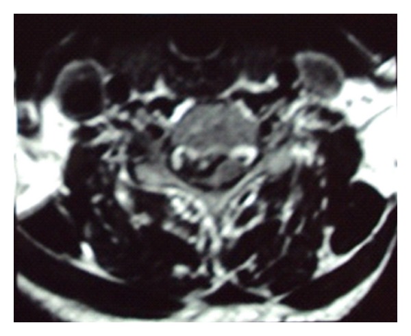
MRIAxial cervical MRI at the C4-C5 level: disc herniation compressing the right side of the spinal cord.
Source : Rustagi et al., Case reports in orthopedics, 2011, figure 2 · CC BY
The correlation between MRI and clinical symptoms is weak. The major systematic review by Brinjikji 2015 (3,110 asymptomatic subjects) quantified the prevalence of degenerative abnormalities in people without pain :
MRI abnormalities in asymptomatic subjects: the lesson from Brinjikji 2015
Prevalence of disc degeneration in the cervical/lumbar spine by age band
Major clinical implication : a 50-year-old patient presenting with cervical radiculopathy will almost always have degenerative changes on MRI, but these are not necessarily the cause of the symptoms. Treat the patient, not the image. Imaging should be requested IF: a red flag, a progressive neurological deficit, or failure after 4-6 weeks of well-conducted conservative treatment.
Key points
- ✅ The prevention of recurrence rests on empowering the patient through education and a personalised exercise programme.
- 💪 Strengthening the deep neck flexors and scapular stabilisers is an essential, proven component.
- 🧠 Education in pain neuroscience reduces kinesiophobia and improves functional outcomes.
- 🤔 Let us stay critical of imaging : we treat a patient, not a radiological image. A disc herniation is not synonymous with pain (Brinjikji 2015).
- 📐 A programme structured in 4 phases over 12 weeks: relief (W1-2) → motor control (W3-6) → strengthening (W6-10) → return to activities (W10-12+).
Bibliography, chapter 4
- Blanpied PR, Gross AR, Elliott JM, et al. Neck Pain: Revision 2017. J Orthop Sports Phys Ther. 2017;47(7):A1-A83. doi:10.2519/jospt.2017.0302.
- Louw A, Nijs J, Puentedura EJ. A clinical perspective on a pain neuroscience education approach to manual therapy. J Man Manip Ther. 2017;25(3):160-168. PMID 28694679.
- Moseley GL, Butler DS. Fifteen Years of Explaining Pain: The Past, Present, and Future. J Pain. 2015;16(9):807-813. PMID 26051220.
- Eubanks JD. Cervical radiculopathy: nonoperative management of neck pain and radicular symptoms. Am Fam Physician. 2010;81(1):33-40. PMID 20052961.
- Liang L, Feng M, Cui X, et al. The effect of exercise on cervical radiculopathy. Medicine (Baltimore). 2019;98(45):e17733. PMID 31702624.
- Cleland JA, Fritz JM, Whitman JM, Heath R. Predictors of short-term outcome in people with a clinical diagnosis of cervical radiculopathy. Phys Ther. 2007;87(12):1619-1632. PMID 17911271.
- Nee RJ, Vicenzino B, Jull GA, Cleland JA, Coppieters MW. Neural tissue management provides immediate clinically relevant benefits without harmful effects for patients with nerve-related neck and arm pain: a randomised trial. J Physiother. 2012;58(1):23-31. PMID 22341379.
- Wong JJ, Cote P, Sutton DA, et al. Effectiveness of Multimodal Rehabilitation Interventions for Management of Cervical Radiculopathy in Adults: An Updated Systematic Review from the OPTIMa Collaboration. J Rehabil Med. 2022. PMID 35797062.
- Brinjikji W, Luetmer PH, Comstock B, et al. Systematic literature review of imaging features of spinal degeneration in asymptomatic populations. AJNR Am J Neuroradiol. 2015;36(4):811-816. PMID 25430861.
- Kuijper B, Tans JT, Beelen A, Nollet F, de Visser M. Cervical collar or physiotherapy versus wait and see policy for recent onset cervical radiculopathy: randomised trial. BMJ. 2009;339:b3883. PMID 19812130.
- Engquist M, Lofgren H, Oberg B, et al. A 5- to 8-year randomized study on the treatment of cervical radiculopathy: anterior cervical decompression and fusion plus physiotherapy versus physiotherapy alone. J Neurosurg Spine. 2017;26(1):19-27. PMID 27564856.
The athlete and return to play: protocol and progression criteria
Cervical radiculopathy in the athlete deserves particular attention because it can sit on a continuum with specific entities: “stingers” and “burners” (transient brachial plexus injuries, common in rugby and American football), post-traumatic cervical instability, and injuries related to repeated axial loading (weightlifting, wrestling). 🏉 The clinical challenge is twofold: (1) treat the radiculopathy effectively so that the athlete can return, (2) avoid a premature return that would expose them to more serious neurological injury.
Specific features of the athlete with cervical radiculopathy
Athletes have specific risk factors:
- Contact sports (rugby, American football, MMA, wrestling, judo, hockey): repeated impacts, tackles and falls, hence a high risk of “stinger” episodes (a transient shock down the arm after trauma) that share their pathophysiology with radiculopathy.
- Axial loading sports (weightlifting, crossfit, gymnastics): repeated axial compression favouring early disc disease.
- Racket and throwing sports (tennis, baseball, javelin): postural asymmetry, unilateral hypertrophy, forced extension and rotation stresses.
- Diving / water polo : cervical impacts on entering the water (Kelsey 1984 identified repeated diving as a risk factor).1
🚩 Red flags specific to the athlete
- More than one “stinger” in the same season → suspect cervical stenosis, MRI mandatory before returning
- Bilateral symptoms (transient or persistent) after cervical trauma → suspect acute myelopathy → URGENT MRI
- Motor weakness persisting > 24h after an acute episode → full neurological work-up
- Painful neck + signs of myelopathy (Lhermitte, balance problems, hyperreflexia) → immediate suspension, urgent MRI
- Cervical canal diameter < 13 mm (Torg-Pavlov measurement) → contraindication to contact sports
How do you plan a safe return to play in 5 phases?
Return to play (RTP) must be progressive and based on functional criteria, not on a fixed timetable.2 🏋️♀️ A premature or poorly managed return is a major risk factor for recurrence and, in contact sports, can expose the athlete to more serious injury.
5-phase RTP protocol: progression criteria
Validation between phases: full range of motion + normal strength + negative provocation tests + functional tolerance
Framework adapted from the general principles of musculoskeletal RTP. No fixed timeline: progress according to the criteria, never the calendar. Monitoring tool : rating of perceived exertion (RPE), weekly NDI, symptom and intensity diary.
Special case: the “stinger / burner”
A stinger is a transient unilateral episode of lightning pain + paraesthesia + temporary weakness (< 24h) typically occurring after a tackle or a fall. It shares the pathophysiology of radiculopathy: compression or stretching of the C5-C6 root during forced side bending. Practical rule : a first isolated stinger that resolves in less than 15 minutes generally allows the player to carry on with the match after examination. Any stinger persisting > 24h, recurrent or bilateral calls for immediate withdrawal and a full work-up (MRI + Torg-Pavlov cervical canal measurement). Congenital cervical stenosis (canal < 13 mm) is a relative contraindication to contact sports according to several expert opinions.
⭐ Key points
- 🏉 Contact sport athletes (rugby, American football, MMA, hockey) have a high risk of “stinger” and “burner” episodes that share the pathophysiology of radiculopathy.
- 🚨 Red flags in the athlete : bilateral symptoms after trauma → URGENT MRI; more than 1 stinger a season → full work-up; cervical canal < 13 mm (Torg-Pavlov) → relative contraindication to contact sports.
- 📐 The 5-phase RTP protocol must be followed on the basis of functional criteria (mobility, strength, negative provocation tests) and not on a fixed calendar.
- 🏋️♀️ Any premature return is a major risk factor for recurrence and, in contact sports, can expose the athlete to more serious injury.
- 🎯 Monitoring tools : weekly NDI, RPE, regular provocation tests, symptom diary.
Bibliography, chapter 5 (athletes)
- Kelsey JL, Githens PB, Walter SD, et al. An epidemiological study of acute prolapsed cervical intervertebral disc. J Bone Joint Surg Am. 1984;66(6):907-914. PMID 6736091.
- Blanpied PR, Gross AR, Elliott JM, et al. Neck Pain: Revision 2017. J Orthop Sports Phys Ther. 2017;47(7):A1-A83. doi:10.2519/jospt.2017.0302.
- Wong JJ, Cote P, Sutton DA, et al. Effectiveness of Multimodal Rehabilitation Interventions for Management of Cervical Radiculopathy in Adults: An Updated Systematic Review from the OPTIMa Collaboration. J Rehabil Med. 2022. PMID 35797062.
- Finucane LM, Downie A, Mercer C, et al. International Framework for Red Flags for Potential Serious Spinal Pathologies. J Orthop Sports Phys Ther. 2020;50(7):350-372. PMID 32438853.
- Cleland JA, Fritz JM, Whitman JM, Heath R. Predictors of short-term outcome in people with a clinical diagnosis of cervical radiculopathy. Phys Ther. 2007;87(12):1619-1632. PMID 17911271.
What do real clinical cases teach us?
Analysing clinical cases published in the scientific literature offers a valuable insight into the presentation, the diagnostic reasoning and the treatment strategies applied to specific individuals. 🧐 Although they rank lower in level of evidence than randomised controlled trials, they illustrate the complexity and variability of everyday clinical practice.
Analysis of a classic case: from assessment to resolution
The reference case series by Cleland, Whitman, Fritz & Palmer (JOSPT 2005, 11 consecutive patients) illustrates an identifiable trajectory.1 The patients showed clinical signs typical of a C5 to C7 radiculopathy, with: pain aggravated by neck movements, positive provocation tests (Spurling and cervical distraction), a motor deficit in the territory concerned and dermatomal loss of sensation.
The treatment approach, based on the evidence emerging at the time and confirmed since, was multimodal. It combined:
- Manual therapy including thoracic manipulations and cervical mobilisations to improve segmental mobility
- Mechanical cervical traction intermittent (10 to 25 lbs, 15-20 min)
- Specific therapeutic exercises : strengthening the deep neck flexors + scapulothoracic stabilisers + neurodynamic mobilisations (median nerve gliding)
The treatment plan was progressive, adapted to the decreasing irritability of the symptoms. 10 of the 11 patients (91%) achieved a clinically significant improvement in 7-8 sessions. Despite its methodological limitations (level 4), this case series was a founding text for evidence-based practice in cervical physiotherapy and inspired the subsequent RCT by Cleland 2007 (Phys Ther, n=96, predictors).2
The diagnostic challenge: the great imitator and double crush syndrome
One of the greatest clinical challenges is that cervical radiculopathy is a great imitator. 🧠 Its symptoms can mimic (and conversely be mimicked by) many musculoskeletal conditions of the upper limb, leading to diagnostic errors and ineffective treatment.
The “double crush syndrome” concept
First described by Upton and McComas in 1973, the double crush syndrome holds that a proximal nerve compression (cervical radiculopathy) makes the nerve more vulnerable to a distal compression (carpal tunnel, cubital tunnel) and vice versa. The concept remains debated but clinically useful: it is a reminder of how important it is to examine the whole course of the nerve. The incidence of a coexisting double crush between cervical radiculopathy and carpal tunnel syndrome in surgical patients has been estimated at about 3.5% in a recent study (Lo 2022, J Hand Surg Am).3 Patients without and with a double crush achieve similar rates of improvement after carpal tunnel release (Patton 2024, n=370+).4
Classic diagnostic errors
- Mimicking a shoulder disorder : C5 radiculopathy often mimics a rotator cuff tendinopathy (lateral deltoid pain, weak abduction). Diagnostic key : normal passive shoulder examination + positive Wainner cluster.
- Mimicking a lateral epicondylalgia : C6-C7 radiculopathy can mimic a tennis elbow. Diagnostic key : positive ULNT1 + no pain on palpation of the epicondyle.
- Mimicking a carpal tunnel syndrome : paraesthesia in the 1st-3rd fingers can arise from a C6-C7 root. Diagnostic key : specific EMG + cervical tests + search for a double crush.
- Mimicking a Pancoast syndrome ⚠️: C8-T1 pain, weakness of the intrinsic hand muscles, unilateral Horner's sign, smoking. Urgent action : chest radiograph + CT.
Study of a complex case: chronicity and central sensitisation
Some cases go beyond the classic picture. Chronic cervical radiculopathy (> 3 months), particularly with documented central sensitisation, calls for a broader approach. Key strategies:
- Pain neuroscience education (Moseley 2015), with explanation of the central mechanisms, taking the drama out of imaging and cognitive reframing.5
- Graded exposure to provocative movements within a reassuring, structured framework.
- Biopsychosocial approach addressing the comorbidities (depression, anxiety, sleep disorders).
- Multidisciplinary collaboration : general practitioner, rehabilitation physician, psychologist specialising in chronic pain, or even a chronic pain team.
Hierarchy of evidence: where do clinical cases sit?
📐 Hierarchy of scientific evidence: where does each type of study belong?
Strength of evidence decreasing from the top (meta-analyses) to the bottom (isolated cases)
A simplified GRADE / Oxford CEBM hierarchy. The length of the coloured bar illustrates the relative strength of evidence. Practical implication: when an appealing clinical case and a meta-analysis diverge, follow the meta-analysis. Clinical cases remain valuable for generating hypotheses, flagging rare presentations or illustrating clinical reasoning.
⭐ Key points
- ✅ The founding case series by Cleland 2005 JOSPT (11 consecutive patients) shows that a multimodal approach (thoracic thrust + traction + exercise + neurodynamics) achieves resolution in 7-8 sessions in most patients.
- ⚠️ Cervical radiculopathy is a great imitator : it can present as shoulder pain, an epicondylalgia, a carpal tunnel syndrome, or even a Pancoast syndrome. A structured cervical examination is essential in the face of any persistent upper limb pain or neurological symptom.
- 🔍 The double crush syndrome concept remains clinically useful: examine the whole course of the nerve. Cervical radiculopathy and carpal tunnel syndrome coexist in about 3.5% of surgical patients.
- 🤯 The complex cases (chronicity, central sensitisation) call for a broader approach including pain education and graded exposure.
- 🔬 The clinical cases are useful for illustration but carry a low level of evidence. Clinical decisions must always be guided first and foremost by meta-analyses and RCTs.
Bibliography, chapter 6 (clinical cases)
- Cleland JA, Whitman JM, Fritz JM, Palmer JA. Manual physical therapy, cervical traction, and strengthening exercises in patients with cervical radiculopathy: a case series. J Orthop Sports Phys Ther. 2005;35(12):802-811. PMID 16848101.
- Cleland JA, Fritz JM, Whitman JM, Heath R. Predictors of short-term outcome in people with a clinical diagnosis of cervical radiculopathy. Phys Ther. 2007;87(12):1619-1632. PMID 17911271.
- The Incidence of Double Crush Syndrome in Surgically Treated Patients. J Hand Surg Am. 2022. PMID 36321208.
- Patients With and Without Double Crush Syndrome Achieve Similar Rates of Clinical Improvement Following Carpal Tunnel Release. J Hand Surg Am. 2024. PMID 38420760.
- Moseley GL, Butler DS. Fifteen Years of Explaining Pain: The Past, Present, and Future. J Pain. 2015;16(9):807-813. PMID 26051220.
- Wainner RS, Fritz JM, Irrgang JJ, Boninger ML, Delitto A, Allison S. Reliability and diagnostic accuracy of the clinical examination and patient self-report measures for cervical radiculopathy. Spine. 2003;28(1):52-62. PMID 12544957.
- Brinjikji W, Luetmer PH, Comstock B, et al. Systematic literature review of imaging features of spinal degeneration in asymptomatic populations. AJNR Am J Neuroradiol. 2015;36(4):811-816. PMID 25430861.
- Greenhalgh T, Howick J, Maskrey N. Evidence based medicine: a movement in crisis? BMJ. 2014;348:g3725. PMID 24927763.
- Foster NE, Anema JR, Cherkin D, et al. Prevention and treatment of low back pain: evidence, challenges, and promising directions. Lancet. 2018;391(10137):2368-2383. PMID 29573872.
How do you apply these recommendations concretely in your practice?
Applying evidence-based recommendations is not limited to knowing the techniques: it also takes in the clinical judgement needed to refer patients, the objective measurement of outcomes and the ability to overcome systemic and individual barriers. 🧐
When and to which other professionals should you refer?
The physiotherapist is often the first point of contact for musculoskeletal disorders, which gives them a crucial responsibility in triage. The decision to refer rests on structured clinical reasoning. 🤝
1) Red flags: immediate medical referral
Detecting red flags is the absolute priority, because it aims to rule out any serious underlying pathology. Signs and symptoms such as intense non-mechanical night pain, unexplained weight loss, persistent fever, progressive neurological deficits or a history of cancer must trigger an immediate medical referral.1 See the detailed table in chapter 1 (Finucane 2020).
2) Yellow flags: biopsychosocial approach
Referral is also indicated in the presence of predominant psychosocial factors (yellow flags) that hinder progress. High levels of catastrophising, fear-avoidance, anxiety or depression are powerful predictors of pain becoming chronic. Collaboration with, or referral to, a psychologist specialising in pain is recommended.
3) Failure of conservative care: specialist referral
Referral to other specialists may be necessary in the event of stagnation or deterioration after 4-6 weeks of well-conducted treatment :
- MPR physician (physical and rehabilitation medicine specialist) : for a change of therapeutic direction, assessment of comorbidities, discussion of a guided epidural injection.
- Rheumatologist : if an inflammatory condition is suspected (spondyloarthritis, rheumatoid arthritis).
- Neurosurgeon / spinal surgeon : if there is a progressive neurological deficit, prolonged failure of conservative care or signs of myelopathy.
- Neurologist : if the picture is atypical or if EMG/ENMG is required.
How do you measure outcomes and overcome barriers to implementation?
The PROMs (Patient-Reported Outcome Measures) validated in cervical radiculopathy
Systematic use of PROMs has become a standard. The tools validated for cervical radiculopathy include:
| PROM | Domain | Scale | MCID | Use |
|---|---|---|---|---|
| NDI (Neck Disability Index) | Neck disability | 0-50 (or %) | 7 points (5-19 depending on context) | Reference standard, measured at each phase transition |
| DASH / QuickDASH | Upper limb disability | 0-100 | 10.2 (DASH) / 8 (QuickDASH) | If upper limb symptoms predominate |
| PSFS (Patient-Specific Functional Scale) | Activities that matter to the patient | 0-10 per activity | 2 points | Patient-centred, ideal for collaborative goals |
| NPRS (Numeric Pain Rating Scale) | Pain intensity | 0-10 | 2 points | Quick, repeated measure |
| GROC (Global Rating of Change) | Subjective perception of improvement | -7 a +7 | +5 = much improved | Anchor outcome at the end of treatment |
| FABQ or TSK-11 | Kinesiophobia / fear-avoidance | variable | variable | Yellow flags, PNE follow-up |
Barriers to evidence-based implementation
Several systematic reviews (Alshehri 2020, Wilkinson 2024) have identified the barriers most frequently cited by physiotherapists:
- Lack of time to read and appraise the literature (cited by 60-80% of practitioners)
- Lack of skills in research and critical appraisal
- Limited access to scientific databases (paywalls)
- Lack of organisational support (private practice, private settings)
- Habits and clinical expertise perceived as superior to the data
Strategies for overcoming the barriers
- Targeted continuing education : workshops on critical appraisal of the literature
- Accessible resources : PEDro (Physiotherapy Evidence Database), the Cochrane Library, JOSPT free access, syntheses such as Physio Learning
- Mentoring and communities of practice : clinical discussion groups, experienced mentoring
- Technological integration : patient record software with built-in PROMs (automation)
- Patient-centred approach : shared decision-making, collaborative goals
Critique and controversies: beyond the recommendations
Several important tensions persist. First: the gap between research and practice. Research, conducted under controlled conditions on homogeneous populations, does not always reflect the complexity of the patients seen in clinic. Applying a recommendation “to the letter” without adapting it can be counterproductive.
Second: the red flag paradox. Screening for them is essential for safety, but most individual red flags have a low positive predictive value. Real skill lies in the ability to group the signs together and to use probabilistic clinical reasoning.
Third: the risk of “box ticking” with PROMs. PROMs are essential but can become a bureaucratic exercise. Measurement must never replace the conversation and a deep understanding of the patient's experience.
⭐ Key points
- 🚨 Red flags → immediate medical referral (Finucane 2020).
- 🟡 Yellow flags (catastrophising, kinesiophobia, depression) → biopsychosocial approach, collaboration with psychology.
- 📅 Failure of conservative care > 4-6 weeks OR progressive deficit → specialist referral (rehabilitation medicine, spinal surgeon).
- 📊 Validated PROMs : NDI (the standard), DASH if the upper limb predominates, PSFS for patient-centred goals, NPRS for pain.
- 🚧 Barriers to EBP : time, skills, access to resources, organisational support. Solutions: training, synthesised resources, mentoring, technological integration.
- 🎯 Evidence-based practice = the integration of 3 pillars : the best available evidence + clinical expertise + the patient's values and preferences.
Bibliography, chapter 7
- Finucane LM, Downie A, Mercer C, et al. International Framework for Red Flags for Potential Serious Spinal Pathologies. J Orthop Sports Phys Ther. 2020;50(7):350-372. PMID 32438853.
- Blanpied PR, Gross AR, Elliott JM, et al. Neck Pain: Revision 2017. J Orthop Sports Phys Ther. 2017;47(7):A1-A83. doi:10.2519/jospt.2017.0302.
- Wong JJ, Cote P, Sutton DA, et al. Effectiveness of Multimodal Rehabilitation Interventions for Management of Cervical Radiculopathy in Adults: An Updated Systematic Review from the OPTIMa Collaboration. J Rehabil Med. 2022. PMID 35797062.
- Bono CM, Ghiselli G, Gilbert TJ, et al. An evidence-based clinical guideline for the diagnosis and treatment of cervical radiculopathy from degenerative disorders. Spine J. 2011;11(1):64-72. PMID 21168100.
- Greenhalgh T, Howick J, Maskrey N. Evidence based medicine: a movement in crisis? BMJ. 2014;348:g3725. PMID 24927763.
- Foster NE, Anema JR, Cherkin D, et al. Prevention and treatment of low back pain: evidence, challenges, and promising directions. Lancet. 2018;391(10137):2368-2383. PMID 29573872.
- Cleland JA, Fritz JM, Whitman JM, Heath R. Predictors of short-term outcome in people with a clinical diagnosis of cervical radiculopathy. Phys Ther. 2007;87(12):1619-1632. PMID 17911271.
- Cote P, Wong JJ, Sutton D, et al. Management of neck pain and associated disorders: A clinical practice guideline from the Ontario Protocol for Traffic Injury Management (OPTIMa) Collaboration. Eur Spine J. 2016. doi:10.1007/s00586-016-4467-7.
- Núñez de Arenas-Arroyo S, Mavridis D, Martínez-Vizcaino V, et al. What components and formats of rehabilitation interventions are more effective to reduce pain in patients with cervical radiculopathy? A Systematic review and component network meta-analysis. Clin Rehabil. 2025;39(10):1296-1310. PMID 40776625.
And after this article?
This article is part of a collection of evidence-based clinical syntheses on musculoskeletal pain. A question, a comment, a correction to suggest? Contact us directly through the WhatsApp button at the bottom right of the screen.

