















In brief
Whiplash (whiplash-associated disorders, WAD) is a cervical acceleration-deceleration injury, classified by the Quebec Task Force into grades 0 to IV, with grade II (musculoskeletal signs without neurological involvement) the most common. The diagnosis is one of exclusion, with the Canadian C-Spine Rule deciding on imaging; the predictors of chronicity are high initial pain, an NDI above 40%, post-traumatic stress and catastrophising, rather than the severity of the crash. First-line management is active: reassuring education, early return to activity and exercise, with the rigid collar advised against. Around 30 to 50% still have symptoms at one year.
Clinical synthesis based on meta-analyses and international consensus statements: Quebec Task Force, JOSPT Neck Pain CPG 2017, OPTIMa 2016, IFOMPT Red Flags 2020, CISG Amsterdam 2023, Häggman-Henrikson 2025.
Clinical synthesis
- Whiplash (whiplash-associated disorders, WAD) is a cervical acceleration-deceleration injury, classified by the Quebec Task Force (Spitzer 1995) into grades 0 to IV: grade II (musculoskeletal signs without neurological involvement) is the most common.
- The Kamper 2008 meta-analysis (38 cohorts, n > 11,000) shows that pain and disability plateau at around 3 months ; between 30 and 50% of patients still have persistent symptoms at 1 year. The trajectory is not linear.
- The predictors of chronicity identified in the literature (Sarrami 2017, Campbell 2018, Walton 2013) are: high initial pain, baseline NDI > 40%, hyperarousal / PTSD, low recovery expectations, catastrophising. Crash severity is a poor predictor on its own.
- The diagnosis is a diagnosis of exclusion. The Canadian C-Spine Rule (Stiell 2001, sensitivity 100%) decides on imaging. The IFOMPT Finucane 2020 framework guides red flag triage (fracture, arterial dissection, myelopathy).
- The standardised examination includes active range of motion, palpation, neurological examination, the deep cervical flexor test (CCFT, Jull 2008), and screening for kinesiophobia (TSK) and post-traumatic stress (IES-R).
- The Ritchie 2015 Clinical Prediction Rule (external validation, JOSPT 45(4):242-250) stratifies prognosis using age, baseline NDI and hyperarousal: the reference tool.
- The first-line treatment is conservative and active : reassuring education, early return to activity, active exercise. The rigid collar is advised against (OPTIMa Côté 2016, Blanpied 2017 JOSPT CPG).
- Exercise (mobility, motor control, strengthening) is the cornerstone. No single type has been shown to be superior : what matters is regularity and progression (Sterling 2019 J Clin Med).
- Manual therapy (mobilisations, manipulations) provides a short-term benefit on pain and mobility (Cochrane Gross 2015), always alongside the active programme.
- In chronic WAD, central sensitisation (Sterling 2003 Pain: objectively demonstrable sensorimotor changes) calls for a modulated approach: pain neuroscience education, graded motor imagery (Bowering 2013), graded exposure.
- The PROMISE trial (Michaleff 2014, Lancet) shows that an intensive exercise programme is no better than structured advice in chronic WAD: a strong argument for self-management.
- The return to sport should follow a stepwise progression based on remaining symptom-free for 24 h after exertion, drawing on the CISG Amsterdam 2023 consensus (BJSM 57(11):695-711).
- Critical differential diagnosis: cervical arterial dissection (Thomas & Rivett 2015 JOSPT): look for the 5D-3N (dizziness, diplopia, dysarthria, dysphagia, drop attacks / nausea, numbness, nystagmus).
- Major comorbidity: post-whiplash temporomandibular disorders : prevalence is already high in the acute phase and persists (Häggman-Henrikson 2025, Eur J Pain, meta-analysis).
- Measure outcomes with validated PROMs: NDI (Vernon 1991), VAS, TSK-11, IES-R. Adopt a reflective evidence-based approach (Greenhalgh 2014 BMJ, “EBM in crisis”).
Contents
- What are the fundamentals to know about whiplash (WAD)?
- How can whiplash be assessed and diagnosed with confidence?
- Which treatment strategies are the most effective?
- Chronic WAD & central sensitisation: how should chronicity be managed?
- How do you secure lasting recovery and prevent flares?
- What do real clinical cases teach us about WAD?
- How do you apply these recommendations concretely in your practice?
What are the fundamentals to know about whiplash (WAD)?
How is WAD defined and what is the reference classification?
The term whiplash refers to the injury mechanism: a transfer of energy to the cervical spine through sudden acceleration-deceleration, classically in a rear-end car collision, but also in contact sports, falls or diving.1 The resulting cluster of symptoms is grouped under the heading of Whiplash-Associated Disorders (WAD), a term adopted internationally since the monograph of the Quebec Task Force published in Spine in 1995, which remains the worldwide classification reference.2
The QTF classification stratifies WAD into five grades according to clinical severity:
- Grade 0: no neck complaint, no physical signs.2
- Grade I: neck pain, stiffness or tenderness, with no objective physical sign.2
- Grade II: neck complaints AND musculoskeletal signs (reduced range of motion, pain on palpation): the most common category in clinical practice.2,3
- Grade III: neck complaints AND neurological signs (reduced reflexes, motor weakness, sensory deficits).2
- Grade IV: neck complaints AND fracture or dislocation: trauma territory, outside the scope of first-line physiotherapy.2
Important note: the everyday French terms “entorse cervicale” (cervical sprain) and “torticolis aigu post-traumatique” (acute post-traumatic torticollis) correspond clinically to WAD grade I-II of the QTF.
Distribution of QTF grades in clinical practice
Typical distribution observed in prospective cohorts after road traffic collisions
Indicative orders of magnitude synthesised from Kamper 2008 and Walton 2013 (prospective cohorts); exact values vary with the medico-legal context and the recruitment method.
What happens in the body and how does WAD evolve naturally?
In the typical mechanism (rear-end car collision), the cervical spine undergoes very rapid two-stage kinematics (< 200 ms): the base of the spine (C5-C7) moves into extension while the head momentarily stays still through inertia, generating a transient S-shaped movement, followed by a phase of global hyperflexion.4 The mechanical stresses affect:
- The zygapophyseal joints (facet joints): the main source of chronic post-WAD pain.
- The intervertebral discs and the cervical ligaments (notably the anterior longitudinal ligament).
- The cervicothoracic muscles and tendons, with objectively demonstrated changes in motor control (Sterling 2003).4
The incidence of WAD remains high in motorised countries. The historical estimates of Ferrari & Russell (1999) vary considerably between countries, in the order of 70 to 188 cases per 100,000 population per year in Australia, Canada and the United States, with major differences linked to the medico-legal framework (insurance, compensation).1 This variability shows that WAD is as much a biomedical reality as a cultural and socio-economic construct.5
Typical recovery trajectory after acute WAD
Kamper 2008 meta-analysis (Pain 138:617-629): 38 prospective cohorts, n > 11,000 patients
How to read this: initial pain is very severe and then falls rapidly. The plateau at 3 months is the key point: beyond it, progress slows considerably. Around 50% of patients still have residual symptoms at 3 months, and 30-40% at 1 year. Source: Kamper SJ et al., Pain. 2008;138(3):617-629.
The predictive factors for chronicity (that is, failure to recover) are identified consistently by several meta-analyses and meta-reviews (Walton 2013 JOSPT; Sarrami 2017 J Orthop Traumatol; Campbell 2018 Clin J Pain):
Major predictors of chronicity after WAD
Strength of association (narrative synthesis: Walton 2013, Sarrami 2017, Campbell 2018)
The key point, often counter-intuitive: the biomechanical severity of the crash is a weak predictor on its own. It is the patient-related factors (pain, function, stress) that dominate. Source: Walton DM et al. JOSPT. 2013;43(2):31-43; Sarrami P et al. J Orthop Traumatol. 2017;18(1):9-16.
- WAD is a cervical acceleration-deceleration injury, classified by the QTF into grades 0-IV. Grade II dominates in clinical practice.
- The kinematics are rapid (< 200 ms) and biphasic (S-shape then hyperflexion), affecting the facet joints, discs, ligaments and cervical muscles.
- Recovery trajectory: rapid improvement over the first 3 months, then a plateau. Around 50% of patients remain symptomatic at 3 months, and 30-40% at 12 months (Kamper 2008).
- The best predictors of chronicity are initial pain (> 5.5/10), the high baseline NDI (> 40%), the PTSD / hyperarousal and catastrophising : not the biomechanical severity of the crash.
Bibliography
- Ferrari R, Russell AS. Epidemiology of whiplash: an international dilemma. Ann Rheum Dis. 1999;58(1):1-5. PMID 10343532.
- Spitzer WO, Skovron ML, Salmi LR, Cassidy JD, Duranceau J, Suissa S, Zeiss E. Scientific monograph of the Quebec Task Force on Whiplash-Associated Disorders: redefining "whiplash" and its management. Spine. 1995;20(8 Suppl):1S-73S. PMID 7604354.
- Walton DM, Carroll LJ, Kasch H, Sterling M, Verhagen AP, Macdermid JC, et al. Risk Factors for Persistent Problems Following Acute Whiplash Injury: Update of a Systematic Review and Meta-analysis. J Orthop Sports Phys Ther. 2013;43(2):31-43. PMID 23322093.
- Sterling M, Jull G, Vicenzino B, Kenardy J, Darnell R. Development of motor system dysfunction following whiplash injury. Pain. 2003;103(1-2):65-73. PMID 12749960.
- Ferrari R, Schrader H. The late whiplash syndrome: a biopsychosocial approach. J Neurol Neurosurg Psychiatry. 2001;70(6):722-726. PMID 11385003.
- Kamper SJ, Rebbeck TJ, Maher CG, McAuley JH, Sterling M. Course and prognostic factors of whiplash: a systematic review and meta-analysis. Pain. 2008;138(3):617-629. PMID 18407412.
- Sarrami P, Armstrong E, Naylor JM, Harris IA. Factors predicting outcome in whiplash injury: a systematic meta-review of prognostic factors. J Orthop Traumatol. 2017;18(1):9-16. PMID 27738773.
- Campbell L, Smith A, McGregor L, Sterling M. Psychological Factors and the Development of Chronic Whiplash-associated Disorder(s): A Systematic Review. Clin J Pain. 2018;34(8):755-768. PMID 29470185.
- Sterling M, Jull G, Vicenzino B, Kenardy J, Darnell R. Physical and psychological factors predict outcome following whiplash injury. Pain. 2005;114(1-2):141-148. PMID 15733639.
How can whiplash be assessed and diagnosed with confidence?
The diagnosis of WAD is above all a diagnosis of exclusion based on the history and the clinical examination. There is no laboratory test and no “positive” imaging finding that confirms WAD: the diagnosis rests on the combination of a cervical trauma context, a compatible clinical picture, and the active exclusion of serious pathology.1,2
Which questions should be asked to understand the patient's history?
The history is the cornerstone. Beyond the usual questions, here are the key points to investigate systematically:
- Mechanism: direction of impact (frontal, lateral, rear), head position at the moment of impact, awareness of the impending collision (element of surprise), estimated speed, seat belt use, type of vehicle. High-energy trauma or an unusual mechanism warrants an active search for fracture or ligamentous instability.3
- Symptom timeline: immediate versus delayed onset (frequently 24-72 h). Delayed onset is compatible with WAD I-II.4
- Initial pain intensity: one initial pain score > 5.5/10 on the NRS is one of the strongest predictors of chronicity, and a parameter to document precisely (Walton 2013, Sarrami 2017).5,6
- Neurological symptoms: paraesthesia, motor weakness, balance disturbance, dizziness, visual disturbance: each of these signs points towards a grade III or calls for a differential diagnosis.1
- Psychosocial factors (yellow flags): assess kinesiophobia (TSK-11 scale), catastrophising (PCS), post-traumatic stress symptoms (IES-R) and recovery expectations. These factors are among the strongest predictors of chronicity (Campbell 2018).7
- Insurance context: ongoing litigation or compensation claim: medico-legal factors worth knowing, without moral judgement, because they statistically influence the recovery trajectory (Ferrari 1999).8
Red flags after cervical trauma
The international IFOMPT (Finucane 2020, JOSPT 50(7):350-372) framework identifies the signs and symptoms that call for immediate or emergency medical referral:
- High-energy mechanism + severe axial pain not relieved by rest → suspected fracture / instability
- Progressive neurological deficit (strength, sensation, reflexes) → cord or root involvement
- 5D-3N : Dizziness, Diplopia, Dysarthria, Dysphagia, Drop attacks / Nausea, Numbness, Nystagmus → suspected arterial dissection (Thomas & Rivett 2015)
- Unusual, sudden headache (“thunderclap”) + central neurological signs → subarachnoid haemorrhage / dissection
- Sphincter disturbance, saddle anaesthesia → cauda equina syndrome (unlikely from a cervical injury but worth noting)
- History of cancer, immunosuppression, persistent fever, unexplained weight loss → infection / metastasis
- Patient > 50 years with progressive neurological deterioration (weakness, unsteadiness, loss of dexterity) → degenerative cervical myelopathy (Davies 2018 BMJ)
⚠️ Any red flag → prompt medical referral (general practitioner, emergency department, neurosurgeon) BEFORE any manual therapy or intensive exercise. Cervical manipulation is contraindicated for as long as vascular suspicion has not been ruled out.
Which clinical tests should be performed and which conditions must be ruled out?
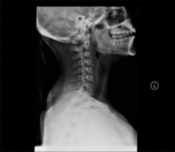
X-rayLateral cervical spine radiograph in a trauma patient: normal vertebral alignment, standard imaging is often unremarkable after whiplash.
Source : Coats et al., The open orthopaedics journal, 2014, figure 5 · CC BY-NC
The physical examination aims to confirm the signs compatible with WAD, rule out serious pathology and stratify severity:
- Imaging decision: Canadian C-Spine Rule (Stiell 2001 JAMA). This clinical decision rule has a sensitivity of 100% for ruling out significant cervical injury in alert, stable trauma patients.9 It is validated internationally and should guide every first-line imaging decision after cervical trauma. If the rule is not satisfied → imaging. If it is satisfied → no routine imaging.
- Active cervical mobility: the reduction in range of motion in several directions is the cardinal sign of WAD II.1 Measure each movement with an inclinometer or a standardised goniometer.
- Palpation: tenderness of the upper trapezius, scalenes, sternocleidomastoid, short suboccipital extensors and spinous processes. Reproduction of the complaint is a diagnostic finding.
- Neurological examination: motor power (MRC 0-5), tendon reflexes (biceps C5, brachioradialis C6, triceps C7), cutaneous sensation (dermatomes), pyramidal signs (Hoffmann, Babinski): mandatory to rule out a grade III or a myelopathy.1
- Deep cervical flexor test (CCFT, Jull 2008): assesses the activation and endurance of the deep flexors (longus colli, longus capitis). Weakness of the deep flexors is documented in chronic WAD and is a treatment target.10
- Sensorimotor assessment (Treleaven 2017 JOSPT): cervical position sense (joint position error), oculomotor control (saccades, smooth pursuit), static and dynamic balance. Often impaired even in low-grade WAD, and it guides treatment.11
Decision algorithm after cervical trauma
Combining the Canadian C-Spine Rule (Stiell 2001) and the IFOMPT framework (Finucane 2020)
Sources: Stiell IG et al. JAMA. 2001;286(15):1841-1848 (CCR); Finucane LM et al. JOSPT. 2020;50(7):350-372 (red flags); Spitzer WO et al. Spine. 1995 (QTF); Ritchie C et al. JOSPT. 2015;45(4):242-250 (CPR).
Should patients be stratified, and with which tool?
Yes: prognostic stratification guides the intensity of management. Two tools should be mastered:
1) The QTF classification (Spitzer 1995) stratifies severity at examination: useful for communication and planning.2
2) The Ritchie 2015 Clinical Prediction Rule (external validation, JOSPT 45(4):242-250) stratifies prognosis. It uses 3 simple variables :12
- Age (< 35 years = good prognosis)
- Baseline NDI (≤ 32% = full recovery likely; ≥ 40% = persistent moderate-to-severe disability likely)
- Hyperarousal symptoms (PCL-S ≥ 6 → poor prognosis)
This rule has good external validity and makes it possible to identify very early (weeks 1-4) the patients at high risk of chronicity, which then justifies a more intensive approach (psychologically informed care, targeted physiotherapy, even multidisciplinary referral).12
| Tool | Use | Level of evidence | Reference |
|---|---|---|---|
| Canadian C-Spine Rule | Imaging decision | Very high (prospective validation, sensitivity 100%) | Stiell 2001 JAMA |
| QTF (grades 0-IV) | Severity classification | Moderate (international consensus, lacks psychosocial integration) | Spitzer 1995 Spine |
| NDI (Vernon Mior) | Disability measure | High (excellent psychometrics) | Vernon & Mior 1991 JMPT |
| CCFT (Jull) | Assessment of deep flexor function | Moderate (acceptable inter-rater reliability) | Jull 2008 JMPT |
| Ritchie CPR | Prognosis for chronicity | High (external validation) | Ritchie 2015 JOSPT |
| IFOMPT Red Flags | Triage of serious pathology | Moderate (international consensus, isolated flags poorly specific) | Finucane 2020 JOSPT |
Critique and controversy
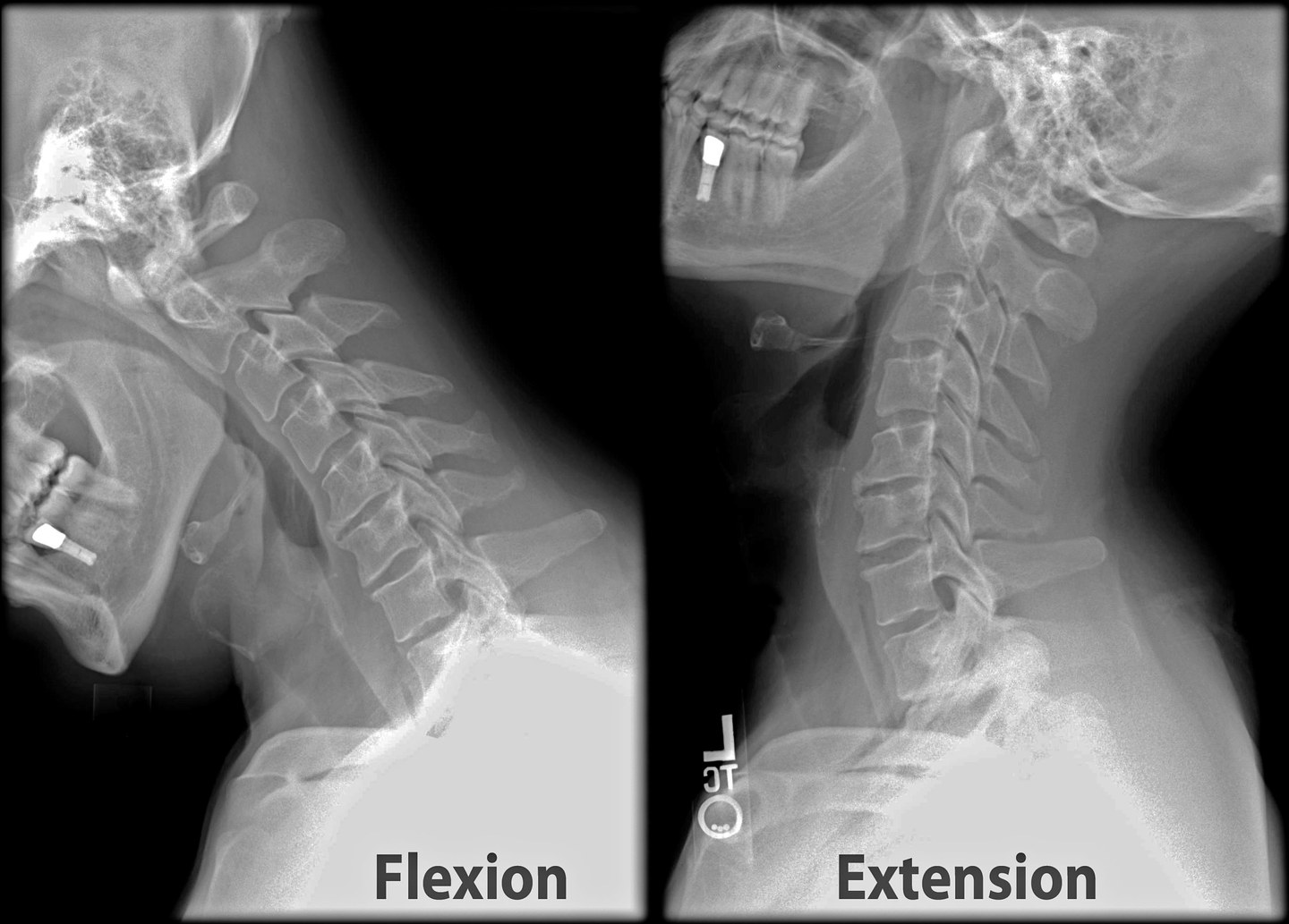
X-rayDynamic lateral cervical spine radiographs in flexion and extension, performed to look for cervical instability after repeated neck trauma.
Source : Stillwaterising, Wikimedia Commons · CC BY-SA
The QTF classification, although useful, is judged too simplistic to capture the biopsychosocial complexity of WAD: a grade II patient with PTSD and high catastrophising has a poorer prognosis than a grade III patient with no psychosocial factors.6,7 This is why the Ritchie 2015 CPR, which integrates a psychological dimension (hyperarousal), is now regarded as an essential complement.12
Moreover, the “diagnosis” of WAD remains a diagnosis of exclusion with no biological marker. This complicates medico-legal decisions and fuels persistent debate about the “somatic versus constructed” nature of the syndrome: a debate that must not obscure the clinical reality of patients' suffering.8
- The diagnosis of WAD is a diagnosis of exclusion based on history plus clinical examination.
- The Canadian C-Spine Rule (Stiell 2001) guides the imaging decision: sensitivity 100% for ruling out a cervical injury in an alert, stable patient.
- The IFOMPT Finucane 2020 framework structures red flag triage, in particular the 5D-3N for arterial dissection.
- The physical examination includes range of motion, palpation, a systematic neurological examination, CCFT (Jull 2008) and sensorimotor assessment (Treleaven 2017).
- The Ritchie 2015 Clinical Prediction Rule stratifies prognosis with 3 variables (age, baseline NDI, hyperarousal): the reference tool.
- Never forget the yellow flags (PTSD, catastrophising, kinesiophobia): they predict outcome more strongly than mechanical severity.
Bibliography
- Blanpied PR, Gross AR, Elliott JM, et al. Neck Pain: Revision 2017. Clinical Practice Guidelines Linked to the International Classification of Functioning, Disability and Health From the Orthopaedic Section of the American Physical Therapy Association. J Orthop Sports Phys Ther. 2017;47(7):A1-A83. doi:10.2519/jospt.2017.0302.
- Spitzer WO, Skovron ML, Salmi LR, et al. Scientific monograph of the Quebec Task Force on Whiplash-Associated Disorders. Spine. 1995;20(8 Suppl):1S-73S. PMID 7604354.
- Finucane LM, Downie A, Mercer C, et al. International Framework for Red Flags for Potential Serious Spinal Pathologies. J Orthop Sports Phys Ther. 2020;50(7):350-372. PMID 32438853.
- Ferrari R, Schrader H. The late whiplash syndrome: a biopsychosocial approach. J Neurol Neurosurg Psychiatry. 2001;70(6):722-726. PMID 11385003.
- Walton DM, Carroll LJ, Kasch H, et al. Risk Factors for Persistent Problems Following Acute Whiplash Injury: Update of a Systematic Review and Meta-analysis. J Orthop Sports Phys Ther. 2013;43(2):31-43. PMID 23322093.
- Sarrami P, Armstrong E, Naylor JM, Harris IA. Factors predicting outcome in whiplash injury: a systematic meta-review of prognostic factors. J Orthop Traumatol. 2017;18(1):9-16. PMID 27738773.
- Campbell L, Smith A, McGregor L, Sterling M. Psychological Factors and the Development of Chronic Whiplash-associated Disorder(s): A Systematic Review. Clin J Pain. 2018;34(8):755-768. PMID 29470185.
- Ferrari R, Russell AS. Epidemiology of whiplash: an international dilemma. Ann Rheum Dis. 1999;58(1):1-5. PMID 10343532.
- Stiell IG, Wells GA, Vandemheen KL, et al. The Canadian C-spine rule for radiography in alert and stable trauma patients. JAMA. 2001;286(15):1841-1848. PMID 11597285.
- Jull GA, O Leary SP, Falla DL. Clinical assessment of the deep cervical flexor muscles: the craniocervical flexion test. J Manipulative Physiol Ther. 2008;31(7):525-533.
- Treleaven J. Dizziness, Unsteadiness, Visual Disturbances, and Sensorimotor Control in Traumatic Neck Pain. J Orthop Sports Phys Ther. 2017;47(7):492-502. PMID 28622488.
- Ritchie C, Hendrikz J, Jull G, Elliott J, Sterling M. External Validation of a Clinical Prediction Rule to Predict Full Recovery and Ongoing Moderate/Severe Disability Following Acute Whiplash Injury. J Orthop Sports Phys Ther. 2015;45(4):242-250. PMID 25827122.
Which treatment strategies are the most effective?
Where do you start? What is the hierarchy of interventions?
Clinical guidelines converge on one essential point: movement and a return to activity should be encouraged as early as possible (Blanpied 2017 JOSPT CPG, Côté 2016 OPTIMa, Sterling 2019 J Clin Med).1,2,3 This approach, sometimes counter-intuitive for the patient, is firmly established: it prevents kinesiophobia and avoidance, two major drivers of chronicity.4
The classic hierarchy of first-line interventions is:
- Patient education (first!): benign nature, favourable prognosis in most cases, the patient's active role, demystifying imaging.2,5
- Early return to activity (daily life, adapted work): no rigid collar beyond 72 h, except on strict medical indication.1,2
- Active exercises that are gentle and progressive (mobility, motor control).3,6
- Manual therapy (mobilisations, manipulations) as an adjunct if a short-term benefit on pain and mobility is expected.7
- Multimodal approach (Sutton 2016 OPTIMa Spine J): education + exercise + manual therapy combined, more effective than each modality on its own.8
Hierarchy of interventions in acute WAD (CPG synthesis)
Pyramid of therapeutic intervention according to Blanpied 2017 JOSPT, OPTIMa Côté 2016, Sterling 2019
Stacked-card format (not triangular) to preserve legibility. Sources: Blanpied PR et al. JOSPT. 2017;47(7):A1-A83; Côté P et al. Eur Spine J. 2016;25(7):2000-2022; Sterling M et al. J Clin Med. 2019;8(8):1219.
What is the role of exercise, and is any one approach superior?
Therapeutic exercise is the cornerstone of WAD treatment for reducing pain and disability.3,9 However, the search for a “superior” type of exercise has proved largely fruitless: no modality (strengthening, endurance, mobility, motor control) shows a clinically significant superiority over the others in the long term.10
The randomised PROMISE trial (Michaleff ZA, Maher CG, Lin CW et al. Lancet. 2014;384(9938):133-141) is emblematic of this finding: in 172 patients with chronic WAD (> 3 months, grades I-II), an intensive programme of 20 sessions of physiotherapy exercise showed no superiority for pain or disability compared with 1 session of structured advice plus a booklet.9 Clinical conclusion: an exhaustive programme is not necessarily more effective than a well-dosed intervention centred on self-management.
Current recommendations therefore favour:
- Exercises for active mobility performed early and gently, within the pain-free range.1,5
- Exercises for cervical motor control (deep flexors: CCFT, deep extensors, scapulothoracic muscles), particularly in patients with sensorimotor control deficits or chronicity (Sterling 2019, Treleaven 2017).3,11
- The graduated progression including the shoulder girdle and the thoracic chain.10
- The individualised dose : frequency, duration and intensity matched to the profile and the phase.
Manual therapies and passive modalities: what is their real effectiveness?
The manual therapy (joint mobilisations, high-velocity manipulations) has a benefit in the short term on pain and mobility, but its specific effect largely disappears in the medium term.7,8 The Cochrane review by Gross 2015 (CD004249, n > 51 RCTs) confirms:
- The mobilisation approach is as effective as manipulation, with a better risk profile.7
- Effectiveness is conditional on being combined with an active programme : manual therapy on its own has not shown clinically relevant long-term effects.7
- High-velocity cervical manipulations are contraindicated in the presence of vascular red flags (suspected arterial dissection).12
As for the passive modalities (TENS, ultrasound, laser, shortwave, mechanical traction): the evidence of effectiveness is weak or absent in WAD.1,13 The JOSPT Neck Pain 2017 CPG and the OPTIMa CPG do not recommend them routinely. The rigid cervical collar beyond 72 h is actively advised against : it encourages inactivity, delays recovery and increases kinesiophobia.1,2
| Intervention | Short-term effect | Long-term effect | Level of evidence (GRADE) | Recommendation |
|---|---|---|---|---|
| Education + return to activity | + | ++ | High | Yes (first line) |
| Active exercises | ++ | + | High | Yes (essential) |
| Manual therapy (mob/manip) | ++ | +/- | Moderate | Yes, as an adjunct |
| Multimodal approach | ++ | ++ | Moderate | Yes |
| Pain education / CBT | + | ++ | Moderate | Yes if yellow flags |
| Rigid collar > 72 h | - | -- | Moderate | No (advised against) |
| TENS / ultrasound / laser | +/- | 0 | Low / Very low | No (no robust evidence) |
| Mechanical cervical traction | 0/+ | 0 | Low | Not routinely |
Critique and controversy
Three debates persist despite the general consensus:
1) Individual stratification versus a standardised approach. Most RCTs apply a uniform protocol to a heterogeneous population. Yet patients at high risk of chronicity (Ritchie 2015 CPR) might benefit from a more intensive and earlier approach, including psychological intervention. The “one-size-fits-all” approach of the guidelines is probably suboptimal for at-risk subgroups.14
2) Placebo effect and regression to the mean. The natural history of WAD shows substantial spontaneous improvement over the first 3 months: part of the effect attributed to interventions is probably due to this natural course rather than to the intervention itself. This strengthens the case for therapeutic parsimony in the acute phase.
3) Choices in a grey zone. When the evidence is moderate or weak (passive modalities, type of exercise), clinical reasoning shared with the patient should take precedence. That does not make everything legitimate: the rule is to avoid high-cost, low-benefit and iatrogenic-risk interventions (prolonged immobilisation, over-imaging, over-medicalisation).
- First line: education, early return to activity, active exercise. No rigid collar beyond 72 h.
- Exercise: the cornerstone, but no single type has been shown to be superior. Engagement counts for more than the modality (Michaleff 2014 PROMISE).
- Manual therapy: short-term benefit on pain and mobility, as an adjunct, never as monotherapy. Mobilisation is as effective as manipulation and safer.
- Passive modalities (TENS, US, laser, traction) not recommended routinely.
- Early biopsychosocial approach for at-risk patients (yellow flags), working with a psychologist where necessary.
Bibliography
- Blanpied PR, Gross AR, Elliott JM, et al. Neck Pain: Revision 2017. J Orthop Sports Phys Ther. 2017;47(7):A1-A83. doi:10.2519/jospt.2017.0302.
- Côté P, Wong JJ, Sutton D, et al. Management of neck pain and associated disorders: A clinical practice guideline from the Ontario Protocol for Traffic Injury Management (OPTIMa) Collaboration. Eur Spine J. 2016;25(7):2000-2022. PMID 26984876.
- Sterling M, de Zoete RMJ, Coppieters I, Farrell SF. Best Evidence Rehabilitation for Chronic Pain Part 4: Neck Pain. J Clin Med. 2019;8(8):1219. PMID 31443149.
- Campbell L, Smith A, McGregor L, Sterling M. Psychological Factors and the Development of Chronic Whiplash-associated Disorder(s): A Systematic Review. Clin J Pain. 2018;34(8):755-768. PMID 29470185.
- Rebbeck T. The Role of Exercise and Patient Education in the Noninvasive Management of Whiplash. J Orthop Sports Phys Ther. 2017;47(7):481-491. doi:10.2519/jospt.2017.7138.
- Jull GA, O Leary SP, Falla DL. Clinical assessment of the deep cervical flexor muscles: the craniocervical flexion test. J Manipulative Physiol Ther. 2008;31(7):525-533.
- Gross A, Langevin P, Burnie SJ, et al. Manipulation and mobilisation for neck pain contrasted against an inactive control or another active treatment. Cochrane Database Syst Rev. 2015;(9):CD004249. PMID 26397370.
- Sutton DA, Côté P, Wong JJ, et al. Is multimodal care effective for the management of patients with whiplash-associated disorders or neck pain and associated disorders? A systematic review by the OPTIMa Collaboration. Spine J. 2016;16(12):1541-1565. PMID 25014556.
- Michaleff ZA, Maher CG, Lin CW, et al. Comprehensive physiotherapy exercise programme or advice for chronic whiplash (PROMISE): a pragmatic randomised controlled trial. Lancet. 2014;384(9938):133-141. PMID 24703832.
- Sterling M, Jull G, Vicenzino B, Kenardy J, Darnell R. Physical and psychological factors predict outcome following whiplash injury. Pain. 2005;114(1-2):141-148. PMID 15733639.
- Treleaven J. Dizziness, Unsteadiness, Visual Disturbances, and Sensorimotor Control in Traumatic Neck Pain. J Orthop Sports Phys Ther. 2017;47(7):492-502. PMID 28622488.
- Thomas LC, Rivett DA, Attia JR, Parsons M, Levi C. Risk Factors and Clinical Presentation of Cervical Arterial Dissection: Preliminary Results of a Prospective Case-Control Study. J Orthop Sports Phys Ther. 2015;45(7):503-511. PMID 25996363.
- Wong JJ, Côté P, Sutton DA, et al. Clinical practice guidelines for the noninvasive management of low back pain: A systematic review by the Ontario Protocol for Traffic Injury Management (OPTIMa) Collaboration. Eur J Pain. 2017;21(2):201-216. PMID 27712027.
- Walton DM, Carroll LJ, Kasch H, et al. Risk Factors for Persistent Problems Following Acute Whiplash Injury. J Orthop Sports Phys Ther. 2013;43(2):31-43. PMID 23322093.
Chronic WAD & central sensitisation: how should chronicity be managed?
What are the mechanisms of central sensitisation in WAD?
The transition from acute WAD → chronic WAD (> 3 months) is not explained by the initial tissue injury alone. The work of Sterling and colleagues (Pain 2003, 2005) demonstrated, early after the crash, objectively measurable sensorimotor changes in the patients who would go on to become chronic: generalised pressure hypersensitivity (lowered PPTs at sites remote from the neck), abnormal responses to cold and thermal stimulation, and altered cervical motor control.1,2 These signs are consistent with a mechanism of sensitisation of the central nervous system : a persistent amplification of the nociceptive signal by central structures (dorsal horn, brainstem, thalamus, cortex).3
Clinically, central sensitisation presents with:
- Hyperalgesia (an exaggerated pain response to a normally painful stimulus)
- Allodynia (pain triggered by a normally non-painful stimulus: light touch, clothing rubbing)
- Spread of pain beyond the injured site (radiating to the shoulder, arm, thoracic back)
- Sleep and cognitive disturbance in association (concentration, memory)
- Emotional hyper-reactivity to pain, marked kinesiophobia
The presence of these signs points the diagnosis towards a dominant central component, which justifies a change of therapeutic strategy: it becomes pointless (and even harmful) to keep looking for a “tissue cause” to be treated with intensive local interventions.4
Which specific interventions for chronic WAD?
The management of chronic WAD calls for a biopsychosocial, neuroscience-informed approach, with four pillars:
1) Pain neuroscience education (PNE). Explain the mechanisms of persistent pain to the patient, decouple pain from tissue damage, reduce the perception of threat. PNE alone has a modest effect but becomes a powerful lever in synergy with exercise and cognitive behavioural therapy.5
2) Graded motor imagery (GMI). A three-stage approach (laterality recognition, explicit motor imagery, mirror therapy) aimed at gradually reorganising the cortical representations of the painful area without provoking pain. The Bowering 2013 meta-analysis (J Pain 14(1):3-13) confirms a benefit of GMI and mirror therapy in chronic pain, with methodological quality that still needs to improve.6
3) Graded exposure to feared activities. Identifying the movements and situations being avoided (TSK-11), exposing the patient gradually outside the panic zone, acknowledging successes: this breaks the fear-avoidance-disability cycle.7
4) Individualised exercise with an educational rationale. Exercise remains central, but with a dose adapted to sensitisation: start below the flare threshold, progress very slowly, explain every step. The aim is as much neurophysiological (modulating the pain matrix) as biomechanical.3,4
Stepped approach to chronic WAD with central sensitisation
Practical synthesis for the clinician
Sources: Sterling M et al. Pain. 2003; Bowering KJ et al. J Pain. 2013; Sterling M et al. J Clin Med. 2019.
And what about comorbidity with temporomandibular disorders?
A critical point that often goes unrecognised: whiplash is associated with a high comorbidity of temporomandibular disorders (TMD). The 2025 Häggman-Henrikson meta-analysis (Eur J Pain 29(3):e4792) confirms:8
- The prevalence of TMD symptoms is significantly higher in patients exposed to whiplash than in control groups, and already in the acute phase.
- There is no decline between the acute and the chronic phase: prevalence remains high in the long term.
- This argues for systematic screening for orofacial complaints in every WAD patient, rather than a “wait-and-see” attitude.
Clinically, the warning signs prompting a search for TMD comorbidity are: jaw or masticatory muscle pain, joint noises (TMJ clicking), limited mouth opening (< 40 mm), temporal headaches. The cervical and mandibular systems share common afferents (spinal nucleus of the trigeminal nerve), hence the value of a joint assessment and, where necessary, of management that includes the mandibular structures.
Warning signs of WAD at high risk of chronicity
- Baseline NDI ≥ 40% and pain ≥ 6/10 at day 7
- IES-R ≥ 33 (probable PTSD)
- TSK-11 ≥ 17 (marked kinesiophobia) or PCS ≥ 30 (catastrophising)
- Explicit negative expectations of recovery (“I will never get better”)
- Generalised hyperalgesia (PPTs lowered bilaterally, at remote sites)
- TMD comorbidity or tension-type headache that persists
- Ongoing medico-legal litigation (to be documented without judgement, but worth knowing)
These patients benefit from an early psychologically informed approach, introducing PNE from the first sessions and referring on to a multidisciplinary team if the situation persists.
- The chronicity of WAD (~30-50% at 1 year, Kamper 2008) often involves central sensitisation, identifiable through generalised hyperalgesia, allodynia and cognitive-emotional disturbance.
- Management combines pain neuroscience education (PNE), graded motor imagery (Bowering 2013), graded exposure and dosed exercise.
- The screening for post-whiplash TMD is imperative from the acute phase (Häggman-Henrikson 2025): high prevalence, no spontaneous decline.
- Identifying at-risk patients early (high NDI, PTSD, catastrophising) makes it possible to adapt the intensity and the nature of management.
- The approach remains patient-centred: collaboration with a psychologist or CBT therapist should be considered if psychosocial factors dominate.
Bibliography
- Sterling M, Jull G, Vicenzino B, Kenardy J, Darnell R. Development of motor system dysfunction following whiplash injury. Pain. 2003;103(1-2):65-73. PMID 12749960.
- Sterling M, Jull G, Vicenzino B, Kenardy J, Darnell R. Physical and psychological factors predict outcome following whiplash injury. Pain. 2005;114(1-2):141-148. PMID 15733639.
- Sterling M, de Zoete RMJ, Coppieters I, Farrell SF. Best Evidence Rehabilitation for Chronic Pain Part 4: Neck Pain. J Clin Med. 2019;8(8):1219. PMID 31443149.
- Treleaven J. Dizziness, Unsteadiness, Visual Disturbances, and Sensorimotor Control in Traumatic Neck Pain. J Orthop Sports Phys Ther. 2017;47(7):492-502. PMID 28622488.
- Kamper SJ, Rebbeck TJ, Maher CG, McAuley JH, Sterling M. Course and prognostic factors of whiplash. Pain. 2008;138(3):617-629. PMID 18407412.
- Bowering KJ, O Connell NE, Tabor A, et al. The effects of graded motor imagery and its components on chronic pain: a systematic review and meta-analysis. J Pain. 2013;14(1):3-13. PMID 23158879.
- Campbell L, Smith A, McGregor L, Sterling M. Psychological Factors and the Development of Chronic Whiplash-associated Disorder(s): A Systematic Review. Clin J Pain. 2018;34(8):755-768. PMID 29470185.
- Häggman-Henrikson B, Lovgren A, Wu WY, Peck CC, Westergren H, List T. Prevalence of Temporomandibular Disorder Symptoms After Whiplash Trauma: A Systematic Review and Meta-Analysis. Eur J Pain. 2025;29(3):e4792. PMID 39921489.
- Walton DM, Carroll LJ, Kasch H, et al. Risk Factors for Persistent Problems Following Acute Whiplash Injury. J Orthop Sports Phys Ther. 2013;43(2):31-43. PMID 23322093.
How do you secure lasting recovery and prevent flares?
How do you make the patient an active participant in their recovery?
Self-management is the cornerstone of durable recovery from WAD.1 Far from being abandonment or an absence of care, it is an active strategy that progressively hands the tools and the confidence over to the patient.
Three pillars of self-management in WAD:
- Clear, reassuring education: the generally benign nature of the injury, a favourable prognosis in most cases, the limited role of imaging (which may instead increase anxiety and promote chronicity: the “VOMIT” phenomenon).2
- Home exercise programme co-designed, simple and progressive:
- Gentle cervical mobility, within the pain-free range (flexion, extension, rotation, side bending)
- Deep flexor activation (the CCFT progresses through 4 levels: 22, 24, 26, 28 mmHg)
- Endurance of the deep cervical extensors
- Scapulothoracic and stabiliser strengthening (middle and lower trapezius, serratus anterior, rhomboids)
- Proprioceptive and oculomotor exercises if sensorimotor disturbance is documented (Treleaven 2017)3
- Basic cognitive behavioural strategies: identify limiting beliefs, encourage the return to valued activities, manage pacing, and take the drama out of normal symptom fluctuations.4
The goal is not the total absence of symptoms but the recovery of functional capacity and improvement in self-efficacy : that is, the patient's confidence in their own ability to manage their condition. It is this self-efficacy that best predicts long-term return to activity and to work.1,4
When and how should a safe return to sport be planned?
The return to sport after a WAD must be progressive, structured and guided by functional criteria : not by a fixed calendar. Although WAD-specific recommendations are less formalised than for concussion, the principles of the international CISG Amsterdam 2023 consensus (Patricios JS, Schneider KJ, Dvorak J, et al. Br J Sports Med. 2023;57(11):695-711)5 can be adapted: a stepwise progression, moving to the next step only after 24 h free of symptoms after the load.
Return to sport after WAD: stepwise progression
Adapted from the CISG Amsterdam 2023 protocol (BJSM 57:695-711) for concussion
Criterion for moving between steps: no cervical symptoms (pain, dizziness, headache) during and after exertion, and for the following 24 h. If symptoms appear, return to the previous step for 48 h before progressing again. Source: adapted from Patricios JS et al. Br J Sports Med. 2023;57(11):695-711 (CISG Amsterdam 2023 consensus).
Secondary prevention and reduction of the risk of recurrence or later complications: in athletes and at-risk individuals (contact sports, motor sports), strengthening of the cervical neuromuscular system is considered a protective factor. The Hrysomallis 2016 review (Sports Med 46(8):1111-1124) summarises the data on the effect of neck strengthening on performance and injury risk: a stronger neck increases cervical stiffness and reduces head acceleration on impact, which is theoretically protective against whiplash and concussion.6 Note that the literature on the specific preventive effect against WAD remains limited and heterogeneous.
The final return-to-play decision should be shared between the patient, the physiotherapist, the doctor and the coach, based on:
- No cervical symptoms on exertion or afterwards (24 h after exertion)
- Restored cervical strength and endurance (CCFT 28 mmHg held 10 s × 10 reps)
- Full, pain-free active ranges of motion
- Restored sensorimotor control (proprioception, oculomotor control)
- The patient's subjective confidence (self-efficacy)
Critique and controversy
1) Individual stratification versus a uniform approach: the guidelines recommend a fairly uniform initial conservative approach. Yet the prognostic data (Ritchie 2015 CPR) show that a proportion of patients might benefit from a more intensive early approach. The literature still lacks RCTs validating this clinical stratification.
2) Manual therapies in the long term: their role and their optimal dosage remain debated. Most studies evaluate multimodal “packages”, which makes it difficult to isolate the specific effect of each component.
3) Extrapolating the CISG criteria (concussion → WAD): there are few WAD-specific RCTs on return to sport. The extrapolation is reasonable given the shared injury mechanisms, but it introduces uncertainty.
- Self-management is the cornerstone: education + home exercise + basic cognitive behavioural strategies (Rebbeck 2017 JOSPT).
- The return to sport must be progressive and stepwise, based on the absence of symptoms 24 h after exertion (CISG Amsterdam 2023 model).
- The secondary prevention in athletes relies on cervical neuromuscular strengthening (Hrysomallis 2016): a moderate level of evidence, but biomechanically coherent.
- A decision shared between the patient, the clinicians and the coach, based on functional criteria and on the patient's confidence, rather than on a fixed time frame.
Bibliography
- Rebbeck T. The Role of Exercise and Patient Education in the Noninvasive Management of Whiplash. J Orthop Sports Phys Ther. 2017;47(7):481-491. doi:10.2519/jospt.2017.7138.
- Finucane LM, Downie A, Mercer C, et al. International Framework for Red Flags for Potential Serious Spinal Pathologies. J Orthop Sports Phys Ther. 2020;50(7):350-372. PMID 32438853.
- Treleaven J. Dizziness, Unsteadiness, Visual Disturbances, and Sensorimotor Control in Traumatic Neck Pain. J Orthop Sports Phys Ther. 2017;47(7):492-502. PMID 28622488.
- Sterling M, de Zoete RMJ, Coppieters I, Farrell SF. Best Evidence Rehabilitation for Chronic Pain Part 4: Neck Pain. J Clin Med. 2019;8(8):1219. PMID 31443149.
- Patricios JS, Schneider KJ, Dvorak J, et al. Consensus statement on concussion in sport: the 6th International Conference on Concussion in Sport, Amsterdam, October 2022. Br J Sports Med. 2023;57(11):695-711. PMID 37316210.
- Hrysomallis C. Neck Muscular Strength, Training, Performance and Sport Injury Risk: A Review. Sports Med. 2016;46(8):1111-1124. PMID 26886474.
- Michaleff ZA, Maher CG, Lin CW, et al. Comprehensive physiotherapy exercise programme or advice for chronic whiplash (PROMISE). Lancet. 2014;384(9938):133-141. PMID 24703832.
- Ritchie C, Hendrikz J, Jull G, Elliott J, Sterling M. External Validation of a Clinical Prediction Rule. J Orthop Sports Phys Ther. 2015;45(4):242-250. PMID 25827122.
What do real clinical cases teach us about whiplash?
Analysis of a typical grade II case: assessment and trajectory
The typical case in clinical practice is WAD II after a low-speed rear-end collision, seen in clinic 7 to 14 days after the crash. Prototypical profile synthesised from the prospective cohorts (Sterling 2003, Walton 2013, Sterling 2019)1,2,3 :
- History: young adult patient, rear-end type collision at modest speed (20-30 km/h), pain appearing 12-24 h later, posterolateral neck pain + occipital headache, generalised stiffness, general tenderness.
- Initial examination: Canadian C-Spine Rule satisfied (no imaging). Active range limited by -30% in all directions, painful paraspinal palpation at C4-C6 and over the upper trapezius. No neurological signs. Baseline NDI 32% (intermediate).
- Ritchie 2015 CPR stratification: age < 35 years + NDI 32% + no hyperarousal → good prognosis profile for full recovery.
- Management: reassuring education (“benign injury, movement = healing”), return to activity + adapted work, home exercises (mobility + progressive CCFT), 4-6 sessions spread over 6-8 weeks. No collar.
- Trajectory: progressive reduction in pain, with the NDI falling to 14% at 6 weeks and 6% at 3 months. Full return to sport at 8-10 weeks.
This profile corresponds to the ~50% of patients who largely recover within the first 3 months (Kamper 2008).4 The key message: early, active and reassuring management is enough for the majority of typical cases.
Diagnostic challenge: when WAD mimics (or masks) serious pathology
Three differential diagnoses that must be known:
1) Cervical arterial dissection (vertebral or carotid) : a life-threatening diagnosis. The prospective case-control study by Thomas, Rivett & Attia 2015 (JOSPT 45(7):503-511, PMID 25996363) characterises the specific red flags:5
- Unusual headache (often occipital) + neck pain, of sudden onset or building over a few days
- Often associated with a minor event (violent coughing, a sudden neck movement, a low-speed road traffic collision)
- Young or middle-aged patient (often < 50 years)
- 5D-3N : Dizziness, Diplopia, Dysarthria, Dysphagia, Drop attacks / Nausea, Numbness, Nystagmus
- Possible focal neurological deficit, ipsilateral Horner syndrome, oculomotor disturbance
If dissection is suspected: immediate referral to neurovascular emergency care, with cervical manipulation contraindicated until it is ruled out.
2) Degenerative cervical myelopathy : often unmasked by a minor event. The educational step-by-step account by Davies BM, Mowforth OD, Smith EK, Kotter MR (BMJ. 2018;360:k186)6 sets out the signs to look for in the patient > 50 years:
- Progressive loss of dexterity (doing up buttons, handwriting)
- Unsteady gait, frequent falls
- Weakness / spasticity of the lower limbs
- Pyramidal signs (Hoffmann, Babinski, hyperreflexia)
- Possibly sphincter disturbance
Cervical MRI is the reference investigation. Every WAD patient > 50 years with progressive deterioration should be assessed for this condition.
3) C1-C2 ligamentous injury / atlantoaxial instability : rare but serious. Suspect it in the face of persistent severe suboccipital pain, a sense of craniocervical instability (“my head is slipping”), clear reproduction on ligamentous integrity tests (alar ligament test, transverse ligament test), and an evolving neurological examination. Dynamic MRI is required.
WAD red flags: 5D-3N for suspected vascular injury
Mnemonic acronym for cervical arterial dissection
Source: Thomas LC, Rivett DA et al. JOSPT. 2015;45(7):503-511 (PMID 25996363); IFOMPT framework Finucane 2020 (PMID 32438853).
Complex case: chronicity, central sensitisation and comorbidities
The case report by Lewis F & Naude B (S Afr J Physiother. 2010;66(2):37-39) illustrates a common situation: chronic post-traumatic neck pain with cervicogenic headache and concurrent temporomandibular dysfunction.7 A 26-year-old male patient with neck pain + headache + TMJ clicking. Multimodal management combining:
- Cervical and TMJ mobilisations (Maitland approach)
- Strengthening of the deep cervical flexors and the scapulothoracic stabilisers
- Trigger point therapy and massage
- Postural correction
Result: significant improvement over 9 sessions spread across 14 weeks (full active cervical and TMJ range, reduced headache intensity, reduced frequency, improved quality of life).7 This case illustrates the value of assessing the cervical and mandibular systems together and confirms the relevance of the Häggman-Henrikson 2025 data on the high prevalence of post-whiplash TMD.8
Limitations of clinical cases: by nature, a case report describes n=1 and cannot establish the effectiveness of an intervention. Clinical cases are illustrations of clinical reasoning, never proof of effectiveness, which requires RCTs and meta-analyses. This distinction is essential in order to avoid over-interpretation.9
- Typical WAD II case: typical profile, favourable recovery in 6-10 weeks with an active approach + education + home exercise.
- Diagnostic challenge: never overlook the red flags. 5D-3N = arterial dissection (Thomas & Rivett 2015). Progressive deterioration > 50 years = cervical myelopathy (Davies 2018 BMJ).
- Complex case: systematically look for TMD comorbidity (Lewis & Naude 2010, Häggman-Henrikson 2025): a multimodal approach covering both regions is often needed.
- Limitation of clinical cases: illustrations of reasoning, never proof of effectiveness. Think meta-analysis and RCT for intervention decisions.
Bibliography
- Sterling M, Jull G, Vicenzino B, Kenardy J, Darnell R. Development of motor system dysfunction following whiplash injury. Pain. 2003;103(1-2):65-73. PMID 12749960.
- Walton DM, Carroll LJ, Kasch H, et al. Risk Factors for Persistent Problems Following Acute Whiplash Injury. J Orthop Sports Phys Ther. 2013;43(2):31-43. PMID 23322093.
- Sterling M, de Zoete RMJ, Coppieters I, Farrell SF. Best Evidence Rehabilitation for Chronic Pain Part 4: Neck Pain. J Clin Med. 2019;8(8):1219. PMID 31443149.
- Kamper SJ, Rebbeck TJ, Maher CG, McAuley JH, Sterling M. Course and prognostic factors of whiplash. Pain. 2008;138(3):617-629. PMID 18407412.
- Thomas LC, Rivett DA, Attia JR, Parsons M, Levi C. Risk Factors and Clinical Presentation of Cervical Arterial Dissection: Preliminary Results of a Prospective Case-Control Study. J Orthop Sports Phys Ther. 2015;45(7):503-511. PMID 25996363.
- Davies BM, Mowforth OD, Smith EK, Kotter MR. Degenerative cervical myelopathy. BMJ. 2018;360:k186. doi:10.1136/bmj.k186.
- Lewis F, Naude B. The effectiveness of physiotherapy in cervicogenic headache and concurring temporomandibular dysfunction: a case report. S Afr J Physiother. 2010;66(2):37-39. sajp.co.za.
- Häggman-Henrikson B, Lovgren A, Wu WY, Peck CC, Westergren H, List T. Prevalence of Temporomandibular Disorder Symptoms After Whiplash Trauma: A Systematic Review and Meta-Analysis. Eur J Pain. 2025;29(3):e4792. PMID 39921489.
- Finucane LM, Downie A, Mercer C, et al. International Framework for Red Flags for Potential Serious Spinal Pathologies. J Orthop Sports Phys Ther. 2020;50(7):350-372. PMID 32438853.
- Bowering KJ, O Connell NE, Tabor A, et al. The effects of graded motor imagery and its components on chronic pain. J Pain. 2013;14(1):3-13. PMID 23158879.
How do you apply these recommendations concretely in your practice?
When and to which other professionals should you refer?
Referral is an essential clinical skill: it protects the patient, avoids diagnostic delay and secures optimal management. Three levels of referral in WAD:
1) Immediate medical referral (emergency department, or the general practitioner promptly):
- Any IFOMPT red flag (Finucane 2020): suspected fracture, progressive neurological deficit, 5D-3N suggesting arterial dissection (Thomas & Rivett 2015), suspected myelopathy (Davies 2018).1,2,3
- High-energy trauma with severe axial pain that is not relieved.
- Altered consciousness, associated head injury (suspected concussion: CISG Amsterdam 2023 protocol).4
2) Multidisciplinary referral (biopsychosocial approach):
- Psychologist / CBT where yellow flags dominate: PTSD (IES-R ≥ 33), severe catastrophising (PCS ≥ 30), marked kinesiophobia (TSK-11 ≥ 17), depression, entrenched limiting beliefs.5
- Pain specialist physician or pain clinic in resistant chronicity with signs of central sensitisation or multiple pain comorbidities.
- Occupational physician where there are difficulties returning to work or adapting the workstation.
- Dentist / TMD specialist where there is mandibular comorbidity (Häggman-Henrikson 2025).6
3) Yellow flags: modifiable psychosocial levers:
The yellow flag concept (“Decade of the Flags”) has shown that early psychosocial factors predict chronicity better than physical signs.5,7 For WAD, the main yellow flags to screen for systematically are:
WAD yellow flags to screen for systematically
- PTSD / hyperarousal (IES-R ≥ 33): the leading predictor of chronicity
- Catastrophising in the face of pain (PCS ≥ 30)
- Kinesiophobia (TSK-11 ≥ 17): fear of movement
- Low recovery expectations (“I will never get better”): a self-fulfilling prophecy
- Depressive symptoms (PHQ-9 ≥ 10) or generalised anxiety
- Passive coping strategies (avoidance, prolonged rest, searching for a cause)
- Ongoing medico-legal context (to be noted without judgement)
Positive yellow flags → adapt management (early PNE, graded exposure, psychologically informed exercise) and consider collaboration with a psychologist or CBT therapist if several flags are present or the intensity is severe.
How do you measure outcomes and overcome barriers to implementation?
Measuring outcomes with validated tools is essential in order to adjust management and demonstrate effectiveness. For WAD, the PROMs (patient-reported outcome measures) recommended are:
- NDI (Neck Disability Index) from Vernon & Mior 1991: the reference tool, 10 items, scored 0-100% as a percentage.8 Interpretation thresholds: 0-8% no disability, 10-28% mild, 30-48% moderate, 50-68% severe, 72-100% complete.
- VAS / NRS (pain 0-10).
- TSK-11 (kinesiophobia 11-44, threshold ≥ 17 for significant kinesiophobia).
- PCS (Pain Catastrophizing Scale, 0-52).
- IES-R (Impact of Event Scale-Revised, PTSD screening, threshold ≥ 33).
- PSFS (Patient-Specific Functional Scale) for individualised goals.
Beyond the tools, the challenge is the real-world implementation of evidence-based practice. Greenhalgh, Howick & Maskrey 2014 (BMJ 348:g3725, “Evidence based medicine: a movement in crisis?”) remind us that EBM is caught between:9
- The industrialisation of research (costly RCTs, conflicts of interest, publication bias)
- The cognitive overload of the clinician (exponential volume of publications)
- The excessive standardisation at the expense of shared patient-clinician decision-making
- The retreat of the clinical and moral dimension of care
The “EBM renaissance” advocated by the authors means recentring practice on:
- The individual experience of the patient (preferences, values, life context)
- The clinical expertise of the practitioner (reasoning, communication)
- The usable evidence (summarised, contextualised, shared)
- The shared decision-making as a collaborative process
The syndrome VOMIT (“Victims of Modern Imaging Technology”) is emblematic of the excesses to avoid: imaging prescribed with no real need, generating patient anxiety + incidental findings + over-medicalisation. In WAD, the Canadian C-Spine Rule should strictly guide the imaging decision, and the clinician must be able to explain why a “normal” MRI does not mean “no problem” and does not mean “a serious problem” either: tissue damage and the experience of pain are not equivalent.
Critique and controversy
1) Isolated red flags are poorly specific: most individual items have a low positive predictive value: what matters is the clinical clustering (cluster) and the context (Finucane 2020).1 The clinician must avoid both under-detection (a missed diagnosis) and over-detection (iatrogenic over-imaging).
2) Tension between standardisation and personalisation: guidelines and Core Outcome Sets standardise measurement and management, which is positive for research and for quality, but they can obscure the patient's individual goals. The right balance is: standardised tools for cohort follow-up + individualised assessment for care.
3) Shared decision-making: a complex ideal. Putting it into practice runs up against lack of time, patients' variable health literacy, and reimbursement systems that reward technical procedures rather than communication. Possible tools: decision aids, motivational interviewing, structured debriefing.
- Immediate medical referral where there are IFOMPT red flags (Finucane 2020): fracture, arterial dissection, myelopathy, progressive deficit.
- Multidisciplinary referral (psychologist, pain specialist, TMD dentist) according to the patient's profile.
- Yellow flags (PTSD, catastrophising, kinesiophobia, low expectations) are major predictors of chronicity and should be screened for systematically.
- WAD PROMs: NDI, VAS, TSK-11, PCS, IES-R, PSFS.
- Evidence-based practice: aim for the balance between standardisation and personalisation, integrate shared decision-making, and avoid the VOMIT syndrome (iatrogenic over-imaging).
- “Renewed” EBM (Greenhalgh 2014) recentres practice on the patient and on the quality of the clinician-patient relationship.
Bibliography
- Finucane LM, Downie A, Mercer C, et al. International Framework for Red Flags for Potential Serious Spinal Pathologies. J Orthop Sports Phys Ther. 2020;50(7):350-372. PMID 32438853.
- Thomas LC, Rivett DA, Attia JR, Parsons M, Levi C. Risk Factors and Clinical Presentation of Cervical Arterial Dissection. J Orthop Sports Phys Ther. 2015;45(7):503-511. PMID 25996363.
- Davies BM, Mowforth OD, Smith EK, Kotter MR. Degenerative cervical myelopathy. BMJ. 2018;360:k186. doi:10.1136/bmj.k186.
- Patricios JS, Schneider KJ, Dvorak J, et al. Consensus statement on concussion in sport: the 6th International Conference on Concussion in Sport, Amsterdam, October 2022. Br J Sports Med. 2023;57(11):695-711. PMID 37316210.
- Campbell L, Smith A, McGregor L, Sterling M. Psychological Factors and the Development of Chronic Whiplash-associated Disorder(s): A Systematic Review. Clin J Pain. 2018;34(8):755-768. PMID 29470185.
- Häggman-Henrikson B, Lovgren A, Wu WY, et al. Prevalence of Temporomandibular Disorder Symptoms After Whiplash Trauma: A Systematic Review and Meta-Analysis. Eur J Pain. 2025;29(3):e4792. PMID 39921489.
- Sarrami P, Armstrong E, Naylor JM, Harris IA. Factors predicting outcome in whiplash injury: a systematic meta-review of prognostic factors. J Orthop Traumatol. 2017;18(1):9-16. PMID 27738773.
- Vernon H, Mior S. The Neck Disability Index: a study of reliability and validity. J Manipulative Physiol Ther. 1991;14(7):409-415. (The original NDI paper, an international reference instrument.)
- Greenhalgh T, Howick J, Maskrey N. Evidence based medicine: a movement in crisis? BMJ. 2014;348:g3725. PMID 24927763.
- Foster NE, Anema JR, Cherkin D, et al. Prevention and treatment of low back pain: evidence, challenges, and promising directions. Lancet. 2018;391(10137):2368-2383. PMID 29573872.
- Wong JJ, Côté P, Sutton DA, et al. Clinical practice guidelines for the noninvasive management of low back pain: A systematic review by the OPTIMa Collaboration. Eur J Pain. 2017;21(2):201-216. PMID 27712027.
- Sterling M, de Zoete RMJ, Coppieters I, Farrell SF. Best Evidence Rehabilitation for Chronic Pain Part 4: Neck Pain. J Clin Med. 2019;8(8):1219. PMID 31443149.
What next, after this read?
This article is part of a collection of evidence-based clinical syntheses. A question, some feedback, a correction to suggest? Contact us directly via the WhatsApp button at the bottom right of the screen.

