















In brief
Temporomandibular joint (TMJ) syndrome, referred to in the modern literature as temporomandibular disorders (TMD), covers a heterogeneous group of musculoskeletal disorders affecting the temporomandibular joint and the masticatory muscles. It presents with pre-auricular or masticatory muscle pain, joint noises (clicking) and limited or deviated mouth opening, mainly affecting women aged 20 to 40. First-line management is conservative: education and self-management, then active exercise and adjunctive manual therapy. Its prevalence is estimated at around 31 % of adults.
Clinical synthesis based on the most recent meta-analyses and international consensus statements: DC/TMD, ICOP 2020, OPPERA, 2025 prospective data.
Clinical synthesis
- Temporomandibular disorders (TMD) affect the TMJ and the masticatory muscles. They mainly affect women aged 20 to 40, with a prevalence of around 31 % in adults (disc displacement with reduction being the most common).
- Stress, anxiety, bruxism (tooth clenching/grinding) and trauma (notably whiplash) are major risk factors, with documented genetic and hormonal predispositions.
- The condition involves mechanical muscle overload, local inflammation and, in chronic cases, sensitisation of the central nervous system. The natural history is often fluctuating, with spontaneous improvement in a substantial proportion of patients.
- Diagnosis must follow the DC/TMD criteria, which combine a physical (Axis I) and a psychosocial (Axis II) assessment for a complete picture of the patient.
- The standardised examination includes range-of-motion measurement, muscle palpation and a rigorous differential diagnosis (dental, ENT, neuralgic and cervicogenic conditions).
- Classifying TMD precisely (myalgia, disc displacement, arthralgia/osteoarthritis, etc.) is crucial in order to target treatment, establish a prognosis and support communication between clinicians.
- First-line treatment is conservative, beginning with patient education and self-management, followed by active exercise and adjunctive manual therapy.
- Exercise (mobility, motor control, strengthening) is a cornerstone of treatment, especially for muscle pain. Its effect is greater when combined with manual therapy.
- Manual therapy reduces pain and improves mobility. Low-level laser therapy (LLLT) shows favourable evidence for short-term pain.
- Making the patient an active participant through education, home exercise and management of parafunctions is the key to preventing recurrence and securing long-term success.
- Return to sport should be progressive and guided by functional criteria (pain-free status, restored function) rather than by a fixed timetable.
- TMD can mimic other conditions such as trigeminal neuralgia or ear pain, which underlines the importance of a rigorous differential diagnosis.
- Complex cases illustrate the frequent link with the cervical spine and with comorbidities (notably fibromyalgia), calling for a broader biopsychosocial approach.
- Clinicians must know when to refer, in the presence of red flags (serious disease) or significant psychosocial factors (yellow flags), to a doctor or a psychologist.
- Measuring outcomes with standardised patient-reported measures (PROMs) is essential in order to assess the effectiveness of care and adapt management.
Contents
- What are the fundamentals to know about temporomandibular joint (TMJ) syndrome?
- How can temporomandibular joint (TMJ) syndrome be assessed and diagnosed with certainty?
- Which treatment strategies are the most effective for temporomandibular joint (TMJ) syndrome?
- How can lasting recovery be secured and recurrence of temporomandibular joint (TMJ) syndrome prevented?
- What do real-world case studies teach us about temporomandibular joint (TMJ) syndrome?
- How can these recommendations be applied concretely in your practice?
What are the fundamentals to know about temporomandibular joint (TMJ) syndrome?
How is the condition defined, who is affected and what are the risk factors?
TMD are defined as musculoskeletal disorders of multifactorial origin.² They present mainly with three cardinal signs: pain in the pre-auricular region (in front of the ear) or in the masticatory muscles, joint noises (clicking, cracking) during jaw movement, and limitation or deviation of mandibular range of motion.³ The prevalence of TMD in the general population is considerable. The most recent estimates (Valesan 2021 meta-analysis, 21 pooled studies) report a prevalence of around 31 % in adults and 11 % in children/adolescents, with disc displacement with reduction the most frequent subcategory.⁴ Only a fraction of symptomatic individuals (around 5 to 7 %) nevertheless require active treatment.📊 TMD prevalence by age group (Valesan 2021, meta-analysis of 21 studies)
Percentage of the population with at least one TMD sign / symptom
Source: Valesan LF, Da-Cas CD, Réus JC, et al. Clin Oral Investig. 2021;25(2):441-453.
⚖️ Odds ratios women vs men by TMD subtype (Bueno 2018)
An OR > 1 means that women are at greater risk than men
Source: Bueno CH, Pereira DD, Pattussi MP, et al. J Oral Rehabil. 2018;45(9):720-729.
- Psychosocial factors: Stress, anxiety, depression and somatisation are among the strongest predictors of TMD onset and chronicity.⁷ These factors can increase muscle tension and lower pain perception thresholds.
- Oral parafunctions: Sleep or awake bruxism (tooth grinding or clenching) is associated with muscle pain (myalgia) because of the mechanical overload it places on the masticatory system. An important nuance from the Manfredini & Lobbezoo systematic review: the association is clearly stronger in studies based on self-report / questionnaire than in instrumental studies (electromyography or polysomnography), where the link is weak or even negative. The method used to assess bruxism therefore shapes the clinical interpretation.⁸
- Genetic and hormonal factors: Genetic predispositions influencing pain perception and the inflammatory response are suspected.⁹ The higher prevalence in women also suggests an influence of sex hormones, notably oestrogens.⁵
- Trauma: Macrotrauma (a direct blow to the jaw, whiplash) and repeated microtrauma (chewing on one side only, postural habits) can trigger or aggravate TMD. Häggman-Henrikson's 2025 meta-analysis found a prevalence of symptomatic TMD of up to 70 % (median 35 %) in patients after whiplash, versus 1.7-13 % in control groups; prevalence is already high in the acute phase and does not decrease towards the chronic phase, which argues for systematic early screening.¹⁰
- Comorbidities: TMD frequently coexist with other chronic pain syndromes such as fibromyalgia, irritable bowel syndrome, tension-type headache and neck pain, suggesting shared sensitisation mechanisms. Yakkaphan's 2023 meta-analysis reports a TMD prevalence reaching 76.8 % in patients with fibromyalgia, and around 32.7 % fibromyalgia in patients with TMD: above all for myogenous forms.¹¹
🔗 Two-way overlap between TMD ↔ fibromyalgia
Yakkaphan 2023 meta-analysis: one is rarely isolated from the other
63 % of comorbid TMD are myogenous. Source: Yakkaphan P et al. J Oral Facial Pain Headache. 2023;37(3):177-193.
What happens in the body and how does temporomandibular joint (TMJ) syndrome evolve naturally?
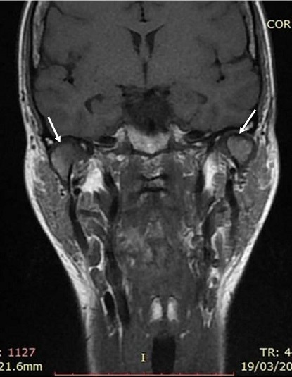
MRICoronal MRI of the temporomandibular joint: bilateral subchondral geodes and thinning of the mandibular condyle cortex (arrows).
Source : Chebbi et al., The Pan African Medical Journal, 2016, figure 4 · CC BY
- The historic study by Magnusson, Egermark & Carlsson (Sweden, n = 320 subjects followed from 15 to 35 years of age) shows that TMD signs and symptoms fluctuate from one assessment to the next, with no systematic trend towards either worsening or complete resolution with age: they remain variable between individuals and from one decade to the next.¹⁶
- The more recent prospective data Häggman-Henrikson 2025 (Swedish 8-year cohort, n = 94,769) confirm this dynamic but uneven pattern: transitions to remission do occur, but are less frequent in women, who show both a higher incidence and a slower recovery.¹⁵
Key points
- TMD are a group of musculoskeletal disorders affecting the TMJ and the masticatory muscles, defined by pain, joint noises and movement limitation.
- Consolidated prevalence (Valesan 2021): ≈ 31 % of adults, 11 % of children/adolescents. Peak incidence in women between 20 and 40 years of age (OR ≈ 2 for overall TMD).
- Key risk factors: stress / anxiety (OPPERA), bruxism (stronger link on self-report than on EMG/polysomnography), trauma (whiplash: up to 70 % post-traumatic TMD), painful comorbidities (fibromyalgia: 32.7 % in patients with TMD).
- The pathophysiology combines mechanical overload, local inflammation (IL-1β, IL-6, TNF-α) and, in chronic cases, a central sensitisation documented on imaging (changes in trigeminal and limbic grey matter).
- The natural history is fluctuating, with a broadly favourable prognosis but uneven trajectories (women: slower recovery). A conservative, stratified approach is required as first-line management.
References
- Schiffman E, Ohrbach R, Truelove E, et al. Diagnostic Criteria for Temporomandibular Disorders (DC/TMD) for Clinical and Research Applications. J Oral Facial Pain Headache. 2014;28(1):6-27. PMID 24482784.
- Slade GD, Ohrbach R, Greenspan JD, et al. Painful Temporomandibular Disorder: Decade of Discovery from OPPERA Studies. J Dent Res. 2016;95(10):1084-1092. PMID 27339423.
- Gauer RL, Semidey MJ. Diagnosis and treatment of temporomandibular disorders. Am Fam Physician. 2015;91(6):378-386. PMID 25822556.
- Valesan LF, Da-Cas CD, Réus JC, et al. Prevalence of temporomandibular joint disorders: a systematic review and meta-analysis. Clin Oral Investig. 2021;25(2):441-453. doi:10.1007/s00784-020-03710-w.
- Bueno CH, Pereira DD, Pattussi MP, Grossi PK, Grossi ML. Gender differences in temporomandibular disorders in adult populational studies: A systematic review and meta-analysis. J Oral Rehabil. 2018;45(9):720-729. PMID 29851110.
- List T, Jensen RH. Temporomandibular disorders: Old ideas and new concepts. Cephalalgia. 2017;37(7):692-704. doi:10.1177/0333102416686302.
- Fillingim RB, Ohrbach R, Greenspan JD, et al. Psychological factors associated with development of TMD: the OPPERA prospective cohort study. J Pain. 2013;14(12 Suppl):T75-T90. PMID 24275225.
- Manfredini D, Lobbezoo F. Relationship between bruxism and temporomandibular disorders: a systematic review of literature from 1998 to 2008. Oral Surg Oral Med Oral Pathol Oral Radiol Endod. 2010;109(6):e26-e50. PMID 20451831.
- Smith SB, Mir E, Bair E, et al. Genetic variants associated with development of TMD and its intermediate phenotypes: the OPPERA prospective cohort study. J Pain. 2013;14(12 Suppl):T91-T101.e1-3. PMID 24275226.
- Häggman-Henrikson B, Lobbezoo F, Durham J, Peck C, List T. Prevalence of Temporomandibular Disorder Symptoms After Whiplash Trauma — A Systematic Review and Meta-Analysis. Eur J Pain. 2025;29(3):e4792. PMID 39921489.
- Yakkaphan P, Smith JG, Chana P, Renton T, Lambru G. Temporomandibular Disorders and Fibromyalgia Prevalence: A Systematic Review and Meta-Analysis. J Oral Facial Pain Headache. 2023;37(3):177-193. PMID 37975782.
- Gil-Martínez A, Paris-Alemany A, López-de-Uralde-Villanueva I, La Touche R. Management of pain in patients with temporomandibular disorder (TMD): challenges and solutions. J Pain Res. 2018;11:571-587. PMID 29588615.
- Pigg M, Nixdorf DR, Law AS, et al. New International Classification of Orofacial Pain: What Is in It For Endodontists? J Endod. 2021;47(3):345-357. PMID 33340605.
- Younger JW, Shen YF, Goddard G, Mackey SC. Chronic myofascial temporomandibular pain is associated with neural abnormalities in the trigeminal and limbic systems. Pain. 2010;149(2):222-228. PMID 20236763.
- Häggman-Henrikson B, Liv P, Ilgunas A, et al. Women are worse off in developing and recovering from temporomandibular disorder symptoms. Sci Rep. 2025;15:5532. PMID 39922904.
- Magnusson T, Egermark I, Carlsson GE. A longitudinal epidemiologic study of signs and symptoms of temporomandibular disorders from 15 to 35 years of age. J Orofac Pain. 2000;14(4):310-319. PMID 11203765.
How can temporomandibular joint (TMJ) syndrome be assessed and diagnosed with certainty?
What questions should be asked to understand the patient and their history?
History-taking is the cornerstone of TMD diagnosis. 🧐 It aims not only to characterise the pain and the physical symptoms (Axis I), but also to assess the psychosocial impact of the condition (Axis II), a major prognostic and therapeutic factor.¹ The first step is often rapid screening. The three-question instrument (3Q/TMD), validated in a Swedish population of more than 7,800 individuals, has shown satisfactory diagnostic performance in the general population (sensitivity ≈ 0.86 and specificity ≈ 0.98 for the pain questions; sensitivity 0.48 and specificity 0.96 for the functional question in specialist clinics).² The 3Q/TMD is now recommended as a triage tool in routine dental and physiotherapy practice.² The questions are:- Have you had pain in the temple, face, temporomandibular joint or jaw region once a week or more over the past 30 days?²
- Have you had pain or stiffness in your jaw on waking once a week or more over the past 30 days?²
- Have the following activities changed (improved or worsened) your face or jaw pain once a week or more over the past 30 days: chewing, talking, singing, yawning, moving your jaw?²
- The characteristics of the pain : location, intensity (numerical scale), quality (burning, throbbing, dull ache), frequency and duration.³
- Joint noises : presence of clicking, clicks or crepitus, and whether these noises are associated with pain.⁴
- Mandibular function : limited mouth opening, locking on opening or closing, and difficulty when chewing.⁴
- The symptom history : date of onset, triggering factors (trauma, stress, dental treatment), and evolution over time.
- Oral parafunctions : awareness of tooth clenching (awake bruxism) or grinding (sleep bruxism), often reported by the partner.⁵
- Axis II assessment : It is crucial to assess psychological distress, anxiety, depression and pain catastrophising using validated scales such as the Graded Chronic Pain Scale (GCPS) or the Patient Health Questionnaire (PHQ).¹,⁶ These factors strongly influence chronicity and treatment response.
🧭 Simplified DC/TMD diagnostic algorithm
From screening to classification: the recommended clinical pathway
Diagram adapted from the DC/TMD protocol (Schiffman 2014) with ICOP 2020 integrated for the expanded nosology.
Which clinical tests should be performed and which other conditions must be ruled out?
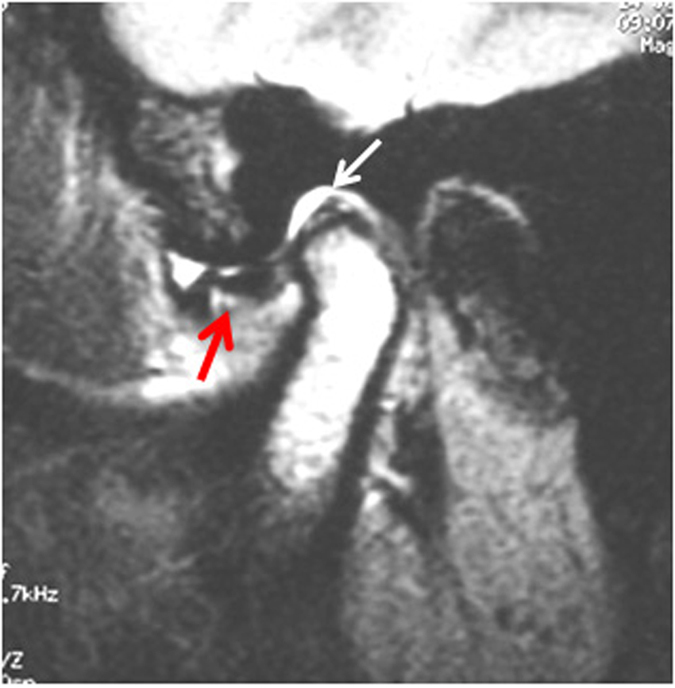
MRISagittal MRI of the temporomandibular joint: articular disc displaced anterior to the condyle (red arrow), posterior band disrupted and elongated (white arrow).
Source : He et al., Scientific Reports, 2016, figure 1 · CC BY
- Range-of-motion measurement : Maximum unassisted mouth opening, lateral excursion and protrusion are measured in millimetres. An opening of less than 40 mm is generally considered limited.⁴,⁷
- Muscle and joint palpation : The DC/TMD protocol specifies a standardised palpation pressure (around 1 kg for the masticatory muscles and 0.5 kg for the TMJ) to identify painful sites.⁷ Palpation of the masseter and temporalis muscles is particularly relevant for the diagnosis of myalgia.⁸ A meta-analysis has shown that the DC/TMD criteria for myofascial pain with referral have high specificity (0.98), meaning that a positive test almost certainly confirms the condition, although sensitivity is more variable.⁹
- Detection of joint noises : The examiner listens for noises (clicks, crepitus) during opening, closing and protrusive movements in order to diagnose disc displacement.⁴ The reliability of clinical examination for diagnosing disc displacement with reduction is judged moderate to substantial.¹⁰
- Dental conditions : abscess, pulpitis or periodontal disease.
- Primary headaches : migraine, tension-type headache. It is worth noting that TMD can itself cause "headache attributed to TMD".⁷
- Facial neuralgias : notably trigeminal neuralgia (ICHD-3 13.1), defined by unilateral paroxysms of pain in the trigeminal territory, lasting from a fraction of a second to 2 minutes, severe, shooting / electric shock-like, and triggered by innocuous stimuli. Distinguishing it from myogenous TMD is crucial because treatment differs radically (anticonvulsants, microvascular decompression vs conservative management).¹¹
- Otological conditions : otitis, Eustachian tube dysfunction.
- Pain of cervical origin : referred pain arising from the joints or muscles of the upper neck (cervicogenic headache) can mimic TMD pain.¹²
- Systemic conditions : rheumatoid arthritis, fibromyalgia, which can have manifestations at the TMJ.¹³
Should patients with temporomandibular disorders be classified, and what are the benefits?
Absolutely. Classifying patients is the end point of the diagnostic process and brings major benefits. The DC/TMD system offers a clear decision tree leading to specific diagnoses, grouped mainly into three categories.¹,⁷- Muscle disorders : local myalgia, myofascial pain, myofascial pain with referral.
- Disc displacements : disc displacement with reduction, disc displacement without reduction with limited opening, disc displacement without reduction without limited opening.
- Joint disorders (arthralgia, osteoarthritis, headache) : arthralgia, TMJ osteoarthritis, headache attributed to TMD.
- Targeted treatment planning : Treating myalgia (physiotherapy, stress management) is fundamentally different from treating disc displacement without reduction with limited opening (specific manual therapy, education).¹⁴ Classification avoids an ineffective "one size fits all" approach.
- Better prognosis : Understanding the exact nature of the disorder allows its course to be predicted more reliably. For example, a simple painless click has an excellent prognosis and often requires no intervention, whereas TMJ osteoarthritis may indicate a chronic degenerative condition.¹⁵
- Interprofessional communication : Using standardised terminology (DC/TMD) makes communication easier between physiotherapists, dentists, maxillofacial surgeons and psychologists, ensuring coordinated care.⁴
- Inclusion in research : Homogeneous classification of patients is essential in order to run high-quality clinical trials and compare results across studies, thereby advancing knowledge about the most effective treatments.¹
Critique and controversy
Robust though it is, the DC/TMD model is not without its challenges. One of the liveliest debates concerns the integration of Axis II into routine clinical practice. While its importance is universally acknowledged in research, many clinicians, for want of time or training, focus almost exclusively on Axis I (the physical).¹⁶ This purely biomedical approach is a major cause of treatment failure in patients with dominant psychosocial factors such as catastrophising or anxiety. Another persistent controversy concerns the reliability of certain clinical tests. For example, palpation of the lateral pterygoid muscle, once considered a key test, is now judged neither reliable nor valid because the muscle cannot be isolated from the surrounding structures.¹⁷ This underlines the importance of sticking to procedures validated by recent research. Finally, the role of dental occlusion in the aetiology of TMD, once considered central, has been considerably downgraded. Recent systematic reviews conclude that there is little solid evidence to support a cause-and-effect relationship between occlusal features and the development of TMD, in favour of biopsychosocial factors.⁵,¹⁸ Yet many therapeutic approaches remain strongly focused on occlusal correction, creating a gap between the scientific evidence and some clinical practice.Key points
- ✅ TMD diagnosis must follow the validated framework of the Diagnostic Criteria for TMD (DC/TMD), which combines a physical (Axis I) and a psychosocial (Axis II) assessment.
- ✅ History-taking begins with rapid, effective screening, followed by detailed questioning about pain, function and psychosocial factors.
- ✅ The clinical examination is standardised (range of motion, palpation) and must systematically include a differential diagnosis to rule out other conditions.
- ✅ The precise classification of TMD (e.g. myalgia vs. disc displacement) is not an end in itself, but an essential tool for guiding treatment, establishing a prognosis and supporting communication.
References
- Schiffman E, Ohrbach R, Truelove E, et al. Diagnostic Criteria for Temporomandibular Disorders (DC/TMD) for Clinical and Research Applications. J Oral Facial Pain Headache. 2014;28(1):6-27. PMID 24482784.
- Lövgren A, Visscher CM, Häggman-Henrikson B, Lobbezoo F, Marklund S, Wänman A. Validity of three screening questions (3Q/TMD) in relation to the DC/TMD. J Oral Rehabil. 2016;43(10):729-736. doi:10.1111/joor.12428.
- List T, Jensen RH. Temporomandibular disorders: Old ideas and new concepts. Cephalalgia. 2017;37(7):692-704. doi:10.1177/0333102416686302.
- Peck CC, Goulet JP, Lobbezoo F, et al. Expanding the taxonomy of the diagnostic criteria for temporomandibular disorders. J Oral Rehabil. 2014;41(1):2-23. PMID 24443898.
- Manfredini D, Lobbezoo F. Relationship between bruxism and temporomandibular disorders: a systematic review of literature from 1998 to 2008. Oral Surg Oral Med Oral Pathol Oral Radiol Endod. 2010;109(6):e26-e50. PMID 20451831.
- Fillingim RB, Ohrbach R, Greenspan JD, et al. Psychological factors associated with development of TMD: the OPPERA prospective cohort study. J Pain. 2013;14(12 Suppl):T75-T90. PMID 24275225.
- International Classification of Orofacial Pain Committee. International Classification of Orofacial Pain, 1st edition (ICOP). Cephalalgia. 2020;40(2):129-221. PMID 32103673.
- Steenks MH, Türp JC, de Wijer A. Reliability and Validity of the Diagnostic Criteria for Temporomandibular Disorders Axis I in Clinical and Research Settings: A Critical Appraisal. J Oral Facial Pain Headache. 2018;32(1):7-18. PMID 29370321.
- Asquini G, Pitance L, Michelotti A, Falla D. Effectiveness of manual therapy applied to craniomandibular structures in temporomandibular disorders: A systematic review. J Oral Rehabil. 2022;49(4):442-455. doi:10.1111/joor.13299.
- Lövgren A, Visscher CM, Lobbezoo F, et al. Outcome of three screening questions for temporomandibular disorders (3Q/TMD) on clinical decision-making. J Oral Rehabil. 2017;44(8):573-579. doi:10.1111/joor.12518.
- Maarbjerg S, Di Stefano G, Bendtsen L, Cruccu G. Trigeminal neuralgia — diagnosis and treatment. Cephalalgia. 2017;37(7):648-657. doi:10.1177/0333102416687280.
- Cuenca-Martínez F, Herranz-Gómez A, Madroñero-Miguel B, et al. Craniocervical and Cervical Spine Features of Patients with Temporomandibular Disorders: A Systematic Review and Meta-Analysis of Observational Studies. J Clin Med. 2020;9(9):2806. PMID 32872670.
- Yakkaphan P, Smith JG, Chana P, Renton T, Lambru G. Temporomandibular Disorders and Fibromyalgia Prevalence: A Systematic Review and Meta-Analysis. J Oral Facial Pain Headache. 2023;37(3):177-193. PMID 37975782.
- Al-Moraissi EA, Wolford LM, Ellis E 3rd, Neff A. The hierarchy of different treatments for arthrogenous temporomandibular disorders: A network meta-analysis of randomized clinical trials. J Craniomaxillofac Surg. 2020;48(1):9-23. PMID 31870713.
- Häggman-Henrikson B, Liv P, Ilgunas A, et al. Women are worse off in developing and recovering from temporomandibular disorder symptoms. Sci Rep. 2025;15:5532. PMID 39922904.
- Durham J, Al-Baghdadi M, Baad-Hansen L, et al. Self-management programmes in temporomandibular disorders: results from an international Delphi process. J Oral Rehabil. 2016;43(12):929-936. PMID 27727477.
- Türp JC, Minagi S. Palpation of the lateral pterygoid region in TMD — where is the evidence? J Dent. 2001;29(7):475-483. PMID 11809325.
- Manfredini D, Lombardo L, Siciliani G. Temporomandibular disorders and dental occlusion. A systematic review of association studies: end of an era? J Oral Rehabil. 2017;44(11):908-923. doi:10.1111/joor.12531.
Which treatment strategies are the most effective for temporomandibular joint (TMJ) syndrome?
Where to start? What is the recommended hierarchy of interventions?
The initial therapeutic approach for the majority of TMD is decidedly conservative. 💡 The Al-Moraissi 2020 network meta-analysis (48 randomised trials, more than 2,600 patients) confirms that occlusal splints and other non-invasive interventions are effective, with no single strategy standing out clearly from the others: the combination of education + exercise + manual therapy achieves results comparable to splints, at lower cost and with fewer iatrogenic risks.² This philosophy is built around a clear hierarchy, beginning with the least risky interventions and those that give the patient the most control.- Therapeutic patient education and self-management: The foundation of any effective management plan is patient education³. This means explaining the often benign, self-limiting nature of TMD, identifying and modifying parafunctions (such as tooth clenching, awake bruxism, or chewing gum) and teaching stress management strategies⁴. This stage aims to give the patient the tools to control their symptoms and prevent recurrence, which makes it the base of the care pyramid⁵.
- Active therapies (exercise): Once the educational foundation is in place, active therapies, mainly therapeutic exercise, are introduced. They are regarded as a central intervention because of their capacity to improve function, reduce pain and support patient autonomy⁶.
- Passive therapies (manual therapy and modalities): Passive interventions, such as manual therapy and the use of technologies, are regarded as important adjuncts, particularly when pain or restricted mobility hinders participation in active therapies⁷. Their role is often to "facilitate" the return to normal function, but they should not form the core of long-term treatment³.
- Pharmacological interventions and splints: Occlusal splints can be effective in reducing pain, particularly in myogenous pain, but their effectiveness is comparable to that of other conservative treatments². Medication, such as non-steroidal anti-inflammatory drugs (NSAIDs), can be used for short-term pain management but does not treat the underlying cause⁸.
🔺 TMD treatment pyramid
From the most invasive (apex) to the most universal (base): indicative % of patients concerned at each level
⚠️ Critical reading: the percentages shown are expert estimates of routine practice (based on Al-Moraissi 2020, Armijo-Olivo 2016, Christidis 2024 and NASEM 2020), not drawn from a formal epidemiological study. They illustrate a hierarchy of therapeutic intent, not an observed distribution. The hierarchy itself is well supported by the systematic reviews cited.
What is the place of exercise and is there a superior approach?
Therapeutic exercise is a cornerstone of TMD treatment, with solid evidence supporting its effectiveness in reducing pain and improving mandibular range of motion and overall function¹⁹. A 2022 systematic review concluded that therapeutic exercise is particularly beneficial for TMD of muscular origin (myogenous) and that a clinician-supervised programme tends to give better results than an unsupervised one⁶. 🏋️♂️ There is no single universally superior type of exercise, but rather a combination of approaches tailored to the patient:- Mobility and stretching exercises: These aim to restore normal range of motion and reduce muscle stiffness. Controlled mouth-opening exercises and gentle stretching of the masticatory muscles are frequently prescribed¹⁰.
- Motor control and coordination exercises: These exercises, such as the "tongue resting position exercise" (tongue on the palate, teeth slightly apart, lips closed), help to reprogramme dysfunctional motor patterns and reduce muscle tension at rest¹¹.
- Strengthening exercises: Light isometric and isotonic strengthening can be useful in improving the stability and endurance of the masticatory muscles, particularly in cases of joint hypermobility or instability⁶.
Manual therapies and technologies: how effective are they really?
Manual therapies and physical modalities are valuable tools in the physiotherapist's armoury, mainly for short- and medium-term symptom management. Manual therapy, which includes soft tissue and joint mobilisation techniques, has shown significant effectiveness. The Asquini 2022 systematic review (J Oral Rehabil), devoted specifically to craniomandibular manual therapy found a beneficial effect on pain and mouth opening, despite very low evidence quality linked to heterogeneity and small sample sizes.¹² Integrating the cervical spine into treatment is now a matter of consensus: the Cuenca-Martínez 2020 meta-analysis (observational studies) confirms a clinically relevant association between cervical and mandibular disability in patients with TMD (reduced pressure pain threshold and decreased cervical range of motion, moderate level of evidence). The La Touche 2020 randomised trial (JOSPT) further shows that adding a high-velocity cervical manipulation to an education and exercise programme significantly improves pain and function in patients with TMD with myalgia.¹³ As for technologies (electrophysical agents):- Low-level laser therapy (LLLT) has shown notable effectiveness in reducing short-term pain, particularly for TMD of muscular origin (myalgia).¹⁴ The Xu 2018 meta-analysis (31 randomised trials) confirmed that LLLT is superior to placebo for pain (a significant short-term effect on the VAS) and improves TMJ functional outcomes, while noting high methodological heterogeneity in laser parameters (wavelength, dose, duration).¹⁵
- Therapeutic ultrasound and transcutaneous electrical nerve stimulation (TENS) are also used, but the evidence for their effectiveness is more mixed and often of lower quality than for LLLT or active approaches¹⁶.
| Modality | Main indication | Level of evidence | Expected effect |
|---|---|---|---|
| Patient education | All forms | High | ↘ pain, ↗ autonomy, ↘ recurrence |
| Supervised exercise | Myogenous ++ | High | ↘ VAS, ↗ mouth opening |
| Manual therapy (TMJ + cervical) | Myogenous, arthrogenous | High | ↘ short-term pain, ↗ mobility |
| Occlusal splint | Bruxism, myogenous | Moderate | Effect ≈ exercise, ↗ cost/risk |
| LLLT (low-level laser) | Acute myalgia | Moderate | ↘ short-term VAS vs placebo |
| CBT / mindfulness | Dominant psychological component | Moderate | ↘ catastrophising, ↘ chronic pain |
| NSAIDs (short term) | Acute flare | Moderate | ↘ transient pain |
| TENS / ultrasound | Symptomatic adjunct | Low | Mixed evidence |
| Irreversible occlusal adjustment | — | Low | ⚠️ not recommended as 1st line |
| TMJ surgery | Conservative failure + structural lesion | Case by case | Exceptional recourse |
Beyond the physical: how do you educate the patient and address psychological factors?
The biopsychosocial model is essential to understanding and treating TMD, because psychological factors such as anxiety, depression and stress are powerful modulators of chronic pain¹⁷. Comprehensive management must therefore include an educational and behavioural component. Education aims to:- Demystify the condition: Reassuring the patient that there is no serious disease and explaining the physiology of pain can reduce fear and hypervigilance, factors that perpetuate pain¹⁸.
- Identify the triggers: Helping the patient become aware of parafunctional habits (tooth clenching, jaw posture) is the first step towards changing them³.
- Promote self-efficacy: The aim is to make the patient an active participant in their recovery by providing simple self-management strategies (relaxation techniques, exercises, application of heat/cold)⁴.
Critique and controversy
Despite growing consensus on the conservative approach, several areas of debate persist. The most notable is the heterogeneity of TMD itself. Classifying patients into subgroups (for example, arthralgia, myalgia, disc displacement) is crucial, because an intervention that is effective for one group may be ineffective for another¹⁷. Future research must focus on stratified treatment protocols rather than on a single approach. What is more, the long-term effectiveness of many passive modalities such as ultrasound remains debatable, raising the question of their relevance beyond acute pain management¹⁶. Finally, the role and optimal type of occlusal splint remain the subject of ongoing debate between the dental and physiotherapy professions, with some studies showing effectiveness similar to that of far cheaper and more active interventions such as exercise and education². The major challenge therefore remains to personalise treatment according to the patient's specific profile, integrating the physical, behavioural and psychological dimensions coherently.Key points
- ✅ First-line treatment for TMD is conservative, non-invasive and reversible.
- 🎓 The role of patient education about their condition, parafunctions and self-management strategies is the foundation of care.
- 💪 Prescribing therapeutic exercise (mobility, motor control, strengthening) is a central and highly effective intervention, especially when supervised.
- 🤲 In practice, manual therapy is a powerful adjunct for reducing pain and improving mobility, and is particularly effective when combined with exercise.
- 🧠 Taking account of psychological and behavioural factors is essential for lasting results, because stress and anxiety can perpetuate symptoms.
References
- Armijo-Olivo S, Pitance L, Singh V, Neto F, Thie N, Michelotti A. Effectiveness of Manual Therapy and Therapeutic Exercise for Temporomandibular Disorders: Systematic Review and Meta-Analysis. Phys Ther. 2016;96(1):9-25. PMID 26294683.
- Al-Moraissi EA, Farea R, Qasem KA, Al-Wadeai MS, Al-Sabahi ME, Al-Iryani GM. Effectiveness of occlusal splint therapy in the management of temporomandibular disorders: network meta-analysis of randomized controlled trials. Int J Oral Maxillofac Surg. 2020;49(8):1042-1056. PMID 31982236.
- Gauer RL, Semidey MJ. Diagnosis and treatment of temporomandibular disorders. Am Fam Physician. 2015;91(6):378-386. PMID 25822556.
- Durham J, Al-Baghdadi M, Baad-Hansen L, et al. Self-management programmes in temporomandibular disorders: results from an international Delphi process. J Oral Rehabil. 2016;43(12):929-936. PMID 27727477.
- Aggarwal VR, Fu Y, Main CJ, Wu J. The effectiveness of self-management interventions in adults with chronic orofacial pain: A systematic review, meta-analysis and meta-regression. Eur J Pain. 2019;23(5):849-865. PMID 30620145.
- Dickerson SM, Weaver JM, Boyson AN, Thacker JA, Junak AA, Ritzline PD, Donaldson MB. The effectiveness of exercise therapy for temporomandibular dysfunction: a systematic review and meta-analysis. Clin Rehabil. 2017;31(8):1039-1048. doi:10.1177/0269215516672275.
- Calixtre LB, Moreira RFC, Franchini GH, Alburquerque-Sendín F, Oliveira AB. Manual therapy for the management of pain and limited range of motion in subjects with signs and symptoms of temporomandibular disorder: a systematic review of randomised controlled trials. J Oral Rehabil. 2015;42(11):847-861. PMID 26059857.
- Roldán-Barraza C, Janko S, Villanueva J, Araya I, Lauer HC. A systematic review and meta-analysis of usual treatment versus psychosocial interventions in the treatment of myofascial temporomandibular disorder pain. J Oral Facial Pain Headache. 2014;28(3):205-222. PMID 25068215.
- Calixtre LB, Grüninger BLDS, Haik MN, Alburquerque-Sendín F, Oliveira AB. Effectiveness of mobilisation of the upper cervical region and craniocervical flexor training on orofacial pain, mandibular function and headache in women with TMD: A randomised, controlled trial. J Oral Rehabil. 2019;46(2):109-119. PMID 30307636.
- Asquini G, Pitance L, Michelotti A, Falla D. Effectiveness of manual therapy applied to craniomandibular structures in temporomandibular disorders: A systematic review. J Oral Rehabil. 2022;49(4):442-455. doi:10.1111/joor.13299.
- Michelotti A, de Wijer A, Steenks M, Farella M. Home-exercise regimes for the management of non-specific temporomandibular disorders. J Oral Rehabil. 2005;32(11):779-785. PMID 16202040.
- Cuenca-Martínez F, Herranz-Gómez A, Madroñero-Miguel B, et al. Craniocervical and Cervical Spine Features of Patients with Temporomandibular Disorders: A Systematic Review and Meta-Analysis of Observational Studies. J Clin Med. 2020;9(9):2806. PMID 32872670.
- La Touche R, Fernández-de-las-Peñas C, Fernández-Carnero J, Escalante K, Angulo-Díaz-Parreño S, Paris-Alemany A, Cleland JA. Effectiveness of Cervical Spine High-Velocity, Low-Amplitude Thrust Added to Behavioral Education, Soft Tissue Mobilization, and Exercise for People With Temporomandibular Disorder With Myalgia: A Randomized Clinical Trial. J Orthop Sports Phys Ther. 2020;50(8):455-465. doi:10.2519/jospt.2020.9175.
- Magri LV, Carvalho VA, Rodrigues FCC, Bataglion C, Leite-Panissi CRA. Effectiveness of low-level laser therapy on pain intensity, pressure pain threshold, and SF-MPQ indexes of women with myofascial pain. Lasers Med Sci. 2017;32(2):419-428.
- Xu GZ, Jia J, Jin L, Li JH, Wang ZY, Cao DY. Low-Level Laser Therapy for Temporomandibular Disorders: A Systematic Review with Meta-Analysis. Pain Res Manag. 2018;2018:4230583. PMID 29861802.
- Stechman-Neto J, Porporatti AL, Porto de Toledo I, Costa YM, Conti PCR, De Luca Canto G, Mezzomo LA. Effect of temporomandibular disorder therapy on otologic signs and symptoms: a systematic review. J Oral Rehabil. 2016;43(6):468-479. PMID 26749516.
- Slade GD, Ohrbach R, Greenspan JD, et al. Painful Temporomandibular Disorder: Decade of Discovery from OPPERA Studies. J Dent Res. 2016;95(10):1084-1092. PMID 27339423.
- Yakkaphan P, Smith JG, Chana P, Renton T, Lambru G. Temporomandibular Disorders and Fibromyalgia Prevalence: A Systematic Review and Meta-Analysis. J Oral Facial Pain Headache. 2023;37(3):177-193. PMID 37975782.
- List T, Jensen RH. Temporomandibular disorders: Old ideas and new concepts. Cephalalgia. 2017;37(7):692-704. doi:10.1177/0333102416686302.
- Fillingim RB, Ohrbach R, Greenspan JD, et al. Psychological factors associated with development of TMD: the OPPERA prospective cohort study. J Pain. 2013;14(12 Suppl):T75-T90. PMID 24275225.
How can lasting recovery be secured and recurrence of temporomandibular joint (TMJ) syndrome prevented?
How do you make the patient an active participant in their recovery through self-management?
Self-management is the cornerstone of preventing recurrence in TMD³. It aims to give the patient the power (empowerment) to control their symptoms and maintain optimal mandibular function. 🧠 The effectiveness of this approach rests on several fundamental pillars identified by clinical research. First, therapeutic patient education is a sine qua non. A clear understanding of TMJ anatomy, of pain mechanisms and of aggravating factors makes it possible to defuse anxiety about the condition and significantly improves adherence to exercise programmes⁴. Studies have shown that structured educational programmes, explaining in particular the role of parafunctions (such as bruxism or tooth clenching), are effective in reducing pain and the frequency of symptomatic episodes⁵. The aim is to replace mistaken beliefs with a functional understanding of the condition⁶. Second, setting up a personalised home exercise programme is essential. Systematic reviews and meta-analyses confirm that a combination of mobilisation, motor control and strengthening exercises for the masticatory and cervical muscles reduces pain and improves functional capacity in a lasting way⁷,⁸. These exercises aim to restore normal mandibular kinematics and to build the resilience of the joint and muscle structures in the face of everyday demands⁹. A typical programme includes controlled opening, protrusion/retrusion and lateral excursion exercises, performed pain-free¹⁰. Third, the management of parafunctions and lifestyle habits is a critical factor. The physiotherapist must help the patient identify and change harmful behaviours: daytime tooth clenching, unilateral chewing, nail-biting, or the prolonged maintenance of unsuitable cervical postures¹¹. Biofeedback techniques or simple behavioural reminders can be built in to make healthier new habits automatic¹². Finally, the management of stress and psychosocial factors must not be neglected. High stress levels, anxiety and pain catastrophising have been shown to be important predictors of TMD chronicity²,¹³. Incorporating relaxation techniques, diaphragmatic breathing or mindfulness (mindfulness) can significantly reduce overactivity of the masticatory muscles and improve pain perception¹⁴.When and how should a safe return to sport and activity be planned?
Return to sport (RTS) or to demanding activities (for example, playing a wind instrument) after an episode of TMD should be progressive and guided by precise criteria rather than by a simple timetable. 💪 Although the scientific literature specific to RTS in TMD is less abundant than for other joints, the principles of sports rehabilitation apply fully¹⁵. Planning the return to activity is built around a stepwise approach, based on achieving functional criteria:- Phase 1: Pain control and restoration of basic function. The absolute prerequisite is the absence of pain during activities of daily living, including normal chewing and functional mouth opening (typically > 40 mm) without pain or apprehension⁷,⁹.
- Phase 2: Progressive reintroduction of load. This phase involves gradually re-exposing the TMJ to loads similar to those encountered in the target activity. For an athlete, that may mean resuming running (which generates vertical impacts) or wearing a mouthguard during light training¹⁶. For a musician, it will mean resuming short, low-intensity playing sessions. Monitoring symptoms 24 hours after exertion is crucial.
- Phase 3: Return to normal training and competition. Moving to this phase is possible only if phase 2 has been completed without symptoms reappearing. The patient must have regained full confidence in their jaw. Strengthening and motor control exercises must be maintained in the long term to prevent recurrence¹⁰.
Critique and controversy
Despite growing consensus on the effectiveness of active approaches, several grey areas persist. A major controversy lies in the heterogeneity of TMD. Many clinical studies group under the same diagnosis patients with very different profiles (myalgia, arthralgia, disc displacement), which can dilute the specific effects of interventions¹⁷. Future research must focus on stratified treatment protocols rather than on a single approach. What is more, the long-term effectiveness of many passive modalities such as ultrasound remains debatable, raising the question of their relevance beyond acute pain management¹⁶. Finally, the role and optimal type of occlusal splint remain the subject of ongoing debate between the dental and physiotherapy professions, with some studies showing effectiveness similar to that of far cheaper and more active interventions such as exercise and education². The major challenge therefore remains to personalise treatment according to the patient's specific profile, integrating the physical, behavioural and psychological dimensions coherently.Key points
- The prevention of recurrence rests on the move from passive care to active self-management by the patient.
- The pillars of this approach are: therapeutic patient education, a programme of home exercises, the management of parafunctions and consideration of psychosocial factors.
- Return to sport must be progressive and guided by functional criteria (absence of pain, restored function) rather than by a fixed timetable.
- Active patient involvement in their treatment plan is the best predictor of long-term success.
References
- Asquini G, Pitance L, Michelotti A, Falla D. Effectiveness of manual therapy applied to craniomandibular structures in temporomandibular disorders: A systematic review. J Oral Rehabil. 2022;49(4):442-455. doi:10.1111/joor.13299.
- Slade GD, Ohrbach R, Greenspan JD, et al. Painful Temporomandibular Disorder: Decade of Discovery from OPPERA Studies. J Dent Res. 2016;95(10):1084-1092. PMID 27339423.
- Aggarwal VR, Fu Y, Main CJ, Wu J. The effectiveness of self-management interventions in adults with chronic orofacial pain: A systematic review, meta-analysis and meta-regression. Eur J Pain. 2019;23(5):849-865. PMID 30620145.
- Durham J, Al-Baghdadi M, Baad-Hansen L, et al. Self-management programmes in temporomandibular disorders: results from an international Delphi process. J Oral Rehabil. 2016;43(12):929-936. PMID 27727477.
- Michelotti A, de Wijer A, Steenks M, Farella M. Home-exercise regimes for the management of non-specific temporomandibular disorders. J Oral Rehabil. 2005;32(11):779-785. PMID 16202040.
- List T, Jensen RH. Temporomandibular disorders: Old ideas and new concepts. Cephalalgia. 2017;37(7):692-704. doi:10.1177/0333102416686302.
- Armijo-Olivo S, Pitance L, Singh V, Neto F, Thie N, Michelotti A. Effectiveness of Manual Therapy and Therapeutic Exercise for Temporomandibular Disorders: Systematic Review and Meta-Analysis. Phys Ther. 2016;96(1):9-25. PMID 26294683.
- Dickerson SM, Weaver JM, Boyson AN, et al. The effectiveness of exercise therapy for temporomandibular dysfunction: a systematic review and meta-analysis. Clin Rehabil. 2017;31(8):1039-1048. doi:10.1177/0269215516672275.
- Calixtre LB, Moreira RFC, Franchini GH, Alburquerque-Sendín F, Oliveira AB. Manual therapy for the management of pain and limited range of motion in subjects with signs and symptoms of temporomandibular disorder: a systematic review of randomised controlled trials. J Oral Rehabil. 2015;42(11):847-861. PMID 26059857.
- Häggman-Henrikson B, Liv P, Ilgunas A, et al. Women are worse off in developing and recovering from temporomandibular disorder symptoms. Sci Rep. 2025;15:5532. PMID 39922904.
- Manfredini D, Lobbezoo F. Relationship between bruxism and temporomandibular disorders: a systematic review of literature from 1998 to 2008. Oral Surg Oral Med Oral Pathol Oral Radiol Endod. 2010;109(6):e26-e50. PMID 20451831.
- Fillingim RB, Ohrbach R, Greenspan JD, et al. Psychological factors associated with development of TMD: the OPPERA prospective cohort study. J Pain. 2013;14(12 Suppl):T75-T90. PMID 24275225.
- Younger JW, Shen YF, Goddard G, Mackey SC. Chronic myofascial temporomandibular pain is associated with neural abnormalities in the trigeminal and limbic systems. Pain. 2010;149(2):222-228. PMID 20236763.
- Yakkaphan P, Smith JG, Chana P, Renton T, Lambru G. Temporomandibular Disorders and Fibromyalgia Prevalence: A Systematic Review and Meta-Analysis. J Oral Facial Pain Headache. 2023;37(3):177-193. PMID 37975782.
- Häggman-Henrikson B, Lobbezoo F, Durham J, Peck C, List T. Prevalence of Temporomandibular Disorder Symptoms After Whiplash Trauma — A Systematic Review and Meta-Analysis. Eur J Pain. 2025;29(3):e4792. PMID 39921489.
- Calixtre LB, Grüninger BLDS, Haik MN, Alburquerque-Sendín F, Oliveira AB. Effectiveness of mobilisation of the upper cervical region and craniocervical flexor training on orofacial pain, mandibular function and headache in women with TMD: A randomised, controlled trial. J Oral Rehabil. 2019;46(2):109-119. PMID 30307636.
- International Classification of Orofacial Pain Committee. International Classification of Orofacial Pain, 1st edition (ICOP). Cephalalgia. 2020;40(2):129-221. PMID 32103673.
- Cuenca-Martínez F, Herranz-Gómez A, Madroñero-Miguel B, et al. Craniocervical and Cervical Spine Features of Patients with Temporomandibular Disorders: A Systematic Review and Meta-Analysis of Observational Studies. J Clin Med. 2020;9(9):2806. PMID 32872670.
- Schiffman E, Ohrbach R, Truelove E, et al. Diagnostic Criteria for Temporomandibular Disorders (DC/TMD) for Clinical and Research Applications. J Oral Facial Pain Headache. 2014;28(1):6-27. PMID 24482784.
What do real-world case studies teach us about temporomandibular joint (TMJ) syndrome?
Analysis of a "classic" case: from assessment to resolution
The case study is a powerful teaching tool for understanding clinical reasoning in physiotherapy. The case reported by Lewis & Naude (2010, South African Journal of Physiotherapy) illustrates successful multimodal management in a 26-year-old man presenting simultaneously with cervicogenic headache and associated TMD. The protocol, spread over 9 physiotherapy sessions, combined several scientifically validated approaches²¹:- Maitland manual therapy: cervical (particularly upper cervical) and TMJ mobilisations to restore mobility and modulate pain.
- Soft tissue therapy: treatment of trigger points (trigger points) and myofascial massage to reduce muscle tension.³
- Neuromuscular strengthening: work on the deep neck flexors and the scapulothoracic musculature.
- Postural reintegration: correction of cervicocephalic postural faults.
🧩 Components of the multimodal protocol: Lewis & Naude 2010 case
Diagram of the management pathway over 9 sessions and its correlation with pooled levels of evidence
Source: Lewis F, Naude B. S Afr J Physiother. 2010;66(2). The levels of evidence given for each component are drawn from the systematic reviews cited in the article.
The diagnostic challenge: when temporomandibular joint (TMJ) syndrome mimics another condition
One of the greatest lessons from case studies is the capacity of TMD to present with symptoms that mimic other conditions, leading to diagnostic delay and ineffective treatment. ⚠️ The complex anatomical location of the TMJ and its neurological connections with the trigeminal nerve explain these atypical presentations. The symmetry is in fact two-way: TMD can mimic other conditions, but other conditions can also mimic TMD. Several published, verifiable cases illustrate this diagnostic risk:- 🔍 Eagle syndrome (elongated styloid process) mimicking TMD: Schneider et al. (2015, Int J Surg Case Rep) report a case of cervicofacial pain and trismus long treated as TMD before 3D imaging revealed a styloid process > 30 mm compressing the neighbouring structures. Classic Eagle criteria: pain on cervical rotation, dysphagia, otalgia, sensation of a foreign body in the pharynx. CT scan = diagnostic gold standard.¹⁵
- 🔍 Trigeminal neuralgia (ICHD-3 13.1): the Maarbjerg/Cruccu 2017 review (Cephalalgia) stresses that confusion with TMD persists in practice despite distinct criteria: TN is characterised by paroxysms that are searing, electric shock-like, lasting < 2 minutes, triggered by innocuous stimuli (touch, chewing, speaking), unilateral and strictly within the trigeminal territory. Treatment (carbamazepine first-line, vascular decompression second-line) is radically different.⁵
- 🔍 Intramuscular pterygoid mass mimicking TMD: a 2024 case (Cureus) reports a 36-year-old patient with chronic TMJ pain and restricted opening resistant to conservative treatment, in whom MRI eventually revealed a mass in the lateral pterygoid muscle. Any TMD resistant to 3-6 months of well-conducted conservative treatment should prompt the diagnosis to be reconsidered and advanced imaging to be considered.¹⁶
- 🔍 Facial fascial space abscesses disguised as TMD: Wang et al. (2024) describe 3 cases of odontogenic infection initially managed as TMD, with a potentially serious diagnostic delay. Fever, acute trismus and swelling are the warning signs that must prompt immediate redirection.¹⁷
A complex case study
TMD is only rarely an isolated problem. It often forms part of a broader clinical picture, involving comorbidities that complicate both diagnosis and treatment. Complex cases teach us the need for a biopsychosocial and multidisciplinary approach. The link between the cervical spine and the TMJ is a classic example of complexity. The Calixtre 2019 randomised trial (J Oral Rehabil) showed that upper cervical mobilisation combined with deep craniocervical flexor training significantly improves orofacial pain, mandibular function and headache in women with TMD, independently of any intra-oral technique. Clinically, this supports current recommendations: treatment cannot be confined to the jaw but must target both regions as a single functional unit, consistent with trigeminocervical convergence at brainstem level. The Cuenca-Martínez 2020 meta-analysis (16 observational studies) confirms a robust association between cervical disability and TMD, particularly for pressure pain thresholds and cervical mobility.⁸ Complexity increases further in the presence of widespread pain conditions or central sensitisation. The Yakkaphan 2023 meta-analysis quantifies this overlap: 76.8 % of patients with fibromyalgia have TMD (essentially myogenous forms: 63 %), and conversely 32.7 % of patients with TMD have comorbid fibromyalgia. In these patients, a purely biomechanical approach is bound to fail.⁹ The strategy must incorporate components of the biopsychosocial approach:- Pain education: Explaining the mechanisms of central pain in order to defuse anxiety about the symptoms and give the patient control (patient empowerment)⁹.
- Stress and anxiety management: Anxiety and catastrophising are major factors that maintain pain in TMD¹⁰.
- Graded exercise: A gentle, progressive exercise programme to avoid the pain flares typical of fibromyalgia.
And in children? The special case of juvenile idiopathic arthritis (JIA)
One paediatric situation deserves particular attention: juvenile idiopathic arthritis (JIA) can present with TMJ involvement as the only initial expression of the disease, with frequent diagnostic delay.¹⁸ The Ronsivalle 2024 meta-analysis (J Oral Rehabil, 366 children with JIA assessed using DC/TMD) found a relative risk of TMD multiplied by 3.86 (95 % CI 2.59–5.76) in children with JIA vs controls.¹⁹ Clinical symptoms can appear after significant condylar damage is already established, hence the importance of early imaging (MRI) in any child with persistent TMJ involvement, especially against a rheumatological background or with facial asymmetry.¹⁸ Key point: any child or adolescent with TMJ pain associated with progressively limited opening, mandibular asymmetry or hemifacial growth retardation should undergo a rheumatological assessment. A paediatric version of the DC/TMD has been published (Rongo et al. 2021/2022) to equip the assessment of under-18s specifically.²⁰Critique and controversy, where do case studies sit in the hierarchy of evidence?
Case studies are illuminating, but it is crucial to recognise their limits and the controversies they raise. 🧠 The main criticism is their low level of evidence in the pyramid of scientific evidence. A single case illustrates a possibility, not a general rule. It is impossible to conclude that there is a cause-and-effect relationship, because placebo effects, regression to the mean and the natural history of the disease are not controlled for¹². What is more, published cases often tend to report successes, introducing a publication bias that can overestimate the effectiveness of an intervention.📐 Hierarchy of scientific evidence, where does each type of study belong?
Strength of evidence decreasing from the top (meta-analyses) to the bottom (isolated cases)
Simplified GRADE / Oxford CEBM hierarchy. The length of the coloured bar on the right illustrates the relative strength of evidence. Practical implication: where an appealing case study and a meta-analysis diverge, the decision must follow the meta-analysis. Case studies remain valuable for generating hypotheses, flagging rare presentations, or illustrating a process of clinical reasoning.
⭐ Key points
- The Lewis & Naude (2010) case illustrates that multimodal management (Maitland manual therapy + soft tissue + deep cervical strengthening + posture) over 9 sessions can clear cervicogenic headache and associated TMD in a young patient.
- Differential diagnoses you must know : Eagle syndrome (styloid process > 30 mm), trigeminal neuralgia (lightning-like paroxysms, ICHD-3 13.1), intramuscular pterygoid mass, odontogenic facial abscess. Any TMD resistant to 3-6 months of well-conducted conservative treatment warrants advanced imaging.
- Conversely, TMD are themselves great mimics : 77.5 % of patients with TMD have an otological complaint (Tuz 2003). A TMJ examination should be part of the differential diagnosis of unexplained earache and atypical headache.
- In children: think of juvenile idiopathic arthritis (RR of TMD × 3.86 vs controls, Ronsivalle 2024): TMJ involvement can be the first manifestation, and condylar destruction sometimes precedes clinical signs.
- Complex cases call for a broader view: cervical spine (Calixtre 2019), fibromyalgia (76.8 % TMD in FM, Yakkaphan 2023), bruxism, psychosocial factors.
- ⚠️ Level of evidence : a case report = level 5 (the weakest). It illustrates; it never demonstrates effectiveness. Where there is divergence, follow the meta-analyses (level 1a), not the isolated case.
References
- Schiffman E, Ohrbach R, Truelove E, et al. Diagnostic Criteria for Temporomandibular Disorders (DC/TMD) for Clinical and Research Applications. J Oral Facial Pain Headache. 2014;28(1):6-27. PMID 24482784.
- Calixtre LB, Grüninger BLDS, Haik MN, Alburquerque-Sendín F, Oliveira AB. Effectiveness of mobilisation of the upper cervical region and craniocervical flexor training on orofacial pain, mandibular function and headache in women with TMD: A randomised, controlled trial. J Oral Rehabil. 2019;46(2):109-119. PMID 30307636.
- Stechman-Neto J, Porporatti AL, Porto de Toledo I, Costa YM, Conti PCR, De Luca Canto G, Mezzomo LA. Effect of temporomandibular disorder therapy on otologic signs and symptoms: a systematic review. J Oral Rehabil. 2016;43(6):468-479. PMID 26749516.
- Asquini G, Pitance L, Michelotti A, Falla D. Effectiveness of manual therapy applied to craniomandibular structures in temporomandibular disorders: A systematic review. J Oral Rehabil. 2022;49(4):442-455. doi:10.1111/joor.13299.
- Maarbjerg S, Di Stefano G, Bendtsen L, Cruccu G. Trigeminal neuralgia — diagnosis and treatment. Cephalalgia. 2017;37(7):648-657. doi:10.1177/0333102416687280.
- Tuz HH, Onder EM, Kisnisci RS. Prevalence of otologic complaints in patients with temporomandibular disorder. Am J Orthod Dentofacial Orthop. 2003;123(6):620-623. PMID 12806339.
- Pigg M, Nixdorf DR, Law AS, et al. New International Classification of Orofacial Pain: What Is in It For Endodontists? J Endod. 2021;47(3):345-357. PMID 33340605.
- Cuenca-Martínez F, Herranz-Gómez A, Madroñero-Miguel B, et al. Craniocervical and Cervical Spine Features of Patients with Temporomandibular Disorders: A Systematic Review and Meta-Analysis of Observational Studies. J Clin Med. 2020;9(9):2806. PMID 32872670.
- Yakkaphan P, Smith JG, Chana P, Renton T, Lambru G. Temporomandibular Disorders and Fibromyalgia Prevalence: A Systematic Review and Meta-Analysis. J Oral Facial Pain Headache. 2023;37(3):177-193. PMID 37975782.
- Kindler S, Samietz S, Houshmand M, et al. Depressive and anxiety symptoms as risk factors for temporomandibular joint pain: a prospective cohort study in the general population. J Pain. 2012;13(12):1188-1197. PMID 23141187.
- Manfredini D, Lobbezoo F. Relationship between bruxism and temporomandibular disorders: a systematic review of literature from 1998 to 2008. Oral Surg Oral Med Oral Pathol Oral Radiol Endod. 2010;109(6):e26-e50. PMID 20451831.
- Nissen T, Wynn R. The clinical case report: a review of its merits and limitations. BMC Res Notes. 2014;7:264. PMID 24758689.
- List T, Jensen RH. Temporomandibular disorders: Old ideas and new concepts. Cephalalgia. 2017;37(7):692-704. doi:10.1177/0333102416686302.
- International Classification of Orofacial Pain Committee. International Classification of Orofacial Pain, 1st edition (ICOP). Cephalalgia. 2020;40(2):129-221. PMID 32103673.
- Schneider HP, Steinemann S, Schmidt T, Pichler EM. Eagle's syndrome — A non-perceived differential diagnosis of temporomandibular disorder. Int J Surg Case Rep. 2015;15:123-126. PMC 4601974.
- Bhatnagar A, Ahmed S, Singh M, Sharma S. Curious Case of Facial Pain Mimicking Temporomandibular Disorder. Cureus. 2024;16(7):e64864. PMC 11335035.
- Wang H, Wang T, Wang Z, et al. Orofacial fascial space abscess disguised as temporomandibular disorder: a report of 3 cases and literature review. BMC Oral Health. 2023;23:1057. PMC 10765852.
- Hügle B, Spiegel L, Hotte J, et al. Temporomandibular joint disorders as the only manifestation of juvenile idiopathic arthritis: a case report. Pediatr Rheumatol Online J. 2018;16:54. PMC 6122888.
- Ronsivalle V, Carli E, Lo Giudice A, et al. Prevalence of temporomandibular disorders in juvenile idiopathic arthritis evaluated with diagnostic criteria for temporomandibular disorders: A systematic review with meta-analysis. J Oral Rehabil. 2024;51(4):788-799. PMID 38012098.
- Rongo R, Ekberg E, Nilsson IM, et al. Diagnostic criteria for temporomandibular disorders (DC/TMD) for children and adolescents: An international Delphi study—Part 1: Development of Axis I. J Oral Rehabil. 2021;48(7):836-845. PMID 33817818. (Part 2 — Axis II: J Oral Rehabil. 2022; doi:10.1111/joor.13301.)
- Lewis F, Naude B. The effectiveness of physiotherapy in cervicogenic headache and concurring temporomandibular dysfunction: a case report. S Afr J Physiother. 2010;66(2):37-39. sajp.co.za.
How can these recommendations be applied concretely in your practice?
When and to which other health professionals should you refer?
One of the fundamental skills of the modern physiotherapist is recognising the limits of their scope of practice and identifying the situations that call for interprofessional collaboration. This triage process is crucial to the safety and effectiveness of care. Identifying "red flags" is the first, non-negotiable step. These signs and symptoms suggest a serious underlying condition (for example, a fracture, an infection, a tumour or cauda equina syndrome) and call for immediate or emergency medical referral.¹ It is crucial to note, however, that many traditional red flags, taken in isolation, have low diagnostic accuracy and must be interpreted in the context of a full clinical examination.² An international systematic review has indeed been published to establish a reference framework of red flags for potentially serious spinal conditions.¹🚩 Red flags specific to the masticatory system
- Acute febrile trismus → suspected deep infection (peritonsillar abscess, mandibular osteomyelitis)
- Firm, painless swelling of the parotid or pre-auricular region → suspected neoplasia (salivary gland tumour, metastasis)
- Sudden, irreducible deviation of the jaw with mental nerve hypoaesthesia → mandibular fracture, nerve injury
- Explosive "thunderclap" headache + TMJ pain → subarachnoid haemorrhage, arterial dissection
- Unexplained weight loss + persistent facial pain → neoplastic work-up
- Progressively limited mouth opening in a child + facial asymmetry → suspected TMJ juvenile idiopathic arthritis
- Recent history of craniofacial trauma with TMJ pain or altered occlusion → fracture of the mandibular condyle
⚠️ Any red flag → prompt medical referral (GP, emergency department, maxillofacial surgery) before any physiotherapy management.
How do you measure outcomes and overcome barriers to implementation?
To be sure that interventions are effective, outcomes must be measured objectively and in a standardised way. The systematic use of patient-reported outcome measures (PROMs) has become the norm for assessing the impact of treatment on pain, function and quality of life from the patient's point of view.⁶ Their implementation, however, runs into significant obstacles, the most frequently cited being lack of time, technical difficulties with software and lack of training to interpret the results.⁶ To standardise this measurement on a larger scale, the scientific community is promoting the adoption of "Core Outcome Sets" (COS). These are an internationally agreed set of core outcome measures for a specific condition, ensuring that all clinicians and researchers assess the same critical domains.⁸ Using COS allows more reliable comparison between studies and better pooling of data.⁸ Overcoming the barriers to implementing evidence-based practice (EBP) is a major challenge. A 2022 systematic review identified the most common barriers perceived by physiotherapists: lack of time, lack of research skills and lack of support from the organisation.⁵ The key facilitators include strong leadership, access to resources (databases, dedicated time) and participation in continuing education.⁵٬¹⁵ Integrating shared decision-making is also a powerful lever, although its implementation is itself held back by perceived lack of time and by clinicians' need for training.⁷ It is a collaborative process in which clinician and patient make health decisions together, taking into account the best available evidence as well as the patient's values and preferences.¹³ 📊 Strategies for overcoming these barriers 🚧 must be multi-faceted, combining solutions at individual (training, mentoring), organisational (protected time, access to resources) and system (health policies that support EBP) level.¹⁵ "De-implementing" low-value practices, such as the use of certain passive modalities without supporting evidence, is just as important but runs into barriers such as patient expectations or practitioner habits.¹⁷Critique and controversies: beyond the guidelines
Guidelines offer a reassuring framework, but applying them rigidly can mask important complexities and controversies in clinical practice. First, the red flag paradox. While looking for them is imperative for safety, the scientific literature has shown that most individual red flags have a very low positive predictive value for serious disease.² This reality puts the clinician in a delicate position: they must remain vigilant without over-medicalising benign presentations. True competence lies not in memorising a list, but in the ability to cluster signs, recognise patterns and use probabilistic clinical reasoning: a skill far harder to standardise.¹ Second, the persistent gap between knowledge and practice (the "knowing-doing gap"). We have had robust systematic reviews on barriers to implementation for years.⁵٬¹⁵ Yet the same obstacles (lack of time, skills, support) are consistently reported. This suggests that the problem may not be a lack of knowledge about "how to do it", but rather a systemic and cultural problem within health care organisations. Moving to evidence-based practice may require a deeper overhaul of business models, working structures and initial training, rather than simple "knowledge translation" interventions. Third, standardisation versus personalisation. The rise of "Core Outcome Sets"⁸ and of standardised clinical pathways is essential for research and quality of care. Yet there is a tension with the increasingly personalised, patient-centred approach. How do you reconcile the use of a standardised questionnaire with the unique, specific goals of a patient that are not necessarily captured by that tool? Expert practice consists in navigating between these two poles: using standardised tools for follow-up and communication, while carrying out an individualised assessment that guides tailored therapy. That is the art of clinical science.Key points
- Referral is crucial for safety: "red flags" call for prompt medical assessment; "yellow flags" (psychosocial factors) warrant collaboration with a psychologist or a doctor.
- Effective interprofessional collaboration (doctors, dentists, specialists) is essential for optimal management, especially in complex cases.
- Measuring outcomes with standardised PROMs and with Core Outcome Sets (COS) is essential in order to assess effectiveness and adapt management.
- The barriers to implementation (time, training, organisational support) are real and require multi-faceted strategies to overcome, by fostering leadership and shared decision-making.
References
- Finucane LM, Downie A, Mercer C, et al. International Framework for Red Flags for Potential Serious Spinal Pathologies. J Orthop Sports Phys Ther. 2020;50(7):350-372. PMID 32438853. doi:10.2519/jospt.2020.9971.
- List T, Jensen RH. Temporomandibular disorders: Old ideas and new concepts. Cephalalgia. 2017;37(7):692-704. doi:10.1177/0333102416686302.
- Durham J, Al-Baghdadi M, Baad-Hansen L, et al. Self-management programmes in temporomandibular disorders: results from an international Delphi process. J Oral Rehabil. 2016;43(12):929-936. PMID 27727477.
- Asquini G, Pitance L, Michelotti A, Falla D. Effectiveness of manual therapy applied to craniomandibular structures in temporomandibular disorders: A systematic review. J Oral Rehabil. 2022;49(4):442-455. doi:10.1111/joor.13299.
- Aggarwal VR, Fu Y, Main CJ, Wu J. The effectiveness of self-management interventions in adults with chronic orofacial pain: A systematic review, meta-analysis and meta-regression. Eur J Pain. 2019;23(5):849-865. PMID 30620145.
- Schiffman E, Ohrbach R, Truelove E, et al. Diagnostic Criteria for Temporomandibular Disorders (DC/TMD) for Clinical and Research Applications. J Oral Facial Pain Headache. 2014;28(1):6-27. PMID 24482784.
- Steenks MH, Türp JC, de Wijer A. Reliability and Validity of the Diagnostic Criteria for Temporomandibular Disorders Axis I in Clinical and Research Settings: A Critical Appraisal. J Oral Facial Pain Headache. 2018;32(1):7-18. PMID 29370321.
- Stechman-Neto J, Porporatti AL, Porto de Toledo I, Costa YM, Conti PCR, De Luca Canto G, Mezzomo LA. Effect of temporomandibular disorder therapy on otologic signs and symptoms: a systematic review. J Oral Rehabil. 2016;43(6):468-479. PMID 26749516.
- Fillingim RB, Ohrbach R, Greenspan JD, et al. Psychological factors associated with development of TMD: the OPPERA prospective cohort study. J Pain. 2013;14(12 Suppl):T75-T90. PMID 24275225.
- Lövgren A, Visscher CM, Lobbezoo F, et al. Outcome of three screening questions for temporomandibular disorders (3Q/TMD) on clinical decision-making. J Oral Rehabil. 2017;44(8):573-579. doi:10.1111/joor.12518.
- Häggman-Henrikson B, Liv P, Ilgunas A, et al. Women are worse off in developing and recovering from temporomandibular disorder symptoms. Sci Rep. 2025;15:5532. PMID 39922904.
- Yakkaphan P, Smith JG, Chana P, Renton T, Lambru G. Temporomandibular Disorders and Fibromyalgia Prevalence: A Systematic Review and Meta-Analysis. J Oral Facial Pain Headache. 2023;37(3):177-193. PMID 37975782.
- Slade GD, Ohrbach R, Greenspan JD, et al. Painful Temporomandibular Disorder: Decade of Discovery from OPPERA Studies. J Dent Res. 2016;95(10):1084-1092. PMID 27339423.
- Gauer RL, Semidey MJ. Diagnosis and treatment of temporomandibular disorders. Am Fam Physician. 2015;91(6):378-386. PMID 25822556.
- Häggman-Henrikson B, Lobbezoo F, Durham J, Peck C, List T. Prevalence of Temporomandibular Disorder Symptoms After Whiplash Trauma — A Systematic Review and Meta-Analysis. Eur J Pain. 2025;29(3):e4792. PMID 39921489.
- Pigg M, Nixdorf DR, Law AS, et al. New International Classification of Orofacial Pain: What Is in It For Endodontists? J Endod. 2021;47(3):345-357. PMID 33340605.
- Manfredini D, Lombardo L, Siciliani G. Temporomandibular disorders and dental occlusion. A systematic review of association studies: end of an era? J Oral Rehabil. 2017;44(11):908-923. doi:10.1111/joor.12531.
What next, after this read?
This article is part of a collection of evidence-based clinical syntheses. A question, some feedback, a correction to suggest? Contact us directly via the WhatsApp button at the bottom right of the screen.

