

















Clinical synthesis on bruxism and temporomandibular disorders (TMD): what bruxism really is (a behaviour, not a disease), its nuanced link with pain, and what physiotherapy can do. Every reference has been checked individually on PubMed.
📝 In brief: clinical synthesis
- Bruxism is not a disease: the international consensus defines it as an activity of the masticatory muscles which, in an otherwise healthy person, is a behaviour, a risk factor (or a protective one) for certain clinical consequences, and not a disorder in itself 1.
- It exists in two distinct circadian forms: sleep bruxism (rhythmic or non-rhythmic muscle activity during sleep) and awake bruxism (repeated or sustained tooth contact, bracing or thrusting of the mandible while awake) 1.
- It is a common behaviour: the worldwide prevalence of bruxism is estimated at 22.22 % (≈21 % for sleep, ≈23 % for awake), roughly one person in four for awake bruxism 4.
- Temporomandibular disorders (TMD) affect about 31.1 % of adults and 11.3 % of children and adolescents, disc displacement with reduction being the commonest subtype 2 ; the diagnosis rests on the validated DC/TMD criteria (sensitivity ≥ 0.86, specificity ≥ 0.98) 3.
- The bruxism–TMD link is an association, not a simple causal relationship: the presence of bruxism multiplies the probability of TMD by ≈2.25 (OR 2.25; 95 % CI 1.94–2.56), but the association weakens markedly when bruxism is measured with instrumental methods (EMG, polysomnography) rather than by self-report questionnaire 67.
- Physiotherapy reduces TMD pain with a moderate effect (SMD = −0.63; 95 % CI −0.95 to −0.31), while the gain in active mouth opening does not reach significance 10 ; manual therapy applied to the cervical spine reduces pain better than placebo (moderate evidence) 9.
- To be honest about the limits: no high-quality evidence establishes the effectiveness of exercise and manual therapy in TMD; the effects are promising but small to moderate, which justifies a cautious, multimodal and evaluated approach 12.
😬 Bruxism and TMD: what exactly are we talking about?
📊 Bruxism and TMD: two common realities in the general population
Bruxism affects nearly one person in five, and temporomandibular disorders nearly one adult in three. Neither is a curiosity.
Bruxism: worldwide meta-analysis, Zieliński et al., 2024 (PMID 39064299). TMD: systematic review, Valesan et al., 2021 (PMID 33409693); disc displacement with reduction is its commonest subtype.
Bruxism, TMD, TMJ… the vocabulary of the masticatory system often mixes very different realities, and that confusion has direct clinical consequences. Confusing a behaviour of muscle with a disease, or making bruxism the automatic cause of jaw pain, leads to poorly calibrated management. Before treatment is discussed, definitions must therefore be set rigorously, from the international consensus statements, and what the epidemiology actually says must be looked at.
Bruxism: a behaviour, not a disease
The international consensus published in 2018 clarified the definition considerably. It describes bruxism as a repetitive activity of the masticatory muscles, characterised by clenching or grinding of the teeth and/or by bracing or thrusting of the mandible (bracing/thrusting) 1. It is therefore not a simple « wearing of the teeth » but a phenomenon that is first of all muscular.
A key point for the clinician: the same consensus states that, in an otherwise healthy person, bruxism should not be regarded as a disorder or a disease but as a behaviour that can be a risk factor, and sometimes a protective one, for certain clinical consequences 1. In other words, presenting bruxism as a condition in itself is a conceptual error. A behaviour is not « treated » the way a lesion is; you assess whether it produces consequences (pain, wear, muscle hypertrophy) and act accordingly.
Bruxism is not a disease to eradicate but a behaviour to understand: sometimes harmful, sometimes neutral, sometimes even protective.
Two distinct circadian entities: sleep and awake
The 2018 consensus insists on another point that is often overlooked: there is not one bruxism but two distinct circadian manifestations, which must not be conflated 1.
| Feature | Sleep bruxism | Awake bruxism |
|---|---|---|
| When it occurs | During sleep | While awake |
| Type of muscle activity | Rhythmic or non-rhythmic | Repeated or sustained tooth contact, and/or bracing or thrusting of the mandible |
| Dominant component | Often grinding (movement) | Often static clenching (bracing) |
| Voluntary control | Involuntary, outside awareness | Partly accessible to awareness |
This distinction is not academic: it shapes both assessment and advice. Awake bruxism, often tied to contexts of concentration or tension, is partly open to awareness and to behavioural strategies; sleep bruxism, being involuntary, follows another logic. Mixing them means offering the same answers to phenomena that share neither their determinants nor their management.
TMD, or SADAM: what is it?
The French term SADAM (syndrome algo-dysfonctionnel de l'appareil manducateur), the historical francophone terminology, corresponds to the temporomandibular disorders (TMD) of the international literature. It covers a set of musculoskeletal conditions affecting the temporomandibular joint (TMJ), the masticatory muscles and the associated structures, typically presenting as pain, joint noises and/or limitation or deviation of mandibular movement.
Unlike bruxism, TMD is not a behaviour but a set of disorders, with joint subtypes (disc displacement, arthralgia) and muscle subtypes (myalgia). In adults, disc displacement is the commonest joint disorder, disc displacement with reduction (DDwR) being the dominant subtype 2.
Making the diagnosis: the DC/TMD criteria
Diagnosing TMD does not rest on a « clinical impression » but on standardised, validated criteria: the Diagnostic Criteria for TMD (DC/TMD) published by Schiffman in 2014. For the commonest painful TMDs these criteria reach a sensitivity ≥ 0.86 and a specificity ≥ 0.98, with excellent inter-examiner reliability (kappa ≥ 0.85) 3.
Another strength of this framework is its two-axis structure. Axis I covers the physical diagnosis (pain, joint involvement), while Axis II assesses the psychosocial and behavioural factors: pain intensity, pain-related disability, psychological distress, mandibular functional limitation and parafunctional behaviours 3. This biopsychosocial architecture speaks directly to the physiotherapist: it legitimises an assessment that goes beyond joint mechanics alone.
Key points
- Bruxism ≠ disease : in a healthy person it is a muscle behaviour (a risk factor or a protective one), not a condition in itself 1.
- Two distinct bruxisms : sleep (rhythmic/non-rhythmic) and awake (clenching, bracing/thrusting); do not confuse them.
- SADAM = TMD : a set of musculoskeletal disorders of the TMJ and the masticatory muscles, not a behaviour.
- Standardised diagnosis : the DC/TMD 3 offer a reliable protocol (physical Axis I + biobehavioural Axis II), not a diagnosis by impression.
Epidemiology: common phenomena in the general population
These situations are far from marginal. A worldwide meta-analysis from 2024 puts the overall prevalence of bruxism (sleep and awake combined) at about 22 %, nearly one person in five 4.
In detail, the prevalence of self-reported sleep bruxism is estimated at about 21 % (95 % CI 18.7–23.5) and that of awake bruxism at about 23 % (95 % CI 18.8–28.5), so that about one person in four may have awake bruxism 4. These figures vary with the measurement method, however: measured by polysomnography, the reference method and a more sensitive one than self-report, sleep bruxism reaches about 43 % 4. An earlier systematic review found consistent orders of magnitude in adults: awake bruxism from 22 to 31 %, « frequent » sleep bruxism more homogeneous at around 12.8 % (± 3.1 %), while stressing that these data often rest on self-report and on limited methodological quality 5.
On the TMD side, a systematic review with meta-analysis reports an overall prevalence of about 31.1 % in adults and older people and 11.3 % in children and adolescents, with disc displacement alone accounting for 19.1 % in adults 2. In other words, the physiotherapist will meet these presentations regularly, including incidentally in patients consulting for neck pain or headache.
The bruxism–TMD link: an association, not a sole cause
This is where caution is needed. The widespread idea that « bruxism causes TMD » is a simplification the data do not support. There is indeed a statistical association: a recent meta-analysis shows that the presence of bruxism multiplies the probability of TMD by about 2.25 (OR = 2.25; 95 % CI 1.94–2.56), with a comparable effect for awake bruxism (OR 2.51) and sleep bruxism (OR 2.06) 6. But the authors themselves frame this result as a positive relationship (association), and not as a demonstrated causal link.
Above all, the strength of this link depends heavily on how one measures bruxism. Studies based on self-report questionnaires or on clinical diagnosis generally find a positive association with TMD pain, whereas instrumental studies (electromyography, polysomnography) find an association that is weaker, and even negative 78. This divergence suggests that the observed association owes something to methodological bias, a patient in pain « feels » like a bruxer, more than to a strong and unequivocal biological relationship.
Bruxism is a risk factor associated with TMD, never their sole cause: the nuance changes everything in clinical reasoning.
This debate is not closed, and it would be dishonest to settle it: depending on the assessment method, bruxism appears now as a contributor, now as a mere co-marker. For the physiotherapist the practical consequence is clear: screening for bruxism in a patient with TMD is useful, but naming it straight away as the culprit, and centring the whole treatment on suppressing it, is not supported by the evidence.
What the physiotherapist needs to understand about the masticatory system
The masticatory system forms a functional unit: the TMJ (a bilateral synovial joint with an interposed disc), the masticatory muscles (masseter, temporalis, pterygoids) and a finely regulated neuromuscular system. Three ideas structure physiotherapy reasoning.
A joint, but a muscular system first. Bruxism is defined by activity of the muscles 1, and a large share of TMD is myogenic in origin. The masseter and the temporalis, powerful and easily palpable muscles, are assessment targets within reach of the manual therapist.
A cervico-mandibular continuity. The masticatory system does not work in isolation from the upper cervical spine: neuroanatomical convergences (the trigeminocervical nucleus) and postural ones explain why the neck is regularly involved in TMD presentations. That justifies including the cervical examination systematically in the assessment, rather than reducing it to a secondary role.
An openly biopsychosocial framework. Axis II of the DC/TMD (distress, disability, parafunctional behaviours) is a reminder that pain, stress and behaviour are intertwined 3. The physiotherapist is therefore not confined to joint mechanics: they assess and act on a whole system, which places them at the heart of conservative, multimodal management, whose effectiveness and limits are the subject of the following sections.
🔍 How is a temporomandibular disorder assessed?
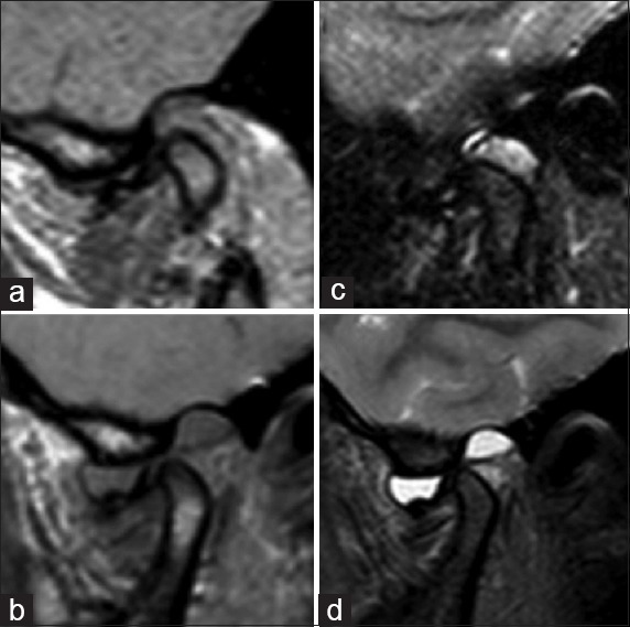
MRISagittal open-mouth MRI of the temporomandibular joint: anterior disc displacement without reduction, before (a, b) and after treatment (c, d).
Source : Ângelo et al., Annals of Maxillofacial Surgery, 2015, figure 3 · CC BY-NC-SA
Temporomandibular disorders (TMD, formerly SADAM) are common: a systematic review with meta-analysis finds a prevalence of about 31.1 % in adults and older people, and 11.3 % in children and adolescents 2. They are frequent reasons for consultation, but their assessment cannot be improvised: it rests on a structured, validated protocol, not on a simple clinical impression. The stake for the physiotherapist, as for the dentist or the first-line doctor, is twofold: to make a reproducible diagnosis, and to spot what is not a common TMD.
The reference framework: the DC/TMD criteria
The diagnosis of TMD rests on the Diagnostic Criteria for Temporomandibular Disorders 3, the validated reference protocol. For the commonest painful TMDs these criteria reach a sensitivity ≥ 0.86 and a specificity ≥ 0.98, with excellent inter-examiner reliability (kappa ≥ 0.85) 3. In other words, correctly applied they let different practitioners reach the same diagnosis, a decisive point in multidisciplinary practice.
The DC/TMD is organised in two complementary axes:
- Axis I, physical diagnosis : identifying the TMD subtype from a structured history and a standardised clinical examination.
- Axis II, the biobehavioural dimension : pain intensity, pain-related disability, psychological distress, functional limitation of the jaw and parafunctional behaviours 3.
This double reading is essential: it recalls that TMD is not only a matter of joint mechanics, and that psychosocial and behavioural factors are an integral part of the assessment, not an optional extra.
Recognising the main subtypes
Speaking of « the » TMD in the singular is misleading: it is a set of distinct presentations that must be told apart, because they do not point to the same management. Disc displacement with reduction is the commonest subtype, disc displacement alone accounting for about 19.1 % in adults 2.
| Subtype | Guiding clinical signs | Landmarks |
|---|---|---|
| Myalgia (muscular) | Pain in the masticatory muscles reproduced on palpation and/or on mandibular movement | The commonest painful TMD in physiotherapy practice |
| Arthralgia (articular) | Pain in the temporomandibular joint reproduced on palpation of the TMJ and on movement | Often associated with myalgia |
| Disc displacement with reduction | A reproducible click on opening and/or closing, with no major limitation | The commonest intra-articular subtype 2 |
| Disc displacement without reduction | Limited opening (locking), deviation, disappearance of the previous click | More marked functional impact |
The colour code above reflects how clear the validated criteria are (painful subtypes: high validity in the DC/TMD) and not a prognosis. The distinction between myalgia, arthralgia and disc involvement drives the reasoning: purely muscular pain, joint conflict and disc locking are not worked on in the same way.
The clinical examination in practice
The assessment follows a standardised sequence, drawn from Axis I of the DC/TMD:
- Structured history : where the pain is and how long it has lasted, triggering and modulating factors (chewing, yawning, speaking, stress), joint noises, episodes of locking, functional impact (eating, sleeping).
- Mandibular ranges : measuring mouth opening (with and without pain), lateral and protrusive movement, and observing the opening path (deviation, deflection).
- Palpation : the masticatory muscles (masseter, temporalis) and the TMJ region, looking for familiar pain , that is, pain that reproduces the patient's complaint, a key DC/TMD criterion.
- Joint noises : looking for a reproducible click (disc displacement with reduction) or for crepitus.
- Dynamic and static provocation tests to distinguish a muscular from an articular origin.
The aim is not to pile up tests but to attach the signs to a DC/TMD subtype, checking systematically that the pain provoked really reproduces the patient's spontaneous symptom.
What the physiotherapist assesses specifically
Beyond diagnosing the subtype, the physiotherapist directs the assessment towards the targets they will be able to treat:
- The cervical spine : its assessment is an integral part of the TMD work-up. Manual therapy applied to the cervical region reduces pain intensity more effectively than placebo or a minimal intervention, with a moderate level of evidence 9 , which justifies testing cervical mobility, pain and pressure thresholds.
- Mandibular function : active range, motor control, concrete functional limitations (chewing, yawning, speaking).
- Parafunctional behaviours, including bruxism. The international consensus recalls that, in an otherwise healthy person, bruxism is not a disease but a behaviour that can be a risk factor, or a protective one, for certain clinical consequences 1. It comes in two distinct entities: sleep bruxism (rhythmic or non-rhythmic activity during sleep) and awake bruxism (repeated or sustained tooth contact, bracing or thrusting of the mandible) 1.
Care with the interpretation of bruxism. Its link with TMD is real but remains an association, not a simple causal relationship. A meta-analysis shows that the presence of bruxism multiplies the risk of TMD by about 2.25 (OR = 2.25; 95 % CI 1.94–2.56) 6. But this link depends heavily on the assessment method : studies based on self-report questionnaires or on clinical examination find a positive association with TMD pain, whereas instrumental studies (electromyography, polysomnography) find a weaker association, and even a negative one 78. The physiotherapist must therefore spot bruxism without making it the sole cause of the picture. This point is still debated and should be presented as such to the patient rather than settled.
Red flags: when it may not be a common TMD
An essential part of the assessment is making sure the picture really is a common TMD, whose validated criteria 3 set the boundary. Some signs, on the contrary, should take the case out of the physiotherapy remit and trigger a medical or specialist opinion:
- Recent, progressive and unexplained pain or swelling in the region, deterioration in general condition, fever.
- An associated neurological deficit, facial sensory disturbance, unusual visual or auditory symptoms.
- Sudden limitation of opening, persistent joint locking or deformity.
- Atypical pain that does not reproduce the criteria for painful TMD, or does not respond to well-conducted conservative management.
- A history or context (trauma, oncological, systemic) incompatible with ordinary TMD.
These situations are not a value judgement about severity but a matter of caution: the role of the DC/TMD is precisely to separate what falls within the field of common TMD from what does not.
Key points
- TMD is common (≈ 31 % in adults) and the assessment rests on a validated protocol, the DC/TMD (sensitivity ≥ 0.86; specificity ≥ 0.98).
- The DC/TMD combines an Axis I (physical diagnosis: myalgia, arthralgia, disc displacement) and an Axis II that is biobehavioural.
- The clinical examination seeks to reproduce the patient's familiar pain and to attach it to a subtype.
- The physiotherapist also assesses the cervical spine and the parafunctions; bruxism is an associated risk factor, not the sole cause, and the link remains debated depending on the measurement method.
- Some red flags require stepping outside the physiotherapy remit and referring for a medical opinion.
🔗 Does bruxism really cause the pain?
🔗 Bruxism doubles the risk of TMD, but it is not the sole cause
The association is real and statistically solid. It does not mean that bruxism « causes » the pain: it is one risk factor among others, within a biopsychosocial model.
Odds ratios: probability of TMD multiplied in the presence of bruxism. Overall OR 2.25 (95 % CI 1.94–2.56). Source: meta-analysis by Mortazavi et al., 2023 (PMID 37474733).
It is one of the most persistent ideas in everyday practice: the patient clenches or grinds their teeth, their jaw hurts, therefore bruxism causes temporomandibular disorders (TMD). The reasoning seems obvious. On the current data it is nonetheless a large oversimplification. Bruxism and TMD are two common phenomena, often concurrent and statistically associated, but the existence of an association has never amounted to a demonstration of causality, and this is exactly where clinical reasoning must stay cautious.
Two common phenomena, and distinct ones
First clarification, often overlooked: bruxism is not a single entity. The 2018 international consensus separates two clearly distinct circadian manifestations, sleep bruxism, masticatory muscle activity during sleep (rhythmic or non-rhythmic), and awake bruxism, characterised while awake by repeated or sustained tooth contact and/or by bracing or thrusting of the mandible 1. These are not two names for the same thing: their determinants, their measurement and, in all likelihood, their relationship with pain differ.
Second point, decisive for everything that follows: the same consensus states that, in an otherwise healthy person, bruxism should not be regarded as a disorder or a disease, but as a behaviour that can be a risk factor, and sometimes a protective one, for certain clinical consequences 1. In other words, bruxism is not a condition in itself to be « treated »; it is a behaviour whose consequences are assessed.
In terms of frequency, these are very widespread behaviours. A worldwide meta-analysis from 2024 puts the overall prevalence of bruxism (sleep and awake combined) at 22.2 %, about 21 % for self-reported sleep bruxism and 23 % for awake bruxism, so roughly one person in four may have awake bruxism 4. Notably, measured by polysomnography, a reference method far more sensitive than self-report, sleep bruxism would reach 43 % 4. In adults, an older review found awake bruxism from 22 to 31 % and « frequent » sleep bruxism more homogeneous at around 12.8 % (± 3.1) 5.
TMD is common too. A systematic review with meta-analysis reports an overall prevalence of about 31 % in adults and older people, and 11 % in children and adolescents, disc displacement being the commonest joint disorder (19.1 % in adults) 2. Two phenomena present in a large share of the general population are mechanically very likely to coexist in the same individual, without that coexistence proving anything about cause and effect.
A real association, but one that depends on the measurement method
That bruxism and TMD are associated is not disputed. A 2023 meta-analysis shows that the presence of bruxism multiplies the probability of TMD by about 2.25 (OR = 2.25; 95 % CI 1.94–2.56), with a comparable effect for awake bruxism (OR 2.51) and sleep bruxism (OR 2.06) 6. The authors themselves conclude that there is a positive relationship , an associated risk factor, and not a demonstrated causal link.
The key point lies elsewhere: the strength of this association depends heavily on how bruxism is measured. Studies based on self-report questionnaires or on clinical diagnosis generally find a positive association with TMD pain; instrumental studies, electromyography and polysomnography, find a clearly weaker association, and even a negative one 7. That observation had already been made in an earlier analysis: self-report gives a positive link, but it collapses as soon as more quantitative and specific diagnostic methods are used 8.
This divergence carries weight. If the association rested on a strong biological mechanism, muscle and joint overload directly generating pain, then the more objective instrumental measures ought to confirm it, or even strengthen it. That they weaken it suggests instead that part of the observed association comes from methodological bias : a patient in pain, more attentive to their jaw, reports more readily that they clench their teeth. Pain might then, at least in part, increase the perception of bruxism as much as the reverse. Bruxism cannot therefore be presented as the sole cause of TMD.
| Method of assessing bruxism | Association with TMD pain | Level of confidence |
|---|---|---|
| Self-report questionnaire / clinical diagnosis | Positive (association found) | Subject to bias and confounding |
| Instrumental methods (EMG, polysomnography) | Weaker, and even negative | More objective, less association |
Leaving the mechanical model: the biopsychosocial reading
Reducing TMD to a mechanical consequence of tooth clenching ignores what the reference diagnostic tools have long incorporated. The Diagnostic Criteria for TMD 3 , a validated protocol that separates the commonest painful TMDs with a sensitivity ≥ 0.86 and a specificity ≥ 0.98 and excellent inter-examiner reliability (kappa ≥ 0.85), are not limited to a physical examination. They are structured in two axes: an Axis I for the physical diagnosis, and an Axis II assessing pain intensity, the disability it causes, psychological distress, mandibular functional limitation and parafunctional behaviours 3.
This double axis is not an academic refinement: it reflects the biopsychosocial nature of TMD. That the reference tool devotes a whole axis to psychosocial and behavioural factors says clearly that temporomandibular pain cannot be understood through joint mechanics alone. Biological, psychological and social factors are intertwined in it, and bruxism is only one thread among others.
And what about stress?
Stress is the spontaneous explanation that patients and clinicians readily put forward to link clenching and pain. Here we must be honest about what the accepted data allow us to state. Awake bruxism, as the 2018 consensus defines it, shows itself in sustained tooth contact and mandibular bracing while awake 1 , behaviours that clinical observation commonly attaches to periods of tension. And Axis II of the DC/TMD formally recognises psychological distress as a dimension to assess in the patient in pain 3. The biopsychosocial framework and the diagnostic tools therefore make explicit room for the psychological component.
That said, none of the work confirmed here quantifies a direct causal relationship « stress → bruxism → TMD ». Caution is required: the psycho-emotional dimension can legitimately be built into assessment and patient education, but without presenting it as an established causal chain. It is a factor to consider, not a demonstrated mechanism.
Key points
- Two distinct entities. Sleep bruxism and awake bruxism are different masticatory muscle activities; in a healthy person bruxism is a behaviour, not a disease 1.
- Association, not causality. Bruxism multiplies the probability of TMD by ~2.25 6, but it remains an associated risk factor, not the sole cause.
- The method changes everything. The association is positive on self-report and weak or even negative on instrumental measurement 7 , a strong signal of bias.
- Biopsychosocial model. The DC/TMD 3 includes a psychosocial Axis II: TMD pain does not reduce to mechanics.
- An openly acknowledged debate. The bruxism↔TMD link is still discussed; better to say so to the patient than to settle it improperly.
In practice, this cluster of nuances has a direct consequence for what is said to the patient. Stating « your pain comes from your bruxism » goes beyond what the evidence allows, and can lock the patient into a purely mechanical and anxiety-provoking reading of their disorder. The honest message is more measured: bruxism is common, it is statistically associated with TMD, and it is probably one risk factor among others, but it is not their sole and demonstrated cause, and temporomandibular pain is understood within a wider biopsychosocial framework. That honesty does not weaken physiotherapy management: it founds it on a realistic understanding, in which pain and function are worked on without over-reading a behaviour whose causal role remains, in 2024, openly debated.
🎯 What can physiotherapy do for TMD?
🎯 What physiotherapy does for TMD pain
The effect on pain is real and of moderate size. The gain in mouth opening, on the other hand, does not reach the threshold of significance, and that should be said honestly.
Standardised mean difference −0.63 (95 % CI −0.95 to −0.31) in favour of physiotherapy; the improvement in active mouth opening was not significant. Manual therapy applied to the cervical spine also reduces pain 9. Pain source: meta-analysis by Paço et al., 2016 (PMID 27472523).
Faced with temporomandibular disorders (TMD), the physiotherapist has a conservative, reversible and low-risk arsenal: orofacial and cervical manual therapy, therapeutic exercise, education, and sometimes dry needling (dry needling). Before any treatment, the assessment gains from resting on a standardised framework: the Diagnostic Criteria for TMD (DC/TMD), whose Axis I separates the commonest painful TMDs with a sensitivity ≥ 0.86 and a specificity ≥ 0.98, while Axis II documents pain intensity, disability, psychological distress, mandibular functional limitation and parafunctional behaviours 3. This biobehavioural foundation points from the outset towards multimodal management rather than an isolated technique.
Physiotherapy acts above all on pain; its effect on mouth opening remains more uncertain.
A measurable effect on pain, a more debatable one on mouth opening
The meta-analysis of randomised trials by Paço 10 is the clearest reference: physiotherapy reduces TMD pain with a moderate and remarkably homogeneous effect size (standardised mean difference SMD = −0.63; 95 % CI: −0.95 to −0.31; 8 studies; I² = 0 %). The gain in active mouth opening range does not reach statistical significance (SMD = 0.33; 95 % CI: −0.07 to 0.72). The authors conclude that physiotherapy « seems » to decrease pain and « may » improve active range, but that the results are not definitive.
This message, a probable analgesic effect and a more fragile effect on mobility, is found at an even higher level of evidence in a recent umbrella review. Arribas-Pascual 11, aggregating several meta-analyses, concludes that manual therapy and therapeutic exercise (as well as low-level laser) are effective at reducing pain intensity and at improving maximum mouth opening, with effects described as moderate for manual therapy and exercise. The divergence from Paço on opening illustrates that the data remain improvable: depending on the corpus and the outcome measures, the functional benefit appears or fades.
Manual therapy and exercise: promising, but with no high-quality evidence
The systematic review with meta-analysis by Armijo-Olivo 12 remains the most honest reading of the field. Its conclusion comes in two parts. First, manual therapy, alone or combined with exercise, at the jaw as well as at the cervical spine, shows « promising effects ». Second, most effect sizes are small to moderate, with no clear indication that exercise is superior to the other conservative treatments, and above all no high-quality evidence was found. The result is « great uncertainty » about the true size of the effect. In other words: these techniques are reasonable to offer, safe and simple, but they must not be oversold.
Concretely, this argues for a combined, individualised approach, mobilisation and soft-tissue techniques for the masticatory muscles, active exercises of controlled opening, coordination and mandibular strengthening, rather than for a single recipe. The superiority of one modality over another is not demonstrated; what counts is the coherence of the programme, its dosage and the patient's adherence.
Do not forget the cervical spine
The neck is not beside the point in TMD. The meta-analysis by La Touche 9 shows that manual therapy applied to the cervical region reduces TMD pain intensity more effectively than placebo or a minimal intervention, with a moderate level of evidence. The clinical effect on pain is large, and an increase in pressure pain thresholds at the masseter is observed, a strong physiological argument for building assessment and treatment of the cervical spine systematically into TMD management. This head-neck convergence also matches Armijo-Olivo's finding that exercises « at the cervical level » contribute to the observed effects.
Dry needling: an option, but on fragile foundations
For the myofascial components (trigger points in the masseter and temporalis), dry needling is sometimes offered. The meta-analysis by Vier 13 is nuanced: dry needling does better than other interventions on pain intensity, and better than sham on the pressure pain threshold. But these conclusions rest on evidence of very low quality, a small effect size and a high risk of bias in several of the included trials. The authors consider that clinicians « may » use it, while stressing the need for larger and better-conducted trials. To be offered, then, with transparency about the level of evidence, and rarely as an isolated first-line treatment.
Education and framing: reframe bruxism, do not demonise it
Education has no quantified « effect size » in the confirmed data here, but it follows directly from what is known of the pathophysiology. The international consensus recalls that, in an otherwise healthy person, bruxism should not be regarded as a disease but as a behaviour that can be a risk factor, and sometimes a protective one, for certain clinical consequences 1. Presenting it to the patient as a condition in itself is therefore inaccurate and potentially anxiety-provoking.
Likewise, the bruxism → TMD link must be explained with caution. There is indeed an association 6, but it depends heavily on the assessment method: strong when bruxism is self-reported, it weakens markedly, and can even reverse, with instrumental measures (electromyography, polysomnography) 87. It is therefore not a simple cause-and-effect relationship, and the physiotherapist should not present it as one. Axis II of the DC/TMD offers the framework for addressing these parafunctional behaviours and psychosocial factors without catastrophising.
Key points
- Pain: physiotherapy reduces TMD pain with a moderate and homogeneous effect 10, supported by an umbrella review 11.
- Mouth opening: the benefit is uncertain, not significant in Paço and positive in the umbrella review; to be announced with reservation.
- Manual therapy + exercise: promising effects but small to moderate effect sizes and no high-quality evidence 12.
- Cervical spine: cervical manual therapy decreases pain 9 , to be built in from the outset.
- Dry needling: possible for myofascial pain, but very low quality evidence and a small effect 13.
- Education: reframe bruxism as a behaviour, not a disease; the bruxism↔TMD link remains debated.
| Intervention | Main effect | Level of evidence |
|---|---|---|
| Physiotherapy (overall), pain | Moderate reduction (SMD = −0.63) | Moderate 10 |
| Manual therapy + exercise | Promising effects, small to moderate | Low / uncertain 12 |
| Cervical manual therapy | Less pain, ↑ masseter thresholds | Moderate 9 |
| Dry needling (myofascial) | Small gain on pain and pressure threshold | Very low 13 |
| Mouth opening (all methods) | Discordant results | Uncertain 10 |
In summary, physiotherapy has its full place in TMD as a first-line conservative treatment, mainly for its analgesic effect. The overall level of evidence remains modest, however: small to moderate effects, heterogeneous protocols, no indisputable standard of effectiveness. The right stance is to offer multimodal management, orofacial and cervical manual therapy, exercise and education, while staying honest about the uncertainty, particularly on mouth opening and on the exact role of bruxism.
🧩 Splints, education, self-management: overall care
Faced with bruxism and temporomandibular disorders (TMD), the logic of care has changed paradigm. The aim is no longer to « correct » an occlusion or to suppress grinding, but to support a common behaviour and a multifactorial pain with simple, conservative and reversible means. This section reviews the three non-invasive first-line pillars, occlusal splints, education and self-management, and how they fit together between physiotherapist, dentist and doctor.
A conservative, reversible first line
The starting point shapes the whole of management: according to the international consensus, in an otherwise healthy person bruxism should not be regarded as a disorder or a disease but as a behaviour that can be a risk factor, and sometimes a protective one, for certain clinical consequences 1. That nuance has a direct consequence: in the absence of any impact (pain, marked wear, functional difficulty), there is no reason to « treat » isolated bruxism. Irreversible interventions (grinding down teeth, heavy occlusal rehabilitation) are not justified for a behaviour that is not in itself pathological.
This behaviour is also very widespread, which argues further for therapeutic restraint: the worldwide prevalence of bruxism is estimated at around 22 %, with about 21 % for sleep bruxism and 23 % for awake bruxism 4, while in adults awake bruxism lies between 22 and 31 % and « frequent » sleep bruxism at around 12.8 % 5.
TMD itself is common, about 31 % in adults and older people and 11 % in children and adolescents 2 , but the diagnosis must stay rigorous. The DC/TMD criteria 3 offer, for the commonest painful TMDs, a sensitivity ≥ 0.86 and a specificity ≥ 0.98: they provide a reliable assessment protocol rather than a clinical impression, and they build in two axes from the outset, a physical Axis I and a biobehavioural Axis II (pain intensity, disability, psychological distress, mandibular functional limitation, parafunctional behaviours). That two-axis architecture alone justifies overall management, in which the splint is only one element among others.
Occlusal splints: a real place, a debated effectiveness
The occlusal splint (interocclusal appliance) remains one of the most prescribed devices in sleep bruxism and TMD. Making it, fitting it and following it up belong to the dentist : the physiotherapist does not prescribe it but is a partner, assessing the muscular and articular impact and coordinating the rest of care. Its main strength is that it is reversible : it protects the tooth surfaces and can change muscle comfort, without any definitive act on the occlusion.
We must nonetheless be honest about the level of evidence. The effect of splints is debated : the available data do not allow us to state that they durably change bruxism activity itself, and their benefit for TMD pain is not settled in the literature. A splint can relieve some patients, essentially by protecting the teeth and improving comfort, but it must not be presented as a causal treatment or as a sufficient solution on its own. That message joins the wider debate on the bruxism–TMD link (see below): acting on bruxism does not guarantee resolving the pain.
Bruxism and TMD: an association, not a sole cause
The received idea that « bruxism wears the joint and causes TMD » deserves strong qualification. The link exists but remains an association, not a simple causal relationship, and it depends heavily on the measurement method. Studies based on self-report questionnaires or on clinical examination find a positive association with TMD pain, whereas instrumental studies (electromyography, polysomnography) find a weaker association, and even a negative one 78. A recent meta-analysis puts the association at an odds ratio of 2.25 (95 % CI 1.94–2.56), comparable for awake bruxism (OR 2.51) and sleep bruxism (OR 2.06), while framing it as a positive relationship and not as a demonstrated causal link 6.
Practical consequence: targeting bruxism alone (with a splint, for instance) to treat painful TMD amounts to betting on an uncertain link. Hence the value of an approach that acts directly on pain, function and the maintaining factors, which is exactly where education, self-management and physiotherapy come into their own.
Education and self-management: acting on the behaviour
Since bruxism is above all a behaviour 1 and since Axis II of the DC/TMD explicitly identifies parafunctional behaviours and functional limitation 3, patient education is a logical cornerstone of first-line care. It is a non-invasive, risk-free and perfectly reversible intervention.
In practice, education aims to:
- Explain and defuse : naming bruxism as a common behaviour rather than a disease, and recalling that the link with pain is neither automatic nor unequivocal, reduces worry and catastrophising.
- Build awareness of daytime parafunctions : spotting repeated or sustained tooth contact and awake clenching during the day, in order to learn to relax the mandible (teeth apart, tongue at rest, lips together).
- Encourage self-management : gentle self-mobilisation, staying within pain-free ranges, managing demands (hard foods, chewing gum, nails) and sleep hygiene.
These measures do not claim to « suppress » bruxism, since no solid data guarantee it, but they tackle the factors that maintain pain and give the patient an active role, which is consistent with the biopsychosocial model carried by the DC/TMD.
Managing stress and psychosocial factors
Axis II of the DC/TMD assesses psychological distress and pain-related disability 3, a sign that the psychosocial dimension is not incidental but an integral part of the diagnosis. Awake bruxism in particular readily expresses itself in contexts of tension. Managing stress (relaxation, regulating tension, spotting triggers) therefore fits naturally into self-management, as a complement to the other measures and not a substitute for them. Here again, caution is needed about promises: these approaches support the patient and modulate the experience of pain, without our being able to claim a specific effect on bruxism activity.
Where physiotherapy sits in this arrangement
Physiotherapy completes this conservative set with a measurable effect on the dominant symptom, pain. A meta-analysis finds an effect in favour of physiotherapy on TMD pain (SMD = −0.63; 95 % CI −0.95 to −0.31; I² = 0 %), while the improvement in active mouth opening does not reach significance 10. Manual therapy, alone or combined with exercise at the jaw or the cervical spine, shows promising effects 12, supported by a more recent umbrella review that concludes there are moderate effects of manual therapy and exercise on pain and on mouth opening 11. Managing the cervical spine has its place: cervical manual therapy is more effective than placebo or a minimal intervention at reducing pain 9.
This effectiveness must nonetheless be presented without overselling it: no high-quality evidence was found, and great uncertainty remains about the true size of the effect, with small to moderate effect sizes 12. That is precisely what justifies a strategy that is multimodal : combining education, self-management, stress management, a splint if the dentist indicates one, and physiotherapy, rather than betting on a single lever.
Key points
- Reversible first. In a healthy person bruxism is a behaviour, not a disease 1 : conservative means come first and the irreversible is avoided.
- Splint = adjunct, not remedy. Made by the dentist, reversible, protective, but its effect on bruxism and on pain remains debated; it is not a causal treatment.
- An uncertain bruxism–TMD link. Association 6, not a simple causal relationship; the association collapses with instrumental measures 8.
- Education and self-management to the fore. Consistent with the biobehavioural Axis II of the DC/TMD 3, they target the maintaining factors and make the patient an active participant.
- Multimodal physiotherapy. An effect on pain 10, promising manual therapy and exercise 1211, cervical spine included 9 , but evidence of limited quality, to be announced honestly.
🗂️ What do concrete case reports teach us?
The two published works that follow carry their PubMed identifier: a case series on rehabilitation, and a case that recalls what must not be taken for a temporomandibular disorder. They serve only to show how physiotherapy reasoning rests on the data currently available: subtyping bruxism, structured diagnosis of the temporomandibular disorder (TMD), reasoned choice of techniques and coordination with the dentist. Every decision refers explicitly to a confirmed piece of data, and the areas of uncertainty are named rather than hidden.
A clinical case is not evidence: it is a thread linking validated data to a decision on the ground.
Published series: an individualised exercise, guided by analysis of mandibular movement
The work. Woo and colleagues report a case series evaluating an exercise intervention that is personalised in temporomandibular disorders14.
The problem they set out, and it is ours. Exercise is strongly recommended in the management of these disorders, but conventional approaches have a limited ability to treat symptoms related to kinematic abnormalities of the mandible, and they often lack enough logical clarity to be reproduced in practice14. The authors add that current diagnostic criteria and imaging mainly assess static anatomical conditions, whereas three-dimensional movement analysis remains hard to implement in routine practice14.
What it is worth. A case series does not demonstrate the effectiveness of a method, and the text above remains the arbiter of what is established. What this work names correctly is the gap between a diagnosis made on static images and a complaint that is, in itself, a problem of movement.
Published case, what must not be taken for a temporomandibular disorder
The case. Hng and colleagues report gout of the left temporomandibular joint with erosion of the skull base15. The diagnosis was suspected on CT and MRI, then confirmed by guided biopsy15.
Why this case matters here. The temporomandibular joint is an unusual site for a first manifestation of gout, and the authors stress that, given its radiological appearance, it can easily be mistaken for other erosive arthropathies or for a tumour15.
What it changes in the session. Temporomandibular pain accompanied by progressive limitation and not responding to well-conducted management is not stubborn bruxism. It is an indication for imaging, and the physiotherapist's role is to say so.
What these two cases bring out
| Element of reasoning | Case 1, Marie (awake, myalgia) | Case 2, Karim (sleep, articular) |
|---|---|---|
| Bruxism subtype | Awake bruxism 1 | Sleep bruxism 1 |
| TMD direction (DC/TMD) | Painful muscular TMD | Suspected disc displacement 2 |
| Treatment priority | Manual therapy + exercise, pain 10 | Function + mobility, multimodal approach 11 |
| Role of the cervical spine | Included 9 | Included 11 |
| Adjunct option | — | Dry needling if myofascial 13 |
| Coordination with the dentist | Dental examination, splint (debated) | Wear, protection, splint (debated) |
Key points
- Subtype first : awake bruxism and sleep bruxism are two distinct entities; bruxism is a behaviour, not a disease in itself 1.
- Diagnose, do not presume : the DC/TMD criteria structure the reasoning, with excellent validity for painful TMD 3.
- Association ≠ causality : bruxism increases the probability of TMD (OR ≈ 2.25) but is not its sole cause, and the link weakens with instrumental measures 67.
- Treat multimodally : manual therapy + exercise for the jaw and the cervical spine reduce pain (SMD = −0.63), with promising effects but acknowledged uncertainty 10129.
- Coordinate : physiotherapist and dentist complement each other; the effect of splints remains debated and must not be oversold.
🧭 How is this applied in practice?
The clinical difficulty of the bruxism–TMD pair does not come from a lack of treatments but from the risk of framing the problem wrongly from the start: confusing a behaviour with a disease, attributing the pain straight away to night-time grinding, or overselling a technique when the evidence remains modest. This section offers a reasoning framework, an algorithm, messages to convey, traps to avoid and tipping points towards referral, to turn the evidence into first-line action.
A five-step decision algorithm
The aim is not to apply a recipe but to sequence the reasoning so that nothing essential is forgotten.
1. Recognise the presentation and tell bruxism and TMD apart. Bruxism is a behaviour, not a symptom to be treated in itself: the international consensus defines it as an activity of the masticatory muscles, distinct according to the circadian moment, sleep bruxism (rhythmic or non-rhythmic) and awake bruxism (repeated or sustained tooth contact, bracing or thrusting of the mandible) 1. Temporomandibular disorders (TMD) group together the pain and dysfunction of the joint and of the masticatory muscles. Confusing the two makes management drift.
2. Frame the complaint with a validated tool. Diagnosing TMD is not done on clinical impression: the Diagnostic Criteria for TMD (DC/TMD) offer, for the commonest painful TMDs, a sensitivity ≥ 0.86 and a specificity ≥ 0.98, with excellent inter-examiner reliability (kappa ≥ 0.85) 3. They structure the examination into an Axis I (physical diagnosis: myalgia, arthralgia, disc displacement…) and a biobehavioural Axis II (pain intensity, disability, psychological distress, functional limitation, parafunctional behaviours). Using this grid means speaking the same language as the dentist and the doctor.
3. Assess the contribution of bruxism without over-reading it. The presence of bruxism is associated with a probability of TMD multiplied by about 2.25 (OR = 2.25; 95 % CI 1.94–2.56), with a comparable effect for awake bruxism (OR 2.51) and sleep bruxism (OR 2.06) 6. But this is an association, not a sole cause: the strong association appears mainly when bruxism is identified by self-report questionnaire, and it weakens or even reverses with instrumental measures (electromyography, polysomnography) 78.
4. Treat painful TMD with a multimodal approach. Physiotherapy has a measurable effect on pain: a meta-analysis of randomised trials finds an effect in favour of physiotherapy (SMD = -0.63; 95 % CI -0.95 to -0.31; I² = 0 %), while the gain in active mouth opening does not reach significance (SMD = 0.33; 95 % CI -0.07 to 0.72) 10. Manual therapy and exercise, at the jaw as well as at the cervical spine, show promising effects 12, and a recent umbrella review supports their place, along with low-level laser, for reducing pain and improving maximum opening 11.
5. Include the cervical spine. Manual therapy applied to the cervical region is more effective than placebo or a minimal intervention at reducing TMD pain intensity (moderate evidence), with an increase in pressure pain thresholds at the masseter 9. The neck is part of the assessment, not just the jaw.
The key messages to convey to the patient
A good part of first-line effectiveness rests on reshaping beliefs. Four messages are worth repeating:
- « Bruxism is not a disease. » In an otherwise healthy person, bruxism is not a disorder but a behaviour that can be a risk factor, and sometimes a protective one, for certain clinical consequences 1. Defusing the drama reduces the anxiety that in turn maintains awake clenching.
- « It is common. » About one person in five to one in four is affected: the worldwide prevalence of bruxism is around 22 %, with ~21 % for sleep and ~23 % for awake bruxism 4. There is nothing abnormal about the patient.
- « Your grinding alone is not what causes your pain. » The bruxism–TMD link is real but complex and debated; presenting bruxism as the sole culprit is misleading and induces guilt.
- « Daytime awareness is a lever. » For awake bruxism, recognisable by repeated or sustained tooth contact, learning to relax the jaw (teeth apart, tongue at rest) several times a day is an accessible self-management route.
Treating TMD pain is not hunting down the grinding; it is reducing the load on a sensitised system.
Common mistakes to avoid
| Common error | What the data say |
|---|---|
| Presenting bruxism as a condition to eradicate | It is not a disorder but a behaviour and risk factor in a healthy person 1 |
| Attributing the pain to sleep bruxism alone | Association, not causality; the effect falls with instrumental measures 7 |
| Promising a quick cure through manual therapy | Promising effects but small to moderate effect sizes, no high-quality evidence 12 |
| Neglecting the cervical spine in the assessment | Cervical manual therapy reduces TMD pain 9 |
| Making a diagnosis « by impression » with no validated grid | The DC/TMD offer Se ≥ 0.86 and Sp ≥ 0.98 3 |
Two uncertainties must be acknowledged rather than settled. First, the bruxism–TMD link: depending on the assessment method, the association ranges from fairly strong (self-report questionnaire) to weak or negative (electromyography, polysomnography) 7. Second, the place of occlusal splints: their effectiveness remains debated, and this text deliberately avoids settling the point; the decision to fit an appliance belongs to a multidisciplinary discussion, not to a certainty. Announcing these grey areas to the patient is more honest, and more solid, than overselling.
Multidisciplinary coordination and the division of roles
TMD is naturally shared ground. The common language of the DC/TMD, with its physical Axis I and its psychosocial Axis II 3, lets each party know what the others are doing:
- The physiotherapist conducts the joint and muscle assessment, applies manual therapy and exercise (jaw + cervical spine), teaches self-management and awake relaxation, and follows the course of pain and function 109.
- The dentist assesses the dental consequences of bruxism, discusses the indication for and the monitoring of a possible splint, and documents wear or occlusal loading.
- The general practitioner or sleep specialist explores the factors associated with sleep bruxism 4, the comorbidities and the treatments that can influence it.
- The psycho-behavioural field (through Axis II) takes over when psychological distress, stress or anxiety feed awake bruxism and pain sensitisation.
Dry needling of the myofascial pain of TMD can complete the arsenal: the meta-analysis shows it superior to other interventions on pain intensity and to placebo on the pressure threshold, but with very low quality evidence and a small effect, usable without making it a cornerstone 13.
When to refer on?
First-line physiotherapy is justified for ordinary painful TMD. Another party should be involved or the patient referred when:
- the complaint does not match a clear DC/TMD diagnosis, or the signs suggest a condition that is not musculoarticular (refer for assessment);
- tooth wear, a fracture or an occlusal problem dominates the picture → dentist ;
- marked sleep bruxism comes with signs of a sleep disorder → doctor or sleep specialist ;
- Axis II reveals significant psychological distress, disability or chronic pain → psycho-behavioural support ;
- progress stalls despite well-conducted multimodal management; the great uncertainty about effectiveness 12 is an invitation to reassess the diagnosis and the plan, not to persist.
Key points
- Two entities, not one: sleep bruxism and awake bruxism are distinct, and bruxism is not a disease but a behaviour and risk factor in a healthy person 1.
- Diagnose with the DC/TMD (Se ≥ 0.86; Sp ≥ 0.98), physical Axis I + psychosocial Axis II, rather than by impression 3.
- The bruxism–TMD link = association (OR ≈ 2.25), not a simple causal relationship ; it depends heavily on the measurement method 67.
- Physiotherapy reduces TMD pain 10, including the cervical spine 9, but with no high-quality evidence: stay cautious and multimodal 12.
- Splints and the causal link remain debated: say so to the patient rather than settling it. Refer on as soon as the picture leaves the musculoarticular field.
Bibliography
Every reference checked individually on PubMed (clickable PMID). 13 sources. Click a superscript note in the text: the bibliography opens and highlights the source.
- Lobbezoo F, Ahlberg J, Raphael KG, et al. (2018). Journal of Oral Rehabilitation. PMID 29926505. doi:10.1111/joor.12663.
- Valesan LF, Da-Cas CD, Réus JC, et al. (2021). Clinical Oral Investigations. PMID 33409693. doi:10.1007/s00784-020-03710-w.
- Schiffman E, Ohrbach R, Truelove E, et al. (2014). Journal of Oral & Facial Pain and Headache. PMID 24482784. doi:10.11607/jop.1151.
- Zieliński G, Pająk-Zielińska B, Ginszt M (2024). Journal of Clinical Medicine. PMID 39064299. doi:10.3390/jcm13144259.
- Manfredini D, Winocur E, Guarda-Nardini L, Paesani D, Lobbezoo F (2013). Journal of Orofacial Pain. PMID 23630682. doi:10.11607/jop.921.
- Mortazavi N, Tabatabaei AH, Mohammadi M, et al. (2023). Evidence-Based Dentistry. PMID 37474733. doi:10.1038/s41432-023-00911-6.
- Manfredini D, Lobbezoo F (2021). Journal of Dentistry. PMID 34090993. doi:10.1016/j.jdent.2021.103711.
- Manfredini D, Lobbezoo F (2010). Oral Surgery, Oral Medicine, Oral Pathology, Oral Radiology and Endodontology. PMID 20451831. doi:10.1016/j.tripleo.2010.02.013.
- La Touche R, Martínez García S, Serrano García B, et al. (2020). Pain Medicine. PMID 32181811. doi:10.1093/pm/pnaa021.
- Paço M, Peleteiro B, Duarte J, Pinho T (2016). Journal of Oral & Facial Pain and Headache. PMID 27472523. doi:10.11607/ofph.1661.
- Arribas-Pascual M, Hernández-Hernández S, Jiménez-Arranz C, et al. (2023). Journal of Clinical Medicine. PMID 36769437. doi:10.3390/jcm12030788.
- Armijo-Olivo S, Pitance L, Singh V, Neto F, Thie N, Michelotti A (2016). Physical Therapy. PMID 26294683. doi:10.2522/ptj.20140548.
- Vier C, Almeida MB, Neves ML, Santos ARS, Bracht MA (2019). Brazilian Journal of Physical Therapy. PMID 30146108. doi:10.1016/j.bjpt.2018.08.008.
- Woo J, Jeon J, Hong J (2026). Kinematic Analysis-Guided Individualized Exercise for Temporomandibular Disorders: A Case Series. Journal of Clinical Medicine. PMID 41598590. doi:10.3390/jcm15020655.
- Hng J, Manchella S, Lekgabe E (2022). Gout of the temporomandibular joint and review of the literature. BJR Case Reports. PMID 36873237. doi:10.1259/bjrcr.20220046.
❓ Frequently asked questions
Is bruxism a disease?
No. The international consensus states that, in an otherwise healthy person, bruxism should not be regarded as a disorder or a disease but as a behaviour that can be a risk factor, and sometimes a protective factor, for certain clinical consequences 1. It is an activity of the masticatory muscles characterised by clenching or grinding of the teeth and/or by bracing or thrusting of the mandible.
What is the difference between sleep bruxism and awake bruxism?
They are two distinct circadian manifestations. Sleep bruxism is masticatory muscle activity occurring during sleep (rhythmic or non-rhythmic); awake bruxism is repeated or sustained tooth contact and/or bracing or thrusting of the mandible while awake 1. They are therefore not one and the same entity.
Is bruxism common?
Yes. A worldwide meta-analysis puts the overall prevalence of bruxism at 22.22 %, with about 21 % for sleep bruxism and 23 % for awake bruxism, roughly one person in four for awake bruxism 4. Measured by polysomnography, the reference method and a more sensitive one than self-report, sleep bruxism reaches about 43 % 4.
Does bruxism cause TMD?
The link is real but it remains an association, not a simple causal relationship. A meta-analysis shows that the presence of bruxism multiplies the probability of TMD by about 2.25 (OR = 2.25; 95 % CI 1.94–2.56), with a comparable effect for awake and for sleep bruxism 6. Above all, the association depends heavily on the method: it is positive with self-report questionnaires but weaker, and even negative, with instrumental methods (electromyography, polysomnography) 7. Bruxism cannot therefore be presented as the sole cause of TMD.
How is a temporomandibular disorder diagnosed?
The diagnosis rests on standardised, validated criteria, the Diagnostic Criteria for TMD (DC/TMD). For the commonest painful TMDs these criteria offer a sensitivity ≥ 0.86 and a specificity ≥ 0.98, with excellent inter-examiner reliability (kappa ≥ 0.85); they comprise an Axis I (physical diagnosis) and a biobehavioural Axis II (pain, disability, psychological distress, functional limitation, parafunctional behaviours) 3. TMD affects about 31.1 % of adults and 11.3 % of children and adolescents 2.
Is physiotherapy effective for TMD?
It tends to reduce pain: a meta-analysis reports an effect in favour of physiotherapy on pain (SMD = −0.63; 95 % CI −0.95 to −0.31), while the gain in active mouth opening does not reach significance (SMD = 0.33; 95 % CI −0.07 to 0.72) 10. Manual therapy applied to the cervical spine reduces pain better than placebo, with a moderate level of evidence 9. Caution is still needed: no high-quality evidence was found and considerable uncertainty remains about the true size of the effect, which justifies a multimodal approach 12.
Also worth reading in the review

