

















A clinical synthesis on cervical spondylosis (cervical osteoarthritis): why the osteoarthritis seen on imaging does not explain the pain, how to manage cervicobrachial neuralgia, how to spot myelopathy, and what exercise and manual therapy are really worth. Every reference has been individually verified on PubMed.
📝 In brief: clinical synthesis
- Cervical osteoarthritis on imaging does not prove the cause of the pain: in 1,211 asymptomatic volunteers, a disc bulge was present in 87.6% (already >73% in the twenties age band) 3.
- Radiographic disc degeneration affects 53.9% of a population of 1,581 people and increases with age, C5/C6 being the most affected level: finding osteoarthritis is commonplace 1.
- Cervical radiculopathy (cervicobrachial neuralgia), most often spondylotic or discogenic, has a favourable natural history: ~83% recover within 24-36 months 8 and 90% are asymptomatic or mildly bothered at last follow-up 7.
- Conservative treatment is first-line: surgery relieves faster in the short term but « is not necessary for patients who do not need rapid relief » 9.
- Major red flag calling for referral: degenerative cervical myelopathy (DCM). Surgery is recommended for moderate to severe forms, graded with the mJOA scale (mild 15-17, moderate 12-14, severe ≤11) 13.
- Exercise is the foundation of treatment: cervico-scapulo-thoracic strengthening is beneficial for chronic pain (SMD −0.71), whereas stretching alone brings no benefit 15.
- Cervical manipulation does no better than mobilisation and carries a rare but serious risk of dissection/stroke: no excess risk demonstrated versus general practice care, but caution and screening for red flags remain in order 19.
🦴 What is cervical spondylosis really?
🧐 What MRI finds in people who have no pain anywhere
In 1,211 strictly asymptomatic volunteers (aged 20-70), imaging finds degenerative signs on a massive scale, proof that « seeing osteoarthritis » does not prove that it hurts.
Prevalence of MRI abnormalities in people WITHOUT any cervical symptom; compression and high signal increase mainly after the age of 50. A degenerative image does not on its own establish the origin of the pain. Source: Nakashima et al., Spine 2015 (PMID 25584950).
Cervical spondylosis, also called cervical osteoarthritis or degenerative cervical spine disease, covers all the degenerative changes of the cervical spine: disc space narrowing, osteophytes (bony spurs), osteoarthritis of the facet joints and narrowing of the intervertebral foramina through which the nerve roots leave. The word is frightening because it suggests irreversible « wear ». Yet what research over the past thirty years has consistently established is that these images are first of all a marker of time passing, and not, on their own, proof of where a pain comes from.
Key points
- The signs of cervical osteoarthritis on imaging are commonplace and increase with age, including in people who have no pain anywhere.
- Seeing osteoarthritis on an MRI or a radiograph does not prove that it explains the neck pain: the image must always be set against the clinical picture.
- The simplistic « wear = pain » model is wrong ; it worries the patient without helping them.
- Imaging is not the first-line investigation for mechanical neck pain without a red flag.
An extremely common reality, largely linked to age
Cervical disc degeneration is one of the most widespread phenomena in the whole musculoskeletal system. In a large population of 1,581 people aged 18 to 97, more than half, 53.9%, had disc degeneration visible on radiographs, the most frequently affected level being C5/C6; the presence and severity of these signs were significantly associated with age, in men as in women 1. In other words, past a certain age, finding cervical osteoarthritis on a film is the rule far more than the exception.
This prevalence climbs steadily decade by decade. On MRI, degenerate cervical discs are found in about 15% of asymptomatic people in their twenties, and in more than 85% of asymptomatic people over 65 2. Cervical osteoarthritis is therefore not a disease that « strikes » certain people: it is a process of tissue ageing that almost all of us go through, at varying rates.
What imaging shows… in people who have no pain anywhere
The decisive point, and the one that ought to change the way we talk to the patient, is that these abnormalities are seen on a massive scale in volunteers without any symptom. This is the landmark study: in a prospective cohort of 1,211 healthy volunteers aged 20 to 70, MRI found a disc bulge in 87.6% of them, spinal cord compression in 5.3% and an intramedullary high signal in 2.3% 3. Disc bulging increased with age in frequency, in severity and in the number of levels involved; strikingly, even the majority of people in their twenties already had bulging discs (73.3% of men, 78.0% of women). Cord compression and high signal, for their part, increased mainly after the age of 50.
This finding is not new: it has been documented for nearly forty years. As early as 1987, cervical MRI in asymptomatic people showed a disc protrusion or herniation in 20% of those aged 45-54 and in 57% of those over 64, with an impression on the cord in 26% of those over 64, all of this without the slightest clinical complaint 4. Three years later, another MRI study concluded that the films were abnormal in 14% of asymptomatic people under 40 and in 28% of those over 40, with foraminal stenosis in about 20% of those over 40; its authors were already warning against « the danger of basing an operative decision on imaging alone without precise correlation with the clinical signs » 5.
The osteoarthritis / pain discordance: the heart of the problem
What should we conclude? That imaging cannot, on its own, tell the person who is in pain from the person who is not. If nearly nine adults in ten have a bulging disc without suffering from it, then finding that same bulge in a patient in pain does not allow us to state that it is the cause. A degenerative abnormality may just as easily be a false positive (visible but silent) as a genuine culprit, and the image alone does not settle the question.
This is exactly the position taken by the imaging guidelines: in neck pain, spondylotic changes are « frequently identified and may constitute both false positives and false negatives » 6. In practice, this means that an « impressive » MRI may belong to someone who is perfectly well, and that a very painful neck may show almost normal imaging. The image and the symptom follow two curves that cross without overlapping.
| Imaging finding | Population | Frequency | Level of evidence |
|---|---|---|---|
| Disc bulge (MRI) | 1,211 healthy volunteers, aged 20-70 | 87.6 % | High |
| Disc degeneration (radiograph) | 1,581 people, aged 18-97 | 53.9 % | High |
| Degenerate discs (MRI), > 65 years | Asymptomatic people | > 85 % | Moderate |
| Protrusion/herniation (MRI), > 64 years | Asymptomatic people | 57 % | Moderate |
| Cord compression (MRI) | 1,211 healthy volunteers | 5.3 % | High |
Why « wear and tear » does not explain everything
The story of mechanical wear, « your neck is damaged, so of course it hurts », is appealing in its simplicity, but it stands up poorly to the facts. If it were true, pain would faithfully follow the degree of osteoarthritis; and yet it does not. Very « osteoarthritic » necks stay painless, and disabling neck pain arises in spines that show little change. Common neck pain is a multifactorial phenomenon that involves tissue sensitivity, load and posture, sleep, stress, physical and muscular fitness, and not only the state of the cartilage or the discs.
This has to be said honestly to the patient: the link between the osteoarthritis visible on imaging and the pain felt is weak. This is not a specialist's nuance, it is a piece of information that changes how the condition is lived. Naming « wear » or « degeneration » on a report, without setting those words back in their everyday context, can on its own worsen anxiety, kinesiophobia (the fear of moving) and, paradoxically, keep the pain going.
What this means in practice for imaging
From this observation follows a rule of clinical common sense, carried by the guidelines: faced with mechanical neck pain, imaging is not routinely justified. Immediate imaging does not improve clinical outcomes in the absence of recent trauma or a red flag 2. Radiographs are the appropriate first-line investigation in the absence of red flags or for stable chronic symptoms; unenhanced MRI, for its part, becomes useful in a new or worsening radiculopathy 6. Imaging too early and too often mainly amounts to collecting commonplace abnormalities which, poorly explained, frighten the patient without changing their management.
This obviously does not mean that imaging is useless: it keeps its full place when warning signs are present. Cervical spondylosis can indeed, more rarely, compress a nerve root (radiculopathy, or cervicobrachial neuralgia) or the spinal cord itself (degenerative cervical myelopathy), situations that warrant a precise work-up and, for myelopathy, a surgical opinion. These pictures, and how to spot them, are set out further on. But the vast majority of age-related neck pain involves osteoarthritis that is a « bystander » rather than a « culprit ».
The underlying message, for the physiotherapist as much as for the patient and their family, comes down to a single sentence: cervical spondylosis is a common and often silent anatomical fact, not a sentence passed. It is by building on that reality, and not on the fear of wear, that active, reassuring and effective management is built.
🔍 How to assess, and when to image?
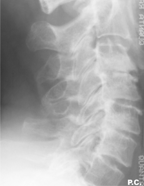
X-rayLateral X-ray of the cervical spine showing disc space narrowing at multiple levels, a radiological hallmark of cervical spondylosis.
Source : Ofiram et al., Journal of orthopaedics and traumatology, 2009, figure 1 · CC BY
🩻 Osteoarthritis on the image follows age, not necessarily pain
In people without symptoms, the frequency of degenerate cervical discs on MRI rises sharply with age.
Proportion of asymptomatic people with degenerative cervical discs on MRI. In the absence of recent trauma or a red flag, immediate imaging does not improve clinical outcomes. Source: Childress et al., American Family Physician 2020 (PMID 32735440).
The diagnosis of cervical spondylosis is almost always clinical, not radiological. The issue at the first consultation is therefore not to « see the wear » on an image, but to answer three concrete questions: is the complaint ordinary mechanical neck pain? Is there involvement of a nerve root (cervicobrachial neuralgia)? And above all, is there a warning sign that calls for prompt referral? Imaging comes in only afterwards, and only when it changes something.
Assessing mechanical neck pain
Neck pain is called mechanical when the pain varies with posture and movement, worsens at certain ranges, settles at rest, and is not accompanied by any diffuse neurological sign. The history establishes how long it has lasted, how it began (traumatic or not), the aggravating and relieving factors, the effect on sleep and work, and the patient's beliefs, because an anxiety-provoking account of « wear » or « vertebrae collapsing » often keeps the pain going as much as the osteoarthritis itself.
The physical examination remains the centrepiece: inspection and posture, palpation of the muscle masses and the facet joints, measurement of active range in the three planes, and reproduction of the usual pain where possible. When root involvement is suspected (pain running down the arm along a defined path, paraesthesia, deficit), a neurological examination of the upper limb is added: segmental strength, reflexes, sensation by dermatome, and provocation manoeuvres. The aim is to link a symptom precisely to a level, because it is this clinical link, and not the image, that guides the decision.
Good news to pass on to the patient from the outset: even when a root is compressed, the course is most often favourable. In the landmark Rochester population study (561 patients), 68.4% of cervical radiculopathies were related to spondylosis (osteoarthritis), to a disc herniation or to both, and at last follow-up 90% of patients were asymptomatic or only mildly bothered 7. A recent systematic review confirms that full recovery occurs in about 83% of patients within 24 to 36 months, with most of the improvement in the first 4 to 6 months 89.
This naturally good prognosis justifies making conservative treatment the first line. It does not mean waiting and seeing: in Kuijper's randomised trial (205 patients), early active management, physiotherapy with home exercises, or a collar and temporary rest, clearly reduced neck and arm pain compared with simple observation 10. Intervening early, rather than « letting things run their course », is the right reflex.
Red flags: when it is NOT just osteoarthritis
Before any mechanical rehabilitation, the physiotherapist must sweep for the warning signals. The most important one in the context of cervical spondylosis is degenerative cervical myelopathy (DCM) : suffering of the spinal cord caused by its compression by the degenerative changes. It is the leading cause of spinal cord dysfunction in adults, and it remains largely under-diagnosed, with an average delay of 2.2 years between the first symptoms and the diagnosis 1112.
The signs to know are not those of ordinary neck pain: clumsiness and loss of hand dexterity (doing up buttons, writing, picking up a coin), numbness, gait and balance disturbance, falls, sometimes bladder and bowel dysfunction. On examination, pyramidal signs are looked for: hyperreflexia, spasticity, clonus, Babinski's sign, Hoffmann's sign, Lhermitte's sign 11. Their presence changes management altogether: manipulation and strengthening are not pressed on with, the patient is referred for MRI and a surgical opinion.
Key points: the signals that call for referral
- Spinal cord signs (DCM) : clumsiness or loss of hand dexterity, gait and balance disturbance, falls, hyperreflexia, Hoffmann, Babinski, Lhermitte, bladder and bowel dysfunction.
- Progressive neurological deficit or a new/increasing radiculopathy with established weakness.
- Recent traumatic context, or general red flags (fever, deterioration in general condition, history of cancer).
- Signs suggesting an arterial dissection : unusual and sudden headache or neck pain, dizziness, neurological signs; not to be manipulated.
Why this vigilance? Because the decision is codified. The international AO Spine / CSRS guidelines grade the severity of myelopathy with the mJOA score (mild 15-17, moderate 12-14, severe ≤ 11) and recommend (strong recommendation) decompression surgery for moderate and severe forms. For mild forms, they leave the choice between surgery and a supervised trial of structured rehabilitation, but require surgery in the event of neurological deterioration 13. Decompression can halt progression, and management within six months offers the best chance of recovery 11 : hence the importance of referring early and monitoring a mild form closely rather than treating it as simple stiffness.
Conversely, in a patient who has cord compression visible on imaging but no sign of myelopathy or radiculopathy, prophylactic surgery is not recommended: monitoring is enough 13. The image alone does not justify operating.
The place and the limits of imaging
This is the most counter-intuitive point for patients: seeing osteoarthritis on an MRI does not prove that it causes the pain. Cervical degenerative abnormalities are extremely common in people who have no pain anywhere. In a prospective cohort of 1,211 asymptomatic volunteers (aged 20-70), MRI found a disc bulge in 87.6%, cord compression in 5.3% and an intramedullary high signal in 2.3% 3. These images increase markedly with age, compression and high signal above all after the age of 50.
The finding is the same whatever the method. On radiographs, in a population of 1,581 people aged 18 to 97, 53.9% had disc degeneration, the most affected level being C5/C6, with a strong association with age 1. The phenomenon has been documented for a long time: as early as 1987, MRI showed a disc protrusion in 57% of asymptomatic people over 64, and an impression on the cord in 26% of them 4. A synthesis for practice sums it up simply: MRI detects degenerative cervical discs in 15% of asymptomatic people in their twenties, and in more than 85% after the age of 65 2.
This mismatch has a direct consequence: routinely imaging mechanical neck pain does not help, and may do harm. Spondylotic changes are so commonplace that they make up false positives (images that explain nothing) just as much as false negatives (a reassuring image that excludes nothing). Childress points out that in the absence of recent trauma or a red flag, immediate imaging does not improve clinical outcomes 2. The radiology appropriateness criteria go the same way: radiographs are appropriate first-line only in the absence of red flags, and unenhanced MRI becomes useful in the case of new or increasing radiculopathy (that is, when the result will guide a decision) 6.
| Clinical situation | Imaging | Justification |
|---|---|---|
| Isolated mechanical neck pain, no red flag | No routine imaging | High-level evidence |
| New or worsening radiculopathy | Unenhanced MRI | Moderate evidence |
| Suspected myelopathy (DCM) or progressive deficit | MRI + surgical opinion | High-level evidence |
| Isolated degenerative image, no matching symptom | Do not decide on the image alone | High-level evidence |
A word of honesty is called for here, because the literature does not allow us to go further. The link between visible osteoarthritis and the pain that is felt is weak and poorly established : we cannot say, in a given patient, what share of their trouble really comes from a particular degenerative change. Boden had already stressed this, insisting on « the danger of basing an operative decision on imaging alone without precise clinical correlation » 5. The message to the patient is therefore neither « your MRI is normal » nor « your osteoarthritis explains everything », but this: the image is one piece of data among others, and it takes on meaning only when set against your examination.
Key points: assessing and imaging
- Cervical spondylosis is diagnosed clinically ; the image rarely confirms, and often misleads.
- No routine imaging in mechanical neck pain without a red flag or trauma 26.
- MRI useful above all for a new/increasing radiculopathy or a suspected myelopathy.
- An isolated degenerative image proves nothing: 87.6% of disc bulges in people without pain 3.
- The red flag never to be missed remains myelopathy (DCM) : screen for it, refer, do not manipulate.
⚡ Cervicobrachial neuralgia: what should be done?
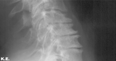
X-rayClose-up lateral X-ray of the cervical spine showing marked posterior osteophytes greater than 4 mm on a vertebral body.
Source : Ofiram et al., Journal of orthopaedics and traumatology, 2009, figure 3 · CC BY
🌿 Cervicobrachial neuralgia most often settles on its own
The natural history of cervical radiculopathy of osteoarthritic origin is broadly favourable: conservative treatment is enough for the great majority.
Clear improvement from the first 4 to 6 months; median follow-up of the landmark cohort ≈ 5 years. Surgery relieves faster in the short term but is not necessary for someone who does not need rapid relief. Sources: Wong et al., The Spine Journal 2014 (PMID 24614255); Radhakrishnan et al., Brain 1994 (PMID 8186959); Huo et al., Global Spine Journal 2022 (PMID 35324370).
When cervical osteoarthritis no longer merely stiffens the neck but radiates into the arm, we speak of cervicobrachial neuralgia, the clinical term for a cervical radiculopathy : the suffering of a nerve root as it leaves the cervical spine. Pain running down into the shoulder, the arm and the forearm as far as the fingers, sometimes tingling, numbness or weakness in a precise territory. It is often this picture, more than simple stiffness, that worries the patient and brings them to consult. The good news, widely documented, is that its spontaneous course is most often favourable.
Where does it come from? A largely osteoarthritic origin
Contrary to a received idea, cervicobrachial neuralgia is not first of all a matter of « disc herniation » in young people. In the landmark epidemiological study conducted at Rochester on 561 patients, 68.4% of cases were related to spondylosis (osteoarthritis), to a disc herniation or to both, an isolated disc protrusion accounting for only 21.9% of cases 7. In other words, in the middle-aged adult it is most often the osteoarthritic narrowing of the intervertebral canal (the foramen through which the root escapes) that compresses or irritates the nerve.
That does not mean, however, that « the wear seen on imaging » explains everything. As noted above, cervical degenerative signs are almost universal with age in people without any symptom. In 1,211 asymptomatic volunteers, MRI found a disc bulge in 87.6% 3 ; on radiographs, 53.9% of a population of 1,581 people had disc degeneration, correlated with age 1. The diagnosis of cervicobrachial neuralgia therefore remains clinical : what counts is the concordance between the painful territory, the neurological examination and, where relevant, the imaging, never the image alone.
Key points
- Cervicobrachial neuralgia is most often osteoarthritic or foraminal, not a simple disc herniation.
- Its natural history is favourable : ~83% recovery within 24-36 months, most of the improvement coming as early as 4 to 6 months.
- The active conservative approach (exercise, education, sometimes traction or a short spell in a collar) is the first line.
- Surgery relieves more quickly in the short term, but makes no further difference beyond one year.
- Only one red flag calls for a prompt surgical opinion: myelopathy (spinal cord signs).
A reassuring natural history
This is the central message to pass on to the patient, who is often frightened by the intensity of the arm pain. A systematic review shows that the time to full recovery ranges from 24 to 36 months in about 83% of patients, with substantial improvement from the first 4 to 6 months 89. In the Rochester cohort, at last follow-up (median 4.9 years), 90% of patients were asymptomatic or only mildly bothered, and only 26% had been operated on 7. A recurrence remains possible (31.7% in that same cohort), but the underlying course is towards improvement.
This favourable natural history is the decisive argument for not rushing : it justifies first-line conservative treatment for the great majority of patients, relying on time, on active management and on support with the pain.
Conservative management, the first line
Doing nothing is not the best option either: intervening early does better than simply waiting. In Kuijper's randomised trial (205 patients), a cervical collar with rest for 3 to 6 weeks, or physiotherapy with home exercises for 6 weeks, clearly reduced neck and arm pain compared with a wait-and-see attitude, in the early phase 10. The collar has a transitional, pain-relieving role here, to get through the acute stage: it must not be prolonged, at the risk of deconditioning.
Beyond the acute phase, the foundation of rehabilitation remains therapeutic exercise. The landmark Cochrane review concludes that specific strengthening and endurance exercises for the cervico-scapulo-thoracic region are beneficial for chronic pain, cervicogenic headache and radiculopathy (pooled SMD for pain immediately after treatment: −0.71; 95% CI −1.33 to −0.10), whereas stretching alone brings no benefit 15. For spondylotic radiculopathy specifically, a conservative programme combining exercise, mechanical cervical traction, TENS, pain management education and a collar, once or twice a week for 3 months, is beneficial in the long term and avoids the risks of surgery 9.
Two honest qualifications must temper the enthusiasm. First, the effect size remains modest and no high-quality evidence exists: the benefit is real but it has to be individualised, and nothing should be oversold 15. Second, there is no « miracle » exercise. Isolated training of the deep cervical flexors (low-load cranio-cervical flexion, the Jull protocol) mainly improves neuromuscular coordination, but strength and endurance at high load little or not at all 16 ; and it is no better than other active exercises at reducing pain 17. It is the fact of moving, strengthening and progressing that carries the effect, within a multimodal programme, not the choice of one particular exercise.
And what about manual therapy? An adjunct, not the engine
Manual therapy is often asked for by patients. The data invite us to put it back in its proper place: it is active exercise that carries most of the benefit. The meta-analysis by Fredin & Lorås (7 trials, grade I-II neck pain) finds only very small and non-significant between-group differences when manual therapy is added to exercise, for pain at rest, disability and quality of life, in the short term, at 6 months and at 12 months 18. Manual therapy can complement exercise, it does not replace it.
As for high-velocity cervical manipulation specifically, two cautions. On effectiveness, the Cochrane review concludes that manipulation and mobilisation give similar results on every outcome; since a risk of rare but serious adverse events exists with manipulation, it argues for preferring mobilisation 15. On safety, the largest population-based study found no excess risk of vertebrobasilar stroke linked to chiropractic care compared with general practice care: the association observed is mainly explained by reverse causation (patients already in the course of an arterial dissection consult for neck pain or headache before their stroke) 19. The event remains very rare, but catastrophic: the American Heart Association/American Stroke Association recommends informing the patient of this statistical association before any cervical manipulation 20. In practice: screen for a dissection picture, weigh the modest benefit against the risk, and prefer mobilisations and exercise.
When should surgery be considered?
For osteoarthritic radiculopathy without myelopathy, surgery is not a race. A systematic review of 6 randomised trials concludes that it is superior to conservative treatment for pain (VAS) and disability (NDI) at under one year (it relieves faster), but that this advantage fades in the medium and long term, where the two approaches converge 9. The authors put it clearly: « surgery is not necessary for patients who do not need rapid pain relief ». The decision therefore depends on the patient's tolerance, on the functional impact and on how quickly they need relief, not on the appearance of the MRI.
| Situation | Course of action | Level of evidence |
|---|---|---|
| Osteoarthritic cervicobrachial neuralgia, no spinal cord sign | Active conservative treatment, 1st line (exercise, education, ± traction or a short collar) | High |
| Disabling radicular pain, need for rapid relief | Discuss surgery (benefit mainly < 1 year) | Moderate |
| Cord compression on imaging, without myelopathy or radiculopathy | Monitoring; no prophylactic surgery | Moderate |
| Moderate to severe myelopathy (mJOA ≤ 14) | Surgical opinion, decompression recommended | High |
The red flag never to be missed: myelopathy
One picture alone radically changes matters and requires stepping outside mechanical reasoning: cervical spondylotic myelopathy (degenerative cervical myelopathy, DCM). It is no longer a matter of an irritated root but of compression of the spinal cord itself. It is the most frequent cause of spinal cord dysfunction in adults, and it remains largely under-diagnosed, with a mean diagnostic delay of 2.2 years 1112.
The signs the physiotherapist must be able to spot: clumsiness and loss of hand dexterity (doing up buttons, writing, picking up coins), gait and balance disturbance, falls, diffuse numbness of the limbs, sometimes bladder and bowel dysfunction; on examination, pyramidal signs such as hyperreflexia, spasticity, clonus, Babinski, Hoffmann's sign and Lhermitte's sign 11. Faced with these signs, mechanical treatment is not pressed on with: the patient is referred for MRI and a surgical opinion.
What is at stake is the prognosis. Surgical decompression can halt the progression of the disease, and management within six months offers the best chance of recovery 11 and hence the importance of referring early. The international AO Spine/CSRS guideline grades severity with the mJOA scale (mild 15-17, moderate 12-14, severe ≤ 11) and recommends (strong recommendation) surgery for moderate and severe forms 13.
For mild forms (mJOA 15-17), the choice remains open between surgery and a supervised trial of structured rehabilitation, but with one imperative rule: operate in the event of neurological deterioration 13. This justifies, when rehabilitation is chosen, close monitoring by the physiotherapist rather than passive symptomatic care. Conversely, in a person whose imaging shows cord compression but without any sign or symptom, prophylactic surgery is not advised: monitoring is enough 13. Another reminder that the image does not decide on its own.
In summary, faced with cervicobrachial neuralgia of osteoarthritic origin, the approach is twofold. On one side, reassure and act: the natural course is favourable, active conservative treatment is effective, surgery is called for only to shorten intense suffering and does not change the long-term result. On the other, stay watchful: screen systematically for spinal cord signs, because they alone turn a physiotherapy problem into a relative spinal surgery emergency.
🚩 Myelopathy: the red flag not to be missed
The great majority of osteoarthritic neck pain runs a favourable course and is a matter for conservative treatment. But there is a rare and formidable complication of cervical spondylosis that the physiotherapist must be able to screen for: cervical spondylotic myelopathy, also called, in current terminology, degenerative cervical myelopathy (DCM). It is no longer a matter of mechanical pain, but of suffering of the spinal cord compressed by the degenerative changes (osteophytes, discs, thickened ligaments). It is the only true « red flag » in this picture: to miss it is to let a neurological condition progress when a surgical procedure at the right moment can halt it.
The central message of this section: faced with spinal cord signs, the physiotherapist does not carry on with mechanical treatment, they refer.
A common condition… and often diagnosed too late
Degenerative cervical myelopathy is the most frequent cause of spinal cord dysfunction in adults 12. Yet it remains largely under-diagnosed: many patients with DCM are not identified, and the mean delay between the first symptoms and the diagnosis reaches 2.2 years (range 1.7 months to nearly 9 years) 11. This delay is not trivial.
Why this delay? Because the early signs are subtle, often put down to « age » or to simple osteoarthritis: a button is fumbled, one trips a little more often, the hands « no longer quite respond ». The patient sometimes consults for ordinary neck pain, and the spinal cord involvement goes unnoticed if the neurological examination is not carried out. The physiotherapist, who sees the patient repeatedly and over a long period, is in the front line for spotting a progressive deterioration that the patient themselves plays down.
The warning signs to spot
Myelopathy shows itself through a set of signs which, taken singly, may seem harmless, but which take on their full meaning together 1112 :
- Clumsiness and loss of hand dexterity : difficulty doing up a shirt, writing, handling coins, gripping small objects. It is often the first sign and the most suggestive.
- Weakness or numbness of the upper limbs, diffuse paraesthesia.
- Gait and balance disturbance : unsteady walking, a feeling of « walking on cotton wool », widening of the base of support.
- Falls and repeated loss of balance, sometimes unexplained.
- Stiffness or weakness of the lower limbs, sensory disturbance in the legs.
- Later on, bladder and bowel dysfunction (urgency, difficulty passing urine).
On clinical examination, the picture combines pyramidal signs (corticospinal tract involvement) that distinguish myelopathy from a simple radiculopathy: brisk tendon reflexes, spasticity, clonus, Babinski's sign, Hoffmann's sign, and sometimes Lhermitte's sign (an electric shock down the spine on neck flexion) 1112. The presence of hyperreflexia or of a positive Hoffmann's sign in a patient who complains of clumsy hands must immediately bring the diagnosis to mind.
Key points: recognising myelopathy
- Three key signs: clumsiness of the hands, gait and balance disturbance, pyramidal signs (hyperreflexia, Hoffmann, Babinski).
- Unlike radiculopathy (pain in a precise territory, reflexes rather diminished), myelopathy gives signs that are diffuse, bilateral and « below » the lesion, with reflexes that are brisk.
- Course typically slow and insidious : it is the deterioration over time that raises the alarm.
- Faced with these signs, mechanical treatment is not pressed on with: the patient is referred for MRI and a surgical opinion.
Beware the opposite trap: the image does not make the diagnosis
Both ends have to be held here. On one side, myelopathy is a genuine red flag not to be missed. On the other, cord compression visible on an MRI does not mean that a myelopathy exists. In 1,211 asymptomatic volunteers, MRI found cord compression in 5.3 % of them and an intramedullary high signal in 2.3 %, these abnormalities increasing after the age of 50, and all of this without any symptom 3.
The diagnosis of myelopathy is therefore clinico-radiological : an image of compression is needed and concordant clinical signs. That is why the guidelines explicitly advise against prophylactic surgery in a patient who has cord compression on imaging but no sign or symptom of myelopathy or radiculopathy: in that case, monitoring is enough 13. In other words, we do not operate on an image, we treat a patient. The physiotherapist's role is not to interpret the MRI, but to spot the clinical picture which does change everything.
Grading severity: the mJOA scale
Once myelopathy is suspected and confirmed, its severity is graded with the mJOA scale (modified Japanese Orthopaedic Association), which assesses upper limb function, lower limb function, sensation and sphincter function. The international AO Spine / Cervical Spine Research Society guidelines 13 define three levels:
| Severity (mJOA) | Score | Recommended course of action |
|---|---|---|
| Mild | 15 – 17 | Surgery or a supervised trial of structured rehabilitation; surgery if neurological deterioration occurs |
| Moderate | 12 – 14 | Decompression surgery recommended |
| Severe | 0 – 11 | Decompression surgery recommended |
For moderate to severe forms, the recommendation in favour of surgery is strong 13. For mild forms, the choice remains open between surgery and a supervised trial of structured rehabilitation, but with one imperative condition: operate in the event of neurological deterioration 13. This justifies, when a conservative trial is chosen, close monitoring by the physiotherapist rather than simple symptomatic care: dexterity, gait and reflexes are followed over time, and the patient is referred back at the slightest sign of worsening.
Why it matters to refer early
What is at stake in early screening is concrete: surgical decompression can halt the progression of the disease, and management within six months of the onset of symptoms offers the best chance of recovery 11. Beyond a certain stage, the spinal cord lesions become partly irreversible. Every month of diagnostic delay, and we have seen that it is often counted in years, is therefore lost potential for recovery.
In practice, any patient presenting with signs and symptoms of myelopathy must be referred to a spinal surgeon for assessment 12. The physiotherapist does not have to decide between surgery and rehabilitation, that decision belongs to the specialist team after MRI, but they have a decisive role: to be the one who identifies the picture and sets the pathway in motion, rather than carrying on with mobilisations or, worse, cervical manipulations on a spinal cord that is already suffering.
In practice: the physiotherapist's reflex
Faced with a patient with cervical spondylosis, a few simple questions and manoeuvres are enough to avoid missing it:
- Ask about dexterity and walking : « Do you find it harder to write, to do up buttons, to pick up small objects? Do you trip, do you feel unsteady when you walk? »
- Test the reflexes and look for pyramidal signs : hyperreflexia, a positive Hoffmann or Babinski sign are alarm signals.
- Follow the course over time : a progressive worsening, even a slow one, is more worrying than a stable picture.
- Refer without waiting in the face of any cluster of spinal cord signs: MRI and surgical opinion. One does not « try » rehabilitation first on a moderate or severe myelopathy.
The golden rule: stay reassuring about ordinary cervical spondylosis (which in the vast majority of cases runs a good course and is a matter for exercise and education), while keeping in mind this rare but serious scenario. Knowing how to recognise myelopathy is precisely what marks out safe management: the clear-headedness to spot the red flag in the middle of an extremely common and usually benign reason for consulting.
💪 What can physiotherapy do?
💪 Exercise really does help, but the effect is modest and there is no magic recipe
Set side by side, the effect sizes of active rehabilitation on chronic neck pain are real… and of moderate size.
Standardised effect sizes (|SMD|) on pain or function versus comparator; a larger value = more effect. Strengthening carries most of the benefit (but the interval around −0.71 remains wide and no high-quality evidence was found). The last bar is a difference BETWEEN two active exercises: −0.10, non-significant, in other words, training the deep flexors specifically does no better than other exercises. Sources: Gross et al., Cochrane Database of Systematic Reviews 2015 (PMID 25629215); Garzonio et al., Physical Therapy 2022 (PMID 35079832).
Cervical spondylosis is frightening because it can be « seen » on radiographs and MRI scans. Yet the image says almost nothing about the pain, and above all it does not dictate the treatment. The great majority of osteoarthritis-related neck pain is a matter for conservative management, of which physiotherapy is the backbone. The honest message from the outset: the benefits are real but modest in size, there is no miracle exercise, and the essential work consists in getting people moving, strengthening and reassured, and in spotting the rare situations that call for referral.
First, take the drama out of the image
Finding cervical osteoarthritis on imaging in an adult is almost the norm, including in people who have no pain anywhere. In a cohort of 1,211 asymptomatic volunteers aged 20 to 70, MRI found a disc bulge in 87.6% of them, cord compression in 5.3% and an intramedullary high signal in 2.3% 3. The frequency climbs with age: about 15% of people without symptoms in their twenties already have degenerative cervical discs, against more than 85% after the age of 65 2. On radiographs, more than one adult in two (53.9% of a population of 1,581 people aged 18 to 97) has disc degeneration, most often at C5/C6 1. As early as 1987, a disc protrusion was already being shown in 57% of asymptomatic people over 64 4.
A major practical consequence: seeing osteoarthritis does not prove that it explains the pain. The imaging guidelines say so clearly: spondylotic changes are so commonplace that they make up false positives just as much as false negatives, and imaging is not indicated as a routine in mechanical neck pain without a red flag 6. The physiotherapist therefore has a decisive first role in education: to reframe the story of « the wear that damages the neck » into that of a neck which stays strong, mobile and trainable.
The « wear » visible on imaging is not the designated culprit: the link between radiological osteoarthritis and pain is weak.
Exercise: the foundation of treatment
This is the best-supported long-term treatment. The landmark Cochrane review concludes that specific strengthening and endurance exercises for the cervico-scapulo-thoracic region are beneficial in chronic neck pain, cervicogenic headache and radiculopathy, whereas stretching alone brings no significant benefit 15. The effect sizes, honestly reported, remain modest to moderate: a moderate to large effect on pain for targeted strengthening (standardised mean difference −0.71; 95% CI −1.33 to −0.10), and a smaller effect for a combined strengthening plus stretching programme (SMD −0.33; 95% CI −0.55 to −0.10) with functional improvement (SMD −0.45; 95% CI −0.72 to −0.18).
An essential and openly stated qualification: the authors point out that no high-quality evidence was found. In other words, exercise really does help, but uncertainty remains about the exact size of the benefit, which is an invitation to individualise rather than to promise.
The deep flexors: useful, but not magic
Training the deep cervical flexors, the low-load cranio-cervical flexion exercise of the Jull protocol, is often presented as the centrepiece. It does indeed have its place: a review of 9 trials finds that 8 of them support the effectiveness of this specific low-load training in correcting deficits of the deep flexor muscles in people with chronic neck pain 21.
But we have to be precise about what it does, and does not do. A review shows solid evidence of an effect on neuromuscular coordination, with, on the other hand, little or no effect on strength and endurance at high load 16. Above all, training these muscles specifically is no better than other active exercises at reducing pain: a meta-analysis finds no significant difference between cranio-cervical work and more conventional cervical flexion exercises 17. The clinical message: it is the fact of moving and strengthening that counts, more than the choice of a « miracle » exercise. The deep flexors belong within a multimodal programme (endurance, scapulo-thoracic strengthening, education), not as an isolated magic wand.
What treats the condition is moving and strengthening, not one « secret » exercise rather than another.
Manual therapy: an adjunct, not the engine
Manual therapy (mobilisations, manipulations) can complement exercise, but it is not its engine. A meta-analysis of 7 trials (grade I-II neck pain) shows that adding manual therapy to exercise produces only very small and non-significant differences in pain at rest, neck disability and quality of life, in the short term, at 6 months and at 12 months 18. Active exercise therefore carries most of the effect; manual therapy is an adjunct, useful for relieving pain and getting people moving again, not a treatment in its own right.
On the question of the manual technique itself, the dedicated Cochrane review concludes that manipulation and mobilisation give similar results on all outcomes in the short and medium term, for a modest benefit and a moderate to very low quality of evidence 15. It stresses one safety point: there is a risk of rare but serious adverse events specific to manipulation, which argues for preferring mobilisation when manual therapy is indicated.
The feared risk is vertebral artery dissection with vertebrobasilar stroke. It has to be put in perspective: the largest population-based study (case-control and case-crossover) found no excess risk of stroke associated with chiropractic care compared with general practice care, the association observed being explained mainly by reverse causation, patients already in the course of a dissection consulting for neck pain or headache before their stroke 19. The event remains very rare. Even so, cervical dissection is an important cause of ischaemic stroke in young people, and the American Heart Association recommends informing the patient of this statistical association before any cervical manipulation 20. In practice: screen for the signs of dissection, weigh a modest pain-relieving benefit against a rare but catastrophic risk, and prefer mobilisations and exercise.
Radiculopathy (cervicobrachial neuralgia): reassure and treat
When osteoarthritis compresses a root and gives pain in the arm, the spontaneous prognosis is broadly favourable, a strong argument for conservative treatment. In the landmark Rochester population study (561 patients), 68.4% of cases were related to spondylosis, to a disc herniation or to both, and at last follow-up (median 4.9 years) 90% of patients were asymptomatic or only mildly bothered; only 26% were operated on 7. A systematic review confirms full recovery within 24 to 36 months in about 83% of patients, with substantial improvement from the first 4 to 6 months 9.
Intervening is better than waiting passively: in Kuijper's trial (205 patients), physiotherapy with home exercises over 6 weeks, like a collar with rest, clearly reduced neck and arm pain compared with a wait-and-see attitude, in the early phase 10. The recommended conservative treatment combines therapeutic exercise, pain management education, cervical traction and TENS, once or twice a week for about 3 months 9. Surgery relieves faster in the short term, but is no longer distinguishable from conservative treatment in the medium and long term, and « is not necessary for patients who do not need rapid relief of pain » 9. It should not therefore be oversold.
The red flag never to be missed: myelopathy
This is the absolute limit of mechanical treatment. Degenerative cervical myelopathy (DCM) is a suffering of the spinal cord from degenerative compression, the most frequent cause of spinal cord dysfunction in adults, and often diagnosed late, with a mean delay of 2.2 years 11. The physiotherapist must be able to screen for it and refer without delay.
Key points: when to refer without waiting
- Warning signs of myelopathy: clumsiness and loss of hand dexterity, numbness, gait and balance disturbance, falls, sometimes bladder and bowel dysfunction.
- On examination: pyramidal signs, namely hyperreflexia, spasticity, clonus, Babinski, Hoffmann's sign and Lhermitte's sign.
- Course of action: faced with these signs, do not carry on with mechanical treatment: refer for MRI and a surgical opinion 1112.
- Why quickly: decompression can halt progression, and management within 6 months offers the best chance of recovery 11.
Severity is graded with the mJOA scale. The international AO Spine guidelines grade it as mild 15-17, moderate 12-14, severe ≤ 11, and recommend (strong recommendation) surgery for moderate and severe forms 13. For mild forms, they leave the choice between surgery and a supervised trial of structured rehabilitation, but require surgery in the event of neurological deterioration, which justifies close monitoring by the physiotherapist rather than simple symptomatic care. Conversely, in a person whose imaging shows cord compression but who has neither myelopathy nor radiculopathy, prophylactic surgery is not advised: monitoring is enough 13.
| Situation | First-line physiotherapy course of action | Level of evidence |
|---|---|---|
| Chronic mechanical neck pain | Active exercise (strengthening, endurance) + education | Moderate |
| Recent cervical radiculopathy | Exercise + education, traction/TENS; intervene early | Moderate |
| Manual therapy as a complement | Mobilisation rather than manipulation; adjunct to exercise | Low |
| Suspected myelopathy (DCM) | Do not treat mechanically: refer (MRI, surgeon) | Strong recommendation |
In short, physiotherapy for cervical spondylosis comes down to a few solid and honest principles: reframe the image, get people moving and strengthening without overrating any single exercise, use manual therapy sparingly and cautiously, reassure about a course that is most often favourable, and stay alert to the rare spinal cord signs that change everything.
⚠️ Cervical manipulation: benefit and safety
The question comes up constantly in consultation: « Can a "crack" of the neck relieve me? » In a person with cervical spondylosis, high-velocity low-amplitude cervical manipulation (the thrust) is part of the historic armoury of manual therapy. But it holds a particular place: it is the only common technique in cervical rehabilitation whose benefit is modest and whose risk, although rare, is potentially catastrophic. This asymmetry calls for honest thinking, which the physiotherapist must be able to set out clearly to their patient.
A real but modest benefit, and no better than mobilisation
Let us start with what manipulation brings. The landmark Cochrane review on cervical manual therapy concludes that manipulation and mobilisation (slower passive movements, without a jolt) give similar results for all outcomes (pain, function) in the short, medium and long term 15. In other words, the fast technique is no more effective than the gentle one. The quality of the evidence there is judged « moderate to very low », which means that the exact size of the benefit remains uncertain.
This finding fits into a broader fact about cervical rehabilitation: it is active exercise that carries most of the effect, manual therapy being only an adjunct. The meta-analysis by Fredin and Lorås 18, covering 7 randomised trials of grade I-II neck pain, finds only « very small and non-significant » between-group differences when manual therapy is added to exercise, whether for pain at rest, disability or quality of life, in the short term, at 6 months and at 12 months. Cervico-scapulo-thoracic strengthening, for its part, has an established benefit (SMD −0.71; 95% CI −1.33 to −0.10 for pain immediately after treatment) even if it remains modest 15.
This point is crucial for setting the balance: when a modest pain-relieving benefit, no better than gentler alternatives, is set against a serious if exceptional risk, logic naturally leans towards the least risky techniques.
The risk: arterial dissection and vertebrobasilar stroke
The feared risk is dissection of a cervical artery (most often the vertebral artery), which can be complicated by a stroke in the vertebrobasilar territory. Cervical artery dissection is an important cause of ischaemic stroke in young and middle-aged people, and most case-control studies find an association between cervical manipulation and vertebral dissection in young people 20.
But « association » does not mean « causation », and this is where the nuance becomes essential. The largest population-based study on the subject, a case-control and case-crossover study of 818 vertebrobasilar strokes, found no excess risk of stroke associated with chiropractic care compared with general practice care 19. The most likely explanation for the association observed is a phenomenon of reverse causation (protopathic bias): a patient whose artery is already dissecting feels neck pain and a headache, the very first symptoms of the dissection, and consults for that, whether a chiropractor, a physiotherapist or their doctor. The stroke then occurs, in the days that follow, whichever professional was seen. The manual technique will merely have coincided with the start of a process already under way.
Key points on the benefit/risk balance
- Modest benefit: manipulation is no better than mobilisation, which is gentler 15.
- Rare but serious risk: vertebrobasilar stroke is a very rare event; the best population-based study shows no excess risk specific to manipulation, the association being explained mainly by a dissection already under way 19.
- Caution maintained: a rare but devastating event justifies preferring mobilisation and exercise when manual therapy is indicated 15.
- Duty to inform: the AHA/ASA recommends informing the patient of the statistical association between dissection and cervical manipulation before any procedure 20.
What the risk data does say, and what it does not
We have to be frank with the reader, physiotherapist and patient alike: the literature on this risk is delicate to interpret and does not lead to a clear-cut certainty. On one side, the Cassidy study 19 detects no excess risk attributable to the technique. On the other, the same study confirms that the event, if it happens, is extremely serious, and the American Heart Association / American Stroke Association guidelines 20 maintain that a statistical association exists in young people, sufficient to justify prior information. These two readings are not contradictory: they say that a cause-and-effect link is not proven, but that neither can it formally be ruled out, and that the seriousness of the outcome commands caution even in the absence of proof of causation.
The Cochrane review itself concludes, despite comparable benefits between the two techniques, that « since the risk of rare but serious adverse events linked to manipulation exists », research should focus on mobilisation 15. That is a reasonable precautionary position, and one this article adopts: when two techniques are equally effective and one carries an additional serious risk, we choose the other.
Setting the balance in practice: screen before manipulating
In concrete terms, safety does not rest on pre-manipulative tests of the vertebral artery (whose predictive value is much debated), but on clinical vigilance in the face of two red flag pictures that the physiotherapist must know.
1. The signs of a dissection in progress. An unusual, sudden headache or neck pain, different from the patient's usual mechanical pain, especially in a young person, must raise the alarm, all the more so because it can precede the stroke. Faced with such a picture, we do not manipulate: we refer.
2. The signs of cervical spondylotic myelopathy (DCM). This is the major red flag of cervical osteoarthritis, and a common-sense contraindication to any manipulation. The signs to spot are clumsiness of the hands and loss of dexterity, gait and balance disturbance, falls, paraesthesia, sometimes bladder and bowel dysfunction, with hyperreflexia and Hoffmann's sign on examination 11. The mean diagnostic delay for DCM reaches 2.2 years, whereas management within 6 months offers the best chance of recovery: the physiotherapist therefore has a genuine sentinel role. Faced with these signs, we refer for MRI and a surgical opinion rather than carrying on with mechanical treatment 11.
Let us also remember that imaging does not settle this question in place of the clinical picture. Spondylotic changes are so frequent that they constitute « both false positives and false negatives »; imaging is not indicated as a routine in mechanical neck pain 6. Seeing osteoarthritis on a film says nothing about the vascular risk, and a « reassuring » image does not replace the history and the neurological examination.
To inform is to respect the patient's choice
Beyond the technique itself, there is an obligation on which everyone agrees: to inform. The American Heart Association / American Stroke Association explicitly recommends that the patient be informed of the statistical association between arterial dissection and cervical manipulation before any manipulation of the cervical spine 20. This information is not a defensive formality: it allows the patient to take part in the decision, in full knowledge, about a technique they can legitimately do without.
The message to pass on can be put simply, without dramatising or trivialising: « Manipulation can relieve pain, but no more than a gentler mobilisation or active work; it carries a very rare but serious risk of injury to an artery in the neck; so I suggest we start with the safest techniques, and keep manipulation, if we use it, for a shared decision. » This approach is consistent with the whole management of cervical spondylosis, where the natural history is often favourable and where active exercise, education and gentle techniques form the foundation of treatment.
In summary for the consultation
- Prefer mobilisation and active exercise : equivalent benefit, lower risk 1518.
- Screen systematically for the red flags: unusual headache or neck pain suggesting a dissection, and signs of myelopathy (clumsy hands, gait disturbance, hyperreflexia, Hoffmann) 11.
- Do not manipulate at the slightest doubt: refer.
- Inform before any procedure of the statistical association between dissection and manipulation, and decide together with the patient 20.
- Do not let imaging decide in place of the clinical picture 6.
Cervical manipulation is therefore not « forbidden », but it is never indispensable in cervical spondylosis. Its modest benefit does not set it apart from gentle techniques, and its risk, exceptional though it is, deserves to be named rather than passed over in silence. The physiotherapist's role is to hold that course: to relieve effectively with what really works, exercise and support, while remaining the first link able to recognise what must leave their field.
📋 What do concrete clinical cases teach us?
Nothing beats two stories for seeing how the reasoning is built in the clinic. The case that follows is published and carries its PubMed identifier. It was chosen because it describes exactly the mistake this chapter exists to avoid. They serve only to illustrate, not to demonstrate: every decision rests on the confirmed data presented earlier in this article, never on an isolated real case. The aim is to show the clinical path, what we look for, what we choose, at what dose, and above all when the patient is no longer a matter for the physiotherapist alone.
Case 1, Mrs R., 58 years old: « my MRI shows osteoarthritis everywhere »
The reason for consulting. Mrs R. consults for mechanical neck pain that has been going on for eight months, worse at the end of the day in front of the computer, with no arm pain, no waking at night, no deterioration in general condition. She arrives worried, an MRI in her hand: « multi-level disc bulges, C5-C6 osteoarthritis, uncovertebral osteoarthritis ». She has understood that her neck is « worn out » and is afraid of making things worse by moving.
The reasoning. This is where the mismatch between image and pain becomes a therapeutic tool. In 1,211 volunteers with no symptom at all, MRI already found a disc bulge in 87.6 % of them 3. On radiographs, in a population of 1,581 people aged 18 to 97, 53.9 % had disc degeneration, most often at C5-C6, exactly the « guilty » level in Mrs R.'s report 1. In other words, at 58, these images are statistically the norm, not an explanation. Imaging describes an anatomical state; it does not measure pain.
The choice. No red flag, chronic mechanical neck pain: the clinical assessment takes precedence over the radiology report. The therapeutic work starts with education, explaining that « visible wear » does not equal « fragile neck », and then with active exercise, which is the recognised foundation. The landmark Cochrane review finds a benefit on pain for cervico-scapulo-thoracic strengthening (SMD −0.71; 95% CI −1.33 to −0.10) and for combined strengthening plus stretching programmes (SMD −0.33 on pain, −0.45 on function), whereas stretching alone brings no significant benefit 15.
The dosage, a realistic framework, to be individualised. Training of the deep cervical flexors (low-load cranio-cervical flexion) is often proposed, and is effective for restoring neuromuscular coordination 21. But we have to be honest about its limits: this exercise mainly improves coordination, and little or not at all strength and endurance at high load 16, and it is no better than other active exercises at bringing pain down 17. The message for Mrs R., and for the physiotherapist, is therefore this: it is the fact of moving and strengthening regularly that counts, more than the choice of a « miracle » exercise. We build a multimodal programme (motor control, endurance, progressive strengthening of the scapular region, reactivation in everyday activities), with supervised sessions completed by almost daily work at home, over several weeks.
What about manual therapy? It can complement, without being the engine of the benefit. Adding manual therapy to exercise brings only a small and non-significant gain in pain, disability and quality of life 18. And if a manual technique is used, mobilisation and manipulation give comparable results, but cervical manipulation carries a rare yet serious risk, which argues for preferring mobilisation 15.
The referral. Here, there is no urgency. The real act of « referral » is an educational one: taking the drama out of the MRI. Reminding her that routinely imaging mechanical neck pain does not improve outcomes in the absence of trauma or a red flag 2, and that spondylotic changes constitute false positives just as much as false negatives 6. Restoring confidence in movement is part of the treatment here.
Case 2, Mr T., 49 years old: a pain that runs down the arm
The reason for consulting. Mr T. describes, over the past three weeks, neck pain radiating into the shoulder and the outer border of the forearm as far as the thumb, with tingling. The pain is sharp, increased by extension and same-side rotation of the head. A picture suggestive of cervicobrachial neuralgia (cervical radiculopathy).
The reasoning. The majority of these radiculopathies are of osteoarthritic or discogenic origin, and their natural history is favourable. In the landmark population study (561 patients, Rochester), 68.4 % of cases were related to spondylosis, to a disc herniation or to both, and at last follow-up 90 % of patients were asymptomatic or only mildly bothered 7. A systematic review confirms that about 83 % of patients recover within 24 to 36 months, with clear improvement from the first 4 to 6 months 89. This trajectory justifies reassuring Mr T. without trivialising his pain.
The choice. Active conservative treatment first-line, and not simple waiting. In a randomised trial (205 patients), physiotherapy with home exercises over six weeks substantially reduced neck and arm pain compared with a wait-and-see attitude, in the early phase 10. The validated conservative programme combines therapeutic exercise, cervical traction, TENS, pain management education, possibly a collar in the acute phase, once or twice a week for about three months 9.
The conversation about surgery. It has to be had without overselling it. Surgery relieves pain and disability faster in the short term (less than a year), but is no longer distinguishable from conservative treatment in the medium and long term: it « is not necessary for patients who do not need rapid relief » 9. For Mr T., whose pain remains manageable, a conservative trial is entirely legitimate.
The screening, the reflex that is not negotiable. A radiculopathy that is running a good course never dispenses us from watching for the major red flag: cervical spondylotic myelopathy (DCM). At every session, the physiotherapist asks again and examines again: clumsiness or loss of hand dexterity (doing up buttons, writing), gait and balance disturbance, falls, bladder and bowel dysfunction, and on examination pyramidal signs (hyperreflexia, Hoffmann's sign, Babinski, Lhermitte). These signs are often overlooked: the mean diagnostic delay for DCM reaches 2.2 years 11.
The turning point. Suppose that in the fifth week, Mr T. reports dropping his keys, feeling unsteady going down stairs, and that examination finds hyperreflexia and a positive Hoffmann. The reasoning shifts: we do not carry on with mechanical treatment. We refer without delay for MRI and a surgical opinion. The international guidelines grade severity with the mJOA score (mild 15-17, moderate 12-14, severe ≤11) and recommend decompression surgery for moderate and severe forms; for mild forms, they leave the choice between surgery and a supervised trial of structured rehabilitation, but require surgery in the event of neurological deterioration 1312. Referring within six months offers the best chance of recovery 11. Recognising this turning point in time is probably the most useful thing the physiotherapist does in this field.
Published case: referred for a carpal tunnel, he had a cervical myelopathy
The case. Avon and colleagues report a man aged 67 years referred to orthopaedic surgery for numbness and weakness of both hands, put down to carpal tunnel syndrome22. The patient also had difficulty walking and rhythmic ankle clonus22.
The diagnosis. This was a cervical spondylotic myelopathy. The authors point out that it is the leading cause of spinal cord dysfunction in older people, that it classically presents with fine clumsiness of the hands and unsteadiness of gait, and that these symptoms are easily masked by age, a crowded medical record and more benign conditions22.
What this changes in the session. Two numb hands is not two carpal tunnels. The sign that settles it is not in the hand: it is in the gait and in the reflexes. A patient referred for a peripheral entrapment condition who walks badly must prompt a search for a central sign before we begin.
And once the central sign has been ruled out? We come back to what the Cochrane review of exercise in neck pain establishes14 : that is the treatment for common cervical spondylosis, and it becomes legitimate only after that sorting.
Key points
- The image does not make the diagnosis. In middle age, osteoarthritis and disc bulges are almost the norm in people without pain, we treat a patient, not a report 31.
- Conservative first. Mechanical neck pain and osteoarthritic radiculopathy alike have a favourable course; active exercise and education are the foundation, surgery is not called for when rapid relief is not required 1579.
- No « miracle » exercise. Strengthening the deep flexors helps coordination but does not replace a multimodal programme 1617.
- Watch for myelopathy at every session. Clumsiness of the hands, gait disturbance, pyramidal signs: faced with these signs, we refer to the surgeon rather than carrying on 1113.
This case sums up the expected stance: systematically set the imaging against the clinical picture, put movement at the heart of the treatment, state honestly the limits of each technique, and keep a permanent hold on the one signal that changes everything, that of the spinal cord.
🧭 How is this applied in practice?
Cervical spondylosis (cervical osteoarthritis, cervical spondylosis) is one of the situations where the gap between what imaging shows and what the patient feels is widest. The physiotherapist's role is therefore not to « repair wear », but to sort (red flags above all), to reassure on a solid basis, and to get people moving. Here is how to turn the evidence into concrete actions.
A three-step reasoning algorithm
Faced with a painful neck with presumed osteoarthritis, or one already « labelled » on imaging, a simple thread helps to miss nothing:
- 1. Screen for the spinal cord red flag. Before any mechanical rehabilitation, actively look for signs of cervical spondylotic myelopathy (see below). Their presence changes everything: this calls for a surgical opinion, not an exercise programme.
- 2. Frame the picture. Isolated mechanical neck pain? Cervicobrachial neuralgia (pain running down the arm, radicular territory)? Both are treated conservatively first, but their natural history and their monitoring differ.
- 3. Treat and rehabilitate actively. Education, progressive active exercise, reassurance, with regular reassessment. We intervene early rather than letting things run: in recent radiculopathy, physiotherapy with home exercises for 6 weeks clearly reduces neck and arm pain compared with waiting 10.
Imaging is not part of the first line. In the absence of a red flag, it does not improve clinical outcomes 2 ; radiographs are enough first-line, and unenhanced MRI is justified above all for a new or worsening radiculopathy 6.
Key messages: taking the drama out of osteoarthritis
The belief that « the neck is worn out » keeps fear, avoidance and pain going. Yet visible osteoarthritis is not synonymous with pain. That is the central therapeutic message, and it rests on robust data.
In this cohort of 1,211 healthy volunteers aged 20 to 70, disc bulging was already present in nearly three people out of four as early as their twenties 3. On radiographs, more than one adult in two (53.9% of 1,581 people aged 18 to 97) has disc degeneration, the C5/C6 level leading, with a frequency that climbs with age 1. And MRI detects degenerate cervical discs in 15% of asymptomatic people in their twenties, against more than 85% after the age of 65 2. These observations chime with older and consistent data 54.
In practice, one can say to the patient: « What the imaging shows is your age and your history, not necessarily the source of your pain. Many people have exactly the same images without ever being in pain. » This reframing is not a denial of the pain, which is real, but a shift of the prognosis towards hope and action.
A second reassuring message: even when the pain runs down the arm, the course is most often favourable. Cervicobrachial neuralgia is mostly of osteoarthritic or discogenic origin (68.4% linked to spondylosis and/or the disc) and, at last follow-up of the landmark Rochester study, 90% of patients were asymptomatic or only mildly bothered 7. About 83% recover within 24 to 36 months, with clear progress from the first 4 to 6 months 89.
What we actually do: exercise at the centre
The foundation of treatment is active exercise, not passive therapy. The landmark Cochrane review shows a benefit on chronic pain, cervicogenic headache and radiculopathy for targeted cervico-scapulo-thoracic strengthening (moderate to large effect size on pain, SMD −0.71), whereas stretching alone brings no benefit 15.
| Intervention | What the evidence says about it | Level of evidence |
|---|---|---|
| Cervico-scapulo-thoracic strengthening | Real benefit on pain and function, modest effect size; no high-quality evidence 15 | Moderate |
| Deep flexor exercise (Jull) | Improves neuromuscular coordination, but strength and endurance little; to be built into a multimodal programme, not used alone 16 | Moderate |
| Stretching alone | No significant effect on pain 15 | Low |
| Manual therapy added to exercise | Small and non-significant additional gain: exercise carries most of the effect 18 | Moderate |
| Cervical manipulation (thrust) | Results similar to mobilisation, but a rare risk of a serious event: prefer mobilisation 1520 | Low |
An honest qualification to pass on: there is no « miracle » exercise. Training the deep neck flexors specifically is no better than other active exercises at reducing pain 17. It is the fact of moving, of strengthening and of regaining confidence that counts, more than the choice of a particular exercise. A multimodal programme (endurance, cervical and scapular strengthening, education, general activity) is preferable to an isolated technique 16.
Common mistakes to avoid
- Routinely imaging mechanical neck pain. Spondylotic changes are frequent false positives and do not guide management in the absence of a red flag 62.
- Attributing the pain to « wear ». That is nocebo and unfounded: the correlation between visible osteoarthritis and pain is weak 35.
- Relying on passive treatment. Sticking to massage, pain-relieving physical therapy or stretching delays the real lever: active exercise 15.
- Overselling surgery. In osteoarthritic radiculopathy, surgery relieves faster in the short term but is no longer distinguishable from conservative treatment in the medium and long term; « it is not necessary for patients who do not need rapid relief » 9.
- Manipulating without weighing the benefit/risk balance. The cervical thrust is no more effective than mobilisation and carries a rare but serious risk of dissection; informing the patient is recommended 1520. The large study by Cassidy 19 found no excess risk of vertebrobasilar stroke specific to manual care compared with general practice, the association being explained mainly by dissections already under way that lead people to consult before the stroke, but the event, rare though it is, remains catastrophic.
- Letting things run. Offering nothing in the early phase of cervicobrachial neuralgia deprives the patient of a documented early benefit 10.
When to refer: the red flag not to be missed
Cervical spondylotic myelopathy (degenerative cervical myelopathy, DCM) is the leading cause of spinal cord dysfunction in adults, and it is often diagnosed late, on average after 2.2 years 1112. The physiotherapist is in the front line for spotting it. Warning signs to look for systematically:
- clumsiness and loss of hand dexterity (buttons, writing, coins);
- gait and balance disturbance, falls;
- paraesthesia or weakness of the limbs, sometimes bladder and bowel dysfunction;
- on examination: hyperreflexia, spasticity, clonus, Hoffmann's sign, Babinski, Lhermitte's sign 11.
Faced with these signs, we do not embark on mechanical treatment: we refer for MRI and a surgical opinion. Severity is graded with the mJOA scale (mild 15-17, moderate 12-14, severe ≤ 11). The international AO Spine guidelines advise (strong recommendation) decompression surgery for moderate and severe forms; for mild forms, surgery or a supervised trial of structured rehabilitation, with surgery required in the event of neurological deterioration 13. Referring early matters: decompression can halt progression, and management within 6 months offers the best chance of recovery 11.
Conversely, cord compression seen on imaging but without any sign of myelopathy or radiculopathy does not justify preventive surgery: monitoring is enough 13. We also refer for a specialist opinion a radiculopathy that is worsening, a progressive motor deficit, or the failure of well-conducted conservative treatment.
Key points
- Osteoarthritis ≠ pain. 87.6% of adults without pain have a disc bulge on MRI 3 : taking the drama out of it is therapeutic, not complacent.
- No routine imaging without a red flag 62.
- Active exercise is the foundation ; manual therapy is only an adjunct and deep strengthening is not a magic wand 151817.
- Cervicobrachial neuralgia = favourable prognosis : 90% little or not at all bothered in the long run 7 ; conservative treatment first-line 9.
- Only one true red flag never to be missed: myelopathy (clumsy hands, unsteady walking, pyramidal signs) → refer without delay 1113.
Bibliography
Every reference verified individually on PubMed (clickable PMID). 21 sources. Click a superscript note marker in the text: the bibliography opens and highlights the source.
- Tao Y, Galbusera F, Niemeyer F, Samartzis D, Vogele D, Wilke HJ (2021). European Spine Journal. PMID 33025192. doi:10.1007/s00586-020-06615-0.
- Childress MA, Stuek SJ (2020). American Family Physician. PMID 32735440.
- Nakashima H, Yukawa Y, Suda K, Yamagata M, Ueta T, Kato F (2015). Spine. PMID 25584950. doi:10.1097/BRS.0000000000000775.
- Teresi LM, Lufkin RB, Reicher MA, Moffit BJ, Vinuela FV, Wilson GM, Bentson JR, Hanafee WN (1987). Radiology. PMID 3588931. doi:10.1148/radiology.164.1.3588931.
- Boden SD, McCowin PR, Davis DO, Dina TS, Mark AS, Wiesel S (1990). Journal of Bone and Joint Surgery. American Volume. PMID 2398088.
- McDonald MA, et al. (Expert Panel on Neurological Imaging) (2019). J Am Coll Radiol. PMID 31054759. doi:10.1016/j.jacr.2019.02.023.
- Radhakrishnan K, Litchy WJ, O'Fallon WM, Kurland LT (1994). Brain. PMID 8186959. doi:10.1093/brain/117.2.325.
- Wong JJ, Côté P, Quesnele JJ, Stern PJ, Mior SA (2014). The Spine Journal. PMID 24614255. doi:10.1016/j.spinee.2014.02.032.
- Luyao H, Xiaoxiao Y, Tianxiao F, Yuandong L, Ping Wang (2022). Global Spine Journal. PMID 35324370. doi:10.1177/21925682221075290.
- Kuijper B, Tans JT, Beelen A, Nollet F, de Visser M (2009). BMJ. PMID 19812130. doi:10.1136/bmj.b3883.
- Davies BM, Mowforth OD, Smith EK, Kotter MR (2018). BMJ. PMID 29472200. doi:10.1136/bmj.k186.
- Milligan J, Ryan K, Fehlings M, Bauman C (2019). Canadian Family Physician. PMID 31515310.
- Fehlings MG, Tetreault LA, Riew KD, et al. (AO Spine/CSRS group) (2017). Global Spine Journal. PMID 29164035. doi:10.1177/2192568217701914.
- Gross A, Kay TM, Paquin JP, Blanchette S, Lalonde P, Christie T, et al. (2015). Cochrane Database of Systematic Reviews. PMID 25629215. doi:10.1002/14651858.CD004250.pub5.
- Gross A, Langevin P, Burnie SJ, Bédard-Brochu MS, Empey B, Dugas E, et al. (2015). Cochrane Database of Systematic Reviews. PMID 26397370. doi:10.1002/14651858.CD004249.pub4.
- Blomgren J, Strandell E, Jull G, Vikman I, Röijezon U (2018). BMC Musculoskeletal Disorders. PMID 30486819. doi:10.1186/s12891-018-2324-z.
- Garzonio S, Arbasetti C, Geri T, Testa M, Carta G (2022). Physical Therapy. PMID 35079832. doi:10.1093/ptj/pzac001.
- Fredin K, Lorås H (2017). Musculoskeletal Science & Practice. PMID 28750310. doi:10.1016/j.msksp.2017.07.005.
- Cassidy JD, Boyle E, Côté P, He Y, Hogg-Johnson S, Silver FL, Bondy SJ (2008). Spine. PMID 18204390. doi:10.1097/BRS.0b013e3181644600.
- Biller J, et al. (2014). Stroke. PMID 25104849. doi:10.1161/STR.0000000000000016.
- Amiri Arimi S, Mohseni Bandpei MA, Javanshir K, Rezasoltani A, Biglarian A (2017). American Journal of Physical Medicine & Rehabilitation. PMID 28225440. doi:10.1097/PHM.0000000000000721.
- Avon JT, Scott GJ, Bowling SM, Jones M (2023). Cervical Spondylotic Myelopathy Presenting as Peripheral Nerve Disease: A Case Report. Cureus. PMID 38022189. doi:10.7759/cureus.47829.
❓ Frequently asked questions
Does seeing cervical osteoarthritis on my MRI mean that it is causing my pain?
No. In completely asymptomatic people, MRI finds a disc bulge in 87.6%, cord compression in 5.3% and an intramedullary high signal in 2.3% 3. A degenerative image therefore does not on its own establish the origin of the pain: imaging has to be set against the clinical picture.
Is radiological cervical spondylosis common, and is it linked to age?
Yes, it is commonplace and increases with age. In a population of 1,581 people aged 18 to 97, 53.9% had radiographic disc degeneration, the most affected level being C5/C6, with a significant association with age in both sexes 1.
Does cervicobrachial neuralgia of degenerative origin resolve without surgery?
Most often, yes. Full recovery occurs within 24 to 36 months in about 83% of patients, with substantial improvement from the first 4 to 6 months 8. In the landmark Rochester study, 90% of patients were asymptomatic or only mildly bothered at last follow-up 7.
Should degenerative cervical radiculopathy be operated on?
Not first-line for most patients. Surgery brings faster relief of pain (VAS) and disability (NDI) at under one year, but « is not necessary for patients who do not need rapid relief »; conservative treatment remains beneficial in the long term 9.
Which signs should raise the alarm and prompt referral to a surgeon?
Degenerative cervical myelopathy (DCM) is the major red flag: clumsiness and loss of hand dexterity, gait and balance disturbance, falls, paraesthesia, sometimes bladder and bowel dysfunction, together with hyperreflexia and Hoffmann's sign. Severity is graded with the mJOA scale (mild 15-17, moderate 12-14, severe ≤11) and surgery is recommended for moderate to severe forms 13.
Which physiotherapy treatment should be preferred for chronic neck pain?
Active exercise is the foundation. Specific cervico-scapulo-thoracic strengthening is beneficial for chronic pain (pooled SMD −0.71; 95% CI −1.33 to −0.10), whereas stretching alone brings no benefit 15. Cervical manipulation does no better than the gentler mobilisation, and carries a rare but serious risk of dissection/stroke 19.
Further reading in the review

