
















On-siteDPCFIFPL

Neurological rehabilitation
Neurological disorders in musculoskeletal practice: screen, assess and treat
Bryan Littré
Go further
Cervical: 83.2 new cases per 100,000 a year, a disc to blame in only 21.9% of radiculopathies. Thoracic: rare, but 86% of operated cases had myelopathy.

A symptomatic thoracic disc herniation is rare and presents with thoracic spine pain or intercostal neuralgia in a band; surgery is only considered for rebel pain or a deficit.
2%of symptomatic disc degeneration are of thoracic origin
What follows covers each of these points in detail, with sources. It is there if you need it.
This topic is taught in a course: Neurological disorders in musculoskeletal practice: screen, assess and treat, on-site with Bryan Littré.
1 / 16
15 article chapters · 64 min in total
Understand◔ 5 min
Three canals, one same lesion: it is the anatomy of the container, not the size of the herniation, that decides the risk.
Imaging◔ 4 min
In subjects without any pain, 87.6 % have a cervical disc bulge, but only 5.3 % have cord compression.
Resorption◔ 4 min
Spontaneous resorption rests on macrophage infiltration and neovascularisation, among other processes.
Recognise◔ 9 min
A specific test turning positive raises the probability of diagnosis: that is the role of Spurling.
Differentiate◔ 3 min
Two families dominate the differential: entrapment syndromes of the upper limb and shoulder pathology.
Deciding on imaging◔ 3 min
Imaging answers a precise question, it does not screen: half of herniations are incidental.
Course◔ 6 min
At 4.9 years follow-up, 90 % of patients are doing well, across all treatments, but 31.7 % recur.
Rehabilitate◔ 4 min
Specific neck training or prescribed physical activity: same result at 24 months, both groups improve.
After 50◔ 4 min
After 50, it is not the frequency of abnormalities that changes, it is the fact that they reach the cord.
Thoracic◔ 4 min
In operated series, 86 % myelopathies: the surgical picture is neurological, not painful.
Screen◔ 4 min
Myelopathy affects the cord: its signs are below the lesion, so in the legs, whatever the level.
Surgery◔ 5 min
The Frankel score is maintained or improved in all patients, with 65.1 % gaining a grade, but 14 % revisions.
Clinical cases◔ 3 min
In three cases out of four, image and clinic evolve at different paces; it is the clinical picture that leads.
In practice◔ 3 min
The first message is that the image does not indicate severity: half of herniations are incidental findings.
FAQ◔ 3 min
Not every herniation disappears: no regression rate has been measured by meta-analysis at cervical level.
Train on this
Neurological rehabilitation
Bryan Littré
Next comes practice: the course that teaches this topic, with Bryan Littré.
Our courses on this topic may be covered by DPC or FIFPL, the French continuing-education funding schemes, depending on your profession and situation.
This page covers the cervical and thoracic levels. For the lumbar level, the most common of the three and one that follows a different triage logic, see lumbar radicular pain from a lumbar disc herniation. Bringing the neck and the back together in one page is not an editorial convenience: these two levels share the same lesion and almost nothing else. The cervical level is common and its course is largely favourable. The thoracic level is rare, most often silent, and when it does speak, it is the spinal cord speaking.
Where disc herniations occur
Distribution by spinal level, and what each level puts at stake
Source: Benditz A, Koehl P, Mahmoud M, Schuh A. Cervical and thoracic disc herniation. Orthopädie 2026;55(1):69-79. PMID 41295983.
The same anatomical lesion, three canals of different calibre, and above all three different neural contents. It is this anatomy, and not the size of the herniation, that decides what the clinician must fear.
A disc herniation is the displacement of disc material beyond the limits of the intervertebral space. The definition is the same from C2 to S1. What changes from one level to another is what lies behind the disc.
In the lumbar spine, below L1-L2, the spinal cord has ended: the canal contains the cauda equina, a bundle of free roots that tolerates a conflict for a long time before suffering as a whole. A herniation there typically compresses one root, and cauda equina syndrome remains the dreaded exception.
In the cervical spine, the spinal cord is present, but the canal is relatively wide and the roots leave through short, oblique foramina. The posterolateral herniation, by far the most common, catches the root along its foraminal course: it produces a cervicobrachial neuralgia. The median herniation, rarer, can catch the spinal cord and produce a cervical myelopathy.
In the thoracic spine, everything narrows. The canal is the narrowest of the whole spine, the cord occupies a larger share of its cross-section, and the spinal cord blood supply is at its most precarious there, in what the vascular literature has long called the watershed zone. To this is added the relative rigidity of the rib cage, which limits disc loading and explains how rare the lesion is, but which offers no protection at all once it occurs. A thoracic herniation of modest volume, in that place, can do what a lumbar herniation three times its size will never do.
The figures follow this anatomy. Symptomatic disc degeneration is distributed as 36% cervical, 2% thoracic and 62% lumbar (Benditz 2026), and thoracic herniations account for fewer than 1% of all spinal herniations (Petrovic 2023). When they do occur, they sit mainly between T8 and L1, that is, at the junction where the rib cage stops stiffening the spine, and they are calcified in 40% of cases (Court 2018).
The lumbar level appears here by cross-reference, because it is the point of comparison every physiotherapist has in mind, and because the difference in prognosis is only visible in a table that sets them side by side.
| Criterion | Cervical | Thoracic | Lumbar (by cross-reference) |
|---|---|---|---|
| Share of herniations | 36 % | 2%, and fewer than 1% in surgical series | 62 % |
| Structure exposed | Mainly the root, the cord if the herniation is median | The spinal cord above all | Root, cauda equina |
| Typical levels | C6-C7 then C5-C6 | T8–L1 | L4-L5, L5-S1 |
| Clinical picture | Cervicobrachial neuralgia: upper limb pain following a dermatome | Thoracic spine pain, intercostal neuralgia, then spinal cord signs | Lumbar radicular pain or femoral neuralgia |
| Incidental finding | Disc bulge in 87.6% of subjects without pain | Herniation in 37% of subjects without pain | Protrusion in 29 to 43% depending on age |
| Leading clinical test | Spurling (specific), the neurodynamic tests combined (sensitive) | No validated provocation test; it is the neurological examination that decides | Straight leg raise (Lasegue), slump test |
| Emergency not to be missed | Cervical myelopathy | Thoracic myelopathy | Cauda equina syndrome |
| Spontaneous regression | Documented, including complete regression, on extrusions | Documented but rarer than at the other two levels | 66.7% of herniations resorb |
| Overall prognosis | 90% asymptomatic or mildly bothered in the medium term | Excellent if asymptomatic; guarded as soon as the spinal cord suffers | Favourable in the majority of cases |
| Dedicated page | This chapter | This chapter | Lumbar radicular pain from a lumbar disc herniation |
The figures in this table come respectively from Benditz 2026 for the distribution, Nakashima 2015 and Wood 1995 and Brinjikji 2015 for the incidental findings, Radhakrishnan 1994 for the cervical prognosis, Zhong 2017 for lumbar resorption and Valluzzi 2021 for thoracic regression.
There is a common misconception, and it matters because it changes what is said to the patient. When a patient consults for arm pain of radicular type, the label that comes to mind is disc herniation. Yet in the only population-based study to have followed an entire community over fifteen years, a confirmed disc herniation was responsible for only 21.9 % of cervical radiculopathies. In 68.4 % of cases, the cause lay with spondylosis, with the disc, or with both together (Radhakrishnan 1994).
In other words, cervicobrachial neuralgia is more often a story of a foramen narrowed by arthritis than a story of an expelled disc. This is anything but anecdotal: the two mechanisms do not have the same time course of onset, nor the same response to time, and the second has no material to resorb. It is also why the site devotes a separate page to cervical spondylosis, and another to cervicobrachial neuralgia taken as a syndrome. The present page joins them from another angle: that of the disc lesion itself, and of what becomes of it when it is left alone.
Cervical radiculopathy in four figures
Rochester population-based cohort, 561 patients followed from 1976 to 1990
Source: Radhakrishnan K, Litchy WJ, O'Fallon WM, Kurland LT. Epidemiology of cervical radiculopathy. A population-based study from Rochester, Minnesota, 1976 through 1990. Brain 1994;117(Pt 2):325-35. PMID 8186959. The figure of 90% refers to patients who were asymptomatic or only mildly bothered at last review, across all treatments, surgery included.
This is the question that should precede any reading of a report. At both levels the answer is the same: a great deal. But it does not carry the same consequence, because the word "herniation" does not describe the same risk at each.
The prevalence of imaging abnormalities in subjects without pain is the piece of data that best protects against overdiagnosis, and it is also the one patients know least about. Three studies document it, each with its own scope.
Nakashima and colleagues examined the cervical spine by MRI in 1,211 volunteers with no symptoms at all, roughly one hundred per decade and per sex, from 20 to 70 years. The result is striking for two opposite reasons (Nakashima 2015).
First, 87.6% of these subjects had a disc bulge. Among those in their twenties, already 73.3% of men and 78.0% of women had one. A cervical disc bulge is therefore not an abnormality: it is a feature of the adult human spine, whose frequency, severity and number of levels involved increase with age.
Second, and this is the other half of the message, spinal cord compression concerned only 5.3% of these subjects, and an intramedullary high signal only 2.3%. These two abnormalities increase markedly after 50. They were located mainly at C5-C6 (41%) and C6-C7 (27%), at a single level in 58% of cases.
The practical conclusion is clear. Faced with a cervical report, the word "bulge" tells us almost nothing: it describes a feature shared by nearly nine adults in ten who are perfectly well. The words that do inform are those describing the spinal cord: cord compression, effacement of the subarachnoid spaces, intramedullary T2 high signal. Those are out of the ordinary.
The reference study is older and smaller, but it has no equivalent, and its figures have never been contradicted. Wood and colleagues reviewed the thoracic MRIs of 90 asymptomatic subjects, blinded to all clinical information (Wood 1995).
Sixty-six of these 90 subjects, that is 73 %, had at least one abnormality. In detail: disc herniation in 33 subjects (37%), disc bulge in 48 (53%), annular tear in 52 (58%), endplate irregularities or Scheuermann-type kyphosis in 34 (38%). And above all, the figure least often quoted although it is the most important one here: a deformity of the spinal cord in 26 subjects, that is 29%.
This last figure deserves a pause, because it cuts both ways. It says that contact between a herniation and the cord, on its own, proves nothing: nearly one pain-free subject in three has one. It also says that the thoracic level brings the cord into contact with the lesion far more readily than the others, which explains why, when the picture does become symptomatic, it becomes a spinal cord picture. The rarity of the disease and the frequency of the image are not contradictory: they describe two different things.
What a pain-free subject's MRI carries
Prevalence of abnormalities in asymptomatic volunteers, by level
Sources: Nakashima H et al. Abnormal findings on magnetic resonance images of the cervical spines in 1211 asymptomatic subjects. Spine 2015;40(6):392-8, PMID 25584950 ; Wood KB et al. Magnetic resonance imaging of the thoracic spine. Evaluation of asymptomatic individuals. J Bone Joint Surg Am 1995;77(11):1631-8, PMID 7593072.
Brinjikji's systematic review, which pooled 33 studies and 3,110 asymptomatic subjects across all levels, gives the general curve: disc degeneration rises from 37% at 20 years to 96% at 80 years, bulging from 30% to 84%, protrusion from 29 %–43 %, annular tears from 19% to 29% (Brinjikji 2015). The authors conclude that many of these images are part of normal ageing and are not associated with pain.
A 2026 review devoted precisely to the two levels of interest here puts it in one sentence that is useful in consultation: around 50% of disc herniations are recognised as incidental findings on imaging and remain asymptomatic (Benditz 2026).
Spontaneous resorption is not a happy accident: it is a described biological process whose actors are known. Knowing which of the two mechanisms is at work changes what can be announced to the patient.
A review devoted to the mechanisms of resorption identifies four processes acting together: the macrophage infiltration, the inflammatory response, the matrix remodelling and neovascularisation. The authors conclude that the regulation, by macrophages, of inflammatory mediators, matrix metalloproteinases and certain cytokines of the intervertebral disc is essential to spontaneous reabsorption (Yu 2022). This work concerns the lumbar spine, where the phenomenon has been most studied, but there is no reason for the cellular mechanism to be specific to one level.
A series that analysed the possible mechanisms of regression offers a complementary reading, with the merit of being clinical: the fragment may move back into the intervertebral space, disappear through dehydration and retraction, be progressively resorbed by phagocytosis and enzymatic degradation triggered by the inflammatory reaction that the extrusion itself provokes, or be pushed back by the pulsation of the cerebrospinal fluid (Martínez-Quiñones 2010).
This is the most important practical consequence of that mechanism, and it is counter-intuitive.
For a disc fragment to be recognised, attacked and digested, it must be exposed to the epidural space, that is, it must have crossed the annulus fibrosus and, better still, the posterior longitudinal ligament. The immune system then no longer meets a disc, but a foreign body: the nucleus pulposus is a tissue sequestered since embryonic life, and its contact with the circulation triggers exactly the inflammatory reaction that ends in its destruction.
That same exposure explains the pain: the inflammatory reaction that digests the fragment is also the one that irritates the root. Intense pain and effective resorption therefore share the same cause.
Symmetrically, a contained protrusion, still under an intact annulus, is not exposed. It does not trigger this reaction, does not resorb in the same way, and can remain stable for years. And a calcified herniation, as 40% of thoracic herniations are (Court 2018), no longer offers degradable material in the same way.
This is what gives meaning to Kobayashi's observation: all the published cases of spontaneous regression of a cervical herniation concerned, at that date, herniations of the extruded type (Kobayashi 2003). The fact that thoracic regression is described as less frequent than at the other levels (Valluzzi 2021) is consistent with the high proportion of calcified herniations at that level, even though no study has formally established this causal link.
Caution is required on one point: none of these data allows us to tell a given patient whether their herniation will resorb. The mechanistic review says so explicitly: the pathogenesis and the physiological indicators that would allow reabsorption to be predicted remain uncertain, which is precisely what prevents clinicians from choosing conservative treatment in an informed way (Yu 2022).
What can honestly be said fits in three sentences. The phenomenon is real and documented on imaging. It preferentially concerns extruded herniations, that is, often the most painful ones. And it unfolds on a timescale that is not that of the pain: in Kobayashi's case, the pain had settled within three weeks and the image took twelve months to normalise.
Cervicobrachial neuralgia is the usual clinical face of cervical disc herniation. Its diagnosis rests on the history, a neurological examination and a few provocation tests whose real values must be known, and they are more modest than their reputation.
Cervicobrachial neuralgia combines neck pain with upper limb pain of radicular distribution, with or without a sensory, motor or reflex deficit. The site devotes a whole page to it as a syndrome, cervicobrachial neuralgia (cervical radiculopathy), as well as a page of dedicated exercises. This chapter approaches it from the angle of the disc lesion and of what the tests really allow us to conclude.
Three elements from the Rochester cohort deserve to be known, because they contradict common expectations (Radhakrishnan 1994).
First, physical exertion or trauma preceded the onset of symptoms in only 14.8% of cases. Cervicobrachial neuralgia therefore has, most often, no triggering event to report, and its absence must not cast doubt on the diagnosis nor point towards another cause.
Second, 41% of patients had a history of lumbar radiculopathy. A history of past sciatica is not off-topic when taking the history of arm pain: it belongs to the background.
Finally, the C7 root was the most often affected, followed by C6, which fits with the most heavily loaded disc levels, C6-C7 and C5-C6, themselves the levels most frequently compressed in asymptomatic subjects.
This is where the literature is at once most useful and most disappointing. The 2026 update of Thoomes's systematic review pooled eight studies assessing six physical examination tests, with bivariate meta-analysis and GRADE appraisal (Thoomes 2026). Here is what it establishes.
The Spurling test, assessed in five studies under slightly different variants, shows a high specificity, from 0.84 to 1.00, and very scattered sensitivity, from 0.38 to 0.98. The authors did not pool these values, precisely because of that heterogeneity. The clinical reading is that of a rule-in test: a positive Spurling, in a patient whose history suggests a radiculopathy, usefully increases the probability of the diagnosis. A negative Spurling does not rule it out.
The upper limb neural tension test number 1 (ULNT 1), median-nerve biased, achieves a pooled sensitivity of 0.70 (95% CI 0.60 to 0.79) and a pooled specificity of 0.71 (95% CI 0.63 to 0.79). Taken on its own, it is therefore a middling test in both directions.
It is the combination of the four neurodynamic tests that changes things, with a pooled sensitivity of 0.97 (95% CI 0.88 to 0.99) for a specificity of 0.51 (95% CI 0.40 to 0.62). Four neurodynamic tests all negative make cervical radiculopathy very unlikely. It is the rule-out tool of the cervical clinical examination.
The shoulder abduction relief test, in which the patient places the hand on the head and sees the arm pain decrease, achieves a sensitivity of 0.49 and a specificity of 0.76.
One reservation must accompany all these figures, and the authors state it themselves without hedging: the data are sparse, the certainty of the evidence is low to very low across all the tests and outcomes, and the pooled estimates hold only for the populations and the test versions actually studied. That is no reason to give up examining; it is a reason not to hang a decision on a single test.
Diagnostic values of the cervical tests
Sensitivity and specificity, 2026 meta-analysis, low to very low certainty
Source: Thoomes EJ, Arvanitidis M, van Geest S et al. Diagnostic accuracy of physical examination tests for painful cervical radiculopathy: update of a systematic review and meta-analysis. BMC Musculoskelet Disord 2026;27(1). PMID 41680685. The Spurling values are not pooled by the authors because of the heterogeneity of the variants; they are shown here as a range. The certainty of the evidence is judged low to very low across all the tests.
The seminal work remains that of Wainner, who put 82 patients suspected of cervical radiculopathy or of carpal tunnel syndrome through a standardised blinded examination, with electrophysiology as the reference (Wainner 2003). Two results have passed into practice.
The first is that a cluster of four variables reaches a point positive likelihood ratio of 30.3, far above what any isolated test gives. The four items are Spurling, cervical distraction, ULTT A and cervical rotation towards the symptomatic side of less than 60 degrees.
The second is that ULTT A was the most useful test for ruling out a cervical radiculopathy, which the 2026 meta-analysis confirms and amplifies by showing that the combination of the four neurodynamic tests reaches a sensitivity of 0.97.
Wainner himself insists on a limitation that must be repeated: the confidence intervals of all the likelihood ratios in his study were wide, and the cluster still needed validating. A likelihood ratio of 30.3 is a point estimate, not a guarantee.
The 2018 review, which predates the update, offered a compatible reading: combine Spurling, axial traction and the arm squeeze test to increase the probability of a cervical radiculopathy, and use four negative neurodynamic tests together with a negative arm squeeze test to rule it out (Thoomes 2018). That review also flagged a gap that persists: no study has assessed the diagnostic value of the most widely used neurological tests, key muscle strength, tendon reflexes and sensory deficits. We perform them because they describe the lesion, not because a study has quantified their yield.
Once a radiculopathy has been accepted, the next question is that of the level. The distribution of the roots is a classic descriptive fact, not a trial result: it describes an anatomy, it predicts nothing. It remains useful because it makes the topography of the pain, the sensory deficit, the motor deficit and the reflex converge, or diverge.
The frequency of the levels, on the other hand, has been measured. In the Rochester cohort, monoradiculopathy of C7 was the most frequent, followed by C6 (Radhakrishnan 1994), which corresponds to the C6-C7 and C5-C6 discs, themselves the levels most often compressed in asymptomatic subjects (Nakashima 2015). A general review takes up this segmental semiology as well as the differential diagnoses to be ruled out (Iyer 2016).
| Root | Disc | Sensory territory | Muscles to test | Reflex |
|---|---|---|---|---|
| C5 | C4-C5 | Shoulder cap, lateral aspect of the arm | Deltoid, supraspinatus and infraspinatus | Biceps, partly |
| C6 | C5-C6 | Lateral aspect of the forearm, thumb and index finger | Biceps brachii, brachioradialis, wrist extensors | Biceps, brachioradialis |
| C7 | C6-C7 | Posterior aspect of the arm, middle finger | Triceps brachii, wrist flexors, finger extensors | Triceps |
| C8 | C7-T1 | Medial border of the forearm, ring and little fingers | Deep finger flexors, intrinsic muscles of the hand | No routine reflex |
| T1 | T1-T2 | Medial aspect of the arm, axilla | Interossei, abductor of the fifth finger | No routine reflex |
Three practical remarks accompany this table, and they matter more than the table itself.
Convergence counts more than any single row. Isolated thumb pain does not make a C6. It is the agreement between the painful territory, the objectified sensory deficit, the weakness of a key muscle and the loss of the corresponding reflex that gives the chosen level its value.
Overlaps are the rule. Sensory territories overlap widely from one person to another, and muscles receive multi-root innervation. An examination that does not fit perfectly with one row of the table does not disprove the diagnosis of radiculopathy.
The diagnostic value of these items has never been measured. The 2018 systematic review states it explicitly: no study has assessed the diagnostic accuracy of the most widely used neurological tests, key muscle strength, tendon reflexes and sensory deficits (Thoomes 2018). We perform them because they describe and localise the lesion, not because any figure guarantees their yield.
Which root is affected, and how often
Segmental distribution and hierarchy of the most affected levels
Sources: Radhakrishnan K et al. Brain 1994;117(Pt 2):325-35, PMID 8186959 for the hierarchy of the levels; Iyer S, Kim HJ. Cervical radiculopathy. Curr Rev Musculoskelet Med 2016;9(3):272-80, PMID 27250042 for the semiology; Thoomes EJ et al. Spine J 2018;18(1):179-189, PMID 28838857 for the absence of diagnostic accuracy data on these items.
No provocation test distinguishes a root compressed by a soft herniation from a root compressed by an arthritic foramen. That distinction, which matters for the prognosis, belongs to imaging and to age. Nor does any of them screen for associated spinal cord involvement, which belongs to a different examination and which the following chapter on myelopathy sets out.
Upper limb pain has many causes, and disc herniation is neither the most frequent nor the first to rule out. A detail from the seminal study on the cervical tests says it better than any argument could.
When Wainner set out to measure the value of the tests for cervical radiculopathy, he did not recruit patients "suspected of radiculopathy". He recruited patients suspected of cervical radiculopathy or carpal tunnel syndrome (Wainner 2003). The choice is not trivial: it acknowledges that these two diagnoses genuinely compete for the same patients in clinical practice, and that telling a root compression from a distal trunk compression is the daily problem, not a textbook case.
A general review of cervical radiculopathy organises the differential around two families, the peripheral entrapment syndromes and shoulder pathology (Iyer 2016). The site devotes a page to each of the entities that follow, and the links below point to them.
The carpal tunnel syndrome is the most common entrapment neuropathy, and its territory, thumb, index and middle finger, overlaps that of the C6 and C7 roots. Two features help: the nocturnal flare with waking at night and shaking of the hand, typical of carpal tunnel and unusual in a radiculopathy, and the absence of neck pain and of any cervical postural factor. A pitfall remains possible in both directions: the two can coexist in the same patient.
The cubital tunnel syndrome, compression of the ulnar nerve at the elbow, mimics C8 involvement. The distinction turns on the boundary of the sensory deficit: ulnar involvement stops at the wrist and spares the medial border of the forearm, whereas a C8 radiculopathy involves it.
The thoracic outlet syndrome is the trickiest differential, because it shares with radiculopathy an upper limb pain of proximal origin. Vascular signs, positional reproduction in sustained abduction and external rotation, and a topography often of the lower trunk, C8-T1, point towards it.
Pain over the lateral aspect of the arm radiating towards the elbow can come from a C5 root or from the shoulder itself. The rotator cuff tendinopathy, the adhesive capsulitis and degenerative rotator cuff tear come top of the list.
Two features settle it in practice. The first is the passive restriction : an adhesive capsulitis restricts passive range, which no radiculopathy does. The second is the shoulder abduction relief test, in which placing the hand on the head relieves the arm pain: its specificity is 0.76 for cervical radiculopathy (Thoomes 2026), and a shoulder problem does not behave that way.
Radiating neck pain does not imply a root conflict. The non-specific neck pain can radiate into the upper limb with no radicular distribution and no neurological sign. The cervical spondylosis accounts, together with the disc, for the vast majority of cervical radiculopathies in population data. The occipital neuralgia and cervicogenic headache concern the high posterior territory. Finally, a whiplash injury changes the context and imposes its own triage rules.
| Diagnosis | What looks alike | What distinguishes it |
|---|---|---|
| Carpal tunnel syndrome | Thumb, index and middle finger territory | Nocturnal flare, no neck pain, deficit limited to the hand |
| Cubital tunnel | Ring and little finger territory, like C8 | Sensory deficit stopping at the wrist, medial forearm spared |
| Thoracic outlet | Proximal upper limb pain | Vascular signs, positional reproduction, C8-T1 involvement |
| Shoulder pathology | Lateral arm pain, like C5 | Passive restriction, no neurological sign, positive shoulder tests |
| Non-specific neck pain | Radiating neck pain | Non-dermatomal radiation, normal neurological examination |
| Myelopathy | Can begin as ordinary neck pain | Lower limb signs, spasticity, sensory level, sphincters |
A methodological remark is called for on this table: the items in the right-hand column are classic semiological landmarks, widely taught, whose diagnostic value has not been quantified in comparative studies, with the exception of the shoulder abduction relief test. They guide the reasoning, they do not conclude it.
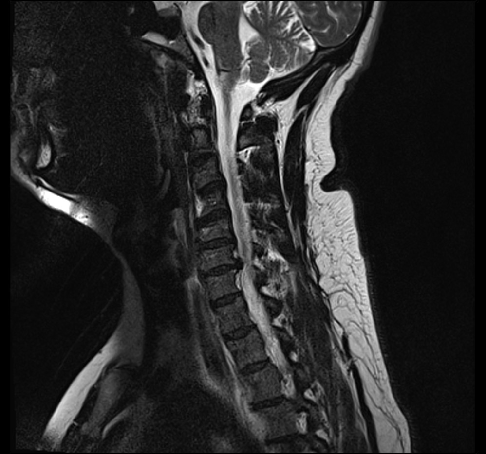
MRISagittal T2-weighted cervical spine MRI showing a disc herniation at C6-C7.
Source : Anthonp, Wikimedia Commons · CC BY-SA
Everything said above about how commonplace these images are leads to an uncomfortable conclusion: imaging is not prescribed to find out whether there is a herniation, since the answer is often yes in people who are perfectly well. It is prescribed to answer a precise question, and the choice of examination depends on the question.
Recalling the figures is enough to set the limit. At the cervical level, 87.6 % of subjects without pain have a disc bulge (Nakashima 2015). At the thoracic level, 37 % have a herniation and 29 % a deformity of the cord (Wood 1995). About half of all disc herniations are incidental findings (Benditz 2026).
Imaging requested without a precise clinical question will therefore produce, with high probability, an abnormality that nobody will know what to do with, and that will weigh lastingly on the patient's beliefs. That is the mechanism of overdiagnosis, and it is particularly easy to set off here.
First question: is the spinal cord compressed? This is the indication that is not up for discussion. The slightest spinal cord sign, at either level, calls for cross-sectional imaging without delay. It is not a matter of confirming a hypothesis but of ruling out a progressive compression.
Second question: does a neurological deficit justify a procedure? In the Rochester cohort, the predictors of the decision to operate were the combination of radicular pain with a sensory deficit, and above all an objective motor deficit (Radhakrishnan 1994). Imaging takes on its meaning when a procedure is being considered, not before.
Third question: does the picture remain unexplained? Pain that does not settle, that does not follow the expected course, or that comes with non-mechanical features justifies imaging aimed at finding a cause, to look for something other than a herniation.
Conversely, a typical cervical radiculopathy, without objective motor deficit, in a patient whose course follows the expected path, does not call for initial imaging: the median duration of symptoms before diagnosis was 15 days in the population-based cohort, and 90% of patients were doing well in the medium term.
This is where the thoracic level imposes a technical exception that has to be known.
MRI is the reference examination for visualising the cord, the roots, the cord signal and the soft tissues. It is MRI that answers the first question.
But in the thoracic spine, 40% of herniations are calcified (Court 2018), and that proportion rises to 38.9% complete calcification and 27.8% partial calcification in a consecutive surgical series, that is two herniations in three carrying calcification (Saway 2022). Now calcification directly determines the surgical strategy: calcified median herniations call for a transthoracic approach, whereas soft lateralised herniations can be reached by a posterolateral approach (Court 2018).
The consequence is that, faced with a symptomatic thoracic herniation, CT provides information that MRI renders poorly: the density and the extent of the calcification. This is not a radiologist's nuance, it is what will determine the surgical approach. The physiotherapist does not prescribe, but knowing this makes it possible to understand why an MRI alone is not enough to settle a thoracic case.
This is the chapter that should be read out to the patient. The natural course of cervical radiculopathy is favourable in the great majority of cases, the herniation itself can regress on imaging, and surgery is not the rule.
The Rochester cohort remains the best available source because it was not recruited in hospital: through the Mayo Clinic records system it took up every case occurring in a community over fifteen years, 561 patients, which protects it from the selection bias of surgical series (Radhakrishnan 1994).
Over a median follow-up of 4.9 years, the main result is that 90% of patients were asymptomatic or only mildly bothered by their cervical radiculopathy at last review. Two qualifications must accompany that figure for it to stay honest. The first is that it includes every treatment received, surgery included: it is not a figure for the course without intervention. The second is that the disease recurs: 31.7% of patients had a recurrence over the duration of follow-up. A good prognosis does not mean the absence of later episodes.
A third figure situates the place of surgery: 26% of patients were operated on. The factors predicting the decision to operate were the combination of radicular pain with a sensory deficit, and the presence of an objective motor deficit. One patient in four, then, and over a decade and a half of North American practice, without today's selection criteria.
The median duration of symptoms before diagnosis was 15 days, a reminder that most of these patients consult early and that the clinician mainly sees recent presentations.
This is the most counter-intuitive point for a patient who has just received an MRI report, and it is documented.
The most solid piece of data, although it concerns the lumbar spine, is a meta-analysis of eleven cohort studies: the overall incidence of spontaneous resorption after lumbar disc herniation is 66.7% (95% CI 51 to 69%) after conservative treatment (Zhong 2017). The authors conclude from it that conservative treatment can become the first choice. That figure belongs to the lumbar level and does not transpose as it stands, but it establishes the phenomenon.
In the cervical spine, the literature is made of case reports and small series rather than cohorts. It nevertheless establishes that the phenomenon exists, including in complete form. Kobayashi reports the case of a 27-year-old man with severe left C6 pain on a C5-C6 disc extrusion, who refused discectomy: the pain disappeared within three weeks, and follow-up MRI showed partial regression at three weeks and a complete regression at twelve months (Kobayashi 2003). The authors note that all the cases of cervical regression published up to then concerned herniations of the extruded type, which fits with what is known of the mechanism at the lumbar level: the expelled fragment, exposed to the epidural space, behaves like a foreign body and undergoes inflammatory and enzymatic resorption.
The 2026 review gives an order of magnitude applicable to the two levels of interest here: disc herniations lose up to 40% of their volume over the course of their evolution, which explains why a non-negligible percentage of patients experience spontaneous improvement of their pain and of mild neurological deficits (Benditz 2026).
A point of rigour is called for here, because it is often overlooked in popular articles. A frequently cited Spanish series covers 37 cases of spontaneous regression, but those 37 cases were identified among 858 patients : it is a series of observed regressions, not a regression rate. The figures of 85% in the lumbar spine and 90% in the cervical spine that this article reports in its introduction are borrowed from other work and presented as context, not as its own result (Martínez-Quiñones 2010). Citing them as a measured rate would be a misreading.
Spontaneous regression by level
What the literature establishes, and with what level of evidence
Sources: Zhong M et al. Incidence of Spontaneous Resorption of Lumbar Disc Herniation: A Meta-Analysis. Pain Physician 2017;20(1):E45-E52, PMID 28072796 ; Kobayashi N et al. Spontaneous regression of herniated cervical disc. Spine J 2003;3(2):171-3, PMID 14589234 ; Valluzzi A et al. J Neurol Surg A 2021;82(2):182-185, PMID 33260243 ; Benditz A et al. Orthopädie 2026;55(1):69-79, PMID 41295983.
Three Swedish randomised trials from the same programme compared anterior cervical decompression and fusion combined with physiotherapy against a structured programme of physiotherapy alone. Reading them requires precision, because they do not say quite the same thing depending on the outcome and on the length of follow-up.
At the longest follow-up, 5 to 8 years in 59 randomised patients, surgery combined with physiotherapy does better than physiotherapy alone on neck disability (NDI reduced by 21% against 11%, p = 0.03) and on neck pain (39 mm against 19 mm on the VAS, p = 0.01). But on arm pain, which is the dominant symptom of a cervicobrachial neuralgia, the difference does not reach the threshold of significance (33 mm against 19 mm, p = 0.10), any more than does quality of life measured by the EQ-5D. The patient's global assessment clearly favours surgery, 93% against 62% of patients declaring themselves improved or much improved. And above all, both groups improved significantly compared with their baseline state, on every outcome (Engquist 2017).
The honest clinical reading is therefore twofold. Surgery is not useless: it brings a measurable additional benefit on disability and neck pain, and clearly greater satisfaction. Nor is it the rule: the benefit is not found on arm pain or on quality of life, and physiotherapy alone also brings a significant and lasting improvement. That fits with the 26% of operated patients observed in the general population.
On minimally invasive techniques, the most recent Cochrane review is clear and negative: four randomised trials totalling 259 participants, all at high risk of bias, do not allow the use of nucleoplasty in cervical radicular pain from a disc herniation to be supported. Compared with conservative treatment, the evidence is of low certainty for a substantial reduction in pain in the short term, but with no difference on function or on quality of life (de Rooij 2025).
The best supported answer is uncomfortable for anyone looking for a protocol: no modality has proved superior to another, and patients improve in every arm of the trials. That result is not a failure of rehabilitation, it is information about the prognosis.
Dedering and colleagues randomised 144 patients with a cervical radiculopathy between a specific neck training programme and prescribed physical activity, both groups also receiving a cognitive behavioural approach, with assessments at 3, 6, 12 and 24 months (Dedering 2018).
In the intention-to-treat as in the per-protocol analysis, there was no group effect and no group by time interaction. Time effects, on the other hand, were significant: both groups improved on every outcome, except on the level of depression. The authors conclude that cervical radiculopathy has a naturally favourable long-term course when patients receive specific neck training and exercise combined with a behavioural approach.
This result deserves to be presented as it is. It does not say that rehabilitation is useless; the trial had no untreated group. It says that the choice between two reasonable active strategies does not change the result at two years, which shifts the question: what counts is probably that the patient moves, is supported and understands the prognosis, more than the exact nature of the exercises.
This is where the literature contradicts itself most visibly, and it would be dishonest to settle the matter in its place.
On the one hand, a meta-analysis of five randomised trials concludes that the literature offers some support for adding traction, mechanical or manual, to the other physiotherapy procedures, for the reduction of pain. The effect sizes reported are substantial for mechanical traction on pain in the short term (g = -0.85, 95% CI -1.63 to -0.06) and in the medium term (g = -1.17, 95% CI -2.25 to -0.10), with an effect on disability in the medium term (g = -1.05). The authors themselves flag as the main limitation the heterogeneity of the diagnostic criteria between the included trials (Romeo 2018).
On the other hand, the most rigorous multicentre randomised trial on the question compared, in 81 patients a programme of manual therapy and exercise with intermittent cervical traction against the same programme with sham traction. Result: no significant difference between the groups on any outcome, primary or secondary, at two or at four weeks, with small effect sizes. The authors conclude that adding mechanical traction to a multimodal programme brings no additional benefit (Young 2009).
The difference probably comes down to what each protocol compares: the meta-analysis pools trials with variable comparators and populations, whereas Young's trial strictly isolates the specific effect of traction against sham traction, within an otherwise identical programme. The workable conclusion is that traction is not the active ingredient of a multimodal programme, and that it does not deserve to be its pivot.
The clinical practice guidelines of the Orthopaedic Section of the American Physical Therapy Association, in their 2017 revision, cover neck pain including with radiating pain and serve as a framework for management (Blanpied 2017). They rest on classification by clinical presentation from the International Classification of Functioning, rather than on the lesion-based diagnosis, which is consistent with everything above: it is the clinical picture, not the image, that guides treatment.
Modalities and level of evidence, cervical radiculopathy
Horizontal cards, from best supported to least supported
Cards drawn from the sources cited in each row. The ranking reflects the strength of the available evidence, not the order of clinical application. None of these modalities has been compared with no treatment in a recent controlled trial.
The practical conclusion of this chapter is not a protocol but a hierarchy. The prognosis is good and must be announced as such, with the figures to back it. Movement, in one form or another, is the foundation. Manual therapy and traction are comfort adjuncts whose own yield is not established. And follow-up really has only one thing to monitor: the appearance of signs that no longer belong to the root.
This is the subgroup that this topic requires to be handled separately. Past fifty, the probability of a pure disc herniation falls, that of a degenerative spinal cord compression rises, and the figure-backed reassurance of the preceding chapters needs qualifying.
The imaging data in asymptomatic subjects give the first curve. Among Nakashima's 1,211 volunteers, disc bulging was already present in those in their twenties, but cord compression and intramedullary high signal only increased markedly after 50, accompanied by a worsening in the severity of the bulging (Nakashima 2015). In other words, what changes with age is not the presence of a disc abnormality, it is the fact that it reaches the cord.
Brinjikji's review gives the backdrop: disc degeneration goes from 37% at 20 years to 96% at 80 years in subjects without pain (Brinjikji 2015). A degenerative image in a patient of 70 therefore has virtually no informative value in itself.
The second curve is that of the disease. Cervical radiculopathy peaks at 202.9 per 100,000 per year between 50 and 54 years, against 83.2 on average across all ages (Radhakrishnan 1994). It is the decade in which the two pictures, radicular and spinal cord, meet most often.
The term degenerative cervical myelopathy has been proposed as an umbrella term covering all the degenerative conditions of the cervical spine responsible for a myelopathy: spondylotic myelopathy, ossification of the posterior longitudinal ligament, ossification of the ligamentum flavum and degenerative disc disease. The non-traumatic degenerative forms of cervical myelopathy represent the most common cause of spinal cord impairment in adults, with an incidence estimated at at least 41 per million and a prevalence of 605 per million in North America; the incidence of hospitalisations related to cervical spondylotic myelopathy is estimated at 4.04 per 100,000 person-years, and surgery rates appear to be rising (Nouri 2015).
That same work describes the mechanisms of injury, which combine a static compression, a malalignment altering the tension and the blood supply of the cord, and dynamic injury mechanisms during movement. It also identifies predisposing conditions: the congenital spinal stenosis, Down syndrome and Klippel-Feil syndrome. A constitutionally narrow cervical canal means that a herniation of ordinary size produces a compression that the same size would not have produced elsewhere.
What age changes in the reasoning
Two distinct curves: how commonplace the image is, and the occurrence of spinal cord involvement
Sources: Brinjikji W et al. AJNR Am J Neuroradiol 2015;36(4):811-6, PMID 25430861 for the degeneration curve; Nakashima H et al. Spine 2015;40(6):392-8, PMID 25584950 for the rise in cord compression after 50; Radhakrishnan K et al. Brain 1994;117(Pt 2):325-35, PMID 8186959 for the incidence peak. The red curve is a qualitative representation of a described trend, and not a series of published values.
The reassurance remains true, but it is worded differently. The 90% of patients with little or no disability in the medium term concern cervical radiculopathy, not myelopathy. Announcing a good prognosis to a patient in whom spinal cord involvement has not been ruled out means getting the wrong disease.
The threshold of suspicion must come down. After 50, a vague complaint of clumsy hands, of unsteadiness when walking or of tiredness in the legs, spontaneously put down to age by the patient, deserves a full neurological examination, not reassurance.
The constitutional context counts. A known history of a narrow cervical canal, Down syndrome or Klippel-Feil syndrome shifts the risk marker straight away, for the same size of herniation.
Imaging does not decide on its own. At 70, almost everyone has degenerative images. What distinguishes the ill patient from the merely old one is the neurological examination, repeated over time.
Rare, most often silent, and yet the one that on its own justifies a red flag box. The paradox resolves as soon as the image is separated from the disease: the image is commonplace, the disease is not.
The orders of magnitude were set out above: 2% of symptomatic disc degeneration (Benditz 2026) and fewer than 1% of all spinal herniations (Petrovic 2023). A physiotherapist can practise an entire career without meeting a characterised symptomatic form. That is precisely what makes the topic dangerous: rarity produces inexperience, and inexperience produces delay.
The figure that measures that delay is the most telling in this whole chapter. In a consecutive series of 43 patients operated on for 54 thoracic herniations, the mean duration of symptoms before surgery was 10.4 months, with a standard deviation of 11.6 months (Saway 2022). Ten months is the time it takes, on average, for a thoracic cord compression to be recognised and treated. Part of that delay belongs to front-line professionals, physiotherapists included.
In that same series of 43 patients, the symptoms present at the time of surgical management were distributed as follows: myelopathy in 86%, motor deficit in 72%, sensory deficit in 65%. This is not a mechanical pain profile, it is a neurological profile. The bias of this statistic must be kept in mind: it is a surgical series, therefore the most severe forms, the ones that are operated on. It does not describe all symptomatic thoracic herniations; it describes those that reach the operating theatre, and it says that the vast majority of them are myelopathies.
The lesion characteristics of this series also shed light on the mechanism: 38.9% of the discs were entirely calcified and 27.8% partly, and 66.7% were so-called giant herniations, occupying more than 40% of the canal.
Court's French surgical review completes the picture: thoracic herniations sit mainly between T8 and L1, are calcified in 40% of cases, and a herniation is called giant when it occupies more than 40% of the spinal canal. The accepted surgical indications are severe thoracic spine pain, intractable intercostal neuralgia, or neurological deficits. And above all, the authors identify giant calcified herniations as the main contributor to myelopathy, to intradural extension and to postoperative complications (Court 2018).
The profile of operated thoracic herniations
Consecutive series of 43 patients, 54 thoracic discectomies
Source: Saway BF, Alshareef M, Lajthia O et al. Ultrasonic spine surgery for every thoracic disc herniation: a 43-patient case series. J Neurosurg Spine 2022;36(5):800-808. PMID 34798611. Surgical series: these proportions describe the operated forms, therefore the most severe ones, and not the whole of symptomatic thoracic herniations.
A thoracic herniation can cause thoracic spine pain and intercostal neuralgia, that is, a girdle pain following a thoracic dermatome. The problem is that these two symptoms are extremely non-specific: ordinary thoracic spine pain is common, and the site devotes a whole page to it, thoracic spine pain, which also sets out the cardiopulmonary and visceral differential diagnosis, unavoidable at this level. Among the differential diagnoses at this level are also Scheuermann's disease, axial spondyloarthritis and thoracolumbar junction syndrome.
In other words: it is not the pain that should bring thoracic herniation to mind. It is the neurological examination of the lower limbs. Ordinary mechanical thoracic spine pain justifies no imaging; thoracic spine pain accompanied by the slightest spinal cord sign justifies imaging without delay.
It would be wrong to conclude that every thoracic herniation belongs in theatre. The literature says the opposite, provided the boundary is drawn in the right place.
Valluzzi reports the case of a thoracic herniation giant and traumatic at T9-T10, spontaneously resorbed within five months, with no treatment at all. The authors draw from it a cautious and precise recommendation: regression of a herniation at the thoracic level can occur even without treatment, and conservative management with clinical and radiological monitoring can be an appropriate first line in patients with no sign of progressive myelopathy and no severe refractory pain. They note in the same text that this spontaneous regression is less frequent at the thoracic level than at the cervical and lumbar levels (Valluzzi 2021).
The condition is therefore the key to this whole chapter. No progressive myelopathy: monitoring is possible. Progressive myelopathy: the question is no longer one of rehabilitation.
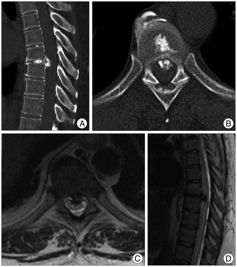
CT scanCT (A, B) and MRI (C, D) of the thoracic spine showing a large, calcified, midline disc herniation at T7-T8, with cord compression clearly visible on the images.
Source : Tan et al., Journal of Korean Neurosurgical Society, 2014, figure 1 · CC BY-NC
This is the most important chapter on this page. Myelopathy almost never presents as a noisy emergency: it sets in through discreet signs, which the patient puts down to age and which the clinician only finds by looking for them.
Myelopathy is suffering of the spinal cord itself, as opposed to radiculopathy, which affects a root as it leaves. Its consequences are not segmental but below the lesion: everything that lies below the level affected is concerned.
Degenerative cervical myelopathy is, according to the reference review, the most common cause of spinal cord impairment in adults, with an incidence estimated at a minimum of 41 per million and a prevalence of 605 per million in North America (Nouri 2015). Its thoracic version is far rarer, but the underlying semiology is the same, with one difference that has considerable localising value: a thoracic myelopathy spares the upper limbs.
It gets missed for three reasons that add up. The first signs are commonplace and slowly progressive, so they are attributed to ageing. The pain, when there is any, occupies the front of the stage and diverts the examination. And the neurological examination of the lower limbs is not routine when faced with high spinal pain.
What to do: any combination of spinal pain with one of these signs calls for cross-sectional imaging and a specialist opinion, without going through a trial of rehabilitation. A rapidly developing deficit or sphincter involvement is an emergency.
There is an error of reasoning that deserves to be known by name, because a published case documents it precisely and because it can cost a second operation.
A spastic paraparesis of the lower limbs naturally points towards a thoracic lesion. But a low cervical lesion can produce exactly the same picture, spared upper limbs included. Sasai's case, set out in the following chapter, is the demonstration: a 48-year-old man carrying two herniations, one at T11-T12 and the other at C6-C7, of which the second produced a spastic paraparesis with no upper limb signs at all (Sasai 2006).
The rule that follows is simple: faced with a spastic paraparesis, the work-up must not stop at the thoracic spine, even if the examination of the upper limbs is normal. The sensory level, when there is one, remains the best clinical localising landmark, but its absence rules nothing out.
Faced with cervical or thoracic spinal pain
Triage tree: what belongs to rehabilitation and what no longer does
Tree built from Nouri 2015 (PMID 25839387) for the spinal cord semiology, Sasai 2006 (PMID 16825057) for the warning about the level, Radhakrishnan 1994 (PMID 8186959) for the predictors of a decision to operate and Valluzzi 2021 (PMID 33260243) for the condition for conservative monitoring.
There is a structural reason why this chapter is addressed first of all to rehabilitation professionals. The physiotherapist is the professional who sees the patient most often and over the longest period. A myelopathy that sets in over months will go unnoticed at an isolated consultation, but it is detectable by comparison between two sessions a few weeks apart.
In practical terms, three repeated observations are enough to screen for it: the quality of walking, including in tandem and with the eyes closed, the fine dexterity of the hands when a cervical level is possible, and the briskness of the reflexes in the lower limbs. None of them requires equipment. All of them deteriorate before the patient complains of them.
The physiotherapist does not decide the indication, but receives the patient before and after. Knowing the surgical approaches, their specific complications and what surgery really recovers changes what can be announced to the patient.
The reference surgical review retains three situations: severe thoracic spine pain, or intractable intercostal neuralgia, or neurological deficits (Court 2018). Conversely, clinical and radiological monitoring remains an appropriate first line in patients with no progressive myelopathy and no severe refractory pain (Valluzzi 2021).
It is worth underlining what these two formulations have in common: neither retains the size of the herniation as a criterion. A giant herniation can be monitored if it does not cause a progressive myelopathy, and a modest herniation may belong in theatre if the cord is suffering.
That choice is described as essential, and it follows a simple logic: reach the herniation without crossing the cord (Court 2018).
The calcified median herniations are approached through a transthoracic incision, because there is no posterior trajectory that reaches them without moving the cord. The soft lateralised herniations can be reached through a posterolateral approach, which is lighter.
The complication rate of transthoracic approaches is higher than that of posterolateral approaches, but the authors immediately explain why: the former are used in the most complex cases. The raw comparison of rates therefore does not set two techniques against each other on equal terms.
The thoracoscopic approach is less invasive but comes with a long learning curve. The retropleural mini-thoracotomy is presented as a possible compromise. A fusion is recommended in the case of multilevel herniations, of a herniation occurring on Scheuermann's disease, when more than 50% of the vertebral body is resected, in patients with preoperative thoracic spine pain, or for a herniation at the thoracolumbar junction.
Besides the complications specific to each approach, the surgical risks identified are the neurological deterioration, the dural tear and subarachnoid-pleural fistulas. Giant calcified herniations are named as the main contributor not only to myelopathy, but also to intradural extension and to postoperative complications (Court 2018).
These three complications have direct practical consequences in postoperative rehabilitation. Postoperative neurological deterioration calls for revising the goals, not for pressing on. A dural tear and a fistula change the instructions on position and early mobilisation, and belong to information passed on by the surgical team: they are things to ask the file for, not to deduce.
The consecutive series of 43 patients and 54 discectomies gives the most detailed results available (Saway 2022).
In terms of decompression, 38 of the 40 levels reviewed on postoperative MRI, that is 95%, showed less than 20% residual disc. Functionally, measured by the Frankel score at more than three months, the result was maintained or improved in every patient, and 28 patients out of 43, that is 65.1%, gained at least one grade.
Two qualifications must accompany these figures. The first is that six patients out of 43, that is 14%, had to be reoperated on : two for recurrent herniation, two for a herniation at an adjacent level, two for wound problems. The second is that the presence of calcification did not degrade the improvement in the Frankel score compared with non-calcified herniations, which is rather reassuring, and that better intraoperative neurological monitoring was associated with a greater functional gain.
These results must be read for what they are: those of a single-surgeon retrospective series, with the selection that this implies. They do not transpose mechanically to every patient and every centre.
What thoracic surgery achieves
Consecutive series of 43 patients, functional results on the Frankel score
Source: Saway BF et al. Ultrasonic spine surgery for every thoracic disc herniation: a 43-patient case series and technical note. J Neurosurg Spine 2022;36(5):800-808. PMID 34798611. The Frankel score grades spinal cord involvement from A, complete, to E, normal.
Three consequences emerge, and none has been the subject of a controlled trial specific to this level, which must be said.
The first is that the delay is the main lever available to the rehabilitation professional. Ten months on average separate the first symptoms from surgery. The only point in that pathway on which a physiotherapist really has any weight is the date at which they recognise spinal cord signs and refer.
The second is that the postoperative prognosis is rather good when surgery does take place: no patient lost a functional grade in this series, and two thirds gained one. That is useful information to give a patient frightened by the idea of spinal cord surgery.
The third is that monitoring does not stop at the operation. Fourteen per cent of revisions, two of them for a herniation at an adjacent level, mean that a reappearance of signs after a period of improvement must be taken seriously, and not put down to poor adherence. Sasai's case, where the relapse at two months in fact came from a second level in the cervical spine, is the extreme illustration of this (Sasai 2006).
Four published observations, chosen because each corrects a different belief. All are referenced by their identifier, and none has been reconstructed for the purposes of the demonstration.
A 48-year-old man, presenting with a gait disturbance with weakness and numbness of the lower limbs. Thoracic MRI shows a disc herniation at T11-T12, removed through a minimally invasive posterior approach under the microscope. The patient improves. Two months after the operation, the numbness and the spasticity recur. No recurrence of the thoracic herniation is found, but cervical MRI reveals a cord compressed at C6-C7. The patient had no neurological signs in the upper limbs at all. The C6-C7 herniation is removed with laminoplasty. The symptoms regress progressively; at two years and nine months from the first operation, walking is stable and the patient has returned to work (Sasai 2006).
What the case corrects: the idea that a spastic paraparesis with no upper limb signs localises the lesion to the thoracic spine. The authors conclude explicitly that spinal cord compression must be looked for not only in the thoracic spine but also in the cervical spine, particularly at C6-C7, even in the absence of upper limb signs.
A 43-year-old man in good health, presenting with a Brown-Séquard syndrome. The initial MRI shows a calcified disc extrusion at T5-T6, with no significant cord compression and no signal abnormality. It is only 48 hours later that an appearance consistent with spinal cord ischaemia emerges. The patient is treated conservatively with aspirin and heparin, later stopped in the face of a negative cardiovascular work-up. The calcified extrusion, recognised retrospectively as the cause of the ischaemia, decreases spontaneously over time, and the patient recovers within a few months (Petrovic 2023, full text in open access under PMC10685598).
What the case corrects: the idea that the seriousness of a thoracic herniation can be read from the degree of mechanical compression. Here, the initial MRI showed no significant compression, and the cord suffered through a vascular mechanism. It also illustrates the vascular precariousness specific to this level, and the fact that a normal early scan does not rule out spinal cord suffering in the making.
Thoracic disc herniation giant and of traumatic origin at T9-T10, which resorbs spontaneously within five months, with no intervention. The authors recall that spontaneous regression is less frequent at this level than in the cervical and lumbar spine, which makes the observation notable, and they draw a course of action from it: clinical and radiological monitoring is a reasonable first line in patients with no progressive myelopathy and no severe refractory pain (Valluzzi 2021).
What the case corrects: the idea that a giant thoracic herniation calls for surgery by its size alone. Size is not the criterion; the neurological course is.
A 27-year-old man, severe pain in a left C6 distribution. MRI shows a lateral disc extrusion at C5-C6. The patient refuses discectomy. His pain disappears within three weeks. The follow-up MRI at three weeks shows partial regression of the extrusion, and the one performed at twelve months shows complete regression. The authors note that all the published cases of spontaneous cervical regression had until then concerned extruded-type herniations, and conclude that non-surgical conservative observation should be considered an option in these patients (Kobayashi 2003).
What the case corrects: the idea that a large extrusion calls for a procedure. It also illustrates the classic dissociation between the clinical timescale and the radiological timescale: the pain settles in three weeks, the image takes twelve months to normalise. It is this dissociation that must be explained to the patient before offering a follow-up MRI.
This chapter condenses the previous ones into consultation actions. It does not replace clinical reasoning, it sets its order.
One. Rule out myelopathy first, before even characterising the radiculopathy. Gait, lower limb reflexes, Babinski, Hoffmann, fine dexterity, questions about the sphincters. It takes two minutes and it cannot be made up for later.
Two. Characterise the territory. The C7 root is the most often affected, followed by C6. The absence of a triggering event is the rule, not the exception: it must not cast doubt.
Three. Use the tests for what they do. Spurling to reinforce a hypothesis already held by the history. The four neurodynamic tests to rule out when they are all negative. The Wainner cluster when a strong argument is wanted, bearing in mind that its confidence interval is wide.
Four. Announce the prognosis, with the figures. Nine patients out of ten are doing well in the medium term, one in four was operated on in an unselected population, and the herniation itself can regress. That information is probably the most profitable intervention of the first session.
Five. Treat with active exercise, in whatever form suits the patient, knowing that no superiority between structured programmes has been demonstrated. Do not build the management around traction.
Six. Monitor. An objective motor deficit that appears or worsens is the documented predictor of a decision to operate: it justifies an opinion, not an intensification of rehabilitation.
One. Remember that symptomatic thoracic herniation is rare, and that ordinary thoracic spine pain is common. The priority differential diagnosis at this level is not the herniation: it is cardiopulmonary and visceral, and the page devoted to thoracic spine pain sets it out.
Two. Examine the lower limbs, routinely. It is the only action that distinguishes ordinary thoracic spine pain from a beginning spinal cord compression, and nothing in the patient's complaint will trigger it spontaneously.
Three. Look for a sensory level if the slightest sign is present. It is the most specific and the most localising sign.
Four. Do not conclude that the level is thoracic. A spastic paraparesis with no upper limb signs can come from a low cervical lesion.
Five. In the absence of any spinal cord sign, treat thoracic spine pain as thoracic spine pain, and reassess at every session. The mean delay of ten months before surgery in the operated series is built up precisely during those months of symptomatic management.
| Clinical situation | Course of action | Timing |
|---|---|---|
| Isolated neck or thoracic pain, normal neurological examination | Rehabilitation, education about the prognosis, reassessment | No imaging |
| Cervical radiculopathy with no objective motor deficit | Active rehabilitation, monitoring of the deficit | Imaging not routine |
| Objective motor deficit, or a deficit that progresses | Specialist opinion, imaging | Prompt |
| One or more spinal cord signs | Cross-sectional imaging, specialist opinion, cervical and thoracic exploration | Without delay |
| Rapidly developing deficit, sphincter disturbance | Urgent referral | Emergency |
| Non-mechanical pain, fever, history of cancer, immunosuppression | Targeted aetiological work-up | Prompt |
Three messages, in this order. The first is that the image does not tell you the severity: half of all disc herniations are incidental findings, and nearly nine pain-free adults in ten have a cervical disc bulge. The second is that the prognosis is good: nine patients out of ten are doing well in the medium term, and the herniation can resorb. The third is the limit of those two messages: certain signs, in the legs, no longer belong to that message, and they must be named to the patient so that they come back if the signs appear.
Yes, and it has been documented on imaging. The clearest case is a C5-C6 extrusion in a 27-year-old man, in partial regression at three weeks and complete at twelve months, with no surgical treatment at all (Kobayashi 2003). Not every herniation disappears, and there is no cervical regression rate measured by meta-analysis as there is in the lumbar spine. What can honestly be said: herniations lose up to 40% of their volume over the course of their evolution (Benditz 2026), and it is the extruded herniations, often the most painful ones, that regress best.
Not as a rule. In an unselected population followed over fifteen years, 26% of patients were operated on (Radhakrishnan 1994). The randomised trial with the longest follow-up shows that at 5-8 years, surgery combined with physiotherapy does better than physiotherapy alone on neck disability and neck pain, but not significantly on arm pain, and that both groups improve markedly (Engquist 2017). The documented predictors of a decision to operate are the combination of radicular pain with a sensory deficit, and above all an objective motor deficit.
Not necessarily, and this is in fact the most common situation. Among 90 subjects with no pain at all, 37% had a thoracic disc herniation and 29% a deformity of the spinal cord (Wood 1995). A thoracic herniation seen on MRI is therefore an unremarkable image. What decides is not the image: it is the neurological examination of the lower limbs. In the absence of any spinal cord sign, watchful waiting is a legitimate course (Valluzzi 2021).
Those that come from the legs, not from the back: difficulty or instability when walking, spastic stiffness, brisk reflexes, a sharp horizontal boundary in trunk or lower limb sensation, and any urinary or sphincter disturbance. These signs reflect suffering of the spinal cord and call for imaging and a specialist opinion without going through a trial of rehabilitation. In surgical series of thoracic herniations, 86% of patients had a myelopathy (Saway 2022).
No. It is specific (0.84 to 1.00) but its sensitivity varies enormously from one study to another (0.38 to 0.98), and the certainty of these estimates is judged low to very low (Thoomes 2026). A positive Spurling reinforces a hypothesis already carried by the history; a negative Spurling does not rule out a radiculopathy. To rule out, the four neurodynamic tests combined are what is needed, with a pooled sensitivity of 0.97.
The honest answer is that the literature contradicts itself. A meta-analysis of five trials reports an effect on pain in the short and medium term (Romeo 2018). But the trial that best isolates its specific effect, comparing it with sham traction within an otherwise identical programme, finds no significant difference on pain, function or disability (Young 2009). In practice: traction is not the active ingredient of a programme, it must not be its pivot, and it can be offered for the comfort it brings some patients.
Because the cervical and thoracic levels share one thing the lumbar spine does not: at these two levels the spinal cord lies behind the disc, and the differential diagnosis between root involvement and spinal cord involvement arises there in the same way. The lumbar spine, below L1-L2, contains the cauda equina and follows a different triage logic: it has its own dedicated page, lumbar radicular pain from a lumbar disc herniation.
No, and this is a documented error. A 48-year-old man was operated on for a T11-T12 herniation causing a spastic paraparesis, improved, then relapsed two months later: the cause was a second herniation at C6-C7, even though he had no upper limb signs at all (Sasai 2006). Faced with a spastic paraparesis, the work-up must cover the cervical spine as much as the thoracic spine.
Behind this article
An author who explains, a reviewer who checks.
How we write and check our content

Physiotherapist · co-founder of Physio Learning
Marked for life by his first four-hour lecture without a single image, he took a master’s in instructional design so that it would never happen to anyone again. He hunts down publication bias and unreadable slides with the same intransigence.

Head of scientific content
Physiotherapist specialising in neuro-musculoskeletal practice and holder of a master’s in public health. He checks the methodological rigour of every article: primary sources, levels of evidence, no exceptions.
