















In brief
Cubital tunnel syndrome, or ulnar neuropathy at the elbow, is the second most common compressive neuropathy of the upper limb after carpal tunnel syndrome; it results from compression of the ulnar nerve in the medial retro-epicondylar groove, whose volume falls sharply in flexion. It combines paraesthesia in the ulnar distribution (the fifth finger and the ulnar half of the fourth), reproduced by sustained elbow flexion, and, in advanced cases, weakness and wasting of the intrinsic muscles of the hand. First-line treatment, run over three to six months, rests on postural education, activity modification and a night splint in near-extension. The incidence is about 30 cases per 100,000 person-years.
Clinical synthesis based on the most recent meta-analyses and international consensus statements: Cochrane Caliandro 2016, Bateman 2025, the McEachan round table 2024, Staples JAAOS 2017, Dy & Mackinnon 2016.
Clinical summary
- Cubital tunnel syndrome is the 2nd most common compressive neuropathy of the upper limb, after carpal tunnel syndrome. Incidence ≈ 30 cases / 100,000 person-years (Osei 2017), a moderate male predominance, peak at 40-60 years.
- Key risk factors: obesity (BMI ≥ 30), diabetes (RR ~2), smoking, and above all prolonged or repeated elbow flexion , which multiplies intraneural pressure sixfold (Werner) and reduces tunnel volume by up to 55 %.
- A cascade pathophysiology: mechanical compression → local ischaemia → segmental demyelination → if chronic, axonal degeneration and wasting of the intrinsic muscles of the hand (Froment's and Wartenberg's signs).
- A bipolar natural course: about 89 % improve spontaneously in mild to moderate cases on splinting and modified activity at 6 months (Svernlöv 2009 RCT). Severe cases with wasting rarely do well without surgery.
- Diagnosis is above all clinical : paraesthesia in the ulnar distribution (4th-5th fingers) reproduced by sustained elbow flexion. No single test is perfect: it is the convergence that counts.
- Useful tests, in order of relevance: elbow flexion + pressure test (sensitivity 91 % combined, Novak 1994), scratch collapse test (sens. 89 % for the cubital tunnel, Cheng 2008), Tinel (sens. 54-70 %, NPV 98 %).
- The differential diagnosis is imperative: C8-T1 radiculopathy, thoracic outlet, compression in Guyon's canal (which spares the dorsum of the hand).
- Classifying (Dellon, or McGowan as modified by Goldberg) is essential: Grade I = conservative; Grade II = to be discussed; Grade III with wasting = a surgical opinion without delay.
- First line for 3 to 6 months: postural education, activity modification, and a night splint in near-extension (30-45°) validated by the Bateman 2025 SR for night symptoms.
- Ulnar neurodynamics: low-quality evidence (Cochrane Caliandro 2016: no clear superiority), to be built into a multimodal approach without being promoted on its own.
- Other passive modalities (ultrasound, laser, TENS): insufficient evidence for a routine recommendation (Wieczorek 2024, critical review).
- Return to sport and work: progressive, based on functional criteria (strength ≥ 90 %, no symptoms), never on a fixed calendar.
- A specific population: the overhead athlete (baseball, javelin, tennis): frequent comorbidity with MCL instability.
- Rare anatomical causes worth knowing: the anconeus epitrochlearis muscle, a ganglion cyst, rheumatoid arthritis.
- The surgical choice (simple decompression vs transposition): equivalent results for mild to moderate cases in meta-analysis (Macadam 2008, Stewart 2024). Transposition is preferred where the nerve dislocates, in recurrence, or in severe cases.
- A weak correlation between symptoms and electrodiagnosis: up to 38 % of clear clinical cases have a normal EDX. The diagnosis remains a convergent body of clinical evidence.
Contents
- What are the fundamentals to know about cubital tunnel syndrome?
- How do you assess and diagnose cubital tunnel syndrome with certainty?
- Which treatment strategies are the most effective for cubital tunnel syndrome?
- How do you secure lasting recovery and prevent recurrence?
- The overhead athlete: why a dedicated chapter?
- What do real clinical cases teach us about cubital tunnel syndrome?
- How do you apply these recommendations concretely in your practice?
What are the fundamentals to know about cubital tunnel syndrome?
How is this condition defined, who does it affect and what are the risk factors?
Clinically, UNE combines paraesthesia in the ulnar distribution (the fifth finger and the ulnar half of the fourth), medial elbow pain and, in advanced cases, motor weakness of the intrinsic muscles of the hand (interossei, hypothenar) with visible wasting.³ The incidence is now better quantified thanks to administrative databases. The American study by Osei and colleagues (2017, national ambulatory database) finds an adjusted incidence of 30.0 cases per 100,000 person-years, with a clear sex effect (male/female ≈ 1.5) and a peak incidence between 40 and 60 years.⁴ Cross-sectional prevalence in a metropolitan sample (An & Boone 2017, n = 1,757) varies with the definition: 5.9 % on a sensitive definition and 1.8 % on a specific one.⁵📊 Incidence of the compressive neuropathies of the upper limb compared
Per 100,000 person-years (US data, Osei 2017 and meta-analyses)
UNE is the 2nd most common compressive neuropathy of the upper limb, about 3 to 4 × less frequent than carpal tunnel syndrome but roughly 10 × more frequent than radial nerve compression. Source: Osei DA et al. Neurosurgery. 2017;80(3):417-420.
- Obesity (BMI ≥ 30) : associated with an increased risk of compressive neuropathy generally.⁵
- Diabetes mellitus : increased nerve vulnerability to compression (a “double crush” through microangiopathy plus metabolic factors). The Scottish cohort study of Stirling et al. (2023, n = 305 operated patients) confirms that patients with diabetes have less favourable functional outcomes after cubital tunnel decompression than those without.⁷
- Smoking : a risk factor identified in several cohorts, probably through the nerve's microvasculature.⁵
- Biomechanical stresses: prolonged or repeated elbow flexion, direct pressure on the medial side (the “desk sign”), heavy manual work, vibration.⁸ The SR of Fadel et al. (2017, Hand Surg Rehabil) finds a body of evidence for intense manual effort as an adverse prognostic factor.⁹
- A history of elbow trauma : medial condyle fracture, elbow dislocation, residual cubitus valgus.¹
⚖️ The main risk factors (approximate order of magnitude)
Estimated relative risk vs the general population, after systematic reviews 2017-2024
⚠️ Indicative values drawn from narrative reviews and heterogeneous observational studies: the exact effect varies with co-exposures and case definitions. Use them as a teaching tool for ranking, not as an individual risk calculator.
What happens in the body, and how does the syndrome evolve naturally?
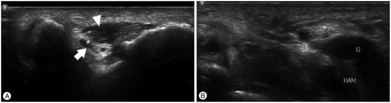
UltrasoundTransverse elbow ultrasound (A): the anconeus epitrochlearis muscle, a supernumerary anatomical variant, in direct contact with a swollen ulnar nerve, a mechanism of cubital tunnel compression, synovial cyst in contact with the ulnar nerve at the wrist (B).
Source : Lee et al., Journal of Korean Neurosurgical Society, 2016, figure 1 · CC BY-NC
📉 Ulnar intraneural pressure by elbow position
Experimental cadaveric data (Werner 1985, Apfelberg 1973)
From 20-30 mmHg upwards, flow in the epineural venules slows; at 60-80 mmHg, capillary flow in the epineurium and perineurium stops. Sustained flexion therefore imposes a chronic functional nerve ischaemia . Source: Werner CO et al. Acta Orthop Scand. 1985; Apfelberg DB et al. Plast Reconstr Surg. 1973.
- For mild to moderate cases (mild Dellon or McGowan I), the course under conservative treatment is broadly favourable. The Swedish randomised trial of Svernlöv et al. (2009, n = 70 patients, 6 months of follow-up) finds objective improvement in 89.5 % of patients whichever arm they were in (night splint, nerve gliding, simple observation): an important result that suggests a strong effect of natural history and of spontaneous activity modification in the less severe cases.¹³
- For severe cases (severe Dellon / McGowan III, with demonstrable wasting), spontaneous recovery is rare. Surgical decompression within a reasonable time is needed to limit permanent deficits, though it does not guarantee complete motor recovery in long-standing cases.¹⁴
- Cubital tunnel syndrome is the 2nd most common compressive neuropathy of the upper limb. US incidence ≈ 30/100,000 person-years (Osei 2017), peak at 40-60 years, a moderate male predominance.
- Key modifiable risk factors: obesity, diabetes, smoking, sustained or repeated elbow flexion. A history of elbow trauma = a major predisposing factor.
- The central mechanism: in flexion the tunnel loses up to 55 % of its volume and intraneural pressure is multiplied by 6-7 (Werner 1985) → chronic ischaemia → demyelination and then axonopathy.
- A bipolar natural course: about 90 % of mild/moderate cases improve on conservative treatment at 6 months; severe cases with wasting rarely do well without surgical decompression.
Bibliography
- Staples JR, Calfee R. Cubital Tunnel Syndrome: Current Concepts. J Am Acad Orthop Surg. 2017;25(10):e215-e224. PMID 28953087.
- Werner CO, Ohlin P, Elmqvist D. Pressures recorded in ulnar neuropathy. Acta Orthop Scand. 1985;56(5):404-406. PMID 3000129.
- Dy CJ, Mackinnon SE. Ulnar neuropathy: evaluation and management. Curr Rev Musculoskelet Med. 2016;9(2):178-184. PMID 27080868.
- Osei DA, Groves AP, Bommarito K, Ray WZ. Cubital Tunnel Syndrome: Incidence and Demographics in a National Administrative Database. Neurosurgery. 2017;80(3):417-420. PMID 28362959.
- An TW, Evanoff BA, Boyer MI, Osei DA. The Prevalence of Cubital Tunnel Syndrome: A Cross-Sectional Study in a U.S. Metropolitan Cohort. J Bone Joint Surg Am. 2017;99(5):408-416. PMID 28244912.
- Palmer BA, Hughes TB. Cubital Tunnel Syndrome. J Hand Surg Am. 2010;35(1):153-163. PMID 20117320.
- Stirling PHC, Harrison SJ, Bowman A, Donald F, McEachan JE. The effect of diabetes mellitus on the outcome of surgery for cubital tunnel syndrome. J Hand Surg Eur Vol. 2023;48(5):419-423. PMID 36524277.
- Descatha A, Leclerc A, Chastang JF, Roquelaure Y. Incidence of ulnar nerve entrapment at the elbow in repetitive work. Scand J Work Environ Health. 2004;30(3):234-240. PMID 15250652.
- Fadel M, Lancigu R, Raimbeau G, Roquelaure Y, Descatha A. Occupational prognosis factors for ulnar nerve entrapment at the elbow: A systematic review. Hand Surg Rehabil. 2017;36(4):244-251. PMID 28528878.
- Andrews K, Rowland A, Pranjal A, Ebraheim N. Cubital tunnel syndrome: Anatomy, clinical presentation, and management. J Orthop. 2018;15(3):832-836. PMID 30140129.
- Mackinnon SE. Pathophysiology of nerve compression. Hand Clin. 2002;18(2):231-241. PMID 12371026.
- Earle AS, Vlastou C. Crossed fingers and other tests of ulnar nerve motor function. J Hand Surg Am. 1980;5(6):560-565. PMID 7430600.
- Svernlöv B, Larsson M, Rehn K, Adolfsson L. Conservative treatment of the cubital tunnel syndrome. J Hand Surg Eur Vol. 2009;34(2):201-207. PMID 19282413.
- Dellon AL. Review of treatment results for ulnar nerve entrapment at the elbow. J Hand Surg Am. 1989;14(4):688-700. PMID 2666496.
How do you assess and diagnose cubital tunnel syndrome with certainty?
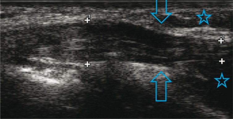
UltrasoundLongitudinal ultrasound of the ulnar nerve at the elbow: a focal notch in the nerve, the notch sign, an ultrasound sign of cubital tunnel compression.
Source : Kowalska, Journal of Ultrasonography, 2014, figure 8 · CC BY-NC-ND
Which questions should you ask to understand the patient and their history? 🤔
A structured history systematically explores:- Location: paraesthesia or numbness strictly in the ulnar distribution: the whole fifth finger + the ulnar half of the fourth + the ulnar border of the dorsum of the hand (the last of which is spared in compression at Guyon's canal).¹
- Behaviour: triggered by sustained elbow flexion (holding the phone, driving, sleeping with the arm bent), by direct pressure on the medial side of the elbow (the desk sign), and by repeated flexion-extension movements.³
- Chronology: intermittent vs constant, whether the patient wakes at night (highly suggestive, since the elbow flexes spontaneously during sleep), and how long it has been going on.¹
- Motor signs: loss of fine dexterity (buttoning, pinching), dropping objects, hand fatigability. These signs point to motor involvement and call for urgent assessment.²
- Past history: elbow fracture or dislocation, cubitus valgus, osteoarthritis, rheumatoid arthritis, diabetes, an exposing occupation, overhead sport.⁴
Which clinical tests should you perform, and which other conditions should you rule out? 🩺
The examination aims to reproduce the symptoms and to document the sensory and motor deficits. The main tests, with the diagnostic values validated in meta-analyses or prospective studies:| Clinical test | Sensitivity | Specificity | Level of evidence | Source |
|---|---|---|---|---|
| Tinel at the elbow (percussion) | 54-70 % | ≈ 98 % NPV | Moderate | Novak 1994; secondary reviews |
| Elbow flexion test (60 s) | 32 % | high | Moderate | Novak 1994 |
| Flexion + pressure combined | 91 % | high | High | Novak 1994 (the historic reference) |
| Scratch Collapse Test (Cheng) | 89 % for the cubital tunnel | 96 % | Moderate-high | Cheng 2008 PMID 18984333 |
| Scratch Collapse Test (SR pooled) | 0.24-0.77 (high heterogeneity) | 0.60-0.99 | Low-moderate | Čebron 2018 SR PMID 33054988 |
| Froment's sign (motor) | variable | high | Moderate | Earle 1980; clinical |
| Static two-point discrimination | late (axonopathy) | high | Moderate | Dellon classification 1989 |
| High-resolution ultrasound | ≈ 80 % | ≈ 90 % | High | Beekman 2004 PMID 15007128; Pelosi 2021 |
| Electrodiagnosis (EDX) | ≈ 60-78 % | high | High | Pelosi 2021; AANEM guidelines |
📊 Sensitivities of the main provocation tests compared
Data from the landmark Novak 1994 study and from Cheng 2008
The combination of tests far outperforms any of them alone. The key is always to pair a dynamic manoeuvre (sustained flexion) with a static one (pressure or Tinel). Source: Novak CB et al. J Hand Surg Am. 1994;19(5):817-820; Cheng CJ et al. J Hand Surg Am. 2008;33(9):1518-1524.
🚩 Differential diagnoses that must be ruled out
- C8-T1 cervical radiculopathy: pain radiating from the neck, symptoms altered by cervical spine movement, a potentially wider deficit (the intrinsic muscles of the hand + flexor carpi ulnaris). A positive Spurling test. → cervical MRI if suspected.
- Thoracic outlet syndrome (true neurogenic TOS): symptoms worsened by overhead arm movement, positive provocation tests (Adson, Roos, Wright), possible hand wasting with axonal involvement. Rare but severe.
- Compression at Guyon's canal (wrist): the palmar ulnar distribution is affected while the dorsum of the hand is spared (the dorsal cutaneous branch arises before the wrist). Tinel localises at the wrist rather than the elbow.
- Early diabetic polyneuropathy: bilateral, in a glove-and-stocking distribution, with poor glycaemic control. Consider checking blood glucose in every UNE patient.
- Lower plexus involvement (Pancoast tumour, traumatic): a deficit spanning several roots, with possible Horner's syndrome.
Should patients be classified, and what are the benefits? 📈
Classifying severity has a direct impact on treatment strategy and prognosis. Two systems coexist and both remain references:| Grade | McGowan 1950 / as modified by Goldberg | Dellon 1989 | Usual approach |
|---|---|---|---|
| I / Mild | Intermittent paraesthesia, no objective motor deficit | Intermittent symptoms, no objective sensory or motor deficit | Conservative for 3-6 months |
| IIA / Moderate | Motor deficit without frank wasting | Persistent symptoms, measurable numbness, early weakness | Cautious conservative treatment, then discuss surgery |
| IIB / Advanced moderate | Early wasting of the intrinsic muscles | – | A surgical opinion without delay |
| III / Severe | Frank wasting, marked weakness, Froment + | Permanent symptoms, a sensory deficit and wasting, a clear Froment's sign | Surgical decompression recommended |
- Diagnosis is above all clinical : paraesthesia in the 4th/5th fingers + reproduction on sustained elbow flexion + night symptoms.
- No single test is perfect. The combination of flexion + pressure (Novak 1994, sens. 91 %) performs best. Tinel and the SCT add information but vary between operators.
- The differential diagnosis is imperative: C8-T1 radiculopathy, neurogenic TOS, Guyon's canal, diabetic polyneuropathy.
- EDX and high-resolution ultrasound confirm and stratify. EDX can be normal in 38 % of clinically obvious cases.
- The classification (McGowan as modified by Goldberg, or Dellon) steers the decision: Grade I = conservative; Grade III with wasting = a surgical opinion without delay.
Bibliography
- Dy CJ, Mackinnon SE. Ulnar neuropathy: evaluation and management. Curr Rev Musculoskelet Med. 2016;9(2):178-184. PMID 27080868.
- Pelosi L, Arányi Z, Beekman R, et al. Expert consensus on the combined investigation of ulnar neuropathy at the elbow using electrodiagnostic tests and nerve ultrasound. Clin Neurophysiol. 2021;132(9):2274-2281. PMID 34229959.
- Staples JR, Calfee R. Cubital Tunnel Syndrome: Current Concepts. J Am Acad Orthop Surg. 2017;25(10):e215-e224. PMID 28953087.
- Palmer BA, Hughes TB. Cubital Tunnel Syndrome. J Hand Surg Am. 2010;35(1):153-163. PMID 20117320.
- Cheng CJ, Mackinnon-Patterson B, Beck JL, Mackinnon SE. Scratch collapse test for evaluation of carpal and cubital tunnel syndrome. J Hand Surg Am. 2008;33(9):1518-1524. PMID 18984333.
- Makanji HS, Becker SJ, Mudgal CS, Jupiter JB, Ring D. Evaluation of the scratch collapse test for the diagnosis of carpal tunnel syndrome. J Hand Surg Eur Vol. 2014;39(2):181-186. PMID 23855039. (Review of accuracy; see also Čebron U and Curtin CM, J Plast Reconstr Aesthet Surg. 2018; SR PMID 33054988.)
- Choi SW, Bae JY, Shin YH, Moon SH, Kim JK. Reliability and validity of the modified McGowan grade in patients with cubital tunnel syndrome. Arch Orthop Trauma Surg. 2022;142(7):1697-1703. PMID 35107635.
- Landau ME, Campbell WW. Clinical features and electrodiagnosis of ulnar neuropathies. Phys Med Rehabil Clin N Am. 2013;24(1):49-66. PMID 23177030.
- Novak CB, Lee GW, Mackinnon SE, Lay L. Provocative testing for cubital tunnel syndrome. J Hand Surg Am. 1994;19(5):817-820. PMID 7806810.
- Beekman R, Schoemaker MC, Van Der Plas JP, et al. Diagnostic value of high-resolution sonography in ulnar neuropathy at the elbow. Neurology. 2004;62(5):767-773. PMID 15007128.
- McGowan AJ. The results of transposition of the ulnar nerve for traumatic ulnar neuritis. J Bone Joint Surg Br. 1950;32-B(3):293-301. doi:10.1302/0301-620X.32B3.293.
- Dellon AL. Review of treatment results for ulnar nerve entrapment at the elbow. J Hand Surg Am. 1989;14(4):688-700. PMID 2666496.
- Aleksenko D, Varacallo M. Guyon Canal Syndrome. In: StatPearls [Internet]. Treasure Island (FL): StatPearls Publishing; 2024. NBK431063.
Which treatment strategies are the most effective for cubital tunnel syndrome?
Where do you start? What is the recommended hierarchy of interventions?
For mild to moderate cases (mild Dellon / McGowan I-IIA), first-line treatment is conservative over 3 to 6 months, in line with the Cochrane SR Caliandro 2016² and the updated recommendations of the McEachan 2024 round table.³ Three pillars structure that protocol. 🎯🗺️ A practical treatment algorithm
Stratification by initial severity and by the response to conservative treatment
The review at 3-6 months (strength, sensation, PROMs) determines the move to surgery. Source: adapted from the McEachan JE et al. round table, J Hand Surg Eur Vol. 2024;49(7):926-932, and Cochrane Caliandro 2016.
- Avoid prolonged, sustained flexion of the elbow (phone, driving, pillow).
- Avoid direct pressure on the medial side of the elbow (the “desk sign”, armrests, leaning).
- Use protection (a sleeve, a pad) where the job exposes the elbow.²
- Sleep hygiene: a pillow to avoid bending the elbow under the head, sometimes a soft, non-occlusive orthosis.
What is the place of exercise and of neurodynamics?
The neurodynamic techniques (gliding, controlled tensioning, sliders and tensioners) aim to restore the excursion of the ulnar nerve within its anatomical interface and to reduce post-inflammatory adhesions.⁷ Their biomechanical rationale is solid (Coppieters & Butler 2008 showed that gliding techniques increase excursion without neural tension, while tensioning techniques increase tension).⁷ However, the level of clinical evidence remains moderate to low. The Cochrane review Caliandro 2016 (updated 2025) concludes that there is no statistically significant difference between splinting, neurodynamics and simple activity modification on functional outcome at 6 months.² That does not mean neurodynamics is useless, but that it does not substitute for education plus the splint, and that it should be used as a multimodal complement, monitored so that no symptomatic worsening occurs (neural irritation from over-dosing).⁸Manual therapies, technologies: how effective are they really?
Several modalities are frequently offered in practice, but their level of evidence for this specific condition is generally low:| Modality | Level of evidence (GRADE) | Recommendation | Comment |
|---|---|---|---|
| Education + activity modification | Moderate to high | Strongly recommended | The pillar of all management, and inexpensive |
| Night splint in near-extension | Low to moderate | Recommended | Bateman 2025 SR; low certainty but a consistent signal |
| Ulnar neurodynamics | Low | Optional as a complement | Cochrane Caliandro 2016: not superior; biomechanically plausible |
| Joint and soft-tissue manual therapy | Very low | Optional | Little UNE-specific data; the effect is probably non-specific |
| Therapeutic ultrasound | Very low | Not recommended on its own | Insufficient data |
| Laser (LLLT) | Very low | Not recommended on its own | Insufficient UNE-specific data |
| Corticosteroids (injection) | Low | Not recommended | No superiority over placebo (NCT trial) |
| Surgical decompression (conservative failure) | Moderate to high | Recommended after failure or in severe cases | Stewart 2024 SR/MA: 4 techniques equivalent in primary cases |
Beyond the physical: patient education and psychosocial factors?
Therapeutic education is the most cost-effective and best evaluated intervention for the compressive neuropathies of the upper limb.⁹ It lets the patient become an active participant in their care, changes their habits durably (sleep, ergonomics, vibration) and reduces acute healthcare use. 💡 The psychosocial dimension must not be neglected. The biopsychosocial models of chronic pain apply here: kinesiophobia, catastrophising, anxiety and depression modulate pain perception and functional disability independently of electrophysiological severity.¹⁰ In a patient with persistent symptoms despite a mild EDX, screening these dimensions with brief tools (PCS, TSK-11) is useful and points towards integrated care (CBT, graded exercise, desensitisation).- First line for mild/moderate cases: multimodal conservative treatment over 3 to 6 months.
- Pillar 1 : postural education + activity modification (moderate to high evidence, the most cost-effective intervention).
- Pillar 2 : a night splint in near-extension (30-45°). Bateman 2025 SR: a consistent signal despite low GRADE certainty.
- Pillar 3 : neurodynamics as a multimodal complement, without promoting it in isolation (Cochrane 2016: no superiority).
- Passive modalities (ultrasound, laser, injections): insufficient evidence to recommend them in isolation.
- Failure at 3-6 months or severity from the outset (wasting, a clear Froment's sign) → a surgical opinion. Simple decompression ≈ transposition for primary cases (Stewart 2024 SR/MA of RCTs).
- Psychosocial factors (kinesiophobia, catastrophising, depression): major modulators, to be screened for and built in.
Bibliography
- Staples JR, Calfee R. Cubital Tunnel Syndrome: Current Concepts. J Am Acad Orthop Surg. 2017;25(10):e215-e224. PMID 28953087.
- Caliandro P, La Torre G, Padua R, Giannini F, Padua L. Treatment for ulnar neuropathy at the elbow. Cochrane Database Syst Rev. 2016;11:CD006839. PMID 27845501.
- McEachan JE, Dahlin LB, Ng CY, Ring D, Ruettermann M. Round table discussion: the management of idiopathic cubital tunnel syndrome. J Hand Surg Eur Vol. 2024;49(7):926-932. PMID 38534139.
- Dy CJ, Mackinnon SE. Ulnar neuropathy: evaluation and management. Curr Rev Musculoskelet Med. 2016;9(2):178-184. PMID 27080868.
- Bateman M, Swaile H, Tambe A. Effectiveness of night splints for cubital tunnel syndrome - A systematic review. Hand Therapy. 2025;30(3):105-112. doi:10.1177/17589983251336157. PMC12081431.
- Svernlöv B, Larsson M, Rehn K, Adolfsson L. Conservative treatment of the cubital tunnel syndrome. J Hand Surg Eur Vol. 2009;34(2):201-207. PMID 19282413.
- Coppieters MW, Butler DS. Do 'sliders' slide and 'tensioners' tension? An analysis of neurodynamic techniques and considerations regarding their application. Man Ther. 2008;13(3):213-221. PMID 17398140.
- Kooner S, Cinats D, Kwong C, Matthewson G, Dhaliwal G. Conservative treatment of cubital tunnel syndrome: A systematic review. Orthop Rev (Pavia). 2019;11(2):7955. PMID 31281598.
- Wieczorek M, Gnat R, Wolny T. The Use of Physiotherapy in the Conservative Treatment of Cubital Tunnel Syndrome: A Critical Review of the Literature. Diagnostics (Basel). 2024;14(11). PMID 38893728.
- Sterling M, de Zoete RMJ, Coppieters I, Farrell SF. Best evidence rehabilitation for chronic pain part 4: neck pain. J Clin Med. 2019;8(8):1219. (A framework transferable to chronic neuropathic pain.) PMID 31443149.
- Abourisha E, Srinivasan AS, Barakat A, Chong HH, Singh HP. Surgical management of cubital tunnel syndrome: A systematic review and meta-analysis of randomised trials. J Orthop. 2024;53:41-48. PMID 38456175.
- Macadam SA, Gandhi R, Bezuhly M, Lefaivre KA. Simple decompression versus anterior subcutaneous and submuscular transposition of the ulnar nerve for cubital tunnel syndrome: a meta-analysis. J Hand Surg Am. 2008;33(8):1314.e1-12. PMID 18929194.
How do you secure lasting recovery and prevent recurrence?
How do you make the patient an active participant in their recovery through self-management?
Empowering the patient is the cornerstone of prevention.¹ The operational components:- Permanent ergonomics: avoid prolonged, sustained flexion and direct pressure on the medial side. Adjust chair height and keyboard angle, use protective sleeves in exposing jobs.²
- Sleep hygiene: wear a light splint or a soft sleeve for 3 to 6 months, until symptoms settle; then wean gradually.³
- A home exercise programme: gentle nerve gliding (sliders), active elbow range exercises, strengthening of the intrinsic muscles of the hand (thumb-index pinch, finger abduction-adduction against light resistance).⁴
- Self-monitoring: respond early to signs of worsening (increasing paraesthesia, a fatigable pinch) by readjusting the loads before symptoms become entrenched.¹
When and how should a safe return to sport and to activity be planned?
🧠 The return to demanding activity (sport, physical work, musical instruments) should be progressive and individualised, based on functional criteria and not on a fixed calendar. The progression criteria validated in clinical practice:- Neurological symptoms resolved at rest and absent during activities of daily living.
- Grip and pinch strength recovered to ≥ 90 % of the other side (dynamometer, pinch meter).⁵
- A functional test reproducing the sporting or occupational movement without triggering symptoms over 24-48 h.
- Sensation restored over the ulnar distribution (Semmes-Weinstein or monofilament).
- Education and permanent modification of the aggravating factors are the basis of preventing recurrence.
- The return to activity is progressive and based on functional criteria (strength ≥ 90 %, no symptoms), never on a fixed calendar.
- A simple home exercise programme (sliders, strengthening the intrinsic muscles) maintains the gains and stabilises the situation.
- The biomechanical analysis of the movement in sport or at work is essential for treating the cause and not only the symptoms.
Bibliography
- McEachan JE, Dahlin LB, Ng CY, Ring D, Ruettermann M. Round table discussion: the management of idiopathic cubital tunnel syndrome. J Hand Surg Eur Vol. 2024;49(7):926-932. PMID 38534139.
- Fadel M, Lancigu R, Raimbeau G, Roquelaure Y, Descatha A. Occupational prognosis factors for ulnar nerve entrapment at the elbow: A systematic review. Hand Surg Rehabil. 2017;36(4):244-251. PMID 28528878.
- Bateman M, Swaile H, Tambe A. Effectiveness of night splints for cubital tunnel syndrome - A systematic review. Hand Therapy. 2025;30(3):105-112. doi:10.1177/17589983251336157.
- Coppieters MW, Butler DS. Do 'sliders' slide and 'tensioners' tension? An analysis of neurodynamic techniques and considerations regarding their application. Man Ther. 2008;13(3):213-221. PMID 17398140.
- Yeoman TFM, Stirling PHC, Lowdon A, Jenkins PJ, McEachan JE. Patient-reported outcomes after in situ cubital tunnel decompression: a report in 77 patients. J Hand Surg Eur Vol. 2020;45(3):232-237. PMID 31663801.
The overhead athlete: why a dedicated chapter?
Chronic valgus mechanism and associated MCL instability
During an overhead throw, in late cocking and acceleration the elbow undergoes valgus stress that can exceed 64 N·m: a load greater than the ultimate strength of the isolated medial collateral ligament in the laboratory.¹ That cyclical stress leads to:- A progressive elongation of the MCL with chronic micro-tears (valgus laxity), and even frank rupture in extreme cases (“UCL injury”).
- A longitudinal traction and dynamic compression of the ulnar nerve as it passes the retro-epicondylar groove, accentuated by the widening of the valgus space at end-extension.
- Sometimes a dynamic anterior dislocation of the ulnar nerve in flexion, audible (“snapping”) and palpable, which is self-perpetuating through friction.²
🚩 Flags specific to the overhead athlete
- Snapping of the ulnar nerve on elbow flexion-extension (dynamic dislocation).
- A loss of throwing velocity > 5-10 % with medial pain = a warning signal (MCL involvement is likely).
- The moving valgus stress test (O'Driscoll) positive (sensitivity 100 %, specificity 75 % for the MCL in the original series).
- Post-exertional paraesthesia persisting at rest for > 24 h or worsening after each training session.
- Early wasting of the first dorsal interosseous or the hypothenar: surgery without delay.
Specific management and pre-surgical stratification
Management in the overhead athlete follows the same conservative → surgical hierarchy, but with specific features:- Temporary rest from sport with a complete stop to throwing for 4 to 6 weeks, maintaining general conditioning and core strength.³
- Specific strengthening of the flexor-pronator muscles (pronator teres, flexor digitorum, flexor carpi ulnaris), which act as dynamic medial stabilisers of the elbow.
- Video analysis of throwing mechanics: looking for faults (the “inverted W”, the arm behind the trunk, contralateral trunk lean) that increase valgus stress.⁴
- Imaging routinely where there is no response at 6 weeks: MR arthrography (looking for a grade ≥ 2 MCL lesion), dynamic ultrasound for snapping.
- The surgical decision coordinated between hand surgeon and sports physician: anterior transposition (subcutaneous or submuscular) rather than simple decompression where there is snapping or established valgus laxity.
- The overhead athlete undergoes extreme cyclical valgus stress (> 64 N·m) that affects the MCL, the flexor-pronator group and the ulnar nerve at once.
- The clinical picture is often mixed (MCL + ulnar neuritis ± dynamic nerve dislocation): always assess both.
- Specific flags: snapping of the nerve in flexion, a loss of throwing velocity, a positive moving valgus stress test.
- Conservative treatment remains first line (rest from sport + flexor-pronator strengthening + video analysis of the movement), but imaging (MRI, dynamic ultrasound) is widely used at 6 weeks where there is no response.
Bibliography
- Werner SL, Fleisig GS, Dillman CJ, Andrews JR. Biomechanics of the elbow during baseball pitching. J Orthop Sports Phys Ther. 1993;17(6):274-278. PMID 8343780.
- Childress HM. Recurrent ulnar nerve dislocation at the elbow. Clin Orthop Relat Res. 1975;108:168-173. PMID 1149958.
- Wilk KE, Macrina LC, Cain EL, Dugas JR, Andrews JR. Rehabilitation of the overhead athlete's elbow. Sports Health. 2012;4(5):404-414. PMID 23016113.
- Davis JT, Limpisvasti O, Fluhme D, et al. The effect of pitching biomechanics on the upper extremity in youth and adolescent baseball pitchers. Am J Sports Med. 2009;37(8):1484-1491. PMID 19633301.
- De Giacomo AF, Keller RA, Banffy M, ElAttrache NS. Ulnar Neuritis and Its Affect on Outcomes of Elbow Ulnar Collateral Ligament Reconstruction. Am J Sports Med. 2022;50(1):224-228. PMID 34797176.
- O'Driscoll SW, Lawton RL, Smith AM. The “moving valgus stress test” for medial collateral ligament tears of the elbow. Am J Sports Med. 2005;33(2):231-239. PMID 15701609.
What do real clinical cases teach us about cubital tunnel syndrome?
Analysis of a typical case: from assessment to conservative resolution
In the night-splint arm of the landmark RCT by Svernlöv et al. (2009, n = 70, mild to moderate cases),¹ patients were assessed before treatment and at 3 and 6 months by: the COPM (Canadian Occupational Performance Measure), pain VAS, grip strength, and ulnar nerve conduction at the elbow. The night splint held the elbow at about 45° of flexion, worn every night for 6 months. Aggregated results (all arms, mild/moderate): 89.5 % of patients improved objectively (VAS, COPM, strength) at 6 months, with no statistically significant difference between arms (splint, gliding, observation). No patient's motor score deteriorated. Nerve conduction improved in 50 % and remained stable in 30 %.¹ 📌 Clinical lessons from this typical case :- For mild to moderate cases, conservative treatment works in the great majority, but the effect of natural history and spontaneous activity modification is probably the major factor, not the splint alone.
- Clinical improvement often precedes electrophysiological improvement, which is why patients should not be followed on nerve conduction studies alone.
- Structured simple observation (with no active therapeutic intervention) remains a legitimate option in the patient who is symptom-free on waking.
The diagnostic challenge: when the cubital tunnel mimics another condition
The cubital tunnel shares sensory territories with other conditions, hence the risk of diagnostic wandering. An updated narrative review stresses that C8-T1 radiculopathy, thoracic outlet syndrome and compression at Guyon's canal are the three major mimics that must be known.² ⚠️ The role of “double crush” deserves emphasis: a patient can have proximal compression (cervical spine, brachial plexus) and distal compression (cubital tunnel) at the same time, each contributing to the symptoms without either being sufficient on its own. That hypothesis, proposed by Upton & McComas in 1973³, remains debated but probably explains why symptoms persist after isolated local surgery in some patients. 📌 Good clinical practice :- Always assess the cervical spine (Spurling, segmental mobility) in a UNE patient.
- Systematically test sensation over the ulnar border of the dorsum of the hand : preserved = compression at Guyon's canal; affected = more proximal compression (including the cubital tunnel).
- Where symptoms and EDX dissociate, widen the work-up: cervical MRI, high-resolution ultrasound along the whole course, blood glucose and HbA1c.
A complex case (a rare anatomical cause or a recurrence)
Several subtypes fall outside the usual pattern: Case 1: Anconeus epitrochlearis muscle (an anatomical variant) : this inconstant accessory muscle occupies the roof of the cubital tunnel and can compress the ulnar nerve. The series of Kim et al. (Hand 2019, n = 13) confirms that this accessory muscle causes a dynamic cubital tunnel, creating problems of symptom localisation in flexion. Myotomy or resection of the muscle alongside decompression resolves most cases.⁴ Diagnosis: dynamic ultrasound (Sookur 2008) or MRI. Case 2: Rheumatoid arthritis with synovitis : RA can compress the ulnar nerve at the elbow through hypertrophic elbow synovitis (and more rarely through eventual mechanical rupture of the nerve, an exceptional case reported by Iyengar 2007).⁵ Treatment combines medical optimisation of the RA (DMARDs, biologic therapy) and, where necessary, surgical decompression with synovectomy. The Stirling 2023 meta-analysis confirms that systemic comorbidities (RA, diabetes) impair postoperative results.⁶ Case 3: Recurrence after in situ decompression : 5-15 % of patients recur after simple decompression. The reviews of revision surgery (Aleem 2014, Krogue 2015) show that a revision subcutaneous or submuscular anterior transposition gives good results in about two thirds of patients, provided the cause of the primary failure is clearly identified (fibrosis, dislocation, continuing activity-related stress).⁷- The typical case responds to conservative treatment in 3 to 6 months (Svernlöv 2009: 89.5 % improve).
- UNE is a great mimic: always include the cervical spine, the plexus and Guyon's canal in the work-up.
- The double crush hypothesis (Upton & McComas 1973) remains useful for explaining failures of isolated local surgery.
- Rare anatomical causes: the anconeus epitrochlearis muscle, a ganglion cyst, rheumatoid arthritis.
- Recurrence after surgery: 5-15 %, managed by revision anterior transposition once the primary cause has been identified.
- ⚠️ A case report = level 5 on the CEBM scale. It illustrates; it never demonstrates efficacy; the recommendations still come from the SRs/MAs.
Bibliography
- Svernlöv B, Larsson M, Rehn K, Adolfsson L. Conservative treatment of the cubital tunnel syndrome. J Hand Surg Eur Vol. 2009;34(2):201-207. PMID 19282413.
- Andrews K, Rowland A, Pranjal A, Ebraheim N. Cubital tunnel syndrome: Anatomy, clinical presentation, and management. J Orthop. 2018;15(3):832-836. PMID 30140129.
- Upton AR, McComas AJ. The double crush in nerve entrapment syndromes. Lancet. 1973;2(7825):359-362. PMID 4124532.
- Kim N, Stehr R, Matloub HS, Sanger JR. Anconeus Epitrochlearis Muscle Associated With Cubital Tunnel Syndrome: A Case Series. Hand (N Y). 2019;14(6):788-792. PMID 29582694.
- Morgenstein A, Lourie G, Miller B. Anconeus epitrochlearis muscle causing dynamic cubital tunnel syndrome: a case series. J Hand Surg Eur Vol. 2016;41(2):227-229. PMID 25409116.
- Stirling PHC, Harrison SJ, Bowman A, Donald F, McEachan JE. The effect of diabetes mellitus on the outcome of surgery for cubital tunnel syndrome. J Hand Surg Eur Vol. 2023;48(5):419-423. PMID 36524277.
- Aleem AW, Krogue JD, Calfee RP. Outcomes of revision surgery for cubital tunnel syndrome. J Hand Surg Am. 2014;39(11):2141-2149. PMID 25169417.
- Krogue JD, Aleem AW, Osei DA, Goldfarb CA, Calfee RP. Predictors of surgical revision after in situ decompression of the ulnar nerve. J Shoulder Elbow Surg. 2015;24(4):634-639. PMID 25660241.
- Cheng CJ, Mackinnon-Patterson B, Beck JL, Mackinnon SE. Scratch collapse test for evaluation of carpal and cubital tunnel syndrome. J Hand Surg Am. 2008;33(9):1518-1524. PMID 18984333.
- Abourisha E, Srinivasan AS, Barakat A, Chong HH, Singh HP. Surgical management of cubital tunnel syndrome: A systematic review and meta-analysis of randomised trials. J Orthop. 2024;53:41-48. PMID 38456175.
How do you apply these recommendations concretely in your practice?
When and to which professionals should you refer?
The fundamental skill of the front-line physiotherapist is to recognise the limits of their scope of practice and to identify the situations that demand collaboration or medical referral. The IFOMPT-JOSPT 2020 framework (Finucane) remains a transferable reference for handling red flags in musculoskeletal practice.¹🚩 Red flags and signals for prompt referral
- Recent elbow trauma (a fall, an accident) with deformity, swelling or loss of function → emergency assessment and radiography.
- A progressive motor deficit (a clear Froment's sign, visible wasting) → a surgical opinion without delay (Dellon III / McGowan III).
- Unexplained weight loss, night sweats, a history of cancer → an oncological work-up (Pancoast, plexus metastasis).
- Associated systemic abnormalities (skin lesions, fever, a widespread deficit) → a broader medical work-up.
- Bilateral or atypically distal-proximal symptoms → consider a polyneuropathy (diabetes, alcohol, vitamin B12, paraprotein).
- Swallowing difficulty, dysarthria or associated ophthalmoplegia → suspect central or extensive plexus involvement.
- Failure of well-conducted conservative treatment at 3-6 months → hand surgeon.²
- Dominant psychosocial factors (severe kinesiophobia, catastrophising, depression, fear of movement) → general practitioner, pain psychologist.
- An overhead athlete or a patient with heavy occupational demands → sports physician, ergonomist, hand surgeon.
- A systemic comorbidity (RA, poorly controlled diabetes, thyroid dysfunction) → general practitioner, for medical optimisation.
How do you measure outcomes and overcome barriers to implementation?
Objective, standardised outcome measurement is indispensable for assessing efficacy and adapting management. 👍 Recommended measurement tools for UNE :- DASH (Disabilities of the Arm, Shoulder and Hand): a general functional questionnaire, internationally validated, with a French version available. Score 0 (normal) to 100 (maximal disability). MCID ≈ 10 points.³
- PRWE / PRUNE (Patient-Rated Wrist Evaluation / Patient-Rated Ulnar Nerve Evaluation): the PRUNE version was developed specifically for ulnar neuropathies (MacDermid 2015).
- BCTQ (Boston Carpal Tunnel Questionnaire): originally for the carpal tunnel but used in practice for upper limb neuropathies; good sensitivity to change.
- Grip and pinch strength (Jamar dynamometer, pinch meter): an objective bilateral comparative measure.
- Pain VAS (visual analogue scale 0-10) and symptom frequency (per day/week).
- Lack of time in consultation.
- Difficulty accessing the scientific databases (PubMed, Cochrane).
- Weak continuing education in critical appraisal and in weighing evidence.
- Entrenched professional habits (“we have always done it this way”).
- Clinical leadership and an institutional culture favourable to EBP.
- Communities of practice and regular journal clubs.
- Building PROMs into the electronic patient record.
- Using checklists and standardised protocols drawn from the SRs/MAs.
- The IFOMPT/Finucane 2020 framework remains the reference for screening red flags in musculoskeletal practice.
- Refer promptly for: recent trauma, a progressive motor deficit, visible wasting, systemic abnormalities, conservative failure at 3-6 months.
- Measure with validated PROMs : the DASH (general), the PRWE/PRUNE (ulnar-specific), grip and pinch strength.
- The psychosocial factors (catastrophising, kinesiophobia) strongly modulate perception and disability: screen for them and address them in the care plan.
- Implementing EBP: barriers (time, access, training) versus facilitators (leadership, communities of practice, integrated tools): the effort must be as much systemic as individual.
Bibliography
- Finucane LM, Downie A, Mercer C, et al. International Framework for Red Flags for Potential Serious Spinal Pathologies. J Orthop Sports Phys Ther. 2020;50(7):350-372. PMID 32438853. doi:10.2519/jospt.2020.9971.
- McEachan JE, Dahlin LB, Ng CY, Ring D, Ruettermann M. Round table discussion: the management of idiopathic cubital tunnel syndrome. J Hand Surg Eur Vol. 2024;49(7):926-932. PMID 38534139.
- Beaton DE, Wright JG, Katz JN; Upper Extremity Collaborative Group. Development of the QuickDASH: comparison of three item-reduction approaches. J Bone Joint Surg Am. 2005;87(5):1038-1046. PMID 15866967.
- MacDermid JC, Grewal R, MacIntyre NJ. Reliability of the Patient-Rated Wrist Evaluation. J Hand Ther. 2007;20(3):220-225. PMID 17631005.
- Al Zoubi FM, Menon A, Mayo NE, Bussières AE. The effectiveness of interventions designed to increase the uptake of clinical practice guidelines and best practices among musculoskeletal professionals: a systematic review. BMC Health Serv Res. 2018;18(1):435. PMID 29884165.
- Yeoman TFM, Stirling PHC, Lowdon A, Jenkins PJ, McEachan JE. Patient-reported outcomes after in situ cubital tunnel decompression: a report in 77 patients. J Hand Surg Eur Vol. 2020;45(3):232-237. PMID 31663801.
- Wieczorek M, Gnat R, Wolny T. The Use of Physiotherapy in the Conservative Treatment of Cubital Tunnel Syndrome: A Critical Review of the Literature. Diagnostics (Basel). 2024;14(11). PMID 38893728.
And after this article?
This article is part of a collection of evidence-based clinical syntheses. A question, a comment, a correction to suggest? Contact us directly through the WhatsApp button at the bottom right of the screen.

