

















In brief
Medial epicondylitis (« golfer's elbow »), more accurately called tendinopathy of the flexor-pronator origin, is a degenerative, and not inflammatory, tendinosis of the common insertion of the flexors and pronators on the medial epicondyle. It shows itself as pain on palpation of the medial epicondyle, reproduced by resisted flexion or pronation, with a peak incidence between 45 and 54 years. Management is tiered: education and load management at the base, then progressive therapeutic exercise, the cornerstone of treatment; corticosteroids are harmful in the medium to long term. Its prevalence is 0.4 % in the general population and 4-5 % in exposed workers.
A clinical synthesis based on the specific systematic reviews and the recent 2023-2026 reviews - Tahir, Konarski, See, Hoogvliet, Cook & Purdam.
Clinical synthesis
- Medial epicondylitis is a degenerative tendinopathy (and not an inflammatory one) of the common flexor-pronator origin on the medial epicondyle. Prevalence 0.4 % in the general population, 4-5 % in exposed workers. Peak at 45-54 years.
- Biomechanical risk factors : forceful work (OR 1.95), awkward postures, vibration. Repetitiveness alone is not an independent factor (Descatha 2003).
- Major systemic factors : type 2 diabetes (OR 11.27 - wide CI), smoking, obesity, age. These comorbidities are therapeutic targets in their own right (De Luca 2025).
- The diagnosis is clinical : painful palpation of the medial epicondyle + positive resisted flexion or pronation. Imaging is reserved for failures > 3-6 months or for suspicion of an associated condition.
- The critical differential diagnosis is ulnar neuropathy at the cubital tunnel (frequent coexistence, 25-50 %). Always look for a positive Tinel and paraesthesiae of the 4th-5th fingers.
- In the overhead thrower, the « medial triad » (tendon + MCL + ulnar nerve) calls for a systematic assessment of all 3 structures. In the manual worker, ergonomics and comorbidity matter as much as pure biomechanics.
- The tendon continuum model (Cook 2009): reactive → dysrepair → degenerative. Distinct therapeutic strategies at each stage.
- The treatment rests on 5 tiers : education + load management at the base, progressive therapeutic exercise at tier 2, adjunct modalities next, then injections and surgery as the last line.
- Progressive exercise is the cornerstone (See 2026, a specific SR; Hoogvliet 2013 BJSM). No robust superiority has been demonstrated between pure eccentric, concentric-eccentric or heavy slow resistance work - the progression of load counts more than the type.
- The corticosteroids are harmful in the medium to long term (recurrences × 2-3 vs exercise). The PRP has conflicting evidence and is not recommended as a first line.
- Self-management of the patient is decisive: a pain-monitoring model (green zone ≤ 4/10), a progressive home programme, a logbook.
- The return to activity is guided by 4 cumulative functional criteria: pain (NPRS), strength (≥ 90 % of the sound side on a dynamometer), function (PRTEE / QuickDASH), load capacity (≥ 80 % of the usual volume without exacerbation).
- The failure of well-conducted treatment must lead to reconsidering the diagnosis (ulnar neuropathy? MCL? another differential) rather than intensifying the same intervention.
- The psychosocial factors (kinesiophobia, catastrophising) are major prognostic determinants - pain education + graded exposure.
- The level of evidence specific to medial epicondylitis is limited (often extrapolated from the lateral side). Be transparent with the patient about the uncertainties.
- Drug history : look for recent exposure to fluoroquinolones (ciprofloxacin, levofloxacin, ofloxacin), and to corticosteroid therapy given systemically or locally. Both raise the risk of tendinopathy and rupture, especially after the age of 60, in weight-bearing tendons and when the two are combined.
Contents
- What are the fundamentals to know about medial epicondylitis (golfer's elbow)?
- Which populations are at high risk of medial epicondylitis?
- How can medial epicondylitis be assessed and diagnosed with certainty?
- What are the most effective treatment strategies for medial epicondylitis?
- How do you ensure durable recovery and prevent recurrence of medial epicondylitis?
- What do concrete clinical cases teach us about medial epicondylitis?
- How do you apply these recommendations concretely in your practice?
What are the fundamentals to know about medial epicondylitis (« golfer's elbow »)?
How is this condition defined, who does it affect and what are the risk factors?
The condition affects primarily the tendon of the pronator teres (musculus pronator teres) and of the flexor carpi radialis (musculus flexor carpi radialis), alone or in combination.⁵ The palmaris longus, flexor carpi ulnaris and flexor digitorum superficialis can also be involved.¹ The prevalence in the general population is low. In the Finnish Health 2000 study (n = 4,783 participants, standardised clinical examination), Shiri et al. find a prevalence of 0.4 % for medial epicondylitis (vs 1.3 % for lateral epicondylitis).⁶ The methodology is of good quality: reproducible physical examination, representative population. In exposed occupational settings, the prevalence rises to 4 - 5 %, with an annual incidence of 1.5 % (Descatha 2003, n = 1,757 French workers followed for 3 years).⁷ US hospital data (Wiggins 2018, national database, 19,856 patients 2007-2014) show a continuous rise in incidence of 3.4 % a year, with a peak in women aged 45-54.⁸(Shiri 2014)
(Descatha 2003)
(Descatha 2003)
(Shiri 2014, Wiggins 2018)
Prevalence of medial epicondylitis by context of exposure
Percentage of the population meeting the clinical criteria (pain on palpation + a positive provocation test)
Sources: Shiri R et al. Am J Epidemiol. 2006;164(11):1065-1074 (PMID 16968862); Descatha A et al. J Occup Environ Med. 2003;45(9):993-1001 (PMID 14506342); Wolf JM, Mountcastle S, Burks R, Sturdivant RX, Owens BD. Mil Med. 2010;175(5):336-339 (PMID 20486505).
- Current smoking : associated with an increased risk of upper limb tendinopathies, through impairment of tendon microvascularisation.¹⁰
- Obesity / high BMI : Gaida et al. (Arthritis Rheum 2009) demonstrated a robust association between adiposity and tendinopathies.¹¹ Mechanisms: low-grade systemic inflammation (TNF-α, IL-6), increased mechanical demand.
- Age : peak incidence at 45-54 years, attributed to tendon ageing (falling water content, progressive collagen disorganisation).⁶,⁸
- Sex : the data are mixed. The US Wiggins 2018 cohort shows a slight female predominance (53 %), whereas the occupational studies show a balance or a slight male predominance.⁸
What happens in the body and how does medial epicondylitis evolve naturally?
Contrary to what the suffix « -itis » suggests, medial epicondylitis is not an inflammatory condition. The reference histological studies (Kraushaar & Nirschl 1999, JBJS) demonstrated on surgical biopsies that the condition corresponds to a degenerative tendinosis, characterised by 🔬:- The disorganisation of the collagen fibres, losing their parallel alignment.¹²
- The fibroblastic hypercellularity with disordered proliferation of the tenocytes.¹²
- The anarchic neovascularisation with growth of poor-quality microvessels and associated hyperinnervation, a mechanism now considered to contribute to the perception of pain.¹³
- The near-total absence of inflammatory cells (lymphocytes, neutrophils), hence the proposed term « tendinosis » to replace « tendinitis ».¹²
The tendon continuum model (Cook & Purdam 2009)
Three successive states with distinct therapeutic implications
The model stresses that the condition is not binary (healthy vs diseased) but progressive, with distinct therapeutic windows at each stage.
- Medial epicondylitis is a degenerative tendinosis of the flexor-pronator origin, not an inflammatory condition (Kraushaar 1999).
- Consolidated prevalence: 0.4 % in the general population (Shiri 2014); 4-5 % in exposed occupational settings with an annual incidence of 1.5 % (Descatha 2003). Peak at 45-54 years.
- Key risk factors: forceful work (OR 1.95), type 2 diabetes (OR 11.27, CI 2.01-63.02, a wide CI), smoking, obesity, age, and overhead sporting activity.
- The continuum model (Cook 2009): reactive → dysrepair → degenerative: management differs at each stage.
- The natural course is generally favourable (80-90 % improvement at 12 months), but 10-20 % become chronic, hence the importance of stratification and of an initial conservative approach.
Bibliography, chapter 1
- Tahir A, Chanian N, Tiwana S, Sahu MA, Blackwell J. Medial Epicondylitis: A Review of Clinical Présentation, Diagnosis, and Management in the United Kingdom. Cureus. 2026;18(1):e102264. PMC12931734.
- Konarski W, Pobozy T, Pobozy K, Domanska J, Konarska K. Current concepts of natural course and in management of medial epicondylitis: a clinical overview. Orthop Rev (Pavia). 2023;15:84275. PMID 37701778.
- Reece CL, Susmarski A. Medial Epicondylitis. In: StatPearls [Internet]. Treasure Island (FL): StatPearls Publishing; 2024 May 2. NBK557869.
- Amin NH, Kumar NS, Schickendantz MS. Medial Epicondylitis: Évaluation and Management. J Am Acad Orthop Surg. 2015;23(6):348-355. PMID 26001427.
- Pitzer ME, Seidenberg PH, Bader DA. Elbow tendinopathy. Med Clin North Am. 2014;98(4):833-849. PMID 24994055.
- Shiri R, Viikari-Juntura E, Varonen H, Heliovaara M. Prévalence and determinants of lateral and medial epicondylitis: a population study. Am J Epidemiol. 2006;164(11):1065-1074. PMID 16968862.
- Descatha A, Leclerc A, Chastang JF, Roquelaure Y. Medial epicondylitis in occupational settings: prévalence, incidence and associated risk factors. J Occup Environ Med. 2003;45(9):993-1001. PMID 14506342.
- Wiggins AJ, Cancienne JM, Camp CL, et al. Disease Burden of Medial Epicondylitis in the USA Is Increasing: An Analysis of 19,856 Patients From 2007 to 2014. HSS J. 2018;14(3):233-237. PMID 30258326.
- Shiri R, Viikari-Juntura E. Lateral and medial epicondylitis: rôle of occupational factors. Best Pract Res Clin Rheumatol. 2011;25(1):43-57. PMID 21663849.
- De Luca P, Grieco G, Bargeri S, et al. The interplay between metabolic disorders and tendinopathies: Systematic review and meta-analysis. J Exp Orthop. 2025;12(3):e70429. PMID 40937086.
- Gaida JE, Ashe MC, Bass SL, Cook JL. Is adiposity an under-recognized risk factor for tendinopathy? A systematic review. Arthritis Rheum. 2009;61(6):840-849. PMID 19479698.
- Kraushaar BS, Nirschl RP. Tendinosis of the elbow (tennis elbow). Clinical features and findings of histological, immunohistochemical, and electron microscopy studies. J Bone Joint Surg Am. 1999;81(2):259-278. PMID 10073590.
- Alfredson H, Ohberg L, Forsgren S. Is vasculo-neural ingrowth the cause of pain in chronic Achilles tendinosis? An investigation using ultrasonography and colour Doppler, immunohistochemistry, and diagnostic injections. Knee Surg Sports Traumatol Arthrosc. 2003;11(5):334-338. PMID 14520512.
- Cook JL, Purdam CR. Is tendon pathology a continuum? A pathology model to explain the clinical presentation of load-induced tendinopathy. Br J Sports Med. 2009;43(6):409-416. PMID 18812414.
- Smidt N, van der Windt DA, Assendelft WJ, Deville WL, Korthals-de Bos IB, Bouter LM. Corticosteroid injections, physiotherapy, or a wait-and-see policy for lateral epicondylitis: a randomised controlled trial. Lancet. 2002;359(9307):657-662. PMID 11879861.
- Bisset L, Beller E, Jull G, Brooks P, Darnell R, Vicenzino B. Mobilisation with movement and exercise, corticosteroid injection, or wait and see for tennis elbow: randomised trial. BMJ. 2006;333(7575):939. PMID 17012266.
Which populations are at high risk of medial epicondylitis?
Throwing (overhead) athletes: the « medial triad » of the elbow
In throwing athletes (baseball, javelin, water polo, tennis with a topspin serve, extreme climbing), medial epicondylitis is rarely an isolated disorder. The common mechanism is the repeated valgus stress during the cocking and acceleration phases of the throw, which simultaneously overloads the three main medial stabilisers of the elbow.¹The thrower's « medial triad », 3 structures, 1 overload mechanism
Chronic valgus stress simultaneously overloads the flexor-pronator tendon, the MCL and the ulnar nerve
Cain EL, Dugas JR, Wolf RS, Andrews JR. Elbow injuries in throwing athletes: a current concepts review. Am J Sports Med. 2003;31(4):621-635 (PMID 12860556).
- Any medial epicondylitis in a thrower calls for systematic screening for MCL instability (valgus stress test at 25° of flexion, moving valgus stress test) and for ulnar neuropathy (Tinel, sustained elbow flexion test, sensation in the 4th-5th fingers).
- Imaging is more often justified : dynamic ultrasound to assess MCL stability under valgus stress, or even MRI if a partial tear of the anterior bundle of the MCL is suspected (the gold standard in the professional thrower).²
- Treating the medial epicondylitis alone is often insufficient : if MCL instability is present, it must be treated first (unloading, surgical reconstruction if that fails). Otherwise, recurrence is almost certain on return to throwing.¹,²
Red flags in the thrower
- 🚩 A « pop » or sharp snapping sensation during a specific throw → suspicion of an acute MCL tear.
- 🚩 Paraesthesiae of the 4th-5th fingers coming on gradually → ulnar neuropathy at the cubital tunnel, frequently associated.
- 🚩 Rapid loss of throwing velocity (> 5 mph) or loss of control → suspicion of an MCL or flexor-pronator mass lesion.
- 🚩 Pain waking the patient at night or at rest → suspicion of a stress fracture of the olecranon (young thrower).
- 🚩 Adolescent thrower with medial epicondylar swelling → suspicion of apophyseal avulsion: routine radiographs.
Manual workers and systemic factors: when biomechanics meets comorbidity
The other over-represented population is that of manual workers : production operatives, electricians, plumbers, mechanics, carpenters, butchers, hairdressers, dentists. The Descatha 2003 meta-analysis identified three occupational exposures independently associated with medial epicondylitis ⚒️:Occupational risk factors for medial epicondylitis
Consolidated odds ratios (Descatha 2003, n = 1,757 workers)
Source: Descatha A, Leclerc A, Chastang JF, Roquelaure Y. J Occup Environ Med. 2003;45(9):993-1001 (PMID 14506342). Confirmed by Shiri R, Viikari-Juntura E. Best Pract Res Clin Rheumatol. 2011;25(1):43-57 (PMID 21663849).
| Systemic factor | Effect on medial epicondylitis | Proposed mechanism | Level of evidence |
|---|---|---|---|
| Type 2 diabetes | OR 11.27 (95 % CI 2.01-63.02) | Collagen glycation, microvasculopathy, diabetic neuropathy | Moderate |
| Obesity (BMI > 30) | Robust association (Gaida 2009) | Low-grade systemic inflammation (TNF-α, IL-6) | Moderate |
| Current smoking | RR ~1.5-2 across studies | Vasoconstriction, tendon hypoxia, altered healing | Low |
| Hypercholesterolaemia | Xanthomatous deposits, weakening | Lipid infiltration of the tendon | Low |
| Occupational vibration | OR 1.5-2.5 depending on intensity | Repeated microtrauma, hypoperfusion | Moderate |
Practical implications: stratify and individualise
Identifying the risk profile early shapes management:- Young or professional thrower : systematic MCL + ulnar nerve assessment, imaging justified, pitch count modulation, throwing technique (video analysis), work on the muscle chains upstream (shoulder, trunk).
- Manual worker : ergonomic analysis of the workstation (tools, postures, loads), temporary modification of the tasks at risk, screening for and referral of comorbidity (general practitioner, occupational physician).
- Patient with metabolic comorbidity : multidisciplinary referral (dietitian, smoking cessation, diabetes control), managing expectations (slower recovery, less favourable prognosis).
- 🎯 In the overhead thrower, isolated medial epicondylitis is rare. The triad « tendon + MCL + ulnar nerve » calls for a systematic assessment of all 3 structures (valgus stress test, Tinel, looking for paraesthesiae).
- ⚒️ In the manual worker, the triplet « force > 5 kg + awkward posture + vibration » multiplies the risk (cumulative OR > 3). Repetitiveness alone is not an independent factor.
- 🩺 The metabolic comorbidities (diabetes OR 11.27, obesity, smoking) are risk factors and factors of therapeutic failure that are often underestimated: the CI is wide but the direction is constant.
- 👶 In the young athlete, suspect first an apophysitis or a medial epicondylar avulsion (« little leaguer's elbow »): routine radiographs.
- 🔄 The return to activity must be stratified by profile: objective functional criteria, never calendar ones.
Bibliography, chapter 2 (high-risk populations)
- Cain EL Jr, Dugas JR, Wolf RS, Andrews JR. Elbow injuries in throwing athletes: a current concepts review. Am J Sports Med. 2003;31(4):621-635. PMID 12860556.
- Vinod AV, Ross G. An effective approach to diagnosis and surgical repair of refractory medial epicondylitis. J Shoulder Elbow Surg. 2015;24(8):1172-1177. PMID 26189803.
- Fleisig GS, Andrews JR. Prévention of elbow injuries in youth baseball pitchers. Sports Health. 2012;4(5):419-424. PMID 23016115.
- Hyman J, Breazeale NM, Altchek DW. Valgus instability of the elbow in athletes. Clin Sports Med. 2001;20(1):25-45, viii. PMID 11227707.
- Descatha A, Leclerc A, Chastang JF, Roquelaure Y. Medial epicondylitis in occupational settings: prévalence, incidence and associated risk factors. J Occup Environ Med. 2003;45(9):993-1001. PMID 14506342.
- Shiri R, Viikari-Juntura E. Lateral and medial epicondylitis: rôle of occupational factors. Best Pract Res Clin Rheumatol. 2011;25(1):43-57. PMID 21663849.
- De Luca P, Grieco G, Bargeri S, et al. The interplay between metabolic disorders and tendinopathies: Systematic review and meta-analysis. J Exp Orthop. 2025;12(3):e70429. PMID 40937086.
- Gaida JE, Ashe MC, Bass SL, Cook JL. Is adiposity an under-recognized risk factor for tendinopathy? A systematic review. Arthritis Rheum. 2009;61(6):840-849. PMID 19479698.
- Ranger TA, Wong AM, Cook JL, Gaida JE. Is there an association between tendinopathy and diabetes mellitus? A systematic review with meta-analysis. Br J Sports Med. 2016;50(16):982-989. PMID 26598716.
How can medial epicondylitis be assessed and diagnosed with certainty?
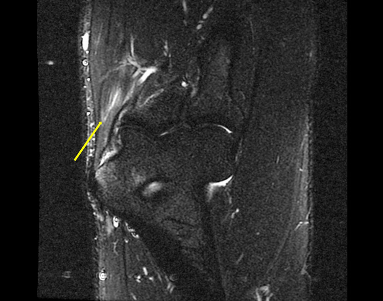
MRICoronal T2-weighted STIR MRI of the elbow: hyperintense signal within the common flexor tendon fibres, a marker line along the affected tendon, medial epicondylitis.
Source : Tahir et al., Cureus, 2026, figure 3 · CC BY
Which questions should you ask to understand the patient and their history?
The history aims to characterise the pain, identify the triggering mechanisms and explore the biopsychosocial context. The following structured questions guide the initial interview 🗣️:- Precise site : exquisite pain over the medial epicondyle or diffuse pain over the medial side of the elbow? Radiation into the flexor forearm? No distal radiation beyond the wrist (otherwise, suspect C8-T1 root involvement).
- Mode of onset : insidious and gradual over several weeks (typical) or acute after trauma (suspect a tear of the common flexor tendon or a medial epicondylar avulsion)?
- Triggering and aggravating factors : wrist flexion against resistance (carrying a load, clenching a fist, screwing), forced pronation (using a screwdriver), prolonged gripping (carrying bags, hand tools), overhead activities (throwing, the tennis smash).¹
- Occupational and sporting activities : the workstation (loads, vibration, postures), hours a day of at-risk activity, time in the post. For athletes: weekly volume, type of movement (throwing, racket, climbing), level of practice. Recent changes (new equipment, intensification, modified technique)?¹,²
- Neurological symptoms : paraesthesiae, numbness or weakness in the 4th-5th fingers (ulnar territory) or the first 3 fingers (median territory)? Waking at night linked to elbow flexion (suspect ulnar neuropathy at the cubital tunnel)?³
- Comorbidity : diabetes (potentiates the risk and the chronicity), rheumatoid arthritis, hypothyroidism, smoking cessation, current medication (statins, fluoroquinolones, a known tendinopathy risk).⁴
- Psychosocial factors : fear of movement (kinesiophobia), catastrophising (« my elbow is fragile, I will never be able to go back »), prolonged sick leave, occupational or insurance conflict, major negative prognostic factors.⁵
Which clinical tests should you run and which other conditions must be ruled out?
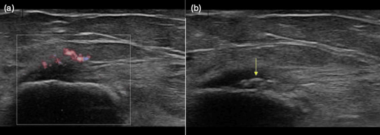
UltrasoundUltrasound of the elbow, two views: reactive hyperaemia on colour Doppler (a) and intratendinous calcification marked by an arrow (b) in a patient with medial epicondylitis.
Source : Tahir et al., Cureus, 2026, figure 2 · CC BY
Inspection and palpation
Inspection is generally unrewarding. Slight swelling over the medial epicondyle is rare, more typical of an avulsion or a bursitis. Palpation is the key manoeuvre. ✅- The medial epicondyle point : exquisite, reproducible pain on point palpation of the medial epicondyle, just anterior and distal to the bony apex. It is the most constant sign.¹,²
- The common flexor origin : the pain can be slightly more distal, at the common tendon, up to 1-2 cm from the epicondyle.
- Side-to-side comparison : essential to calibrate the patient's normal sensitivity (reproducible palpation pressure).
Validated provocation tests
The provocation tests seek to reproduce the pain by putting the flexor-pronator group under active or passive tension. The scientific literature on the diagnostic validity of these tests for medial epicondylitis is less robust than for the lateral side (few specific studies published): interpretative caution is called for.⁶- Resisted wrist flexion test : with the elbow extended and the forearm supinated, the clinician resists wrist flexion. Reproduction of the pain at the medial epicondyle makes it positive. It is the test most used in clinical practice.²
- Resisted pronation test : with the elbow flexed to 90°, the clinician resists active forearm pronation. Often positive if the pronator teres is involved.²
- Passive stretch test : passive extension of the wrist and fingers with the elbow extended. It reproduces the pain if the flexor tendon is loaded. Moderate sensitivity.²
- Functional provocation : reproduce the patient's triggering movement (gripping a heavy object, simulating the golf swing, a grip position specific to the job), a pragmatic tool that is often revealing.
Differential diagnosis: the trap not to be missed
⚠️ Medial elbow pain is NOT always a medial epicondylitis. A rigorous differential diagnosis is essential, because treatment diverges sharply according to the condition.Decision tree: medial elbow pain
5 conditions to assess systematically in every patient with medial elbow pain
Sources: Amin NH, Kumar NS, Schickendantz MS. J Am Acad Orthop Surg. 2015;23(6):348-355 (PMID 26001427); Tahir A et al. Cureus. 2026;18(1):e102264 (PMC12931734).
- Ulnar neuropathy at the cubital tunnel: The most important. Often associated with medial epicondylitis (up to 25-50 % coexistence across series), it can also be the sole cause of the symptoms. Tests: positive Tinel at the cubital tunnel, sustained elbow flexion test (3 minutes), assessment of sensation and of the strength of the intrinsic muscles. EMG if in doubt.³ ⚠️
- Instability of the medial collateral ligament (MCL): In the thrower above all. Valgus stress test at 25° of flexion, moving valgus stress test (maximum pain between 80° and 120° of flexion under sustained valgus). MRI or dynamic ultrasound for confirmation.⁷
- Pronator teres syndrome: Compression of the median nerve by the pronator teres. Pain on the anterior aspect of the proximal forearm, paraesthesiae of the first 3 fingers (including the radial half of the ring finger), resisted pronation reproduces the pain. Rarer but a diagnostic trap.⁸
- C8-T1 cervical radiculopathy: Pain referred from the medial aspect of the arm towards the 5th finger. Positive Spurling test, cervical distraction relieves the symptoms. Routine cervical assessment if in doubt.⁹
- Elbow osteoarthritis / intra-articular loose bodies: Older patient, diffuse stiffness, crepitus on flexion-extension, loss of range (in extension in particular). Standard radiographs are generally sufficient.¹
The place of imaging: rarely first-line
Imaging is not needed to confirm a typical medial epicondylitis. It is reserved for 4 precise indications 🔍:- Failure of well-conducted conservative treatment over 3-6 months (consider a partial tear of the tendon).
- Clinical suspicion of an associated condition (MCL, severe ulnar neuropathy, occult fracture in the young athlete).
- An atypical history (acute trauma, intense night pain, a red flag).
- A surgical decision under consideration.
Should patients with medial epicondylitis be classified, and for what benefit?
Beyond the positive diagnosis, classifying the patient on the tendinopathy continuum (Cook & Purdam 2009) guides the therapeutic strategy and the prognosis.¹¹| Continuum stage | Typical patient profile | Priority therapeutic strategy |
|---|---|---|
| Reactive tendinopathy | Patient with a recent acute overload (a weekend of DIY, a new sporting movement). Symptoms < 2 weeks. | Reduce the load (without complete rest). Analgesic isometrics. Ergonomic modification. Excellent prognosis. |
| Tendon dysrepair (failed healing) | Symptoms 2 weeks to 3 months. Prolonged unresolved overload. | Progressive heavy slow isotonic strengthening. Education about progressive loading. Education about pain management. |
| Degenerative tendinopathy | Symptoms > 3 months, often > 1 year. Older patient (> 50 years). Frequent comorbidity. | Increase the load capacity of the remaining healthy tendon. Accepting that the structure will not « heal », the objective is functional. Multimodal. |
- Calibrate the patient's expectations (recovery time, likelihood of recurrence).
- Adapt the exercise dose : a reactive tendon tolerates intense exercise poorly; a degenerative tendon needs a high load stimulus to adapt.
- Avoid over-treatment in patients in the reactive phase (who often recover spontaneously) and under-treatment in chronic patients (who need a prolonged structured programme).¹¹
- ✅ The diagnosis of medial epicondylitis is clinical : painful palpation of the medial epicondyle + positive resisted flexion or pronation + no ulnar neurological symptoms.
- 🧠 The key differential diagnosis is ulnar neuropathy at the cubital tunnel: frequent coexistence (25-50 %). Always look for a positive Tinel and paraesthesiae of the 4th-5th fingers.
- 🚩 The 4 other differentials to assess: MCL instability (thrower), pronator teres syndrome, C8-T1 radiculopathy, elbow osteoarthritis.
- 🔍 Imaging is not routine : reserved for failures > 3-6 months, suspicion of an associated condition, an atypical history or a surgical decision.
- 📊 The tendon continuum classification (Cook 2009) guides the exercise dose and the prognostic expectations: reactive vs degenerative = opposite strategies.
Bibliography, chapter 3
- Amin NH, Kumar NS, Schickendantz MS. Medial Epicondylitis: Évaluation and Management. J Am Acad Orthop Surg. 2015;23(6):348-355. PMID 26001427.
- Tahir A, Chanian N, Tiwana S, Sahu MA, Blackwell J. Medial Epicondylitis: A Review of Clinical Présentation, Diagnosis, and Management in the United Kingdom. Cureus. 2026;18(1):e102264. PMC12931734.
- Cutts S, Gangoo S, Modi N, Pasapula C. Tennis elbow: A clinical review article. J Orthop. 2020;17:203-207. PMID 31889742.
- Khan KM, Cook JL, Bonar F, Harcourt P, Astrom M. Histopathology of common tendinopathies. Update and implications for clinical management. Sports Med. 1999;27(6):393-408. PMID 10418074.
- Coronado RA, Bialosky JE. Manual physical therapy for chronic pain: the complex whole is greater than the sum of its parts. J Man Manip Ther. 2017;25(3):115-117. PMID 28694673.
- Zwerus EL, Somford MP, Maissan F, Heisen J, Eygendaal D, van den Bekerom MP. Physical examination of the elbow, what is the evidence? A systematic literature review. Br J Sports Med. 2018;52(19):1253-1260. PMID 28249855.
- O'Driscoll SW, Lawton RL, Smith AM. The "moving valgus stress test" for medial collatéral ligament tears of the elbow. Am J Sports Med. 2005;33(2):231-239. PMID 15701609.
- Olehnik WK, Manske PR, Szerzinski J. Median nerve compression in the proximal forearm. J Hand Surg Am. 1994;19(1):121-126. PMID 8169356.
- Thoomes EJ, van Geest S, van der Windt DA, et al. Value of physical tests in diagnosing cervical radiculopathy: a systematic review. Spine J. 2018;18(1):179-189. PMID 28823584.
- Walz DM, Newman JS, Konin GP, Ross G. Epicondylitis: pathogenesis, imaging, and treatment. Radiographics. 2010;30(1):167-184. PMID 20083592.
- Cook JL, Purdam CR. Is tendon pathology a continuum? A pathology model to explain the clinical presentation of load-induced tendinopathy. Br J Sports Med. 2009;43(6):409-416. PMID 18812414.
What are the most effective treatment strategies for medial epicondylitis?
Where do you start? What is the recommended hierarchy of interventions?
The consensus therapeutic pyramid (a synthesis of Hoogvliet 2013, Konarski 2023, Tahir 2026) places the interventions in this order 🥇:Therapeutic pyramid for medial epicondylitis
From the base (universal interventions, strong evidence) to the top (last-line interventions, limited evidence)
The pyramid is read from the bottom (universal intervention, the most solid evidence) to the top (specialist interventions, limited evidence, last line).
Tier 1: Education and load management (universal)
The education is the first-line intervention, inseparable from any other. It aims to:- Reframe the condition : a tendinopathy (load insufficiently tolerated) rather than an « inflammation » (which induces fear of movement and a search for anti-inflammatories).³
- Set realistic expectations : gradual improvement over 3-6 months (sometimes 9-12), a generally favourable natural course, acceptable pain during exercise (≤ 4/10).⁴
- Identify and modify the triggering activities without imposing complete rest (which is counterproductive : it deconditions the tendon and makes the symptoms chronic).
- Engage the patient in self-management : an activity diary, pain monitoring, a home programme.
Tier 2: Therapeutic exercise (the central intervention)
Exercise is the cornerstone of treatment. The 2026 systematic review specific to medial epicondylitis (See, Loo, Jaafar - Complement Ther Med) included 5 studies (n = 143 patients, RCTs + pre-post + comparative studies) and concludes: « eccentric exercise is associated with a significant reduction in pain and an improvement in function. Between-group superiority was demonstrated in 1 RCT. The overall certainty of evidence remains low: larger, higher-quality randomised trials are needed ».⁵ ⚠️ More broadly, the Cochrane-like systematic review Hoogvliet 2013 (BJSM) on lateral and medial epicondylalgia concludes that exercise programmes (concentric, eccentric, isometric) are effective, with modest to moderate effects on pain and function.⁶ The Clifford 2020 meta-analysis on isometrics (BMJ Open Sport Exerc Med, n = 10 studies, all tendinopathies combined) did not demonstrate the superiority of isometrics over isotonics for pain or function in the short term.⁷ This conclusion qualifies the « isometrics for acute pain » dogma without contradicting it entirely: isometrics can be useful in a very painful phase, but they must not replace progressive isotonic strengthening.| Exercise modality | Main indication | Typical parameters | Level of evidence |
|---|---|---|---|
| Isometrics | Very painful phase (reactive) | 5 × 30-45 sec, 70 % MVC, 2-3×/day | Low |
| Heavy slow isotonic (HSR) | Subacute and chronic phase | 3 × 8-12 reps, 70-80 % 1RM, 6 sec/rep, 3×/week | Moderate |
| Pure eccentric | Chronic phase, degenerative tendon | 3 × 15 slow eccentric reps, 1-2×/day | Moderate |
| Concentric-eccentric | Return-to-sport phase | Progressively towards sport-specific movements | Low |
Tier 3: Adjunct modalities (as a complement)
- Extracorporeal shockwave therapy (ESWT) : no robust meta-analysis specific to medial epicondylitis. The data are extrapolated from studies on lateral epicondylitis, where ESWT shows moderate effectiveness in addition to exercise.⁸ To be reserved for chronic cases in which exercise alone has failed.
- Manual therapy : mobilisation with movement (Mulligan MWM), soft-tissue techniques, neural mobilisation (if associated ulnar neuropathy). A short-term analgesic effect is demonstrated. It must be integrated into an active programme, not used alone.⁹
- Dry needling : on the trigger points of the flexor-pronator muscles. Short-term effectiveness on pain (Navarro-Santana 2020 review on epicondylalgia in general).¹⁰ Moderate level of evidence.
- Taping (Kinesio Taping) : possible very short-term symptomatic relief, without evidence of an effect on the underlying condition. Useful as a transitional adjunct.
- Therapeutic ultrasound / low-level laser : little high-quality evidence. Not recommended routinely.¹
Tier 4: Injections (in the event of documented failure)
Injections must be discussed with caution and reserved for documented failures after 3-6 months of well-conducted conservative management.- Corticosteroids : An analgesic effect that is spectacular in the short term (2-6 weeks), but harmful in the medium to long term. The data on lateral epicondylitis (Bisset 2006, Coombes 2013 BMJ) show a risk of recurrence multiplied by 2-3 at 12 months versus exercise or watchful waiting.¹¹,¹² Cautious extrapolation to the medial side. ⚠️
- Platelet-rich plasma (PRP) : For medial epicondylitis specifically, no systematic review of randomised trials was found: the data are limited to small series and non-randomised comparisons, insufficient to conclude. The only substantial data concern lateral epicondylitis, where a retrospective series reports a reduction in symptoms and in recourse to surgery after PRP.¹³ The randomised literature on the lateral side remains contradictory (the Xu 2024 AJSM meta-analysis favourable, the Schoffl 2017 placebo-controlled trial negative). No strong first-line recommendation.
- Others (autologous blood, prolotherapy) : limited evidence, not recommended routinely.
Tier 5: Surgery (the last line)
Reserved for failures with symptoms persisting > 6-12 months despite an optimal conservative programme. Techniques: open or arthroscopic debridement of the common flexor tendon, with or without anteromedial transposition of the ulnar nerve (if severe associated ulnar neuropathy). Success rates of 80-95 % in the surgical series (low level of evidence: mostly case series).¹⁴Where does exercise fit in, and is there a superior approach?
The question of the « best » exercise modality (pure eccentric vs concentric-eccentric vs HSR vs isometric) has divided the literature for 20 years. The current state of the evidence, synthesised by See 2026 and the Cullinane 2014 meta-analysis (on the lateral side), can be summarised as follows 💡:- All types of supervised active strengthening are superior to placebo or to watchful waiting in the short and medium term.⁵,⁶
- Within-modality superiority (eccentric vs concentric) is not robustly demonstrated for medial epicondylitis (See 2026: a single RCT showing eccentric superiority).⁵
- The total load applied (volume × intensity × frequency) seems more decisive than the type.⁴
- Long-term adherence (8-12 weeks minimum, ideally 6 months) is the most powerful prognostic factor.⁵
Manual therapies and technologies: how effective are they really?
Passive therapies have a legitimate but limited place. They can:- Temporarily reduce pain, opening a therapeutic window for active exercise.
- Transiently improve mobility and grip strength (an effect that is often immediate, declining at 24-48 h).⁹
- Strengthen the therapeutic alliance and patient satisfaction.
- Change the tendon structure (shockwave would be a partial exception, weak evidence).
- Replace the need for progressive strengthening.
- Guarantee medium to long-term results without associated exercise.
Beyond the physical: how do you educate the patient and address psychological factors?
Chronic medial epicondylitis is not only a matter of tendon. The psychosocial factors are major prognostic determinants 🧠:- Kinesiophobia (fear of movement): associated with a poorer functional outcome and with chronicity. Measured with the TSK (Tampa Scale of Kinesiophobia).¹⁵
- Catastrophising (« I will never be able to use my arm normally again »): an independent risk factor for chronicity, measured with the PCS (Pain Catastrophizing Scale).¹⁵
- Low self-efficacy : the conviction that one will not manage to carry out the programme ; it predicts dropping out of treatment.
- Occupational / insurance context : prolonged sick leave, conflict with the employer, an occupational-disease claim, all aggravating factors.
- Pain education (Pain Neuroscience Éducation) : reconceptualising pain as an alarm signal that is sometimes misleading (central sensitisation), not as a direct marker of tissue damage. Demonstrated to reduce kinesiophobia.¹⁶
- Graded exposure to feared movements: a programme of activities gradually reintroduced, validated by the patient's own experience (« this breaks nothing »).
- Referral for comorbid anxiety and depression : referral to the GP or to a psychologist if screening is positive (PHQ-9, GAD-7).
- A biopsychosocial approach that is integrated: do not separate the « physical » from the « psychological ».
- 🥇 A pyramid : education + load management at the base, therapeutic exercise at tier 2, adjunct modalities next, then injections and surgery as the last line.
- 💪 Progressive exercise is central. The 2026 systematic review specific to medial epicondylitis (See, Loo, Jaafar) confirms the effectiveness of eccentric work (5 studies, n=143, low certainty).
- ⚖️ No robust superiority demonstrated between pure eccentric, concentric-eccentric, or HSR (heavy slow resistance): the progression of load matters more than the type.
- 💉 Corticosteroids : a spectacular short-term effect but harmful in the medium to long term (recurrences × 2-3). PRP: conflicting evidence, no strong recommendation.
- 🧠 The psychosocial factors (kinesiophobia, catastrophising) are major prognostic determinants that are often underestimated. Pain education + graded exposure.
Bibliography, chapter 4
- Tahir A, Chanian N, Tiwana S, Sahu MA, Blackwell J. Medial Epicondylitis: A Review of Clinical Présentation, Diagnosis, and Management in the United Kingdom. Cureus. 2026;18(1):e102264. PMC12931734.
- Konarski W, Pobozy T, Pobozy K, Domanska J, Konarska K. Current concepts of natural course and in management of medial epicondylitis: a clinical overview. Orthop Rev (Pavia). 2023;15:84275. PMID 37701778.
- Cardoso TB, Pizzari T, Kinsella R, Hope D, Cook JL. Current trends in tendinopathy management. Best Pract Res Clin Rheumatol. 2019;33(1):122-140. PMID 31431267.
- Silbernagel KG, Thomeé R, Eriksson BI, Karlsson J. Continued sports activity, using a pain-monitoring model, during rehabilitation in patients with Achilles tendinopathy: a randomized controlled study. Am J Sports Med. 2007;35(6):897-906. PMID 17307888.
- See ZH, Loo CE, Jaafar Z. Eccentric exercise therapy for medial epicondylitis: A systematic review of clinical outcomes. Complément Ther Med. 2026;98:103132. PMID 41887339.
- Hoogvliet P, Randsdorp MS, Dingemanse R, Koes BW, Huisstede BM. Does effectiveness of exercise therapy and mobilisation techniques offer guidance for the treatment of lateral and medial epicondylitis? A systematic review. Br J Sports Med. 2013;47(17):1112-1119. PMID 23709519.
- Clifford C, Challoumas D, Paul L, Syme G, Millar NL. Effectiveness of isometric exercise in the management of tendinopathy: a systematic review and meta-analysis of randomised trials. BMJ Open Sport Exerc Med. 2020;6(1):e000760. PMID 32818059.
- Karanasios S, Tsamasiotis GK, Michopoulos K, Sakellari V, Gioftsos G. Clinical effectiveness of shockwave therapy in lateral elbow tendinopathy: systematic review and meta-analysis. Clin Rehabil. 2021;35(10):1383-1398. PMID 33813913.
- Bisset L, Coombes B, Vicenzino B. Tennis elbow. BMJ Clin Evid. 2011;2011:1117. PMID 21708051.
- Navarro-Santana MJ, Sanchez-Infante J, Fernandez-de-Las-Penas C, Cleland JA, Martin-Casas P, Plaza-Manzano G. Effectiveness of Dry Needling for Myofascial Trigger Points Associated with Neck Pain Symptoms: An Updated Systematic Review and Meta-Analysis. J Clin Med. 2020;9(10):3300. PMID 33066556.
- Bisset L, Beller E, Jull G, Brooks P, Darnell R, Vicenzino B. Mobilisation with movement and exercise, corticosteroid injection, or wait and see for tennis elbow: randomised trial. BMJ. 2006;333(7575):939. PMID 17012266.
- Coombes BK, Bisset L, Brooks P, Khan A, Vicenzino B. Effect of corticosteroid injection, physiotherapy, or both on clinical outcomes in patients with unilatéral lateral epicondylalgia: a randomized controlled trial. JAMA. 2013;309(5):461-469. PMID 23385272.
- Hastie G, Soufi M, Wilson J, Roy B. Platelet rich plasma injections for lateral epicondylitis of the elbow reduce the need for surgical intervention. J Orthop. 2018;15(1):239-241. PMID 29657476.
- Vinod AV, Ross G. An effective approach to diagnosis and surgical repair of refractory medial epicondylitis. J Shoulder Elbow Surg. 2015;24(8):1172-1177. PMID 26189803.
- Coronado RA, Brintz CE, McKernan LC, et al. Psychologically informed physical therapy for musculoskeletal pain: current approaches, implications, and future directions from récent randomized trials. Pain Rep. 2020;5(5):e847. PMID 33490842.
- Louw A, Diener I, Butler DS, Puentedura EJ. The effect of neuroscience éducation on pain, disability, anxiety, and stress in chronic musculoskeletal pain. Arch Phys Med Rehabil. 2011;92(12):2041-2056. PMID 22133255.
How do you ensure durable recovery and prevent recurrence of medial epicondylitis?
How do you make the patient an actor in their own recovery through self-management?
Self-management is a major prognostic determinant. It rests on 4 skills to be transferred to the patient:1. Understanding the condition
The patient must have taken in the key ideas: tendinopathy ≠ acute inflammation, an evolution over several months, the major role of load (neither too much nor too little), the notion of load capacity. These concepts allow an autonomous self-adjustment in the face of symptom fluctuation.³2. Mastering the pain-monitoring model
The Silbernagel model (developed initially for the Achilles tendon but widely transferable to upper limb tendinopathies) is the reference 📊:Pain-monitoring model (adapted from Silbernagel)
Simple markers to guide the daily load
The « zero pain » principle is counterproductive in tendinopathies: tolerable pain is compatible with therapeutic progression.
3. Adhering to the home programme
The home exercise programme must be realistic: 10-15 minutes maximum, 1 to 2 times a day, minimal equipment (an elastic band, light dumbbells, possibly a TheraBand FlexBar). The simplicity and the integration into the daily routine shape adherence over 8-12 weeks.| Phase of management | Typical home programme | Frequency / duration | Criterion for progression |
|---|---|---|---|
| Phase 1 (W1-W2) : analgesia | Wrist flexor isometrics 5 × 30 sec at 70 % MVC | 2-3×/day, 14 days | Rest pain < 3/10 |
| Phase 2 (W3-W6) : strengthening | Wrist flexion with dumbbells 3 × 10-15 slow reps (3 sec eccentric) | 1×/day, progressive loads | Increase the load by 10-15 % a week if in the green zone |
| Phase 3 (W7-W12) : capacity | HSR: wrist flexion 3 × 8 heavy reps (6 sec/rep), 70-80 % 1RM | 3×/week | Grip strength at 90 % of the sound side |
| Phase 4 (M3-M6) : sport / work | Progressive specific movements (partial swing, task simulation) | According to discipline / post | Carrying out 80 % of the usual volume without exacerbation |
4. Keeping a logbook
A simple logbook (on paper or in an app) records: morning pain (0-10), pain during activity, activities carried out, exercises done, observations. This tool:- Objectifies the trends (sometimes invisible to subjective memory).
- Identifies the triggers (workstation, type of movement, emotional context).
- Strengthens self-efficacy by showing the real progress.
When and how do you plan a safe return to sport and to activity?
The return to activities (sport, manual work, leisure) is the phase most at risk of recurrence. The timetable must not be temporal (« 6 weeks after the diagnosis ») but functional : validated by measurable criteria. ✅Criteria for return to activities: a pragmatic synthesis
Functional criteria for the return to activities
4 cumulative conditions to be met before a full return
A pragmatic synthesis: a criterion of clinical common sense. Adapt to the specifics of the sport or the workstation. The PRTEE is validated in French (Rompe 2007).
Progression strategy by discipline
- Golfer : return through putting → chipping → half swings → full swing with wedges → full swing with all irons → woods (with a stop after each step for 48-72 h to assess the tendon's reaction). Check the grip (a neutral grip, avoid the strong grip), consider a technique lesson. Weekly increase of 25-50 % in volume if in the green zone.⁴
- Thrower (baseball, javelin) : the off-season is ideal. A Crow Hop programme, progressive Long Toss, a return to competitive throwing over 8-12 structured weeks (Wilk-type programmes). Strict pitch count monitoring. MCL assessment before competitive return.⁵
- Manual worker : progressive return (50 % load / 50 % duration initially, then increments). Durable ergonomic modifications (ergonomic tools, task rotation, breaks). Discussion with the occupational physician if the post is high-risk.⁶
- Leisure activities (gardening, DIY) : return through short sessions (30 min) with breaks. Favour ergonomic tools, avoid prolonged vibration, break heavy tasks into parts.
How do you correct the predisposing technical and ergonomic factors?
The initial cause of medial epicondylitis is rarely random: it almost always results from a mechanical overload linked to a movement, a workstation or a piece of equipment. Without correction, recurrence is likely. 🔧For the athlete
- Video analysis of the movement : identifying technical faults (poor elbow placement, an unsuitable grip, a lack of stabilisation of the muscle chains upstream).
- Equipment : a suitable racket or club grip (a grip that is too small increases the demand on the flexor tendon), less tightly strung racket for tennis.
- Programme : avoid abrupt increases in volume (the 10 % weekly rule).
- Work on the chains upstream : strengthening the rotator cuff, the scapular stabilisers, the trunk, to reduce the distal demand.⁷
For the worker
- Ergonomic analysis of the workstation by a specialist (occupational physician, ergonomist): height of the work surface, type of tools, constrained postures.
- Ergonomic tools : anti-vibration handles, size adapted to the hand, assisting springs for repetitive tools.
- Task rotation : avoid repeating at-risk movements for more than 2 consecutive hours.
- Active breaks : brief stretches and micro-rests (1-2 min) every 30-45 minutes.
- Smoking cessation and control of metabolic comorbidity : interventions in their own right, sometimes decisive.
How do you prevent chronicity and recurrence?
Secondary prevention rests on 4 complementary levers 🛡️:- Maintaining tendon strengthening after clinical resolution: 1-2 sessions a week of isotonic strengthening of the flexors-pronators for at least 6 months after resolution. The « healed » tendon takes 6-12 months to regain optimal load capacity, even after the pain has gone.²
- Watching for the early signals : pain on palpation of the epicondyle, morning stiffness, unusual fatigue of the flexors, early intervention (a temporary reduction in load, analgesic isometrics) limits the recurrence to a « minor relapse ».
- Continuing the ergonomic / technical modification : do not let the corrections slip after resolution.
- Addressing the residual psychosocial factors : persistent kinesiophobia, excessive self-imposed restrictions, anxiety about recurrence. Continuing education and reassurance.
- 🎯 Self-management of the patient is decisive: understanding the condition, mastering the pain-monitoring model (green zone ≤ 4/10), adhering to the home programme.
- 📊 The return to activities is guided by 4 cumulative functional criteria: pain (VAS), strength (90 % of the sound side), function (PRTEE / QuickDASH), load capacity (80 % of the usual volume without exacerbation).
- 🔧 The correction of the predisposing factors (technique, ergonomics, equipment) is essential, without it, recurrence is likely.
- 🛡️ The strengthening after resolution must be kept up for at least 6 months (a tendon remodels slowly).
- 🧠 The psychosocial factors that persist (residual kinesiophobia) must be addressed continuously to avoid chronicity.
Bibliography, chapter 5
- Tahir A, Chanian N, Tiwana S, Sahu MA, Blackwell J. Medial Epicondylitis: A Review of Clinical Présentation, Diagnosis, and Management in the United Kingdom. Cureus. 2026;18(1):e102264. PMC12931734.
- Konarski W, Pobozy T, Pobozy K, Domanska J, Konarska K. Current concepts of natural course and in management of medial epicondylitis: a clinical overview. Orthop Rev (Pavia). 2023;15:84275. PMID 37701778.
- Cardoso TB, Pizzari T, Kinsella R, Hope D, Cook JL. Current trends in tendinopathy management. Best Pract Res Clin Rheumatol. 2019;33(1):122-140. PMID 31431267.
- McHardy A, Pollard H. Lower back pain in golfers: a review of the literature. J Chiropr Med. 2005;4(3):135-143. PMID 19674657.
- Reinold MM, Wilk KE, Reed J, Crenshaw K, Andrews JR. Interval sport programs: guidelines for baseball, tennis, and golf. J Orthop Sports Phys Ther. 2002;32(6):293-298. PMID 12061709.
- Roquelaure Y, Bodin J, Ha C, et al. Personal, biomechanical, and psychosocial risk factors for rotator cuff syndrome in a working population. Scand J Work Environ Health. 2011;37(6):502-511. PMID 21706122.
- Lucado AM, Dale RB, Vincent J, Day JM. Do joint mobilizations assist in the recovery of lateral elbow tendinopathy? A systematic review and meta-analysis. J Hand Ther. 2019;32(2):262-276.e1. PMID 29705077.
- Silbernagel KG, Thomeé R, Eriksson BI, Karlsson J. Continued sports activity, using a pain-monitoring model, during rehabilitation in patients with Achilles tendinopathy: a randomized controlled study. Am J Sports Med. 2007;35(6):897-906. PMID 17307888.
- Rompe JD, Overend TJ, MacDermid JC. Validation of the Patient-rated Tennis Elbow Évaluation Questionnaire. J Hand Ther. 2007;20(1):3-10. PMID 17254903.
What do concrete clinical cases teach us about medial epicondylitis?
Case 1: CrossFit, tissue flossing and functional chains (Lane 2025)
Reference : Lane E et al. Treatment of Medial Epicondylalgia Using Tissue Flossing, Local Eccentric Exercise, and Addressing Functional Chain Asymmetries: A Case Report. JOSPT Cases. 2025;5(1):32-40. DOI 10.2519/josptcases.2025.0062. Presentation : A 34-year-old man, a CrossFit athlete, with pain on the medial side of the right elbow for 2 weeks, worsened by high-intensity workouts. Pain described as dull, sometimes sharp, radiating into the anterior aspect of the forearm. Examination: pain on palpation of the medial epicondyle, weakness of wrist flexion and of elbow movements. No ulnar neurological symptoms. Functional asymmetry observed (a deficit of glenohumeral and thoracic mobility on the symptomatic side).¹ Therapeutic strategy :- Phase 1: activity modification (reducing forced flexion/pronation movements), education about tendinopathy.
- Phase 2: a structured programme of progressive eccentric exercises for the wrist flexors.
- Phase 3: tissue flossing (an elastic compression wrapping technique) in addition, applied to the forearm to improve soft-tissue mobility.
- Phase 4: addressing the asymmetries of the functional chain (thoracic, glenohumeral, scapular mobility).
- The multimodal approach (exercise + manual therapy + addressing the chains upstream) goes beyond purely local management.
- Tissue flossing is an emerging tool: a low level of evidence but consistent with other soft-tissue manual therapy modalities.
- The functional chain (shoulder, thorax, scapula) must be assessed systematically in the athlete: a deficit upstream mechanically overloads the elbow.
Case 2: The « chronic patient » and the multidisciplinary decision (a synthesis of Mederake / Konarski 2023)
Reference : Konarski W, Pobozy T, et al. Current concepts of natural course and in management of medial epicondylitis: a clinical overview. Orthop Rev (Pavia). 2023;15:84275. PMID 37701778. Typical profile described (a synthesis of the cases analysed in the Konarski 2023 review): A patient of 50-55, a manual worker (carpenter, plumber or factory operative), symptoms > 12 months, several therapeutic attempts already failed (short physiotherapy, corticosteroid injections, long-term NSAIDs). Often with type 2 diabetes and/or a smoker. Pain 6/10 at rest, 8-9/10 on loading. Prolonged sick leave in 30-40 % of cases. PRTEE score > 60/100. High catastrophising and kinesiophobia.² Strategy recommended by the authors :- Routine imaging at this stage (ultrasound or MRI): looking for a partial tear, assessment of the MCL, assessment of the ulnar nerve.
- A multidisciplinary approach: physiotherapist + occupational physician + general practitioner (diabetes control, smoking cessation) + possibly a psychologist (severe catastrophising).
- An exercise programme longer than average : 4-6 supervised months, with acceptance of a slower improvement.
- Discussion of a PRP injection if it fails at 6 months (a low level of evidence but a reasonable option at this stage).
- A surgical indication if the whole thing fails > 12 months: debridement + ulnar nerve transposition if there is an associated neuropathy.
- The chronic patient is qualitatively different from the acute patient: standard protocols often fail there.
- The metabolic comorbidities (diabetes, smoking, obesity) must be treated as therapeutic targets in their own right.
- The imaging becomes relevant at this stage in order to stratify the options (surgery vs continuing conservative care).
- The purely biomedical approach fails in catastrophising patients: the psychosocial dimension is decisive.
Case 3: The diagnostic trap: coexisting ulnar neuropathy
A synthesised case built from the literature (Konarski 2023, Vinod & Ross 2015, Cain 2003): a teaching illustration of a frequent clinical situation, NOT taken from a single case report. Typical presentation : A 48-year-old woman, a secretary and heavy computer user, with pain on the medial side of the left elbow for 4 months. Initial diagnosis of medial epicondylitis by the general practitioner, treated with NSAIDs + rest then « classic » physiotherapy (ultrasound, stretching, transverse friction) without improvement over 3 months.²,³ A revealing further history :- Pain waking her at night (elbow flexion during sleep).
- Discreet paraesthesiae of the 5th finger and of the ulnar border of the ring finger, ignored at first.
- Fatigue of the hand at the end of the day, a sense of « clumsiness » for fine movements (holding a cup, doing up a button).
- Palpation of the medial epicondyle: pain ++.
- Resisted wrist flexion: pain ++.
- Positive Tinel at the cubital tunnel ++.
- Sustained elbow flexion test for 1 minute: reproduces the ulnar paraesthesiae.
- Grip strength slightly reduced (Jamar: 22 kg vs 28 kg on the sound side).
- Discreet atrophy of the first dorsal interosseous.
- Education: reframing the condition (two coexisting disorders).
- Ergonomic modification of the workstation (keyboard, mouse, elbow position).
- Avoiding prolonged positions of elbow flexion (sleeping with the elbow flexed, holding the phone with the elbow bent).
- An exercise programme specific to both conditions: progressive strengthening of the flexors-pronators + neural mobilisation of the ulnar nerve (« nerve gliding »).
- Reassessment at 3 months: symptomatic improvement but partial persistence of the paraesthesiae → surgical discussion (anteromedial transposition of the ulnar nerve).
- The coexistence of medial epicondylitis + ulnar neuropathy is frequent (25-50 % across series). Always look for ulnar symptoms, even discreet ones.
- The failure of a well-conducted « standard » treatment must lead to reconsidering the diagnosis, not to intensifying the same intervention.
- The paraesthesiae of the 4th-5th fingers are a flag to look for actively, often ignored by the patient if they are not asked.
- The surgery of ulnar nerve transposition combined with debridement of the flexor tendon is the reference treatment if a symptomatic neuropathy persists after conservative treatment.⁴
Critique and controversy: the limits of clinical cases
Using clinical cases to guide practice has important methodological limits ⚠️:- Publication bias : successful cases are over-represented. Failures are rarely published. The « apparent » literature is therefore more optimistic than clinical reality.
- A low level of evidence : clinical cases sit at level 5 of the CEBM pyramid. They generate hypotheses, they do not demonstrate the effectiveness of an intervention.
- Limited generalisability : what works for one patient may not work for another. The individual factors (motivation, context, comorbidity) are rarely detailed.
- Confusing correlation with causation : a patient improves after an intervention. That does not mean the intervention caused the improvement (a frequently favourable natural course, non-specific effects of therapeutic attention).
- ✅ The diagnosis is the key : medial elbow pain is not always a medial epicondylitis. Always assess the ulnar nerve and the cervical spine.
- 💪 The active approach (Lane 2025) with eccentric exercises + manual therapy + functional chains works in the acute athlete.
- 🧩 The context is decisive : young athlete vs chronic worker with comorbidity, very different strategies.
- ⚠️ Caution with isolated passive approaches (ultrasound, transverse friction alone), often without documented effectiveness, they must always be combined with an active programme.
- 🔄 The failure of well-conducted treatment must lead to reconsidering the diagnosis (ulnar neuropathy? MCL? another differential) rather than intensifying the same intervention.
Bibliography, chapter 6
- Lane E, et al. Treatment of Medial Epicondylalgia Using Tissue Flossing, Local Eccentric Exercise, and Addressing Functional Chain Asymmetries - A Case Report. JOSPT Cases. 2025;5(1):32-40. doi:10.2519/josptcases.2025.0062.
- Konarski W, Pobozy T, Pobozy K, Domanska J, Konarska K. Current concepts of natural course and in management of medial epicondylitis: a clinical overview. Orthop Rev (Pavia). 2023;15:84275. PMID 37701778.
- Sutton EG, Wong WCT, Yiu ELK. Ultrasound in the Differential Diagnosis of Medial Epicondylalgia and Medial Elbow Pain — Imaging Findings and Narrative Literature Review. Ultrasonography. 2022;41(4):641-655. PMC9407887.
- Vinod AV, Ross G. An effective approach to diagnosis and surgical repair of refractory medial epicondylitis. J Shoulder Elbow Surg. 2015;24(8):1172-1177. PMID 26189803.
- Cain EL Jr, Dugas JR, Wolf RS, Andrews JR. Elbow injuries in throwing athletes: a current concepts review. Am J Sports Med. 2003;31(4):621-635. PMID 12860556.
- OCEBM Levels of Evidence Working Group. The Oxford 2011 Levels of Evidence. Oxford Centre for Evidence-Based Medicine. cebm.ox.ac.uk.
How do you apply these recommendations concretely in your practice?
When, and to which other health professionals, should you refer?
Referral is not an admission of failure, it is an essential clinical skill. Four clinical situations call for a discussion of referral:1. Red flags = a medical emergency
Red flags calling for immediate referral
- 🚩 Acute trauma with deformity or severe functional impairment → orthopaedic emergency (fracture? avulsion?)
- 🚩 Intense night pain with no mechanical trigger → GP + imaging (tumour? infection?)
- 🚩 Fever + localised pain + erythema → emergency (septic arthritis? cellulitis?)
- 🚩 Progressive neurological symptoms (marked motor weakness, muscle atrophy, extensive paraesthesiae) → neurologist / specialist surgeon
- 🚩 Adolescent thrower with acute pain + swelling → urgent radiographs (apophyseal avulsion)
- 🚩 A history of cancer + new bone pain → GP + imaging (metastasis?)
2. Failure of well-conducted conservative treatment > 3-6 months
If the structured exercise programme, the education and the load modification bring no significant improvement after 3-6 months, consider:- Sports physician / rehabilitation physician : diagnostic reassessment, discussion of injections (PRP, corticosteroids as a last resort).
- Upper limb specialist surgeon : if a partial tear or severe ulnar neuropathy is suspected, or if a surgical indication is to be discussed.
- Imaging specialist (sonographer, radiologist) : to clarify the structural state of the tendon, of the MCL and of the ulnar nerve.
3. Active medical comorbidity
- Poorly controlled diabetes → GP / endocrinologist.
- Current smoking → a smoking cessation consultation.
- Obesity with metabolic syndrome → GP / dietitian.
- Inflammatory rheumatological conditions suspected → rheumatologist.
4. Dominant psychosocial factors
- Severe catastrophising (PCS > 30), marked kinesiophobia (TSK > 40), active depression / anxiety → psychologist specialising in chronic pain, GP.
- Occupational / insurance conflict → occupational physician.
How do you measure outcomes and overcome barriers to implementation?
The PROMs (patient-reported outcome measures) are essential to objectify the effectiveness of the interventions. For medial epicondylitis, the validated and recommended tools are 📏:| Measurement tool | Domain assessed | Format | MCID (minimal clinically important difference) |
|---|---|---|---|
| NPRS (Numerical Pain Rating Scale) | Pain | 0-10 scale | 2 points |
| PRTEE (Patient-Rated Tennis Elbow Évaluation) | Pain + elbow-specific function | 15 items, score 0-100 | 11 points |
| QuickDASH (short Disabilities of the Arm, Shoulder, Hand) | Overall upper limb function | 11 items, score 0-100 | 8-15 points depending on the context |
| GROC (Global Rating of Change) | Overall perception of change | Scale -7 to +7 | ≥ 4 = significant improvement |
| Grip strength (Jamar dynamometer) | Objective strength | kg | ~5-7 kg or 20 % of baseline |
Evidence pyramid: how much confidence in the recommendations
Applying the recommendations must be modulated by the quality of the evidence that underpins them. The GRADE pyramid (5 levels) offers a clear framework 🔬:GRADE pyramid of the evidence available for medial epicondylitis
From the top (the most solid evidence) to the bottom (the weakest)
The overall picture: medial epicondylitis suffers from a shortage of specific high-quality studies. Many recommendations are extrapolated from the literature on lateral epicondylitis. This limit must be transparent in clinical practice.
Barriers to implementing the recommendations
The Scurlock-Evans 2014 systematic review (Physiotherapy) on evidence-based practice in physiotherapy identified the recurring obstacles 🚧:- Lack of time (cited by 70-80 % of practitioners): difficulty in fitting the reading and critical appraisal of articles into everyday practice.¹
- Lack of research skills : difficulty in assessing the methodological quality of an article, in understanding statistical concepts.¹
- Lack of organisational support : no resources, no mentor, no time set aside for continuing education.
- The inertia of old practices : continued use of modalities that are not recommended (ultrasound) out of habit.
- Patients' expectations : specific requests (« I want ultrasound ») can influence practice.
- An executive summary : rely on recent systematic reviews and meta-analyses rather than on individual primary articles.
- Targeted continuing education : stay up to date on the major changes (for example: the decline of corticosteroids as a first line, the rise of progressive exercise).
- A professional network : share resources, discuss complex cases with peers.
- Patient education : turn requests into teaching opportunities (« here is why I suggest this instead »).
Critique and controversy: beyond the guidelines
Recommendations are not dogma. Several areas of tension persist in the literature 🤔:- Standardisation vs personalisation : a standardised protocol improves reproducibility but may not suit every patient. Clinical expertise consists in modulating the recommendations according to the individual context.
- Evidence-based medicine vs clinical experience : EBM integrates 3 pillars: the best available evidence + the clinician's expertise + the patient's values. Evidence alone is never enough.
- The insufficiency of the specific literature : most of the data come from lateral epicondylitis. This extrapolation is reasonable but carries uncertainty: the scientific community must produce more studies specific to the medial side.
- The limits of PROMs : standardised questionnaires capture part of the patient's reality but can miss important individual dimensions (personal goals, values).
- Tension between levels of evidence and practice : some common practices (taping, ultrasound, transverse friction) persist despite weak evidence. Why? Because they have a contextual effect (therapeutic alliance, expectation, the ritual of care): « clinical evidence » does not reduce to intrinsic effectiveness.
- 🚨 Refer without hesitating in the presence of red flags, of failure > 3-6 months, of active comorbidity, or of dominant psychosocial factors.
- 📊 Measure systematically with validated PROMs (PRTEE, QuickDASH, NPRS, GROC), at least at 3 time points (initial, intermediate, final).
- 🔬 The GRADE pyramid specific to medial epicondylitis is thin: much of it at level 3-4. Be transparent with the patient about the uncertainties.
- 🚧 The barriers to EBP (time, training, organisation) are real: strategies: recent systematic reviews, targeted continuing education, a professional network.
- ⚖️ The Authentic EBP integrates 3 pillars: evidence + clinical expertise + the patient's values. Evidence alone does not dictate the decision.
Bibliography, chapter 7
- Scurlock-Evans L, Upton P, Upton D. Evidence-based practice in physiotherapy: a systematic review of barriers, enablers and interventions. Physiotherapy. 2014;100(3):208-219. PMID 24780633.
- Rompe JD, Overend TJ, MacDermid JC. Validation of the Patient-rated Tennis Elbow Évaluation Questionnaire. J Hand Ther. 2007;20(1):3-10. PMID 17254903.
- Beaton DE, Wright JG, Katz JN; Upper Extremity Collaborative Group. Development of the QuickDASH: comparison of three item-réduction approaches. J Bone Joint Surg Am. 2005;87(5):1038-1046. PMID 15866967.
- Kamper SJ, Maher CG, Mackay G. Global rating of change scales: a review of strengths and weaknesses and considerations for design. J Man Manip Ther. 2009;17(3):163-170. PMID 20046623.
- Guyatt GH, Oxman AD, Vist GE, et al. GRADE: an emerging consensus on rating quality of evidence and strength of recommendations. BMJ. 2008;336(7650):924-926. PMID 18436948.
- Sackett DL, Rosenberg WM, Gray JA, Haynes RB, Richardson WS. Evidence based medicine: what it is and what it isn't. BMJ. 1996;312(7023):71-72. PMID 8555924.
- Konarski W, Pobozy T, Pobozy K, Domanska J, Konarska K. Current concepts of natural course and in management of medial epicondylitis: a clinical overview. Orthop Rev (Pavia). 2023;15:84275. PMID 37701778.
- Tahir A, Chanian N, Tiwana S, Sahu MA, Blackwell J. Medial Epicondylitis: A Review of Clinical Présentation, Diagnosis, and Management in the United Kingdom. Cureus. 2026;18(1):e102264. PMC12931734.
What next after reading this?
This article is part of a collection of evidence-based clinical syntheses. A question, a comment, a correction to suggest? Contact us directly through the WhatsApp button at the bottom right of the screen.

