















In brief
Thoracic outlet syndrome refers to compression of the neurovascular structures (brachial plexus, subclavian artery and/or vein), between the base of the neck and the axilla. It covers three unequal forms: neurogenic (about 95 % of cases), venous (3-5 %) and arterial (less than 1 %). The neurogenic form mainly affects young women (20-50) and combines cervicoscapular pain, paraesthesia along the ulnar border and fatigability of the upper limb with the arms raised. The diagnosis is clinical and one of exclusion. First-line management is multimodal conservative care over 3 to 6 months: education, postural correction, diaphragmatic breathing, stretching of the scalenes and pectoralis minor, scapular strengthening.
An evidence-based clinical review: Thoracic Outlet Syndrome: why the neurogenic form dominates (~95 %), how to avoid the diagnostic traps, and why 3 to 6 months of structured rehabilitation precede any surgery.
Clinical summary
- The Thoracic outlet syndrome (TOS) covers three very unequal entities: the neurogenic (~95 %), venous (3-5 %) and arterial (<1 %) forms.
- Typical profile: a young adult woman (20-50, ratio ≈ 3:1), often with a history of trauma (whiplash, clavicle fracture), a forward head posture, or repeated microtrauma (overhead sport, carrying loads).
- Compression occurs in three anatomical zones in series : the interscalene triangle, the costoclavicular space, the subpectoral space.
- The diagnosis is clinical and one of exclusion : there is no gold standard; cervical radiculopathy, carpal tunnel syndrome and cuff pathology must be ruled out.
- The provocation tests (Roos, Adson, Wright, Cyriax) have poor specificity in isolation: use them as a cluster, never on their own.
- Classification into three types determines the urgency : neurogenic = conservative treatment for 3-6 months; venous (Paget-Schroetter) and arterial = surgical emergencies.
- First-line treatment for neurogenic TOS is multimodal conservative care over 3 to 6 months: education, posture, diaphragmatic breathing, stretching (scalenes, pectoralis minor), strengthening of the scapular stabilisers.
- And Botulinum toxin to the scalenes is a possible adjunct in resistant patients (very weak evidence, Fouasson-Chailloux 2023, Woodworth 2024).
- Building patient independence (a home programme, postural awareness, stress management) is the key to preventing recurrence.
- The published clinical cases (Kohen 2013, Suwal 2022, Yoo 2009, Balram 2024) illustrate the great heterogeneity of TOS and the importance of the vascular emergencies.
- The overall quality of the evidence remains weak to moderate : few RCTs, a predominance of observational studies.
Contents
- What are the fundamentals to know about thoracic outlet syndrome?
- How can thoracic outlet syndrome be assessed and diagnosed with certainty?
- Which treatment strategies are the most effective?
- How do you ensure lasting recovery and prevent recurrences?
- What do real clinical cases teach us?
- How can these recommendations be applied in practice?
What are the fundamentals to know about thoracic outlet syndrome?
The thoracic outlet syndrome (TOS, in French syndrome du défilé thoracique, SDT) is an umbrella term for a set of symptoms resulting from compression of the neurovascular structures (brachial plexus, subclavian artery and/or subclavian vein), along their course between the base of the neck and the axilla.1 Its apparent rarity hides considerable heterogeneity: one name covers diffuse neurogenic pain, acute venous thrombosis in an athlete, and arterial ischaemia that is sometimes serious. 🩺
How is this condition defined, who is affected and what are the risk factors?
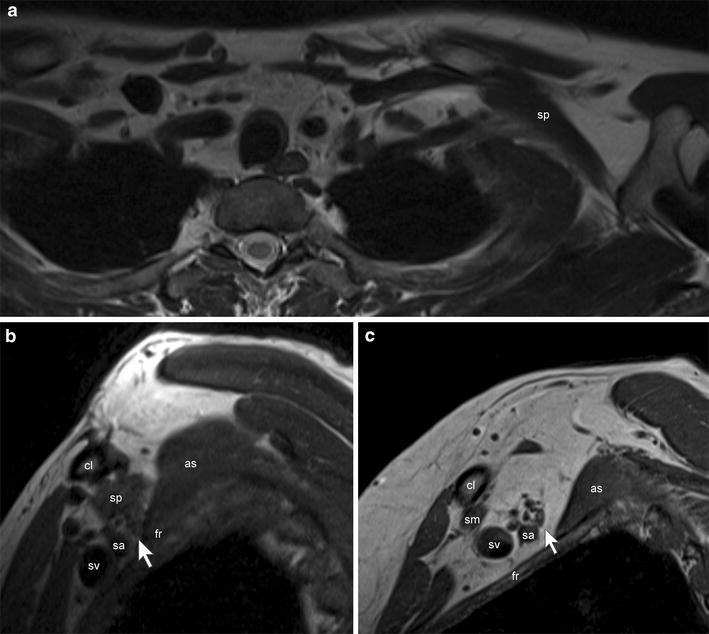
MRI3-tesla brachial plexus MRI, arm adducted, axial section (a) and sagittal sections (b, c): an accessory subclavius posticus muscle (sp) in contact with the subclavian artery and brachial plexus in the costoclavicular space, arrows.
Source : Muellner et al., BMC Research Notes, 2015, figure 1 · CC BY
Three clinical entities are distinguished according to the structure compressed, with very unequal prevalences:
📊 Distribution of the three types of thoracic outlet syndrome
Neurogenic TOS accounts for almost all of physiotherapy practice
Consensus estimates. The exact prevalence remains uncertain in the absence of a diagnostic gold standard. Source: Cavanna 2022, Jones 2019.
- Neurogenic TOS (nTOS): ~95 % of cases. Compression of the brachial plexus, mainly in the lower trunk territory (C8-T1). Vague symptoms: cervicoscapular pain, paraesthesia in the hand (most often the ulnar border), fatigability of the upper limb during activities with the arms raised, sometimes weakness of grip.111
- Venous TOS (vTOS): 3 to 5 % of cases. Compression of the subclavian vein. Can lead to effort thrombosis or Paget-Schroetter syndrome in the young overhead athlete. Massive swelling, cyanosis, acute heaviness of the arm.8
- Arterial TOS (aTOS): < 1 % of cases, but the most serious. Compression of the subclavian artery, often associated with a cervical rib. Risk of post-stenotic aneurysm, distal embolisation, acute ischaemia.10
Epidemiological profile
TOS preferentially affects young adult women, with a female-to-male ratio of about 3:1 in adult populations, and 1.84:1 in children and adolescents (2024 paediatric meta-analysis).3 The typical age of onset is 20 to 50.4 The exact prevalence in the general population remains poorly established because there is no reference diagnostic test.
Risk factors
Anatomical / congenital 🦴 :
- Cervical rib or an elongated C7 transverse process (~0.5-1 % of the population, a major factor in aTOS).10
- Scalene muscle anomalies (hypertrophy, abnormal insertion of the anterior or middle scalene).1
- Variations in the costoclavicular or subpectoral space (anatomical narrowness).11
Acquired / functional 🏋️ :
- Trauma: whiplash , clavicle fracture (particularly a malunion), acute brachial injury.9
- Repeated microtrauma: overhead sports (swimming, baseball, volleyball, throwing), occupations with repetitive movements in elevation.12
- Prolonged forward head posture, rounded shoulders (long periods of screen work, sedentary living).1
What happens anatomically and how does TOS progress?
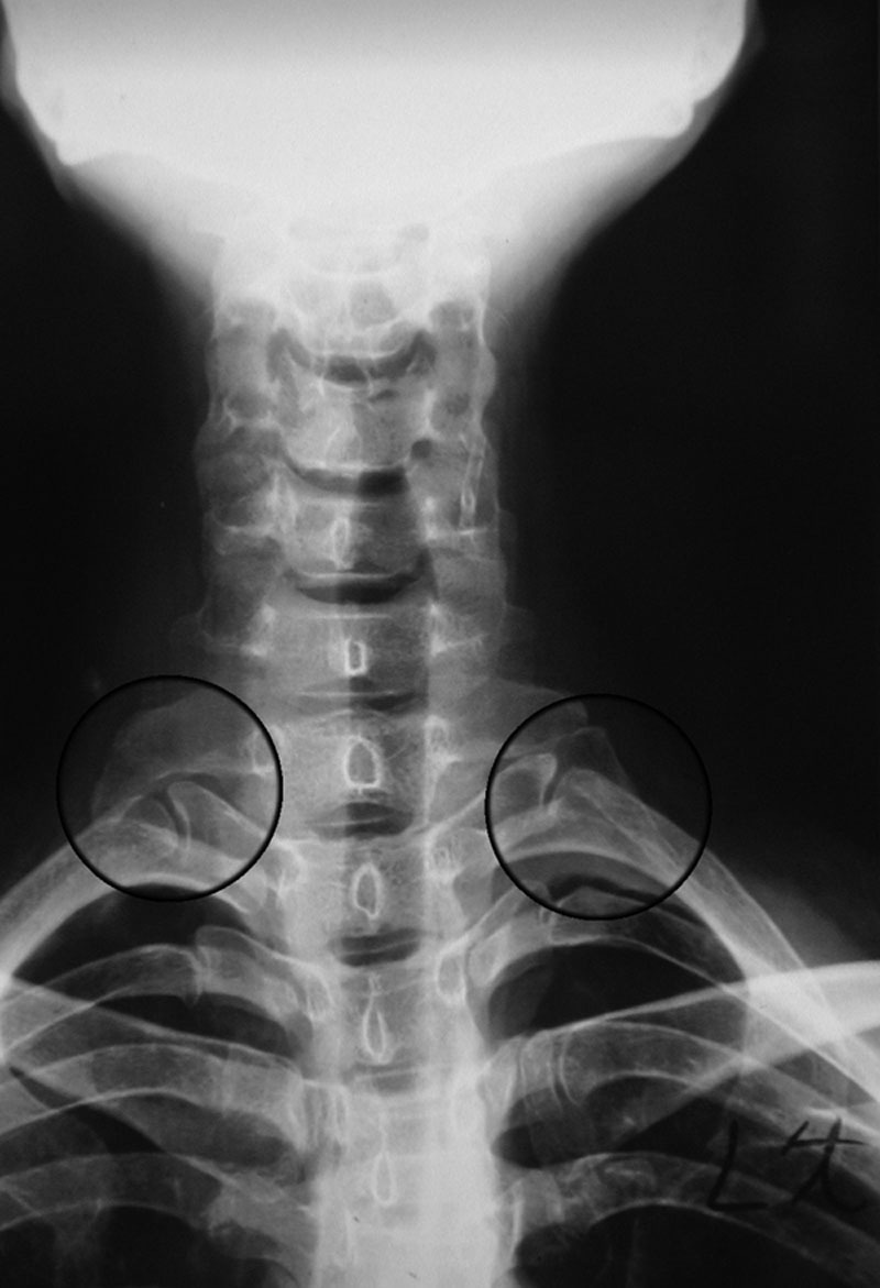
X-rayAnteroposterior cervical spine X-ray showing two bilateral supernumerary cervical ribs, circled, a classic bony cause of thoracic outlet syndrome.
Source : Chwei-Chin Chuang et al., Plastic and Reconstructive Surgery. Global Open, 2016, figure 3 · CC BY-NC-ND
Compression can occur in three anatomical zones in series, from proximal to distal:
🧭 Three anatomical zones of compression, from the neck to the axilla
Identifying the zone helps target treatment (stretching, mobilisation, scapular control)
Source: Jones 2019, Hooper 2010. The most common zone of neurogenic compression: the interscalene triangle (zone 1).
In neurogenic TOS, chronic nerve compression sustains a cycle of inflammation and fibrosis : repeated microtrauma or scalene overactivity → inflammation of the scalene muscles and surrounding tissue → formation of scar tissue (fibrosis) → perineural adhesions → restricted nerve gliding → worsening symptoms.13 This mechanism explains the insidious, chronic character of the condition.
Natural history
- nTOS: slow progression over months to years. Without intervention, chronic pain, loss of function and muscle wasting are possible in advanced cases. The majority of patients (estimated at 60 to 90 %) respond to well-conducted multimodal physiotherapy at 3-6 months; the evidence, however, remains of low to moderate quality.6
- vTOS: an immediate risk of deep vein thrombosis of the upper limb (Paget-Schroetter), with long-term complications (post-thrombotic syndrome).8
- aTOS: a risk of aneurysm and distal embolisation that can threaten the viability of the limb.10
Vascular red flags
- Massive swelling of the arm ± cyanosis ± acute heaviness → suspected vTOS / Paget-Schroetter → urgent referral (Doppler ultrasound, possible thrombolysis).
- A cold, pale hand, arm claudication on effort, acute weakness, a necrotic finger → suspected aTOS → immediate surgical referral (CT angiography, decompression).
- A palpable cervical rib + asymmetrical vascular signs → urgent imaging.
- A history of clavicle fracture with malunion + progressive neurogenic symptoms → orthopaedic consultation.
- Three very unequal entities: neurogenic ~95 %, venous 3-5 %, arterial <1 %.
- Women aged 20-50 are over-represented (adult ratio ~3:1).
- Anatomical factors (cervical rib, hypertrophic scalenes) + acquired ones (trauma, posture, overhead microtrauma).
- 3 compression zones in series (interscalene triangle → costoclavicular → subpectoral).
- nTOS progresses slowly and responds well to rehabilitation; the vascular forms are emergencies.
Bibliography: chapter 1
- Jones MR, Prabhakar A, Viswanath O, Urits I, Green JB, Kendrick JB, Brunk AJ, Eng MR, Orhurhu V, Cornett EM, Kaye AD. Thoracic Outlet Syndrome: A Comprehensive Review of Pathophysiology, Diagnosis, and Treatment. Pain Ther. 2019;8(1):5-18. PMID 31037504 · doi:10.1007/s40122-019-0124-2.
- Illig KA, Donahue D, Duncan A, Freischlag J, Gelabert H, Johansen K, Jordan S, Sanders R, Thompson R. Reporting standards of the Society for Vascular Surgery for thoracic outlet syndrome. J Vasc Surg. 2016;64(3):e23-e35. PMID 27565607.
- Abdalla NS, Hijaz T, Abdalla M, et al. Pediatric thoracic outlet syndrome: a systematic review. Pediatr Surg Int. 2024;40(1):207. PMID 39003407. Paediatric F:M ratio 1.84:1.
- DiLosa KL, Humphries MD. Epidemiology of thoracic outlet syndrome. Semin Vasc Surg. 2021;34(1):65-70. PMID 33757638.
- Povlsen S, Povlsen B. Diagnosing Thoracic Outlet Syndrome: Current Approaches and Future Directions. Diagnostics (Basel). 2018;8(1):21. PMID 29558408.
- Hock CC, Whitt JL, Tarara D. Current Clinical Concepts: Rehabilitation of Thoracic Outlet Syndrome. J Athl Train. 2024;59(7):669-676. PMID 39048118.
- Fouasson-Chailloux A, Menu P, Daley P, Crenn V, Le Sant G, Dauty M. Efficacy of botulinum toxin injection on neurogenic thoracic outlet syndrome: a systematic review. Eur J Phys Rehabil Med. 2023;59(6):706-713. PMID 37737048.
- Moore R, Lum YW. Venous thoracic outlet syndrome. Vasc Med. 2015;20(2):182-189. PMID 25832605.
- Laulan J, Fouquet B, Rodaix C, Jauffret P, Roquelaure Y, Descatha A. Thoracic outlet syndrome: definition, aetiological factors, diagnosis, management and occupational impact. J Occup Rehabil. 2011;21(3):366-373. PMID 21193950.
- Pitcher GS, Sen I, Bjarnason H, et al. Thirty-year single-center experience with arterial thoracic outlet syndrome. J Vasc Surg. 2022;76(2):523-530. PMID 35378247.
- Cavanna AC, Giovanis A, Daley A, Feminella R, Chipman R, Onyeukwu V. Thoracic outlet syndrome: a review for the primary care provider. J Osteopath Med. 2022;122(11):587-599. PMID 36018621.
- Ferrante MA. The thoracic outlet syndromes. Muscle Nerve. 2012;45(6):780-795. PMID 22581530.
- Hooper TL, Denton J, McGalliard MK, Brismée JM, Sizer PS Jr. Thoracic outlet syndrome: a controversial clinical condition. Part 1: anatomy, and clinical examination/diagnosis. J Man Manip Ther. 2010;18(2):74-83. PMID 21655389.
How can thoracic outlet syndrome be assessed and diagnosed with certainty?
Diagnosing TOS is notoriously difficult : no test or investigation serves as a gold standard, particularly for the neurogenic form.514 The approach rests on a rigorous history, a structured clinical examination, and reasoning by exclusion. More than 90 % of cases are neurogenic, which makes the diagnosis even more dependent on clinical assessment than on imaging.
What questions should be asked to understand the patient and their history properly?
🔍 The history is the cornerstone of the diagnosis. Four axes structure the questioning:
- Nature and topography of the symptoms: pain, paraesthesia, heaviness, weakness, change in colour. A C8-T1 distribution (4th-5th fingers, ulnar border) is common in nTOS.9
- Triggering and aggravating factors: arms raised (doing your hair, reaching shelves, painting a ceiling), prolonged driving, carrying a heavy rucksack or a shoulder bag, repetitive occupational or sporting movements in elevation.11
- Chronology: sudden vs insidious onset, remissions, total duration, any link with a traumatic event.
- History of trauma: cervical spine trauma (whiplash), clavicle fracture (particularly a malunion), 1st rib fracture, sporting or road traffic accident. Always ask about these actively.9
Which clinical tests should be carried out and which other conditions must be ruled out?
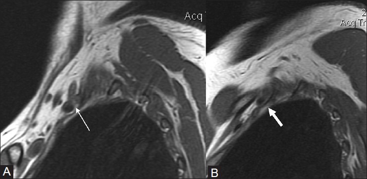
MRISagittal T1-weighted shoulder MRI, comparing the arm in neutral position and hyperabduction: compression of the subclavian artery and brachial plexus in the costoclavicular space due to a cervical rib.
Source : Lawande et al., The Indian Journal of Radiology & Imaging, 2012, figure 16 · CC BY-NC-SA
The clinical examination aims to reproduce the patient's symptoms and to exclude other conditions. Most of the provocation tests have limited reliability when used in isolation and must be interpreted as a cluster.513
📊 Approximate diagnostic performance of the provocation tests
Values vary across studies: use as a cluster, never on their own
Approximate values; the primary studies are heterogeneous and the overall quality is low. Adson: a high prevalence of false positives in asymptomatic subjects. No single test is sufficient.5
📋 The most-used tests
- Roos test (EAST, Elevated Arm Stress Test): arms at 90° abduction and 90° external rotation, opening and closing the fists for 3 minutes. Reproduction of the symptoms = positive. Regarded as the most useful for nTOS, but with sensitivity/specificity varying across studies.13
- Adson's manoeuvre: loss or reduction of the radial pulse on cervical extension + ipsilateral rotation with deep inspiration. False positives are very common in asymptomatic subjects (up to 50 % in some series).5
- Wright test (hyperabduction): arm hyperabducted/externally rotated; looking for reproduction of symptoms or loss of the radial pulse. Specificity disputed.5
- Cyriax test (costoclavicular): shoulders drawn back and down (the « soldier's position »), looking for reproduction. Used within the combined clinical battery.
- Palpation of the scalenes and pectoralis minor: looking for trigger points and muscle tension reproducing the symptoms, completing the assessment.
Essential differential diagnoses
| Condition | Distinguishing clues | Discriminating test / investigation | Level of evidence |
|---|---|---|---|
| Cervical radiculopathy | Strict dermatomal distribution, positive Spurling sign, axial neck pain, scapular pain | Spurling, cervical MRI, segmental EMG | Moderate |
| Carpal tunnel syndrome | Symptoms in the thumb/index/middle finger, at night, relieved by shaking the hand | Phalen, Tinel at the wrist, distal EMG | High |
| Rotator cuff / frozen shoulder | Mechanical shoulder pain, restricted active/passive range, without distal paraesthesia | Hawkins, Neer, Jobe, ultrasound | Moderate |
| Myofascial syndrome | Trigger points in the scalenes / trapezius / pectoralis, reproducible referred pain | Targeted direct palpation | Weak |
| Ulnar nerve compression at the elbow | Symptoms isolated to the 4th-5th fingers, worse on elbow flexion | Tinel at the elbow, segmental EMG | High |
| Primary Raynaud's phenomenon | Bilateral and symmetrical, triggered by cold, with no sign of compression | Vascular workup, capillaroscopy | Moderate |
Should patients with TOS be classified, and for what benefit?
🤔 Classification into three types is essential because it entirely determines the treatment strategy and the urgency of management (the 2016 SVS consensus, still in force).2
| Type | Frequency | Manifestations | Management | Urgency |
|---|---|---|---|---|
| Neurogenic (nTOS) | ~95 % | Paraesthesia, pain, weakness, fatigability, sometimes thenar/hypothenar wasting | Conservative for 3-6 months → surgery if that fails | No |
| Venous (vTOS) | 3-5 % | Swelling, cyanosis, heaviness; thrombosis (Paget-Schroetter) | Thrombolysis + surgical decompression (1st rib) | Emergency |
| Arterial (aTOS) | <1 % | Pallor, coldness, claudication, distal embolism, aneurysm | Surgical decompression ± reconstruction | Emergency |
⚠️ Critique and controversy: the challenge of diagnostic certainty
The absence of a diagnostic gold standard for nTOS is the major controversy in this condition.14 The provocation tests have poor specificity (a positive Adson in many asymptomatic people) and EMG is generally normal in « non-specific » nTOS, which is the most common form.12 The diagnosis therefore rests on a « constellation of signs and symptoms » and on a rigorous process of exclusion. The literature acknowledges both over-diagnosis (a default label applied to patients with chronic pain) and under-diagnosis (genuine cases that wander for years before the right hypothesis is raised).14
High-field (3T) MR neurography is a promising emerging tool for visualising brachial plexus compression, but its standardisation and availability remain limited (Szaro 2023 scoping review).15
- The diagnosis is clinical and one of exclusion, with no gold standard.
- The history targets the aggravating positions and activities, particularly overhead arm movements.
- The provocation tests (Roos, Adson, Wright, Cyriax) are to be interpreted as a cluster, never on their own (Adson: massive false positives).
- Systematically rule out cervical radiculopathy, carpal tunnel syndrome, cuff pathology, myofascial syndrome.
- Classification into 3 types dictates the urgency : neurogenic = conservative; venous and arterial = surgical emergencies.
Bibliography: chapter 2
- Povlsen S, Povlsen B. Diagnosing Thoracic Outlet Syndrome: Current Approaches and Future Directions. Diagnostics (Basel). 2018;8(1):21. PMID 29558408. (= ref 5).
- Hooper TL, Denton J, McGalliard MK, Brismée JM, Sizer PS Jr. Thoracic outlet syndrome: a controversial clinical condition. Part 1: anatomy, and clinical examination/diagnosis. J Man Manip Ther. 2010;18(2):74-83. PMID 21655389. (= ref 13).
- Illig KA, Donahue D, Duncan A, et al. Reporting standards of the Society for Vascular Surgery for thoracic outlet syndrome. J Vasc Surg. 2016;64(3):e23-e35. PMID 27565607. The SVS diagnostic consensus, the reference classification.
- Ferrante MA. The thoracic outlet syndromes. Muscle Nerve. 2012;45(6):780-795. PMID 22581530. A review of the « true » vs « non-specific » nTOS controversy.
- Szaro P, McGrath A, Ciszek B, Geijer M. The MRI features of thoracic outlet syndrome: a scoping review. Front Physiol. 2023;14:1208888. PMID 37920804.
- Kaplan J, Kanwal A. Thoracic Outlet Syndrome. StatPearls. Treasure Island (FL): StatPearls Publishing; 2023. NBK557450.
Which treatment strategies are the most effective?
For the neurogenic form (which accounts for > 95 % of the patients seen in physiotherapy), the international consensus is unequivocal: first-line treatment is conservative, structured over 3 to 6 months, before any invasive option is considered.26 A recent review confirms that a majority of patients report clear or complete improvement at 6 months, but the quality of the primary studies remains heterogeneous and of low to moderate level.6
Where do you start? What hierarchy of interventions?
🪜 Treatment hierarchy for neurogenic TOS
A conservative progression in 6 steps: education and posture are prerequisites, not an add-on
Adapted from Hock 2024 (J Athl Train, current concepts) and Watson 2010 (Manual Therapy part 2). A step that skips the foundations (education, posture, breathing) generally compromises the steps that follow.
What is the place of exercise, and is there a superior approach?
Exercise is the core of treatment. A one-size-fits-all approach is ineffective: the superior strategy is personalised and progressive, based on an assessment of the patient's individual deficits.6
Key components of an effective programme
- Stretching the tight muscles : scalenes (anterior, middle), pectoralis minor, upper trapezius, levator scapulae. Gentle, sustained stretching (30 s × 3-5 repetitions, 2-3 times/day).617
- Strengthening the scapular stabilisers : serratus anterior, lower and middle trapezius, rhomboids. Restores posture and opens the costoclavicular and subpectoral spaces.18
- Neurodynamic mobilisation (« nerve gliding ») : brachial plexus gliding techniques to restore the compliance of the nerve interfaces. Evidence of very low level (old case series).19
- Diaphragmatic breathing : reduces overactivity of the accessory scalenes, a useful accompaniment from the start and throughout the programme.20
- Cervicothoracic mobility : upper thoracic stiffness increases the load on the 1st rib and the scalenes; work on thoracic mobility into extension/rotation is often beneficial.6
Manual therapies, botulinum toxin, technologies: how effective are they?
Passive therapies are adjuncts, never treatments in isolation.
- Manual therapy: mobilisation of the 1st rib, of the upper cervical and thoracic spine, of the acromioclavicular joint; soft-tissue release (scalenes, pectoralis minor) alongside active stretching.13
- Botulinum toxin (BoNT-A) 💉: ultrasound-guided injections into the scalenes or pectoralis minor in resistant patients. The Fouasson-Chailloux 2023 systematic review (10 studies, ~555 patients) reports benefits at 1-3 months but poor methodological quality and considerable heterogeneity.7 The Woodworth 2024 SR confirms those cautious conclusions.21 To be regarded as a therapeutic window of 3-6 months for optimising rehabilitation, not as a treatment in its own right.
- Ultrasound, EMG biofeedback: possible use for postural and breathing work, but the evidence specific to TOS is very limited.
- Neuromuscular taping / kinesiotaping: a possible proprioceptive adjunct, weak level of evidence.
Summary table: modalities × level of evidence (GRADE)
| Modality | Indication | Strength of recommendation | Quality of evidence |
|---|---|---|---|
| Education + postural correction | All nTOS | Strong | Weak |
| Diaphragmatic breathing | All nTOS | Strong | Weak |
| Strengthening the scapular stabilisers | All nTOS | Strong | Moderate |
| Stretching scalenes / pectoralis minor | All nTOS | Moderate | Weak |
| Neurodynamic mobilisation | nTOS adjunct | Weak | Very weak |
| Manual therapy (1st rib, scapula) | nTOS adjunct | Weak | Weak |
| Botulinum toxin to the scalenes | Resistant nTOS | Weak (conditional) | Very weak |
| Surgery as first-line for nTOS | Not recommended | Against | Moderate |
| Urgent surgery for vTOS / aTOS | Vascular emergency | Strong | Moderate |
Beyond the physical: how do you educate the patient and address the psychological factors?
🧠 TOS is often chronic and frustrating. Ignoring the psychosocial factors is a major cause of failure.
- A clear explanation of the condition: demystifying nerve compression, taking the drama out of it without minimising it.
- Identifying the aggravating factors and coping strategies (desk ergonomics, how a bag is carried, sporting technique, sleep).
- Adherence to the programme: improvement is gradual (weeks to months), and the home exercises are indispensable: short but regular.
- Spotting kinesiophobia, catastrophising and anxiety: predictors of chronicity to be built into management.22
⚠️ Critique and controversy
Despite the consensus on the conservative approach, the literature suffers from heterogeneous protocols (« physiotherapy » covers very variable content from one study to another).6 The 2023-2024 reviews all acknowledge low to moderate methodological quality and a lack of RCTs comparing the components (strengthening vs neurodynamics vs stretching alone).7 The effectiveness figures (« 60 to 90 % » improvement) rest on heterogeneous series with no clear control group. That uncertainty calls for transparency with the patient about timescales and the variability of individual responses.
- First-line care is conservative, a structured programme over 3-6 months.
- Hierarchy: education → posture → breathing → stretching → strengthening → functional.
- Scapular strengthening (serratus anterior, lower/middle trapezius) + stretching of the scalenes/pectoralis minor = the best-supported combination.
- Manual therapy and botulinum toxin = adjuncts, never used alone.
- A biopsychosocial approach is indispensable: spot kinesiophobia and catastrophising.
- Overall quality of the evidence: weak to moderate (few RCTs, many heterogeneous series).
Bibliography: chapter 3
- Illig KA et al. Reporting standards SVS for thoracic outlet syndrome. J Vasc Surg. 2016;64(3):e23-e35. PMID 27565607. (= ref 2).
- Hock CC, Whitt JL, Tarara D. Current Clinical Concepts: Rehabilitation of Thoracic Outlet Syndrome. J Athl Train. 2024;59(7):669-676. PMID 39048118. (= ref 6).
- Fouasson-Chailloux A, Menu P, Daley P, Crenn V, Le Sant G, Dauty M. Efficacy of botulinum toxin injection on neurogenic thoracic outlet syndrome: a systematic review. Eur J Phys Rehabil Med. 2023;59(6):706-713. PMID 37737048. (= ref 7).
- Watson LA, Pizzari T, Balster S. Thoracic outlet syndrome part 2: conservative management of thoracic outlet. Man Ther. 2010;15(4):305-314. PMID 20382063. A founding paper; its rehabilitation principles still hold.
- Kibler WB, McMullen J. Scapular dyskinesis and its relation to shoulder pain. J Am Acad Orthop Surg. 2003;11(2):142-151. PMID 12670140.
- Wehbé MA, Schlegel JM. Nerve gliding exercises for thoracic outlet syndrome. Hand Clin. 2004;20(1):51-55. PMID 15005384.
- Levine NA, Rigby BR. Thoracic Outlet Syndrome: Biomechanical and Exercise Considerations. Healthcare (Basel). 2018;6(2):68. PMID 29921751.
- Woodworth GE, Smith RD, Lischwe Mueller N, et al. Botulinum toxin injections for neurogenic thoracic outlet syndrome: a systematic review. Muscle Nerve. 2024;69(5):530-539. PMID 38529885.
- Nicholas MK, Linton SJ, Watson PJ, Main CJ; "Decade of the Flags" Working Group. Early identification and management of psychological risk factors ("yellow flags") in patients with low back pain: a reappraisal. Phys Ther. 2011;91(5):737-753. PMID 21451099.
How do you ensure lasting recovery and prevent recurrences?
Resolving the initial symptoms is not enough: nTOS readily recurs when the patient returns to their previous mechanical loads without having taken on the lasting corrections. Long-term success rests on two pillars:self-management and a structured return to activity.6
How do you make the patient an active participant in their recovery through self-management?
🧠 Building independence is central. Conservative treatment achieves positive results in about 60-90 % of patients; the patient's engagement with their home programme is a key determinant. Without that ownership, the initial benefits erode quickly once supervised care ends.6
- In-depth therapeutic education: simplified anatomy, the mechanisms of compression, at-risk postures targeted at the patient's own daily life (workstation, sleep, mode of transport, how a bag is carried). Defusing fear of movement.22
- Postural awareness: learning to recognise and correct rounded shoulders and a forward head. The aim is underlying strengthening that makes « good posture » spontaneous, rather than an exhausting permanent vigilance.
- Diaphragmatic breathing done independently to manage peaks of tension and stress, at least twice a day (morning and evening, 5 minutes).20
- A personalised home programme: concise, effective, realistic (10-15 minutes maximum). A few key stretches (scalenes, pectoralis minor) + 2-3 scapular strengthening exercises + breathing. Modified progressively according to tolerance.
- Self-monitoring: identifying the early signs (paraesthesia, heaviness, early fatigue of the arm), adjusting activities without absolute rest, being extra careful during periods of stress or heavy work.
- Workstation ergonomics: screen height, armrest position, alternating sitting and standing, active breaks every 45-60 minutes.
When and how should a safe return to sport and activities be planned?
🏃 The return to demanding activities must be based on functional criteria, not on a fixed calendar. A contemporary review of the return to activity after surgical decompression confirms the importance of staged progression and of objective criteria before each step up in intensity.23
🏃 Return to sport in 5 phases: symptom-guided
No stage is passed without meeting the criteria of the previous phase
Adapted from Talutis 2024 and Hock 2024. Rigour matters more than speed: a rushed return carries a strong risk of recurrence.
⚠️ Critique and controversy
The overall quality of the evidence on preventing recurrence and returning to sport remains weak to moderate: few RCTs, a predominance of case studies and non-randomised series. The published return-to-sport protocols (particularly in overhead athletes) remain essentially expertise-based rather than grounded in controlled clinical trials.23 The place of psychosocial factors in chronicity and recurrence is increasingly recognised but remains under-studied.22 You have to be able to admit uncertainty in front of the patient and adapt management to their context (amateur vs professional sport, physical vs sedentary occupation).
- Lasting prevention = building patient independence (education, posture, home programme).
- Return to sport that is progressive, by functional criteria, not by calendar.
- Underlying strengthening of the scapular stabilisers + daily stretching of the scalenes/pectoralis minor.
- Diaphragmatic breathing = a simple, effective tool for reducing regional tension.
- Acknowledge the uncertainty (quality of evidence weak to moderate) and adapt to the patient's profile.
Bibliography: chapter 4
- Hock CC, Whitt JL, Tarara D. Current Clinical Concepts: Rehabilitation of Thoracic Outlet Syndrome. J Athl Train. 2024;59(7):669-676. PMID 39048118. (= ref 6).
- Levine NA, Rigby BR. Thoracic Outlet Syndrome: Biomechanical and Exercise Considerations. Healthcare (Basel). 2018;6(2):68. PMID 29921751. (= ref 20).
- Nicholas MK et al. Early identification of psychological risk factors. Phys Ther. 2011;91(5):737-753. PMID 21451099. (= ref 22).
- Talutis SD, Caron E, Becerra A, Lawrence PF, Gelabert HA. Return to physical activity following thoracic outlet decompression: a contemporary review. Ann Vasc Surg. 2024;101:325-332. PMID 37931887.
What do real clinical cases teach us?
Studying clinical cases published on PubMed/PMC anchors the theoretical principles in reality. The cases below are all verified publications, selected to illustrate the main variants: effort-related Paget-Schroetter, post-traumatic from a malunion, and vascular mimicry. 🧐
The level of evidence of clinical cases
The case reports represent the lowest level of evidence (Oxford CEBM level 5 / GRADE very low). They are pedagogically useful but cannot support a general treatment recommendation. Conclusions are never generalisable from a single case.
Case 1: Paget-Schroetter in a college lacrosse player (Kohen 2013)
📚 A college lacrosse player presents with massive swelling and cyanosis of the dominant upper limb after a run of intensive training. Doppler ultrasound confirms axillo-subclavian vein thrombosis (Paget-Schroetter syndrome). Management combines pharmacological thrombolysis and surgical decompression by resection of the 1st rib.24
Clinical lesson : venous TOS is an emergency in the young overhead athlete. Acute swelling + cyanosis after effort must raise the diagnosis immediately and trigger a vascular Doppler ultrasound. The first-contact physiotherapist (particularly in college sport) must recognise this red flag and must not offer mobilisation before vascular causes are excluded.
Case 2: Paget-Schroetter in a young swimmer (Suwal 2022)
A young adult, a recreational swimmer, presents with acute swelling of the dominant arm. Imaging reveals thrombosis of the axillo-subclavian vein on costoclavicular compression. The case, published open access in Cureus, sets out the diagnostic sequence (Doppler → MR angiography) and the treatment sequence (anticoagulation → thrombolysis → delayed surgical decompression).25
Clinical lesson : vTOS is not confined to professional athletes. Any patient doing regular overhead sport (swimming, volleyball, throwing) with unusual swelling should be considered at risk. The thrombolysis → delayed decompression sequence is the one favoured today.
Case 3: Post-traumatic TOS from clavicle malunion (Yoo 2009)
💥 A patient progressively develops neurogenic symptoms several months after a clavicle fracture that healed with a hypertrophic callus. Compression of the neurovascular bundle by the callus reduces the costoclavicular space. Management requires surgical resection of the callus in addition to rehabilitation.26
Clinical lesson : faced with a patient with progressive neurogenic symptoms and a history of clavicle fracture (even an old one, even one regarded as « healed »), palpate the clavicle and image it if necessary. A malunion is a structural cause that will respond only partly to rehabilitation alone.
Case 4: Arterial TOS presenting as Raynaud's phenomenon (Balram 2024)
🩸 A patient initially referred for presumed primary Raynaud's phenomenon (asymmetrical cooling of the fingers). Examination shows an inter-arm blood pressure asymmetry, and CT angiography reveals compression of the subclavian artery with a cervical rib and a post-stenotic aneurysm: arterial TOS. This 2024 case report illustrates how vascular presentations can be missed and mimic other conditions.27
Clinical lesson : faced with an asymmetrical« Raynaud », especially with a palpable cervical rib or a diminished radial pulse, consider aTOS and refer for a vascular workup before any symptomatic vasodilator treatment.
The diagnostic challenge: when TOS mimics another condition (Hooper 2010 review)
🤔 The clinical reviews are a reminder that a patient with C8-T1 paraesthesia can be wrongly diagnosed and treated for a C7-T1 cervical disc herniation, with treatment failure. Symptoms that are positional and overhead, a history of whiplash or clavicle fracture, and the absence of clear findings on cervical MRI should point towards assessment of the thoracic outlet.13 Conversely, TOS can also mimic carpal tunnel syndrome (distal paraesthesia) or cuff pathology (scapular pain), hence the importance of a systemic assessment rather than one focused on a single joint.
⚠️ Critique and controversy: the value and limits of clinical cases
Published cases carry a major publication bias : treatment successes are over-represented relative to failures. Over-interpretation of the provocation tests (Adson, Wright, Roos) remains common: their sensitivity/specificity in isolation is poor and using them as a cluster is more useful.5 Case reports must never replace systematic reviews in guiding standard treatment decisions.
📐 The hierarchy of scientific evidence, where does each type of study sit?
Strength of evidence decreasing from the top (meta-analyses) to the bottom (isolated cases)
A simplified GRADE / Oxford CEBM hierarchy. The length of the coloured bar illustrates the relative strength of evidence. Practical implication: where an appealing clinical case and a meta-analysis diverge, follow the meta-analysis. Clinical cases remain valuable for generating hypotheses, flagging rare presentations, or illustrating a line of reasoning.
- The published real cases demonstrate the great heterogeneity of TOS: classic neurogenic, a venous emergency in the athlete (Kohen, Suwal), post-traumatic (Yoo), vascular mimicry (Balram).
- vTOS (Paget-Schroetter) and aTOS are emergencies, particularly in the young athlete. The first-contact physiotherapist must know how to recognise them and must not manipulate.
- A history of trauma (clavicle fracture, even an old one) should raise the suspicion of post-traumatic TOS from a malunion.
- Faced with an asymmetrical Raynaud, consider arterial TOS before any vasodilator treatment.
- ⚠️ Level of evidence : a case report = level 5. It illustrates; it never demonstrates effectiveness. Where they diverge, follow the meta-analyses (level 1a), not the isolated case.
Bibliography: chapter 5 (verified PMC clinical cases)
- Kohen D, Hanhan S, Bellah R. Paget-Schroetter syndrome in a lacrosse player. Del Med J. 2013;85(3):77-79. PMID 23631108.
- Suwal A, Rijal SS, Shrestha M, Munankami S, Basnet S. Paget-Schroetter Syndrome: A Rare Case of Upper Extremity Deep Vein Thrombosis in a Young Swimmer. Cureus. 2022;14(6):e26060. PMID 35865424.
- Yoo MJ, Seo JB, Kim JP, Lee JH. Surgical treatment of thoracic outlet syndrome secondary to clavicular malunion. Clin Orthop Surg. 2009;1(1):54-57. PMC2766689.
- Balram B, Pothier L, Lee CJ. A case of misdiagnosed arterial thoracic outlet syndrome as primary Raynaud's phenomenon. J Vasc Surg Cases Innov Tech. 2024;10(2):101441. PMID 38812728.
- Hooper TL et al. J Man Manip Ther 2010;18(2):74-83. PMID 21655389. (= ref 13).
- Povlsen S, Povlsen B. Diagnostics 2018;8(1):21. PMID 29558408. (= ref 5).
How can these recommendations be applied in practice?
Applying the recommendations runs along three axes: knowing when to refer in good time, measuring outcomes objectively, and overcoming the obstacles to implementing good practice. 🧭
When, and to which other professionals, should you refer?
One of the physiotherapist's fundamental roles, particularly in direct access, is knowing how to identify situations that fall outside their scope of practice. That decision rests mainly on screening for pathological or psychosocial « flags ».
Screening for red flags 🚩
Warning signals requiring medical referral
- Acute massive swelling of the arm + cyanosis ± sudden heaviness → suspected vTOS / Paget-Schroetter → urgent vascular Doppler ultrasound.
- Pallor, coldness, acute claudication of the arm on effort, a necrotic finger → suspected aTOS → urgent CT angiography, vascular/surgical consultation.
- A palpable cervical rib + asymmetrical neurovascular signs → targeted imaging.
- Visible muscle wasting of the hand (thenar, hypothenar), a clear motor deficit → urgent EMG.
- Failure of well-conducted treatment over 4-6 weeks (pain, weakness not improved) → medical reassessment, cervical and thoracic imaging.
- A recent history of clavicle fracture with malunion + neurogenic symptoms → orthopaedic consultation (suspected compressing malunion).
- General red flags (weight loss, persistent fever, a history of cancer, night sweats, severe non-postural night pain) → refer to the GP.
⚠️ No single sign is sufficient: it is the grouping that counts. But faced with a vascular emergency (massive swelling, cyanosis, claudication), referral takes priority over any further physiotherapy assessment.
A prospective study is a reminder that serious pathology remains rare in first-contact care.29 Verhagen's review also stresses that many classic red flags lack solid empirical validation when taken in isolation.28 The right reflex is therefore the cluster of signs, weighted by the clinical context.
Identifying yellow, blue and black flags 🟡
Psychosocial factors are major predictors of chronicity, sometimes more so than the biomechanical factors.22
- Yellow : beliefs, emotions, behaviours (catastrophising, fear-avoidance, kinesiophobia).
- Blue : work-related perceptions (low satisfaction, conflictual relationships, a sense of not being supported by management).
- Black : contextual and systemic obstacles (an insurance dispute, medico-legal litigation, rigid legislation).
The STarT Back Screening Tool, designed for low back pain, transfers as a principle of stratification: risk-stratified care is more effective and more cost-effective than standard care.30 Where psychosocial factors predominate, refer to a psychologist, an occupational physician or a social worker.
How do you measure outcomes and overcome the barriers to implementation?
Measure in order to progress 📈
- Validated PROMs : VAS / NRS for pain (0-10); DASH (30 items) or the QuickDASH (11 items) for upper limb function; SPADI for the shoulder.31
- Performance tests : grip strength (dynamometer), goniometric cervical and scapular range, quantified Roos test (maximum time without symptoms), isometric strength of the scapular stabilisers.
- Longitudinal follow-up at fixed intervals (week 1, 6, 12, 24): this supports shared patient-therapist decision-making and makes progress objective even where subjective improvement is slow.32
Overcoming the barriers to EBP 🚧
The implementation reviews identify the same barriers across countries:33
| Obstacle | Suggested lever | Feasibility |
|---|---|---|
| Lack of time | PROMs via digital forms before the session, dedicated slots for keeping up with the literature | High |
| Lack of confidence in appraising the literature | Journal clubs, PEDro training, clinical synopses (Cochrane, BMJ EBM) | Moderate |
| Lack of organisational support | Practice leadership, shared access to databases, EBP quality indicators | Variable |
| Long-standing treatment habits | Clinical audit, peer feedback, structured protocols | Moderate |
| Difficulty transferring it to the patient | Shared decision tools, plain language, written/video materials32 | High |
A fundamental methodological review is also a reminder that critically appraising a randomised controlled trial goes through validated scales (PEDro for physiotherapy).34 Translating knowledge into practice requires active strategies: the mere availability of guidelines is not enough.35
⚠️ Critique and controversy
🧠 While EBP is universally valued, putting it into practice raises tensions:
- The « tyranny of red flags » can lead to over-ordering of costly, anxiety-provoking imaging. The prevalence of serious pathology in first-contact care is low.29 The challenge: caution without over-medicalisation.
- Enthusiasm for PROMs can paradoxically dehumanise care if you treat the score rather than the person.
- And Interprofessional collaboration remains more theoretical than operational in siloed systems with uncoordinated payment. Breaking down those silos requires structural change beyond individual behaviour alone.
- Spot the vascular red flags (massive swelling, cyanosis, claudication) → urgent referral, no mobilisation.
- Screen early for the yellow flags (kinesiophobia, catastrophising); refer to psychology where they predominate.
- Use validated PROMs (DASH, QuickDASH, VAS) and performance tests to make outcomes objective.
- Overcome the barriers to EBP through continuing education, journal clubs, organisational leadership.
- And Shared decision-making with the patient is central: no recommendation replaces an informed conversation.
Bibliography: chapter 6
- Verhagen AP, Downie A, Maher CG, Koes BW. Most red flags for malignancy in low back pain guidelines lack empirical support: a systematic review. Pain. 2017;158(10):1860-1868. PMID 28708761.
- Henschke N, Maher CG, Refshauge KM, et al. Prevalence of and screening for serious spinal pathology in patients presenting to primary care settings with acute low back pain. Arthritis Rheum. 2009;60(10):3072-3080. PMID 19790051.
- Hill JC, Whitehurst DG, Lewis M, et al. Comparison of stratified primary care management for low back pain with current best practice (STarT Back): a randomised controlled trial. Lancet. 2011;378(9802):1560-1571. PMID 21963002.
- Kyte DG, Calvert M, van der Wees PJ, ten Hove R, Tolan S, Hill JC. An introduction to patient-reported outcome measures (PROMs) in physiotherapy. Physiotherapy. 2015;101(2):119-125. PMID 25620440.
- Hoffmann T, Légaré F, Simmons MB, et al. Shared decision making: what do clinicians need to know and why should they bother? Med J Aust. 2014;201(1):35-39. PMID 24999896.
- Scurlock-Evans L, Upton P, Upton D. Evidence-based practice in physiotherapy: a systematic review of barriers, enablers and interventions. Physiotherapy. 2014;100(3):208-219. PMID 24780633.
- Maher CG, Sherrington C, Herbert RD, Moseley AM, Elkins M. Reliability of the PEDro scale for rating quality of randomized controlled trials. Phys Ther. 2003;83(8):713-721. PMID 12882612.
- Yost J, Ganann R, Thompson D, et al. The effectiveness of knowledge translation interventions for promoting evidence-informed decision-making among nurses in tertiary care: a systematic review and meta-analysis. Implement Sci. 2015;10:98. PMID 26169063.
- Nicholas MK et al. Yellow flags reappraisal. Phys Ther. 2011;91(5):737-753. PMID 21451099. (= ref 22).
Enjoyed this review?
Physio Learning publishes monthly evidence-based physiotherapy updates. Get the next audits straight to your inbox.

