















In brief
Occipital neuralgia, historically known in France as Arnold's neuralgia, is a secondary headache (ICHD-3 13.4) characterised by paroxysmal stabbing pain in the territory of the greater occipital nerve, often unilateral, with dysaesthesia or allodynia and tenderness on palpation of the nerve. The diagnosis is clinical and confirmed by temporary relief after an anaesthetic block of the nerve concerned, considered the gold standard. First-line management is conservative: pain education, ergonomic correction, exercise for the deep cervical flexors and cervical extensors, and manual therapy. It affects 73 % women, of mean age 56.
Clinical synthesis based on the ICHD-3 criteria (13.4), the Melchior 2025 epidemiological meta-analysis, the Deuel 2024 review of conservative management and the most recent meta-analyses (Oliveira 2024, Demont 2023, Núñez-Cabaleiro 2022, Nguyen 2025).
Clinical summary
- The condition known as occipital neuralgia is an ICHD-3 13.4 secondary headache characterised by paroxysmal stabbing pain in the territory of the greater occipital nerve (GON), the lesser occipital nerve (LON) or the third occipital nerve (TON), often unilateral (81 %, Melchior 2025).
- Epidemiology: it affects women preferentially (73 %), mean age 56; incidence historically reported at 3.2 per 100,000 person-years (Koopman 2009, a Dutch cohort of 360,000 subjects), with a revision expected from the 2025 data.
- Aetiology: mechanical compression or irritation of the GON along its course (as it passes through semispinalis and trapezius, Janis 2010), post-traumatic (whiplash), degenerative (C2-C3), vascular (occipital artery), tumour (schwannoma), inflammatory or post-herpetic.
- Pathophysiology: focal neuropathy plus peripheral and central sensitisation (Treede 2008, Finnerup 2016); trigeminocervical convergence explains the fronto-orbital radiation.
- Course: chronic and recurrent, alternating attacks and remissions; the natural history is insufficiently documented by longitudinal cohorts.
- Clinical diagnosis = ICHD-3 13.4: paroxysmal pain in the territory + dysaesthesia/allodynia + tenderness on palpation + temporary relief from an anaesthetic block of the nerve concerned.
- The key examination: palpation reproducing the pain about 2-3 cm lateral and inferior to the external occipital protuberance; Tinel's sign may be present. No validated specific score exists to date.
- An anaesthetic GON block is the diagnostic gold standard according to ICHD-3; its therapeutic value is documented (Cohen 2015 RCT, Shauly 2019 SR/MA).
- Differential diagnosis is essential: cervicogenic headache (ICHD-3 11.2.1), migraine (1), tension-type headache (2), trigeminal neuralgia (13.1), post-traumatic headache, TON involvement (Bogduk 2009).
- Primary vs secondary classification: the secondary form (post-traumatic, compressive, tumour) calls for treatment of the cause as well.
- Self-management is the cornerstone: pain education, ergonomic correction (forward head posture), sleep hygiene, stress management.
- Therapeutic exercise: training the deep neck flexors (craniocervical flexion test, Jull 2008), endurance of the cervical extensors, scapular stabilisers (Demont 2023 PM&R).
- Manual therapy: C0-C1-C2 mobilisations, soft-tissue techniques for the suboccipital muscles; efficacy documented for cervicogenic headache (Núñez-Cabaleiro 2022, Jull 2002).
- A stepped treatment algorithm: physiotherapy/education → GON block ± steroid → repeated blocks → pulsed radiofrequency (Oliveira 2024) → decompression surgery (Nguyen 2025) as a last resort.
- Return to activity is driven by functional criteria (pain < 3/10, full cervical range) rather than by a fixed delay.
- Red flags (Finucane 2020): an unusual or thunderclap headache, fever, neurological deficit, a history of cancer, unexplained weight loss, immunosuppression.
- Recommended PROMs: VAS / NRS, Neck Disability Index (NDI), HIT-6, MIDAS, PCS for catastrophising, TSK-11 for kinesiophobia.
- Overall level of evidence: moderate (GRADE) for physiotherapy in cervicogenic headache, and low to moderate for the interventions specific to occipital neuralgia, given how few dedicated RCTs exist (Deuel 2024).
Contents
- What are the fundamentals to know about occipital neuralgia?
- How do you assess and diagnose occipital neuralgia with certainty?
- Which treatment strategies are the most effective for occipital neuralgia?
- How do you secure lasting recovery and prevent recurrence of occipital neuralgia?
- What do real clinical cases teach us about occipital neuralgia?
- How do you apply these recommendations concretely in your practice?
What are the fundamentals to know about occipital neuralgia?
How is this condition defined, who does it affect and what are the risk factors?
ICHD-3 defines occipital neuralgia as unilateral or bilateral paroxysmal pain in the territory of the greater occipital nerve (GON), the lesser occipital nerve (LON) or the third occipital nerve (TON), with dysaesthesia and/or allodynia, with tenderness on palpation of the nerve concerned, and relieved by a local anaesthetic block.¹ The pain is typically described as an electric shock, a stab or a burn, lasting from a few seconds to a few minutes per attack, often followed by a residual dull ache.² 🎯 Consolidated epidemiology 2025. The systematic meta-analysis by Melchior et al. (Cephalalgia 2025, 15 pooled studies, n=579 patients) is now the most solid epidemiological reference.² It reports:- 73 % of the patients diagnosed are women (a proportion consistent across studies);
- Mean age 56 years (median 53), with a peak incidence in the fifth and sixth decades;
- Presentation unilateral in 81 % of cases;
- A shooting or stabbing character in 59 %, severe in 54 %;
- Marked heterogeneity in the incidence estimates from one study to the next, underlining the need for uniform diagnostic criteria.
- Mechanical / compressive : compression by semispinalis capitis or trapezius at the point where the nerve pierces the aponeurosis (Janis 2010, the reference anatomical study of the compression points, PRS Part II)⁴; osteoarthritis of the C1-C2 or C2-C3 facet joints.
- Traumatic : whiplash, direct craniocervical trauma, repeated microtrauma (occupational posture, previous posterior cervical surgery).
- Vascular : a loop of the occipital artery in contact with the GON, aneurysms, malformations (see the case PMC5346380, Choi 2017)⁵; a C2 cavernoma (PMC5898127).⁶
- Tumour : schwannoma of the GON or of the C2 root (Krishnan 2015, PMID 26752925)⁷; metastasis to the occipital condyle (PMC9985513).⁸
- Inflammatory / infectious : occipital shingles with post-herpetic neuralgia; inflammatory arthritis of the upper cervical joints.
- Malformation : associated Chiari I malformation (PMC5788399).⁹
🚩 Specific red flags to consider in occipital headache
- A thunderclap headache or one of sudden onset → subarachnoid haemorrhage, vertebral dissection
- Fever + neck stiffness → meningitis, osteomyelitis
- A focal neurological deficit (weakness, ataxia, visual disturbance) → posterior fossa stroke, Chiari malformation, tumour
- A known history of cancer or unexplained weight loss → metastasis, neoplastic infiltration
- Immunosuppression or a context of recent shingles → post-herpetic neuralgia, opportunistic infection
- Progressive worsening over a few weeks with resistance to well-conducted treatment → a compressive mass (schwannoma, metastasis)
- Presentation in a child or a young adult → secondary causes are more frequent (Villeneuve 2023, paediatric cohort)¹⁰
⚠️ Any red flag → cervical and brain MRI before any invasive procedure, and prompt medical referral (neurologist, or the emergency department depending on the context).
What happens in the body, and how does occipital neuralgia evolve naturally?
🧠 Neuropathic pathophysiology. The greater occipital nerve, the dorsal branch of the C2 spinal nerve, runs upwards through layers of muscle (obliquus capitis inferior, semispinalis capitis) before becoming superficial as it pierces the trapezius aponeurosis about 2-3 cm lateral and inferior to the external occipital protuberance , the key anatomical landmark of the clinical examination.⁴ Cesmebasi 2015 (Clin Anat) lists the many potential sites of mechanical conflict: the muscular passage, the aponeurotic point, and vascular contact with the occipital artery.¹¹ Chronic irritation of the nerve produces focal demyelination, peripheral sensitisation (a lowered activation threshold in the nociceptors) and, beyond that, central sensitisation. Treede et al. (Neurology 2008) set out the operational definition of neuropathic pain with a possible/probable/definite grading, updated by Finnerup et al. (Pain 2016).¹²,¹³ This central sensitisation, mediated by hyperexcitability of the neurons of the trigeminocervical complex, explains:- Scalp allodynia (pain triggered by a normally non-painful stimulus, such as brushing the hair or contact with the pillow);
- The radiation towards the frontal, orbital and even maxillary regions, through trigeminocervical convergence, described precisely by Bogduk & Govind (Lancet Neurology 2009) in their review of cervicogenic headache.¹⁴
- Persistence of the mechanical factors (posture, occupational movements);
- An unidentified secondary cause (a progressive structural compression);
- Neuropathic or inflammatory comorbidity;
- Psychosocial factors (kinesiophobia, catastrophising, sleep disturbance), major modulators of pain becoming chronic (Su 2022 on central sensitisation in migraine, applicable by analogy).¹⁵
Key points
- The ICHD-3 13.4 definition : paroxysmal pain in the territory of an occipital nerve + dysaesthesia/allodynia + tenderness on palpation + relief from an anaesthetic block.
- The 2025 epidemiology (Melchior, SR/MA): 73 % women, mean age 56, 81 % unilateral, 59 % stabbing.
- Aetiologies : compressive (muscular, degenerative), traumatic (whiplash), vascular, tumour (schwannoma), inflammatory (shingles) or idiopathic.
- Pathophysiology : focal neuropathy plus peripheral and central sensitisation through the trigeminocervical complex.
- Course : chronic and recurrent; the gap in longitudinal data should be flagged to the patient.
Bibliography
- Headache Classification Committee of the International Headache Society (IHS). The International Classification of Headache Disorders, 3rd edition. Cephalalgia. 2018;38(1):1-211. PMID 29368949. doi:10.1177/0333102417738202.
- Melchior AG, Al-Khazali S, Christensen RH, Al-Khazali HM, Ashina H. Epidemiology and clinical features of occipital neuralgia: A systematic review and meta-analysis. Cephalalgia. 2025;45(2):3331024251317595. PMID 40017062.
- Koopman JS, Dieleman JP, Huygen FJ, de Mos M, Martin CG, Sturkenboom MC. Incidence of facial pain in the general population. Pain. 2009;147(1-3):122-127. PMID 19783099.
- Janis JE, Hatef DA, Reece EM, McCluskey PD, Schaub TA, Guyuron B. The anatomy of the greater occipital nerve: Part II. Compression point topography. Plast Reconstr Surg. 2010;126(5):1563-1572. PMID 20639804.
- Choi JG, Kang YJ, Kim BC, Lee CG, Choi SK. Hemifacial Pain and Hemisensory Disturbance Referred from Occipital Neuralgia Caused by Pathological Vascular Contact of the Greater Occipital Nerve. Case Rep Neurol Med. 2017;2017:3827230. PMC 5346380.
- Occipital neuralgia from C2 cavernous malformation. Surg Neurol Int. 2018. PMC 5898127.
- Krishnan A, Kartikueyan R, Chowdhury SR, Das S. Schwannoma of the greater occipital nerve: An uncommon cause of occipital neuralgia. J Neurosci Rural Pract. 2015;6(4):634-636. PMID 26752925.
- Occipital condyle syndrome as the initial presentation of recurrence of metastatic breast cancer. Case report. 2023. PMC 9985513.
- Occipital Neuralgia in Chiari I Malformation. Acta Neurochir Suppl. 2018. PMC 5788399.
- Villeneuve LM, Coulibaly NJ, Raza SM, Poinson B, Chrusciel D, Desai VR. Surgical management of pediatric occipital neuralgia: a single-center experience of an uncommon pathology. J Neurosurg Pediatr. 2023;32(4):514-521. PMID 37548543.
- Cesmebasi A, Muhleman MA, Hulsberg P, Gielecki J, Matusz P, Tubbs RS, Loukas M. Occipital neuralgia: anatomic considerations. Clin Anat. 2015;28(1):101-108. PMID 25244129.
- Treede RD, Jensen TS, Campbell JN, et al. Neuropathic pain: redefinition and a grading system for clinical and research purposes. Neurology. 2008;70(18):1630-1635. PMID 18003941.
- Finnerup NB, Haroutounian S, Kamerman P, et al. Neuropathic pain: an updated grading system for research and clinical practice. Pain. 2016;157(8):1599-1606. PMID 27115670.
- Bogduk N, Govind J. Cervicogenic headache: an assessment of the evidence on clinical diagnosis, invasive tests, and treatment. Lancet Neurol. 2009;8(10):959-968. PMID 19747657.
- Su M, Yu S. Central Sensitization in Migraine: A Narrative Review. J Pain Res. 2022;15:2673-2682. PMC 9464439.
How do you assess and diagnose occipital neuralgia with certainty?
Which questions should you ask to understand the patient and their history?
Structured questioning explores six key dimensions:- ⚡ Quality of the pain : paroxysmal in character (“a stab”, “an electric shock”, “a burn”), brief attacks lasting seconds to minutes, sometimes in salvoes. According to Melchior 2025, a shooting character is reported by 59 % of patients.³
- Location : pain beginning in the suboccipital region and radiating towards the vertex, sometimes to the forehead, the orbit or the ear (trigeminocervical convergence). Unilateral in 81 % of cases.³
- Triggers : neck movements (rotation, extension), pressure on the occiput (chair back, pillow), direct palpation along the course of the nerve, brushing the hair (allodynia).
- Associated symptoms : dysaesthesia (abnormal sensations, tingling, burning) and scalp allodynia are close to pathognomonic. Mild visual discomfort (photophobia) is possible, but without the nausea typical of migraine.
- Relevant past history : whiplash, posterior cervical surgery, head injury, recent shingles in the territory, a history of cancer, recent vaccination (cases reported after COVID-19 vaccination, but the evidence is weak).⁴
- Yellow flags and contextual factors : kinesiophobia, catastrophising (Pain Catastrophizing Scale), sleep disturbance, level of occupational activity, workstation ergonomics.
Which clinical tests should you perform, and which other conditions should you rule out?
Standardised physical examination
- Posture : looking for a forward head posture and a straightened or kyphotic upper cervical spine.
- Cervical mobility : active and passive ranges, particularly C1-C2 rotation (the flexion-rotation test, the reference in cervicogenic headache, Hall 2010; not specific to ON).
- The key diagnostic palpation : firm, sustained pressure at the emergence point of the GON, about 2-3 cm lateral and inferior to the external occipital protuberance, at the point where it pierces the trapezius aponeurosis.⁵ A reproduction of the typical pain is the most telling clinical finding.
- Tinel's sign : gentle percussion along the course of the nerve, triggering an electric-shock sensation radiating towards the vertex.
- Allodynia testing : light stroking of the scalp in the occipital territory, compared with the contralateral side.
- Neurological assessment : sensation, motor function, reflexes. Any abnormality should prompt a rethink of the diagnosis and lead to imaging.
The greater occipital nerve block: the diagnostic gold standard
Under ICHD-3, criterion C for the neuralgias (including 13.4) requires the pain to be temporarily relieved by an anaesthetic block of the nerve concerned.¹ In practice the procedure means injecting a local anaesthetic (lidocaine 1-2 %, sometimes with a corticosteroid) at the emergence point of the nerve. The block can be performed using anatomical landmarks or guided by ultrasound, a technique increasingly used to improve accuracy and safety (Gürsoy & Tuna 2024, comparative RCT).⁶ The GON block also has a documented therapeutic value : Shauly 2019 (SR/MA, PMID 31568309)⁷ showed its efficacy in migraine headaches with occipital tenderness, and Cohen 2015 (RCT, PMID 26447705) compared block plus corticosteroid with pulsed radiofrequency in occipital neuralgia, with favourable results for both interventions.⁸ ⚠️ Limits of the block : a positive response may result from the anaesthetic diffusing to neighbouring pain-generating structures (C2-C3 facet joints, suboccipital muscles), producing false positives. Standardisation of the volume injected, the puncture point and the duration of post-block assessment remains imperfect, a limit acknowledged in the literature.⁹Differential diagnosis
| Condition | Distinguishing features | Level of evidence for the differential |
|---|---|---|
| Cervicogenic headache (ICHD-3 11.2.1) | Unilateral pain referred from the upper cervical spine; reproduced by neck movement or facet palpation; the C1-C2 flexion-rotation test is relevant; an anaesthetic block of the facets or of the TON relieves it. Often confused with ON. | High (Bogduk 2009, Lancet Neurol)¹⁰ |
| Migraine (ICHD-3 1) | Throbbing pain, lasting 4-72 h untreated, nausea or vomiting, photophobia and phonophobia, worse with activity. Allodynia is possible but without stabbing paroxysms. | High (ICHD-3)¹ |
| Tension-type headache (ICHD-3 2) | Bilateral, vice-like pain, not paroxysmal, not worsened by activity. | Moderate |
| Trigeminal neuralgia (ICHD-3 13.1) | V territory (face), electric paroxysms, perioral trigger zones. | Moderate |
| Third occipital nerve (TON) involvement | Unilateral upper cervical pain, reproduced by C2-C3 movement; a TON block relieves it specifically. | Moderate (Bogduk)¹⁰ |
| Chronic post-traumatic headache | A clear history of head injury; a highly variable clinical picture; it can coexist with ON. | Low (nosological overlap) |
Should patients with occipital neuralgia be classified, and what are the benefits?
Yes. The distinction between primary (idiopathic) and secondary shapes the direction of treatment:- The primary form : management targeted on pain control, cervical rehabilitation (physiotherapy, exercise) and, depending on the course, blocks or pulsed radiofrequency.
- The secondary form : treatment of the cause in parallel (muscular decompression, tumour removal, treatment of shingles, treatment of facet osteoarthritis, and so on). Imaging is essential.
Critique and controversy around the diagnosis
🧐 Several major limits still burden the diagnosis of occipital neuralgia in everyday practice:- Variability of the clinical presentations and overlap with cervicogenic headache and migraine. The Melchior 2025 meta-analysis stresses the heterogeneity of the cohorts included, some of which do not follow ICHD-3 strictly.²,³
- False positives from the anaesthetic block through diffusion to neighbouring pain-generating structures (see Bogduk 2009).⁹,¹⁰
- The absence of a non-invasive clinical test with high sensitivity and specificity; palpation remains subjective.
- Frequent confusion with cervicogenic headache in the French-language literature, with variable terminology and therapeutic consequences that are not always appropriate.¹⁰
Key points
- 📌 The ICHD-3 13.4 diagnosis : paroxysmal pain in the territory + dysaesthesia/allodynia + tenderness on palpation + relief from a block.
- 📌 The key examination : palpation at the emergence point of the GON (2-3 cm lateral and inferior to the external occipital protuberance) reproducing the typical pain; Tinel's sign may be present.
- 📌 The anaesthetic block is the diagnostic gold standard (and the start of treatment), ideally ultrasound-guided.
- 📌 Differential diagnosis is mandatory : cervicogenic headache above all, migraine, tension-type headache, trigeminal neuralgia, TON involvement.
- 📌 Primary/secondary classification is essential; a secondary cause calls for treating the cause and usually for imaging.
Bibliography
- Headache Classification Committee of the International Headache Society (IHS). ICHD-3, 3rd edition. Cephalalgia. 2018;38(1):1-211. PMID 29368949.
- Djavaherian DM, Guthmiller KB. Occipital Neuralgia. StatPearls [Internet]. Treasure Island (FL): StatPearls Publishing; 2024. NBK538281.
- Melchior AG, Al-Khazali S, Christensen RH, Al-Khazali HM, Ashina H. Epidemiology and clinical features of occipital neuralgia: A systematic review and meta-analysis. Cephalalgia. 2025;45(2):3331024251317595. PMID 40017062.
- Dougherty C. Occipital neuralgia. Curr Pain Headache Rep. 2014;18(5):411. PMID 24737457.
- Janis JE, Hatef DA, Reece EM, et al. The anatomy of the greater occipital nerve: Part II. Compression point topography. Plast Reconstr Surg. 2010;126(5):1563-1572. PMID 20639804.
- Gürsoy S, Tuna AT. Comparison of two methods of greater occipital nerve block in chronic migraine: ultrasound-guided and landmark-based. BMC Neurol. 2024;24:294. doi:10.1186/s12883-024-03816-8.
- Shauly O, Gould DJ, Sahai-Srivastava S, Patel KM. Greater Occipital Nerve Block for the Treatment of Chronic Migraine Headaches: A Systematic Review and Meta-Analysis. Plast Reconstr Surg. 2019;144(4):943-952. PMID 31568309.
- Cohen SP, Peterlin BL, Fulton L, et al. Randomized, double-blind, comparative-effectiveness study comparing pulsed radiofrequency to steroid injections for occipital neuralgia or migraine with occipital nerve tenderness. Pain. 2015;156(12):2585-2594. PMID 26447705.
- Cesmebasi A, Muhleman MA, Hulsberg P, et al. Occipital neuralgia: anatomic considerations. Clin Anat. 2015;28(1):101-108. PMID 25244129.
- Bogduk N, Govind J. Cervicogenic headache: an assessment of the evidence. Lancet Neurol. 2009;8(10):959-968. PMID 19747657.
Which treatment strategies are the most effective for occipital neuralgia?
Where do you start? What is the recommended hierarchy of interventions?
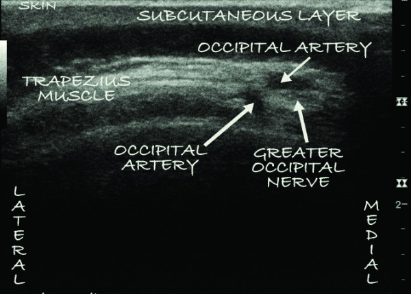
UltrasoundUltrasound of the neck locating the greater occipital nerve just next to the occipital artery, beneath the trapezius, used as a landmark for the anaesthetic block.
Source : Skaribas et al., Journal of medical case reports, 2011, figure 2 · CC BY
What is the place of exercise, and is there a superior approach?
Exercise is central to management, but the literature specific to occipital neuralgia remains limited. The recommendations rest largely on extrapolation from the data on cervicogenic headache, a neighbouring condition sharing upper cervical mechanisms. 💪 🎯 The reference cervical programme. The Demont 2023 meta-analysis (PM&R, PMID 35596553) of physiotherapy RCTs in cervicogenic headache concludes that manual therapy is beneficial in the short term and cervical exercise in the long term.⁶ The pivotal trial Jull 2002 (Spine, PMID 12221344, n=200) showed that a programme of specific exercises (deep flexor training through the craniocervical flexion test, CCFT, Jull 2008 PMID 18804003) and cervical manipulation significantly reduced headache frequency and intensity at 12 months.⁷,⁸ Recommended components (extrapolated from cervicogenic headache):- Deep neck flexor training (longus colli, longus capitis): a progressive CCFT protocol up to 10 seconds × 10 repetitions at 26-30 mmHg on biofeedback.⁸
- Cervical extensor endurance : isometric and endurance exercise.
- Scapular stabilisation : strengthening middle and lower trapezius, the rhomboids and serratus anterior.
- Stretching / mobility : upper trapezius, levator scapulae, scalenes, suboccipitals.
- Global motor control : postural integration, awareness of forward head posture.
Manual therapies, technologies: how effective are they really?
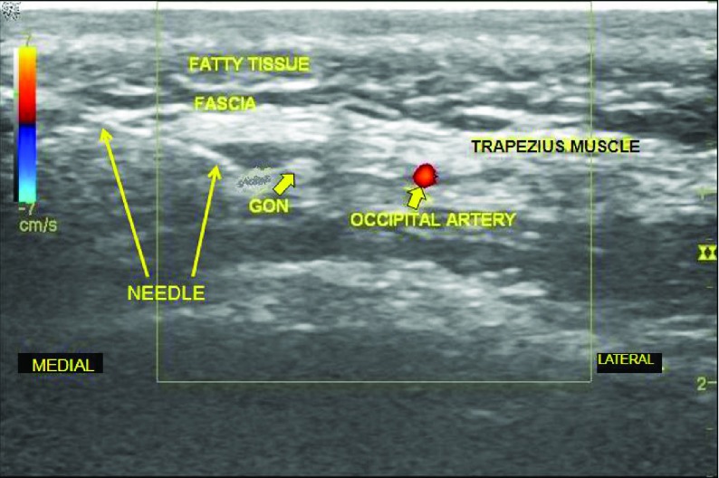
UltrasoundUltrasound of the neck showing the needle positioned just next to the greater occipital nerve, between the occipital artery and the trapezius muscle, before the anaesthetic injection.
Source : VanderHoek et al., Anesthesiology and pain medicine, 2013, figure 2 · CC BY
- TENS, ultrasound, low-level laser : indirect evidence, low to moderate, usable as symptomatic adjuncts.
- Pulsed radiofrequency (PRF) : a medical intervention (not physiotherapy), with robust evidence (Oliveira 2024).²
- Botulinum toxin : update 2025 (Toxicon) rates the efficacy in occipital neuralgia as “indeterminate”, for want of double-blind controlled studies of sufficient quality.¹¹
- Acupuncture / dry needling : the data are still insufficient specifically for ON.
| Modality | Level of evidence (GRADE) | Recommendation |
|---|---|---|
| GON block ± steroid (Shauly 2019, Cohen 2015) | Moderate to high | Recommended (diagnostic + therapeutic) |
| Pulsed radiofrequency (Oliveira 2024 SR) | Moderate (6-10 month follow-up) | Recommended as second or third line |
| Specific cervical physiotherapy (Demont 2023, Jull 2002) | Moderate (extrapolated from CGH) | Recommended first line |
| Manual therapy (Núñez-Cabaleiro 2022) | Moderate (short term) | Recommended alongside exercise |
| TENS, ultrasound, LLLT | Low | Possible adjunct, not as monotherapy |
| Botulinum toxin (Toxicon 2025) | Very low (indeterminate) | No formal recommendation |
| ONS / surgical decompression (Nguyen 2025) | Low to moderate (refractory cases) | Reserved for multimodal failure |
Beyond the physical: how do you educate the patient and address psychological factors?
Therapeutic pain education is a pillar of management. It aims to reframe pain as a modifiable neurobiological phenomenon (Su 2022, Treede 2008), to reduce fear of movement (kinesiophobia) and catastrophising, and to build self-efficacy.¹²,¹³ Validated strategies:- Explaining the mechanisms (central sensitisation, trigeminocervical convergence, distinguishing migraine / ON / CGH);
- Identifying personal triggers (sustained posture, stress, lack of sleep);
- Relaxation and stress-management techniques (paced breathing, mindfulness);
- Sleep hygiene (Finan 2013, on the sleep-pain relationship, PMID 24290442);¹⁴
- Cognitive behavioural therapy (CBT) where catastrophising, anxiety or depression coexist (Ehde 2014, Am Psychol, meta-analysis of CBT in chronic pain, PMID 24547801).¹⁵
Critique and controversy around treatment
🧐 The therapeutic field suffers from several limits:- Few RCTs devoted specifically to occipital neuralgia ; most of the literature is extrapolated from cervicogenic headache or from migraine with occipital tenderness.¹,⁶
- Heterogeneous protocols (volumes injected, frequency of blocks, duration of PRF follow-up), which limits comparison.
- A moderate level of evidence at best for most interventions (Deuel 2024 says so explicitly).¹
- No standardised consensus on the threshold for moving from conservative to invasive treatment (how many blocks? after how many months?).
- A non-negligible risk of iatrogenic harm from repeated interventions (systemic corticosteroids, ONS, surgery).
Key points
- ✅ A stepped algorithm : physiotherapy/education → GON block → pulsed radiofrequency → surgery.
- 💉 The GON block is both diagnostic and therapeutic (Shauly 2019, Cohen 2015), ideally ultrasound-guided.
- 💪 Specific cervical physiotherapy : deep flexor training (CCFT), C0-C2 manual therapy, counter-extension exercise.
- 🧠 Pulsed radiofrequency is effective for 6-10 months (Oliveira 2024 SR/MA).
- 🗣️ Therapeutic education + CBT are non-negotiable where the condition becomes chronic.
Bibliography
- Deuel D, Sandgren A, Nelson EO, Cropes M, Deacon A, Houdek T, Abd-Elsayed A. Conservative Management of Occipital Neuralgia Supported by Physical Therapy: A Review of Available Research and Mechanistic Rationale to Guide Treatment. Curr Pain Headache Rep. 2024;28(12):1321-1331. PMID 38958920.
- Oliveira K, Dhondt N, Englesakis M, Goel A, Hoydonckx Y. Pulsed Radiofrequency Neuromodulation of the Greater Occipital Nerve for the Treatment of Headache Disorders in Adults: A Systematic Review. Can J Pain. 2024;8(1):2355571. PMID 38915302.
- Nguyen AT, Li RA, Galiano RD, Ellis MF. Nerve Decompression in Occipital Neuralgia: A Systematic Review and Meta-analysis. J Craniofac Surg. 2025 (Epub ahead of print). PMID 40367499.
- Shauly O, Gould DJ, Sahai-Srivastava S, Patel KM. Greater Occipital Nerve Block for the Treatment of Chronic Migraine Headaches: A Systematic Review and Meta-Analysis. Plast Reconstr Surg. 2019;144(4):943-952. PMID 31568309.
- Cohen SP, Peterlin BL, Fulton L, et al. Randomized, double-blind, comparative-effectiveness study comparing pulsed radiofrequency to steroid injections for occipital neuralgia or migraine with occipital nerve tenderness. Pain. 2015;156(12):2585-2594. PMID 26447705.
- Demont A, Lafrance S, Gaska C, et al. Efficacy of physiotherapy interventions for the management of adults with cervicogenic headache: A systematic review and meta-analyses. PM&R. 2023;15(5):613-628. PMID 35596553.
- Jull G, Trott P, Potter H, et al. A randomized controlled trial of exercise and manipulative therapy for cervicogenic headache. Spine. 2002;27(17):1835-1843. PMID 12221344.
- Jull GA, O'Leary SP, Falla DL. Clinical assessment of the deep cervical flexor muscles: the craniocervical flexion test. J Manipulative Physiol Ther. 2008;31(7):525-533. PMID 18804003.
- Núñez-Cabaleiro P, Leirós-Rodríguez R. Effectiveness of manual therapy in the treatment of cervicogenic headache: A systematic review. Headache. 2022;62(3):271-283. PMID 35294051.
- Xu Y, Ling Y. Comparative safety and efficacy of manual therapy interventions for cervicogenic headache: a systematic review and network meta-analysis. Front Neurol. 2025;16:1566764. PMC 12123087.
- Update on botulinum toxin for occipital neuralgia. Toxicon. 2025. doi:10.1016/j.toxicon.2025.01.011.
- Su M, Yu S. Central Sensitization in Migraine: A Narrative Review. J Pain Res. 2022;15:2673-2682. PMC 9464439.
- Treede RD, Jensen TS, Campbell JN, et al. Neuropathic pain: redefinition and a grading system. Neurology. 2008;70(18):1630-1635. PMID 18003941.
- Finan PH, Goodin BR, Smith MT. The association of sleep and pain: an update and a path forward. J Pain. 2013;14(12):1539-1552. PMID 24290442.
- Ehde DM, Dillworth TM, Turner JA. Cognitive-behavioral therapy for individuals with chronic pain: efficacy, innovations, and directions for research. Am Psychol. 2014;69(2):153-166. PMID 24547801.
How do you secure lasting recovery and prevent recurrence of occipital neuralgia?
How do you make the patient an active participant in their recovery through self-management?
1. Pain education. The Deuel 2024 review stresses that pain neuroscience education is a prerequisite for any effective rehabilitation in occipital neuralgia.¹ It covers explaining central sensitisation, distinguishing neuralgia from migraine and cervicogenic headache, and dismantling limiting beliefs (“I must protect my neck”, “the more it hurts, the more damage I am doing”). 2. Ergonomics and posture. Forward head posture increases the mechanical stress on the upper cervical segments and the cervicothoracic junction. The classic recommendations:- Screen at eye height, at arm's length;
- Regular breaks (20-20-20: every 20 minutes, for 20 seconds, look 20 feet away, about 6 m);
- Less cumulative time in cervical flexion over a smartphone (“text neck”);
- A pillow that allows neutral cervical alignment.
- Deep flexor activation (head nodding against biofeedback or in lying);
- Cervical extensor endurance (prone neck holds);
- Scapular strengthening (Y/T/W, rowing);
- Gentle stretching (upper trapezius, levator scapulae, suboccipitals), avoiding aggressive stretching while symptoms are active;
- Upper thoracic mobility (cat-camel, extensions over a foam roller).
- Regular hours, a cool, dark environment;
- Less screen time in the evening;
- Relaxation techniques (paced breathing, body scan, mindfulness);
- Regular physical activity (but not late in the evening);
- Limiting stimulants (no caffeine after 2 pm).
When and how should a safe return to activity and to sport be planned?
🏃 The return to activity should be guided by functional criteria, not by a fixed calendar; there is no published standard specific to occipital neuralgia. Suggested progression criteria:- Pain at rest < 3/10 on the VAS;
- Pain-free functional cervical ranges (rotation, flexion-extension);
- No symptoms during activities of daily living for 1-2 weeks.
- Phase 1 : low-impact aerobic activity (walking, upright cycling); no cervical stress.
- Phase 2 : sport-specific movements at moderate intensity and without impact (light jogging, swimming with a board to limit cervical extension).
- Phase 3 : progressively increasing complexity and intensity; introduction of specific strengthening. The non-aggravation rule: any significant increase in pain the following day → back to the previous phase.
- Phase 4 : full return, without restriction, once every sport-specific movement is tolerated at full intensity.
Critique and controversy
🧐 The scientific literature has several limits:- Considerable heterogeneity of rehabilitation protocols, most of them extrapolated from cervicogenic headache or from common neck pain.¹,³
- A lack of longitudinal studies specific to preventing recurrence in occipital neuralgia.
- No consensus on the criteria for returning to sport or to high-risk occupational activity.
- Under-assessment of psychosocial factors in the classic treatment studies, a point Deuel 2024 emphasises at length.¹
Key points
- Autonomy comes through pain education, ergonomics and a home exercise programme.
- A cervical programme built on the CCFT (Jull 2008) plus extensor endurance, scapular work and stretching.
- Sleep hygiene + stress management modulate the sensitivity of the nervous system (Finan 2013).
- Return to activity on functional criteria, not on a fixed delay.
- ⚠ Longitudinal data and return-to-sport criteria for ON specifically are a major gap in the literature.
Bibliography
- Deuel D, Sandgren A, Nelson EO, et al. Conservative Management of Occipital Neuralgia Supported by Physical Therapy. Curr Pain Headache Rep. 2024;28(12):1321-1331. PMID 38958920.
- Jull G, Trott P, Potter H, et al. A randomized controlled trial of exercise and manipulative therapy for cervicogenic headache. Spine. 2002;27(17):1835-1843. PMID 12221344.
- Demont A, Lafrance S, Gaska C, et al. Efficacy of physiotherapy interventions for the management of adults with cervicogenic headache: SR and meta-analyses. PM&R. 2023;15(5):613-628. PMID 35596553.
- Finan PH, Goodin BR, Smith MT. The association of sleep and pain: an update and a path forward. J Pain. 2013;14(12):1539-1552. PMID 24290442.
- Jull GA, O'Leary SP, Falla DL. Clinical assessment of the deep cervical flexor muscles: the craniocervical flexion test. J Manipulative Physiol Ther. 2008;31(7):525-533. PMID 18804003.
- Núñez-Cabaleiro P, Leirós-Rodríguez R. Effectiveness of manual therapy in cervicogenic headache: SR. Headache. 2022;62(3):271-283. PMID 35294051.
- Page P. Cervicogenic headaches: an evidence-led approach to clinical management. Int J Sports Phys Ther. 2011;6(3):254-266. PMC 3201065.
What do real clinical cases teach us about occipital neuralgia?
Analysis of a “classic” case: from assessment to conservative resolution.
The literature offers few rigorously published physiotherapy case reports on isolated occipital neuralgia. The Deuel 2024 review (Curr Pain Headache Rep), the first mechanistic synthesis devoted to physiotherapy management of ON, identifies no specific RCT and rests on extrapolation from cervicogenic headache data.² The typical features of a successful conservative pathway (drawn together from Deuel 2024 and the Jull 2002 / Demont 2023 protocols):- Phase 1 (weeks 1-2) : pain education, identifying triggers, ergonomics, gentle isometric neck exercise;
- Phase 2 (weeks 3-6) : manual therapy (C0-C2 mobilisations, suboccipital soft tissue), progression of the CCFT, scapular integration;
- Phase 3 (weeks 7-12) : full strengthening, global motor control, return to activity;
- Phase 4 (beyond) : autonomous maintenance, telephone follow-up or a consultation every 3-6 months to adjust prevention.
The diagnostic challenge: when occipital neuralgia mimics another condition.
🎯 Published case 1: vascular compression mimicking hemifacial migraine. Choi et al. (Case Rep Neurol Med 2017, PMC5346380) report a case of hemifacial pain with referred hemihypoaesthesia, initially attributed to migraine and finally diagnosed as occipital neuralgia secondary to pathological vascular contact between the occipital artery and the GON.⁵ Surgical neurovascular decompression resolved the symptoms. This case illustrates trigeminocervical convergence and the risk of misattributing the diagnosis. 🎯 Published case 2: occipital neuralgia after COVID-19 vaccination. Several open-access case reports (2022-2023) have described series of patients developing occipital neuralgia in the days after a second dose of an mRNA vaccine.⁶ The proposed mechanism is inflammatory or autoimmune, with no formal proof of causation. The evidence remains low level (case series, reporting bias), but the signal is worth documenting for pharmacovigilance. 🎯 Published case 3: an atypical presentation with maxillary radiation. Anatomical variations in the course of the GON (Cesmebasi 2015) explain why some patients have radiation into distant territories (maxilla, external ear), which can wrongly point towards trigeminal neuralgia or an ENT condition.⁷A complex case: serious secondary causes.
🧬 Published case: schwannoma of the C2 root. Krishnan et al. (J Neurosci Rural Pract 2015, PMID 26752925) report a benign schwannoma of the C2 root in a patient with refractory occipital neuralgia.⁸ The anaesthetic blocks gave only fleeting relief, which prompted the cervical MRI that revealed the mass. Surgical resection brought complete resolution. This case is the paradigmatic illustration of the “resistance to well-conducted treatment” red flag. 🧬 Published case: C2 cavernoma. An intramedullary vascular malformation at C2 (PMC5898127) was found in a patient with chronic refractory ON, underlining that MRI must include the upper cord and the craniocervical junction.⁹ 🧬 Published case: Chiari I malformation. The association between ON and Chiari I is documented (PMC5788399); management requires a dedicated neurosurgical approach.¹⁰ 🧬 Published case: occipital condyle metastasis. One case (PMC9985513) shows a bone metastasis revealed by an occipital condyle syndrome mimicking ON, in a patient with a history of cancer.¹¹ It confirms the importance of the “history of cancer” red flag. 🧬 Published case: the paediatric population. Villeneuve et al. (J Neurosurg Pediatr 2023, PMID 37548543) report a series of paediatric ON cases requiring surgical management, illustrating that a secondary cause should be sought more systematically in children and young adults.¹²Critique and controversy
🧐 Published clinical cases have inherent limits:- Publication bias : spectacular cases (exceptional treatment successes, highly atypical presentations) are over-represented.¹
- A low level of evidence : CEBM 5 / GRADE very low. A case illustrates, it never proves.
- The absence of longitudinal cohorts on occipital neuralgia specifically; the Melchior 2025 meta-analysis stresses this gap.¹³
- Heterogeneous diagnostic criteria between the published cases (some do not apply ICHD-3, others have no confirmatory anaesthetic block).
Key points
- The clinical cases show the range of causes : compressive (muscular, vascular), tumour (schwannoma), malformation (Chiari), metastatic, post-traumatic.
- The Resistance to well-conducted conservative treatment is the major red flag calling for MRI.
- Occipital neuralgia is a great imitator (migraine, cervicogenic headache, trigeminal neuralgia) → rigorous differential diagnosis.
- In children and young adults: look more systematically for a secondary cause (Villeneuve 2023).
- ⚠ The level of evidence of a case report is 5 (the weakest): follow the meta-analyses for treatment decisions.
Bibliography
- Nissen T, Wynn R. The clinical case report: a review of its merits and limitations. BMC Res Notes. 2014;7:264. PMID 24758689.
- Deuel D, Sandgren A, Nelson EO, et al. Conservative Management of Occipital Neuralgia Supported by Physical Therapy. Curr Pain Headache Rep. 2024;28(12):1321-1331. PMID 38958920.
- Shauly O, Gould DJ, Sahai-Srivastava S, Patel KM. Greater Occipital Nerve Block for the Treatment of Chronic Migraine Headaches: SR/MA. Plast Reconstr Surg. 2019;144(4):943-952. PMID 31568309.
- Cohen SP, Peterlin BL, Fulton L, et al. RCT comparing pulsed radiofrequency to steroid injections for occipital neuralgia. Pain. 2015;156(12):2585-2594. PMID 26447705.
- Choi JG, Kang YJ, Kim BC, Lee CG, Choi SK. Hemifacial Pain and Hemisensory Disturbance Referred from Occipital Neuralgia. Case Rep Neurol Med. 2017;2017:3827230. PMC 5346380.
- Occipital Neuralgia after COVID-19 Vaccination: Case report (open access). 2023.
- Cesmebasi A, Muhleman MA, Hulsberg P, et al. Occipital neuralgia: anatomic considerations. Clin Anat. 2015;28(1):101-108. PMID 25244129.
- Krishnan A, Kartikueyan R, Chowdhury SR, Das S. Schwannoma of the greater occipital nerve. J Neurosci Rural Pract. 2015;6(4):634-636. PMID 26752925.
- Occipital neuralgia from C2 cavernous malformation. Surg Neurol Int. 2018. PMC 5898127.
- Occipital Neuralgia in Chiari I Malformation. Acta Neurochir Suppl. 2018. PMC 5788399.
- Occipital condyle syndrome, metastatic breast cancer (case report PMC). 2023. PMC 9985513.
- Villeneuve LM, Coulibaly NJ, Raza SM, Poinson B, Chrusciel D, Desai VR. Surgical management of pediatric occipital neuralgia: a single-center experience of an uncommon pathology. J Neurosurg Pediatr. 2023;32(4):514-521. PMID 37548543.
- Melchior AG, Al-Khazali S, Christensen RH, et al. Epidemiology and clinical features of occipital neuralgia: SR/MA. Cephalalgia. 2025;45(2):3331024251317595. PMID 40017062.
How do you apply these recommendations concretely in your practice?
When and to which other health professionals should you refer?
Red flags call for prompt medical referral. The IFOMPT framework of Finucane et al. (JOSPT 2020, PMID 32438853) remains the French and international reference for spinal red flags.¹ Verhagen et al. (Pain 2017, PMID 28708761) showed, however, that most red flags taken in isolation have a low positive predictive value, hence the need to read them as a cluster of findings.² The Cochrane review Han 2023 (CD014461) confirms this probabilistic logic for vertebral fracture in low back pain: combining several flags significantly raises the post-test probability, whereas the absence of red flags reasonably rules out serious disease.³ Specifically for occipital neuralgia, the situations calling for medical referral (GP, neurologist, pain specialist or the emergency department depending on context) are:- A thunderclap headache or one of sudden onset → subarachnoid haemorrhage, arterial dissection;
- A focal neurological deficit (weakness, ataxia, visual disturbance);
- A known history of cancer or unexplained weight loss;
- Fever + neck stiffness → infection;
- Resistance to well-conducted conservative treatment (4-6 weeks) → cervical and brain MRI;
- Recurrence after posterior cervical surgery → suspected postoperative fibrosis or iatrogenic injury.
- A GP or neurologist (differential diagnosis, prescribing, imaging);
- A pain specialist (nerve blocks, pulsed radiofrequency);
- A neurosurgeon (surgical cases: decompression, ONS);
- A psychologist or psychiatrist (CBT, managing comorbidity);
- An ergonomist or occupational physician (workstation adaptation).
How do you measure outcomes and overcome barriers to implementation?
📈 Measuring outcomes with validated PROMs. Standardised follow-up should combine:- VAS / NRS for pain (at rest, on exertion, during an attack);
- The Neck Disability Index (NDI) for cervical functional impact;
- HIT-6 or MIDAS for headache-specific impact;
- The Pain Catastrophizing Scale (PCS) and the Tampa Scale of Kinesiophobia (TSK-11) for the psychological factors;
- Physical performance measures: cervical ranges, CCFT score (mmHg of pressure held × seconds).
- Targeted continuing education on critical appraisal of papers and applied statistics;
- Using evidence syntheses (Cochrane Reviews, BMJ Best Practice, the Physio Learning syntheses) to save time;⁴
- Audit and feedback on your own practice (rate of PROM use, adherence to the recommendations);
- Clinical leadership and mentoring by “EBP champions”;
- Technological integration (patient record software with PROMs built in).
Critique and controversy: beyond the checklist
🧐 Three critical tensions run through the application of EBP in practice:- Reductionism. Red flags can lead to an anxiety-driven “hunt for pathology” that neglects the biopsychosocial context. Conversely, a PROM score captures only part of the human experience. Expert competence uses these tools without being confined to them.
- The knowing-doing gap. The barriers identified (time, skills, support) have been strikingly stable for 20 years. That suggests the problem is not purely individual: the health systems (reimbursement models, administrative burden, initial training) need rethinking to favour EBP.
- Standardisation vs personalisation. Core Outcome Sets and standardised care pathways are essential for research and quality, but they can pull against a patient-centred approach. Clinical artistry lies in reconciling the two.
Key points
- The red flags should be read as a cluster (Verhagen 2017, Han 2023); the Finucane 2020 framework remains the reference.
- The yellow flags (kinesiophobia, catastrophising) call for collaboration with a psychologist.
- The interprofessional collaboration (doctor, neurologist, pain specialist, psychologist, neurosurgeon) is central in complex cases.
- Measuring outcomes with validated PROMs (VAS, NDI, HIT-6, MIDAS, PCS, TSK-11) is non-negotiable.
- Overcoming the barriers to EBP requires a multifaceted approach (training, evidence syntheses, audit, leadership).
- Beyond the checklist: holistic clinical reasoning, shared decision-making, and attention to the systems that hold EBP back.
Bibliography
- Finucane LM, Downie A, Mercer C, Greenhalgh SM, Boissonnault WG, Pool-Goudzwaard AL, et al. International Framework for Red Flags for Potential Serious Spinal Pathologies. J Orthop Sports Phys Ther. 2020;50(7):350-372. PMID 32438853.
- Verhagen AP, Downie A, Maher CG, Koes BW. Most red flags for malignancy in low back pain guidelines lack empirical support: a systematic review. Pain. 2017;158(10):1860-1868. PMID 28708761.
- Han CS, Hancock MJ, Downie A, et al. Red flags to screen for vertebral fracture in people presenting with low back pain. Cochrane Database Syst Rev. 2023;Issue 8:CD014461. doi:10.1002/14651858.CD014461.pub2.
- Cumpston M, Li T, Page MJ, et al. Updated guidance for trusted systematic reviews: a new edition of the Cochrane Handbook for Systematic Reviews of Interventions. Cochrane Database Syst Rev. 2019;10:ED000142. PMID 31643080.
- Deuel D, Sandgren A, Nelson EO, et al. Conservative Management of Occipital Neuralgia Supported by Physical Therapy. Curr Pain Headache Rep. 2024;28(12):1321-1331. PMID 38958920.
- Núñez-Cabaleiro P, Leirós-Rodríguez R. Effectiveness of manual therapy in cervicogenic headache: SR. Headache. 2022;62(3):271-283. PMID 35294051.
And after this article?
This article is part of a collection of evidence-based clinical syntheses. A question, a comment, a correction to suggest? Contact us directly through the WhatsApp button at the bottom right of the screen.

