















In brief
Rotator cuff calcific tendinopathy (RCCT) is an active cellular process marked by deposits of calcium hydroxyapatite crystals within the tendon, above all supraspinatus (about 80 % of cases), and not simple passive wear. It alternates between a chronic form with mechanical pain and an acute, highly painful form corresponding to the resorptive phase; diagnosis combines an anteroposterior radiograph with Doppler ultrasound, and radiographic and clinical findings frequently dissociate. First-line management rests on therapeutic exercise, shockwave therapy and ultrasound-guided lavage, the course often being self-limiting. Prevalence in the general population ranges from 2.7 to 22 %.
Clinical synthesis based on the most recent meta-analyses and international consensus statements: the EFORT Open Reviews network meta-analysis 2025, the Guido 2025 scoping review, Ricci Diagnostics 2022, the Brindisino 2024 ESWT SR/MA, Louwerens JSES 2014, and the 2018-2025 epidemiological data.
Clinical summary
- Rotator cuff calcific tendinopathy (RCCT) is an active cellular condition characterised by deposits of calcium hydroxyapatite crystals within the tendon, and not simple degenerative wear (Uhthoff 1997, Sansone 2018).
- Prevalence in the general population is between 2.7 % and 22 %, much of it asymptomatic. The peak falls between 40 and 60 years, with a female predominance, and it is more frequent in sedentary workers than in manual workers (Sansone 2015 Rheumatol Int).
- The supraspinatus tendon is the most frequent site (about 80 % of cases), followed by infraspinatus and subscapularis (Guido 2025).
- Established metabolic risk factors: diabetes (HR 1.27 over 8 years, Su 2021), thyroid disorders and the metabolic syndrome (Bechay 2020 Phys Sportsmed).
- The Uhthoff pathophysiological model (1997) describes three phases: precalcific (silent), calcific (formation, resting, resorption) and postcalcific. The resorptive phase triggers intense acute pain through an inflammatory reaction.
- The course is often self-limiting : Cho 2010 shows clinical improvement in most patients under conservative treatment, with complete or partial resorption of the deposits in about two thirds of cases in the medium term.
- The diagnosis rests on a triad: the history (acute resorptive form vs chronic formative form), a clinical examination of low specificity (Neer and Hawkins impingement tests are frequently positive), and imaging with an anteroposterior radiograph in 3 rotations plus Doppler ultrasound as the reference (Ricci 2022 Diagnostics).
- The Gartner classification (1993), or Molini's, correlated with the clinical phase, guides the decision: type I, dense and homogeneous (formation/resting), is often little symptomatic, while type III, fluffy (resorption), is very painful but carries an excellent spontaneous prognosis.
- The radiographic-clinical dissociation is frequent : the size of the deposit does not correlate with symptom severity (Cho 2010, Drummond 2021).
- The EFORT 2025 network meta-analysis (Liu, 33 RCTs, 26 modalities) puts full physiotherapy with high-energy ESWT first for function, radial SWT with PT first for pain, and UGN plus subacromial injection first for resorption.
- Therapeutic exercise is non-negotiable: cuff strengthening (infraspinatus, subscapularis), scapular stabilisers and load management. No specific protocol is superior to another (Pieters 2020 JOSPT, Michener 2020).
- By contrast, corticosteroid injections give weak, transient relief (Mohamadi 2017 CORR, meta-analysis of 11 studies). To be reserved for refractory hyperalgesic flares.
- Similarly, conventional therapeutic ultrasound has no robust evidence in RCCT (Page Cochrane 2016 CD012225).
- The clinical presentation can mimic septic arthritis : acute pain, fever, leucocytosis, raised CRP: a serious trap to know about (Aljumaan 2025 Cureus).
- Red flags and differential diagnoses: frozen shoulder, rotator cuff tear, acromioclavicular osteoarthritis, tumour, infection.
- The return to sport should rest on objective criteria (pain, range of motion, strength ≥ 90 % of the healthy side, functional tests), not on elapsed time (Wilk 2018 IJSPT, Schwank 2022).
- Recurrence after successful treatment is reported at between 6 and 20 % in the long term, which is why prevention through self-management belongs in the plan.
Contents
- What are the fundamentals to know about calcific tendinopathy of the shoulder?
- How do you assess and diagnose calcific tendinopathy of the shoulder with certainty?
- Which treatment strategies are the most effective for calcific tendinopathy of the shoulder?
- Where do you start? What is the recommended hierarchy of interventions?
- What is the place of exercise, and is there a superior approach?
- Shockwave, ultrasound-guided lavage, manual therapies: how effective are they really?
- Beyond the physical: how do you educate the patient and address psychological factors?
- How do you secure lasting recovery and prevent recurrence of calcific tendinopathy of the shoulder?
- What do real clinical cases teach us about calcific tendinopathy of the shoulder?
- How do you apply these recommendations concretely in your practice?
What are the fundamentals to know about calcific tendinopathy of the shoulder?
How is this condition defined, who does it affect and what are the risk factors?
The narrative review by Sansone and colleagues 2018, published in Orthopedic Research and Reviews, and the Guido 2025 scoping review (50 studies included, Shoulder & Elbow) converge on the definition: RCCT is characterised by deposits of calcium phosphate crystals (essentially hydroxyapatite, Ca₁₀(PO₄)₆(OH)₂) within the rotator cuff tendons.²,³ It is neither a chronic inflammatory process nor simple wear: it is ectopic chondrogenesis mediated by the tendon cells, whose exact trigger remains only partly elucidated (ischaemic hypothesis, metabolic hypothesis, repeated mechanical hypothesis).¹,² Epidemiologically, the recent data consolidate several findings:- Prevalence in the general population is between 2.7 % and 22 % depending on the population and the imaging methods used; a large share is entirely asymptomatic and found incidentally.²,³
- Peak incidence between 40 and 60 years, with a clear sex difference: female predominance (a ratio of about 2:1).²,³,⁴
- More frequent in sedentary workers than in manual workers, a strong argument against repeated mechanical overload as the sole cause.²,⁵
- The supraspinatus tendon is by far the most often involved (about 80 % of cases), followed by infraspinatus (15 %) and subscapularis (5 %).³
- First, diabetes significantly increases the risk: Su's 2021 matched cohort from the Taiwanese national database (42,915 patients with diabetes vs 171,660 controls) finds an adjusted HR = 1.27 (95 % CI 1.18-1.38) at 8 years of developing RCCT.⁵
- Second, thyroid disorders (mainly hypothyroidism) have had an established association since Harvie 2007, confirmed in several more recent cohorts.²,⁶
- Third, the metabolic syndrome as a whole (obesity + hypertension + dyslipidaemia + insulin resistance) correlates with RCCT and with a more prolonged course (Bechay 2020 Phys Sportsmed).⁶
📊 Anatomical distribution of the calcific deposits in the rotator cuff
Synthesis of Guido 2025 (scoping review, 50 studies) and Sansone 2018
Supraspinatus accounts for 4 cases in 5. That predominant location is explained by its critical vascular zone (about 1 cm downstream of the insertion on the greater tuberosity) and by the biomechanical stresses of the acromial arch.
What happens in the tendon, and how does the condition evolve naturally?
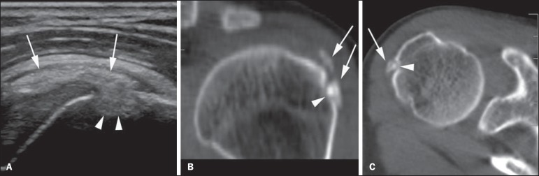
UltrasoundShoulder ultrasound followed by CT: intratendinous calcification of the infraspinatus with adjacent cortical bone erosion, arrows on the calcific deposit and the intraosseous migration.
Source : Nogueira-Barbosa et al., Radiologia Brasileira, 2015, figure 1 · CC BY
- Precalcific phase (silent): fibrocartilaginous metaplasia of the tenocytes, with no visible deposit yet and no notable symptoms.¹
- Calcific phase divided into:
- Formation : calcium accumulates in a dense, chalky form, the deposit is well demarcated, with a clear acoustic shadow on ultrasound. The patient is often little symptomatic, or reports chronic mechanical discomfort.¹,⁸
- Resting : a stable phase, with a possible plateau lasting several months.
- Resorption ⚠️: the deposit becomes pasty (“toothpaste”), loses its outline and recruits an intense inflammatory reaction mediated by phagocytes (macrophages, neutrophils). This is the most painful phase (acute, sleep-disturbing, throbbing pain) but paradoxically the signal of imminent self-resolution.¹,⁷,⁹
- Postcalcific phase : tendon remodelling, healing, return to homeostasis. Often painless, but it can leave partial tendon sequelae.¹,⁷
📈 Uhthoff's three-phase model (1997) and the trajectory of pain
Pain peaks in the resorptive phase, paradoxically the signal of healing
The resorptive phase (maximal pain intensity, often VAS 8-10) corresponds to the peak of neovascularisation visible on power Doppler and is paradoxically associated with an excellent spontaneous prognosis.⁷,⁹
Key points
- RCCT is an active cellular process (ectopic chondrogenesis mediated by the tenocytes), not passive wear (Uhthoff 1997).
- Prevalence 2.7-22 %, peak at 40-60 years, female predominance, ~80 % supraspinatus, more frequent in sedentary than in manual workers.
- Established metabolic risk factors: diabetes (HR 1.27), hypothyroidism, metabolic syndrome.
- The three-phase model (precalcific, calcific with formation/resting/resorption, postcalcific) illuminates the clinical picture.
- The resorptive phase is the most painful but marks imminent self-resolution: a key message to pass on to the patient.
- The course is often self-limiting: clinical improvement in most patients under conservative treatment (Cho 2010), but a calcification > 1 cm predicts failure (Drummond 2021).
Bibliography
- Uhthoff HK, Loehr JW. Calcific Tendinopathy of the Rotator Cuff: Pathogenesis, Diagnosis, and Management. J Am Acad Orthop Surg. 1997;5(4):183-191. PMID 10797220.
- Sansone V, Maiorano E, Galluzzo A, Pascale V. Calcific tendinopathy of the shoulder: clinical perspectives into the mechanisms, pathogenesis, and treatment. Orthop Res Rev. 2018;10:63-72. PMC6209365.
- Guido F, Venturin D, De Santis A, Giovannico G, Brindisino F. Clinical features in rotator cuff calcific tendinopathy: A scoping review. Shoulder Elbow. 2025. doi:10.1177/17585732241244515 · PMC11562322.
- Speed CA, Hazleman BL. Calcific Tendinitis of the Shoulder. N Engl J Med. 1999;340(20):1582-1584. doi:10.1056/NEJM199905203402011.
- Su YC, Chung CH, Wang CH, et al. Increased risk of shoulder calcific tendinopathy in diabetes mellitus: A nationwide, population-based, matched cohort study. Int J Clin Pract. 2021;75(8):e14549. doi:10.1111/ijcp.14549.
- Bechay J, Lawrence C, Namdari S. Calcific tendinopathy of the rotator cuff: a review of operative versus nonoperative management. Phys Sportsmed. 2020;48(3):241-246. PMID 31893972.
- Drummond Junior M, Ayinon C, Rodosky M, Vyas D, Lesniak B, Lin A. Predictive factors for failure of conservative management in the treatment of calcific tendinitis of the shoulder. JSES Int. 2021;5(3):469-473. PMID 34136856.
- Cho NS, Lee BG, Rhee YG. Radiologic course of the calcific deposits in calcific tendinitis of the shoulder: does the initial radiologic aspect affect the final results? J Shoulder Elbow Surg. 2010;19(2):267-272. PMID 19800263.
- Ricci V, Mezian K, Chang KV, Ozcakar L. Clinical/Sonographic Assessment and Management of Calcific Tendinopathy of the Shoulder: A Narrative Review. Diagnostics (Basel). 2022;12(12):3097. PMID 36553104.
- Merolla G, Singh S, Paladini P, Porcellini G. Calcific tendinitis of the rotator cuff: state of the art in diagnosis and treatment. J Orthop Traumatol. 2016;17(1):7-14. PMID 26163832.
How do you assess and diagnose calcific tendinopathy of the shoulder with certainty?
Which questions should you ask to understand the patient and their history?
The history is the most discriminating step. 🤔 It should explore:- Patient profile : a woman between 40 and 60 years, occupation (sedentary vs manual), metabolic comorbidities (diabetes, thyroid disorder), personal or family history of RCCT.²,³
- Characteristics of the pain : distinguish two distinct pictures:
- Chronic form : dull mechanical pain, discomfort on elevation, painful side-lying, a course over several months. Often corresponds to a formation or resting phase.²
- Acute hyperalgesic form : pain of abrupt onset, throbbing, sleep-disturbing, sometimes described as one of “the worst pains ever experienced”. The patient holds the arm in a sling, with near-total loss of function. Corresponds to the inflammatory resorptive phase.²,⁴
- Cyclical history : previous painful episodes, periods of spontaneous remission, earlier traumatic episodes.²,⁵
- Aggravating and relieving factors : triggering movements (elevation above 60-120°), night positions, specific ADLs.²
Which clinical tests should you perform, and which other conditions should you rule out?
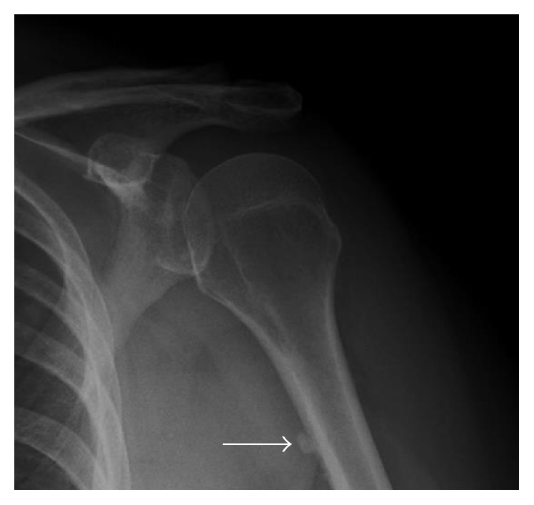
X-rayAnteroposterior shoulder radiograph: linear calcification along the anterior cortex of the humeral diaphysis, arrow, calcific tendinopathy of pectoralis major.
Source : Beckmann, Radiology Research and Practice, 2016, figure 9 · CC BY
- Impingement tests are frequently positive in RCCT through the mass effect of the deposit, but their positivity is not specific.²,⁶
- Cuff muscle tests (Jobe, lift-off, belly-press) can be painful without an associated tear, so do not conclude too early.⁶
- most clinically useful sign is exquisite, very focal pain on palpation over the greater tuberosity or the supraspinatus insertion.⁶
- Plain radiography first line: anteroposterior views in neutral, internal and external rotation plus a scapular Y view. Visualisation of the deposit, its location and the measurement of its size.²,⁷
- Ultrasound as the reference for characterisation: excellent sensitivity, no ionising radiation, allows dynamic assessment, precise measurement, analysis of morphology (arciform, fragmented, nodular) and evaluation of power Doppler around the deposit: pericalcific hyperaemia is strongly associated with the resorptive phase (Ricci 2022, Chiou 2010).⁷,⁸
- MRI second line only, where an associated tear is suspected or a preoperative work-up is needed.²
Red flags and differential diagnoses not to be missed
- Fever + leucocytosis + raised CRP : septic arthritis vs the pseudo-septic arthritis of the resorptive phase, a critical distinction (see Aljumaan 2025, chapter 5). Any doubt → joint aspiration and prompt specialist opinion.
- Non-mechanical night pain + weight loss : rule out bone tumour, metastasis, lymphoma.
- Established motor deficit : rule out massive cuff tear, axillary nerve palsy (following an injection or procedure), brachial plexus involvement.
- Frozen shoulder : global passive restriction (external rotation above all), which differs from simple antalgic restriction of active movement.
- Acromioclavicular or glenohumeral osteoarthritis : referred pain, crepitus, specific radiographic changes.
- Referred neck pain : test the cervical spine systematically.
Should patients be classified, and what are the benefits?
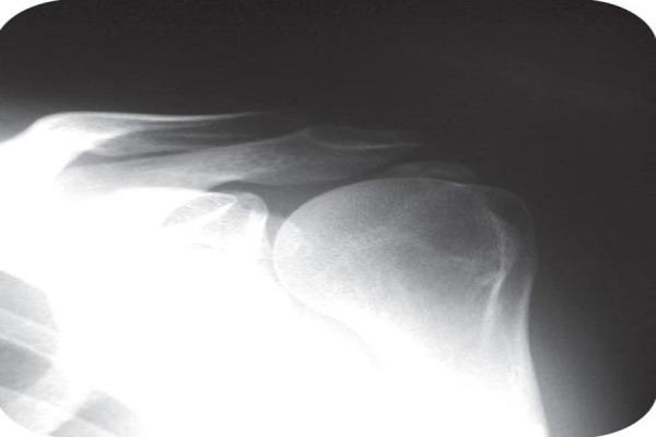
X-rayAnteroposterior radiograph of the left shoulder: calcification overlying the supraspinatus tendon, above the humeral head, a classic radiographic sign of calcific tendinopathy.
Source : Schuh et al., Journal of Orthopaedic Case Reports, 2011, figure 2 · CC BY-NC-SA
- Gartner's classification (1995, Orthopade), based on the radiograph:⁹
- Type I : a dense, homogeneous deposit with sharp outlines and a clear acoustic shadow on ultrasound. Corresponds to the formation/resting phase. Often little symptomatic, or chronically painful. Slower spontaneous prognosis; the better target for mechanical thermolysis (UGN, focused ESWT).
- Type II : a dense deposit with less sharp outlines, fragmented or multilobed. A transitional phase.
- Type III : a fluffy, translucent, poorly defined deposit. Corresponds to the resorptive phase. Very painful but with an excellent spontaneous prognosis and a favourable response to needle aspiration and lavage.
- The Molini / Sconfienza sonographic classification : precise morphology (arciform, fragmented, nodular, cystic) correlated with Gartner's types.⁷
⚙️ Diagnostic decision algorithm for suspected RCCT
The hierarchy of investigations and the clinical decision points
If the radiograph shows a characteristic deposit → Doppler ultrasound to stratify the phase (formation/resorption) and the exact site. If in doubt → ultrasound first line.
Critique and controversy
The correlation between the size or type of the calcification and the intensity of symptoms is notoriously weak.⁵,⁸ Bulky Type I calcifications can be asymptomatic, while small Type III deposits can trigger disabling pain. The transition from one type to another is not linear and its timing is unpredictable. The clinician must therefore reason on morphology and clinical picture together, not on imaging alone. Furthermore, the frequent positivity of impingement tests (Neer, Hawkins, painful arc) in RCCT historically led to excessive indications for surgical acromioplasty, a procedure whose efficacy is strongly called into question for this indication by recent trials (Beard 2018 CSAW, Paavola 2018 FIMPACT) and meta-analyses.⁶,¹⁰Key points
- Diagnosis rests on a triad : history (acute vs chronic pain), clinical examination (of low specificity) and imaging (confirmation).
- Doppler ultrasound is the reference investigation for characterising the deposit and assessing the active phase (pericalcific hyperaemia = resorption).
- The Gartner classification (Type I dense, Type II transitional, Type III fluffy) correlates with the clinical phase and guides treatment.
- Beware the frequent radiographic-clinical dissociation : the size of the deposit is not correlated with the severity of symptoms.
- Red flags: fever + leucocytosis + raised CRP (an emergency: rule out septic arthritis), weight loss, established motor deficit.
Bibliography
- Ricci V, Mezian K, Chang KV, Ozcakar L. Clinical/Sonographic Assessment and Management of Calcific Tendinopathy of the Shoulder: A Narrative Review. Diagnostics (Basel). 2022;12(12):3097. PMID 36553104.
- Sansone V, Maiorano E, Galluzzo A, Pascale V. Calcific tendinopathy of the shoulder: clinical perspectives into the mechanisms, pathogenesis, and treatment. Orthop Res Rev. 2018;10:63-72. PMC6209365.
- Guido F, Venturin D, De Santis A, Giovannico G, Brindisino F. Clinical features in rotator cuff calcific tendinopathy: A scoping review. Shoulder Elbow. 2025. doi:10.1177/17585732241244515.
- Aljumaan IM, Alzahrani AS, Khan HA, et al. Shoulder Calcific Tendinitis Presenting as Septic Arthritis: A Case Report. Cureus. 2025. PMC12659715.
- Speed CA, Hazleman BL. Calcific Tendinitis of the Shoulder. N Engl J Med. 1999;340(20):1582-1584. doi:10.1056/NEJM199905203402011.
- Hegedus EJ, Goode AP, Cook CE, Michener L, Myer CA, Myer DM, Wright AA. Which physical examination tests provide clinicians with the most value when examining the shoulder? Update of a systematic review with meta-analysis of individual tests. Br J Sports Med. 2012;46(14):964-978. PMID 22773322.
- Serafini G, Sconfienza LM, Lacelli F, Silvestri E, Aliprandi A, Sardanelli F. Rotator cuff calcific tendinopathy: short-term and 10-year outcomes after two-needle us-guided percutaneous treatment - nonrandomized controlled trial. Radiology. 2009;252(1):157-164. PMID 19561254.
- Drummond Junior M, Ayinon C, Rodosky M, Vyas D, Lesniak B, Lin A. Predictive factors for failure of conservative management in the treatment of calcific tendinitis of the shoulder. JSES Int. 2021;5(3):469-473. PMID 34136856.
- Merolla G, Singh S, Paladini P, Porcellini G. Calcific tendinitis of the rotator cuff: state of the art in diagnosis and treatment. J Orthop Traumatol. 2016;17(1):7-14. PMID 26163832 · contains the synthesis of the Gartner and Molini classifications.
- Beard DJ, Rees JL, Cook JA, et al. Arthroscopic subacromial decompression for subacromial shoulder pain (CSAW): a multicentre, pragmatic, parallel group, placebo-controlled, three-group, randomised surgical trial. Lancet. 2018;391(10118):329-338. PMID 29169668.
Which treatment strategies are the most effective for calcific tendinopathy of the shoulder?
Where do you start? What is the recommended hierarchy of interventions?
🎯 The evidence-based therapeutic hierarchy in 2026 is:- First line: education + exercise + occasional NSAIDs : information about the self-limiting nature, temporary activity modification, a personalised exercise programme (cuff strengthening plus scapular stabilisers).¹,²
- Second line (after 3-6 months of failed conservative treatment) :
- High-energy (focused) ESWT : Brindisino 2024 (Physiother Res Int, a recent RCCT-specific SR/MA) confirms a significant effect on pain and function, and greater fragmentation of the deposits than with low-energy protocols.³,⁴
- Ultrasound-guided lavage (UGN, barbotage, US-PICT) : Brindisino 2024 (SR/MA), Liu 2025 (NMA). The EFORT NMA ranks UGN plus subacromial injection first for resorption of the deposits, with rapid relief.¹
- Third line (after the previous modalities have failed) : arthroscopy with removal of the deposit plus cuff repair where a tear is associated. Systematic acromioplasty is no longer recommended (Beard 2018 CSAW).⁵
⚖️ Treatment modalities for RCCT: efficacy and level of evidence
GRADE / Oxford CEBM synthesis. Sources: EFORT NMA 2025 Liu, Brindisino 2024 SR/MA, Mohamadi 2017, Page Cochrane 2016, Pieters 2020 JOSPT
| Modality | Main indication | Expected effect | Level of evidence |
|---|---|---|---|
| Education + supervised exercise | Universal first line | ↓ pain + ↑ function | HIGH (Pieters 2020) |
| Focused high-energy ESWT | 2nd line, chronic deposit > 5 mm | ↓ pain, ↑ function, fragmentation | HIGH (Brindisino 2024, Liu 2025) |
| UGN / barbotage / US-PICT | 2nd line, highly symptomatic deposit | ↓ pain rapidly + better resorption | HIGH (Brindisino 2024, Liu 2025) |
| Radial (low-energy) ESWT | Alternative to focused ESWT where unavailable | ↓ pain (a more modest effect) | MODERATE (Liu 2025) |
| Subacromial corticosteroid injection | Short hyperalgesic flare | ↓ pain transiently (≤ 6 weeks) | MODERATE (Mohamadi 2017) |
| Manual therapy (mobilisations) | Adjunct to exercise | ↑ range of motion, ↓ pain | MODERATE (Pieters 2020) |
| Oral NSAIDs | Short acute phase | ↓ symptomatic pain | LOW (a symptomatic effect only) |
| High-intensity laser therapy (HILT) | Under evaluation | Promising effects but limited evidence | LOW |
| Not recommended | No evidence of efficacy | LOW / Cochrane 2016 | |
| Systematic acromioplasty | Not recommended as routine | No better than placebo (CSAW 2018) | HIGH evidence against (Beard 2018) |
GRADE synthesis adapted from the Liu 2025 NMA (33 RCTs, 26 modalities), Brindisino 2024 (RCCT-specific SR/MA), Mohamadi 2017 (MA of 11 corticosteroid studies) and Cochrane 2016 (electrotherapy). HIGH levels presuppose a robust effect, replicated across several good-quality studies and with an acceptable I².
What is the place of exercise, and is there a superior approach?
💪 Therapeutic exercise is a non-negotiable pillar of management, not because it dissolves the deposit but because it treats the subacromial pain component (SAPS) that accompanies RCCT almost systematically, and restores optimal shoulder function.²,⁹ The reference systematic review by Pieters & Lewis 2020 (JOSPT 50(3):131-141) synthesised 16 earlier systematic reviews on subacromial pain and strongly recommended therapeutic exercise as first line.² The programme targets:- Rotator cuff strengthening, particularly infraspinatus (external rotation) and subscapularis (internal rotation) for humeral head centring.²,⁹
- Strengthening the scapular stabilisers : lower trapezius, serratus anterior, rhomboids, to optimise scapulothoracic rhythm.⁹
- Glenohumeral and scapulothoracic mobility.
- Education in progressive load management.
Shockwave, ultrasound-guided lavage, manual therapies: how effective are they really?
Extracorporeal shockwave therapy (ESWT). The recent RCCT-specific SR/MA by Brindisino 2024 (Physiotherapy Research International) confirmed a significant effect of ESWT on pain, function and resorption of the deposit. Focused high-energy ESWT is superior to low energy for fragmenting the deposit. Radial ESWT is a valid alternative, more widely available but with a more modest effect on resorption.³,⁴ Ultrasound-guided lavage (UGN, barbotage, US-PICT). This minimally invasive technique consists of fragmenting and aspirating the calcification with saline under continuous ultrasound guidance. The EFORT 2025 NMA by Liu (33 RCTs, 26 modalities) ranks UGN plus subacromial injection in first place for resorption of the deposits, with faster relief than with ESWT. The Brindisino 2024 SR/MA confirms that superiority for short-term resorption.¹,³ Manual therapy. Joint mobilisations (glenohumeral, scapulothoracic, upper thoracic segment) can be integrated alongside exercise: improved mobility, pain modulation. Evidence of a direct effect on the calcific deposit is non-existent ; mobilisations remain a behavioural and functional adjunct.²Beyond the physical: how do you educate the patient and address psychological factors?
🧠 The psychosocial dimension is decisive.Therapeutic education is the first step: explaining the self-limiting nature of the condition reduces anxiety and catastrophising, which are established predictors of persistent pain in musculoskeletal disorders.² Distinguishing pain from tissue damage makes it possible to encourage the maintenance of adapted activity and to avoid deconditioning through kinesiophobia. The biopsychosocial approach should include:- Identifying mistaken beliefs (for example: “I am going to need surgery”, “calcium means irreversible wear”).
- Setting realistic functional goals (recovering sleep, returning to ADLs, a progressive return to sport).
- Self-management strategies and recognition of the signs of overload.
- Assessment of the moderating factors: stress, sleep quality, metabolic health.
Critique and controversy
Three grey areas persist. First, phenotyping patients remains a challenge: why do some respond brilliantly to a simple exercise programme while others need UGN? Predictive biomarkers or clinical characteristics are not established: Drummond 2021 finds only size > 1 cm to be a reliable predictor of failure.⁷ Second, the heterogeneity of ESWT protocols (energy, number of shocks, frequency) makes comparisons difficult and prevents any “gold standard” protocol being established.³ Finally, the strong tendency to spontaneous resolution is a confounding factor in any assessment of efficacy: trials must be read in the light of the natural history.⁸Key points
- Management is progressive : education plus supervised exercise as first line (HIGH evidence).
- If that fails at 3-6 months, focused high-energy ESWT and/or ultrasound-guided UGN/barbotage are the reference options (HIGH evidence: EFORT 2025 NMA, Brindisino 2024 SR/MA).
- UGN has the best efficacy for rapid resorption ; focused ESWT has the advantage of being non-invasive.
- The corticosteroids give weak, transient relief (Mohamadi 2017 CORR), to be reserved for short hyperalgesic flares.
- Conventional therapeutic ultrasound has no evidence of efficacy (Cochrane 2016) and should not be the default prescription.
- Systematic acromioplasty is no longer recommended (CSAW 2018, Lancet).
- Patient education about the self-limiting nature is key to reducing anxiety and kinesiophobia.
Bibliography
- Liu Y, et al. Treatments for rotator cuff calcific tendinitis: a systematic review and network meta-analysis of randomized-controlled trials. EFORT Open Rev. 2025;10(7):EOR-2024-0078. PMID 40591667 · PMC12232402.
- Pieters L, Lewis J, Kuppens K, Jochems J, Bruijstens T, Joossens L, Struyf F. An Update of Systematic Reviews Examining the Effectiveness of Conservative Physical Therapy Interventions for Subacromial Shoulder Pain. J Orthop Sports Phys Ther. 2020;50(3):131-141. doi:10.2519/jospt.2020.8498.
- Brindisino F, et al. The effectiveness of extracorporeal shock wave therapy for rotator cuff calcific tendinopathy. A systematic review with meta-analysis. Physiother Res Int. 2024. doi:10.1002/pri.2106.
- Schmitz C, Csaszar NB, Milz S, et al. Efficacy and safety of extracorporeal shock wave therapy for orthopedic conditions: a systematic review on studies listed in the PEDro database. Br Med Bull. 2015;116:115-138. PMID 26585999 · PMC4674007.
- Beard DJ, Rees JL, Cook JA, et al. Arthroscopic subacromial decompression for subacromial shoulder pain (CSAW): a multicentre, pragmatic, parallel group, placebo-controlled, three-group, randomised surgical trial. Lancet. 2018;391(10118):329-338. PMID 29169668.
- Mohamadi A, Chan JJ, Claessen FMAP, Ring D, Chen NC. Corticosteroid Injections Give Small and Transient Pain Relief in Rotator Cuff Tendinosis: A Meta-analysis. Clin Orthop Relat Res. 2017;475(1):232-243. PMC5174041.
- Drummond Junior M, Ayinon C, Rodosky M, Vyas D, Lesniak B, Lin A. Predictive factors for failure of conservative management in the treatment of calcific tendinitis of the shoulder. JSES Int. 2021;5(3):469-473. PMID 34136856.
- Page MJ, Green S, Mrocki MA, Surace SJ, Buchbinder R. Electrotherapy modalities for rotator cuff disease. Cochrane Database Syst Rev. 2016;(6):CD012225. doi:10.1002/14651858.CD012225.
- Cools AM, Cambier D, Witvrouw EE. Screening the athlete's shoulder for impingement symptoms: a clinical reasoning algorithm for early detection of shoulder pathology. Br J Sports Med. 2008;42(8):628-635. PMID 18523035.
- Sansone V, Maiorano E, Galluzzo A, Pascale V. Calcific tendinopathy of the shoulder: clinical perspectives into the mechanisms, pathogenesis, and treatment. Orthop Res Rev. 2018;10:63-72. PMC6209365.
How do you secure lasting recovery and prevent recurrence of calcific tendinopathy of the shoulder?
How do you make the patient an active participant in their recovery through self-management?
Self-management is the cornerstone of preventing recurrence. It aims to turn the patient into an expert in their own condition, able to recognise the warning signs and adapt their activities accordingly. Therapeutic patient education significantly improves understanding, adherence and the sense of self-efficacy.² Building in a personalised home exercise programme is documented as effective in the literature on subacromial pain.² The aims are:- Cuff strengthening : infraspinatus and subscapularis for optimal humeral head centring.²
- Strengthening the scapular stabilisers : lower trapezius, serratus anterior, rhomboids.
- Maintaining full, pain-free range of motion.
- Learning progressive load management : modulating the volume, intensity and frequency of activities to avoid overloading the tendon, while applying enough stress for it to adapt.²,³
When and how should a safe return to sport and to activity be planned?
🎯 Return to sport (RTS) or to demanding occupational activity should be a progressive process based on objective criteria, not on elapsed time alone. Returning too early is a major risk factor for recurrence. RTS after RCCT has no specific international consensus (unlike instability or cuff tears), but the general principles for cuff disorders apply. The decision-making process should be shared between the patient, the physiotherapist and the physician, and rest on several criteria being met:- No pain : at rest, during ADLs, or on palpation of the tendon.
- Recovery of range of motion : active and passive, symmetrical with the other side, with no pain at end range.
- Recovery of muscle strength : abductors, external and internal rotators at at least 90 % of the healthy side, measured objectively (handheld dynamometer).
- Functional tests passed without pain or apprehension: sport-specific tests (simulated throwing for overhead athletes, push-up for weight-bearing sports, and so on).
- PROM scores close to normal : SPADI, ASES, Constant or DASH, depending on the tool used.
📋 Objective return-to-sport criteria after RCCT
A five-step clinical checklist, a synthesis based on shoulder RTS principles
Five cumulative conditions, ideally assessed before each step up. Failing a single criterion should postpone RTS and prompt the rehabilitation to be readjusted.
Critique and controversy
Despite a growing consensus on the importance of active rehabilitation, clinical practice faces two challenges. First, the frequent mismatch between imaging and symptoms : a patient can become entirely asymptomatic while the calcification is still visible (Cho 2010), and the reverse. The fundamental question is therefore: is the therapeutic goal eradication of the calcification (a radiographic goal) or restoration of pain-free function (a clinical goal)? Excessive focus on the image sometimes leads to needlessly aggressive treatment. Second, the “passive treatment bias” : effective procedures such as ESWT or UGN can be perceived as a “miracle cure”, reducing the motivation to engage in active rehabilitation. Failure to prevent recurrence is then often due not to the ineffectiveness of the initial treatment, but to the absence of any correction of the biomechanical deficits.¹,⁴Key points
- Recurrence runs at 6-20 % after successful treatment → prevention through self-management is essential.
- Home exercise programme that is personalised, built around the cuff, the scapular stabilisers and load control.
- RTS based on objective criteria in five steps: pain, range of motion, strength ≥ 90 %, functional tests, PROMs.
- ESWT and UGN do not correct the underlying biomechanical factors: active rehabilitation remains indispensable.
- Think function rather than image : a pain-free patient with a residual calcification can be considered recovered.
Bibliography
- Louwerens JK, Veltman ES, van Noort A, van den Bekerom MPJ. The effectiveness of high-energy extracorporeal shockwave therapy versus ultrasound-guided needling versus arthroscopic surgery in the management of chronic calcific rotator cuff tendinopathy: a systematic review. Arthroscopy. 2016;32(1):165-175. PMID 26382637.
- Pieters L, Lewis J, Kuppens K, Jochems J, Bruijstens T, Joossens L, Struyf F. An Update of Systematic Reviews Examining the Effectiveness of Conservative Physical Therapy Interventions for Subacromial Shoulder Pain. J Orthop Sports Phys Ther. 2020;50(3):131-141. doi:10.2519/jospt.2020.8498.
- Liu Y, et al. Treatments for rotator cuff calcific tendinitis: a systematic review and network meta-analysis of randomized-controlled trials. EFORT Open Rev. 2025;10(7):EOR-2024-0078. PMID 40591667.
- Brindisino F, et al. The effectiveness of extracorporeal shock wave therapy for rotator cuff calcific tendinopathy. A SR/MA. Physiother Res Int. 2024. doi:10.1002/pri.2106.
- Drummond Junior M, Ayinon C, Rodosky M, Vyas D, Lesniak B, Lin A. Predictive factors for failure of conservative management in the treatment of calcific tendinitis of the shoulder. JSES Int. 2021;5(3):469-473. PMID 34136856.
- Su YC, Chung CH, Wang CH, et al. Increased risk of shoulder calcific tendinopathy in diabetes mellitus: A nationwide, population-based, matched cohort study. Int J Clin Pract. 2021;75(8):e14549. doi:10.1111/ijcp.14549.
- Cools AM, Cambier D, Witvrouw EE. Screening the athlete's shoulder for impingement symptoms: a clinical reasoning algorithm for early detection of shoulder pathology. Br J Sports Med. 2008;42(8):628-635. PMID 18523035.
- Page MJ, Green S, McBain B, et al. Manual therapy and exercise for rotator cuff disease. Cochrane Database Syst Rev. 2016;(6):CD012224. doi:10.1002/14651858.CD012224.
- Cho NS, Lee BG, Rhee YG. Radiologic course of the calcific deposits in calcific tendinitis of the shoulder. J Shoulder Elbow Surg. 2010;19(2):267-272. PMID 19800263.
- Bechay J, Lawrence C, Namdari S. Calcific tendinopathy of the rotator cuff: a review of operative versus nonoperative management. Phys Sportsmed. 2020;48(3):241-246. PMID 31893972.
What do real clinical cases teach us about calcific tendinopathy of the shoulder?
Analysis of a classic case: from assessment to resolution.
A typical case often involves a woman of 40-60 years presenting with shoulder pain of abrupt onset, intense (VAS 8-10), sleep-disturbing, with no history of trauma. Clinical examination finds near-total loss of function, exquisite tenderness on palpation of the greater tuberosity, and all the impingement tests (Neer, Hawkins) positive but poorly discriminating.¹ Imaging settles it: the radiograph shows a dense opacity above the humeral head.Ultrasound characterises the deposit, often fluffy, fragmented, with pericalcific hyperaemia on power Doppler, suggesting the resorptive phase.² Faced with such a flare, several treatment options can be brought to bear:- Ultrasound-guided needle aspiration and lavage (UGN, Aina 2001, Radiology): Aina and colleagues described this minimally invasive technique in 30 consecutive shoulders (23 women / 7 men, mean age 47.4 years), with chronic pain of 43 months on average refractory to medical treatment. The method fragments and then aspirates the pasty (“toothpaste”) deposit. Pain was reduced almost immediately and function recovered rapidly in most patients.³
- Two-needle technique (Serafini 2009, Radiology): a technical refinement using two needles, with non-randomised follow-up at 10 years confirming long-term efficacy.⁴
- Focused high-energy ESWT : a non-invasive alternative with documented efficacy, particularly for dense chronic deposits (Gartner Type I).⁵
The diagnostic challenge: when RCCT mimics an emergency.
🦠 The most dangerous diagnostic trap is that of pseudo-septic arthritis. The resorptive phase can be accompanied by a systemic inflammatory reaction mimicking infection. The case report by Aljumaan 2025 (published in Cureus) illustrates that trap perfectly:⁶- A patient presenting to the emergency department with acute shoulder pain, redness, swelling and fever.
- Bloods: leucocytosis, raised CRP.
- Initial suspicion: septic arthritis , a surgical emergency.
- Empirical IV antibiotics started.
- MRI (or ultrasound depending on the setting) : a calcific deposit in supraspinatus plus reactive bursitis, with no infected effusion.
- Antibiotics stopped, the diagnosis reattributed to an acute hyperalgesic resorptive flare of RCCT.
Warning signs: do not confuse a resorptive flare with septic arthritis
- Persistent fever > 38.5 °C plus a massive biological inflammatory syndrome: think septic arthritis first, with urgent joint aspiration to rule it out.
- Recent history of shoulder surgery, injection, bacteraemia or immunosuppression → tips the balance towards infection.
- Ultrasound is the pivotal investigation: a visible calcific deposit plus pericalcific hyperaemia → resorptive RCCT. A suspicious joint effusion → aspiration.
- Never inject a joint until septic arthritis has been formally ruled out.
- If doubt persists, admit for monitoring, MRI and guided aspiration.
A complex case: intraosseous migration, giant forms, bilateral disease.
RCCT can take rare but clinically significant atypical forms:- Intraosseous migration : a rare phenomenon in which the calcific material migrates from the tendon into the underlying bone. Marinetti and colleagues (2018, Skeletal Radiology) described two cases with cortical erosion and intramedullary migration. The MRI appearance can be confused with a bone tumour (osteoblastoma, osteoid osteoma) or with osteomyelitis. Knowing about this entity avoids unnecessary biopsies.⁷,⁸
- Bilateral forms : bilateral disease, synchronous or asynchronous, suggests a systemic predisposition (metabolic, genetic). Its frequency remains poorly estimated.
- Giant calcifications (> 2-3 cm): these pose particular challenges through their mechanical mass effect and the difficulty of treating them with simple ultrasound-guided lavage. A size > 1 cm is in itself a predictor of failure of conservative treatment (Drummond 2021, OR 2.86).⁹
- RCCT outside the rotator cuff : deposits in pectoralis major, along the neck, along the epicondyle: rare entities that present with atypical pain (cases related to Aljumaan's in the literature).
Critique and controversy
Analysing clinical cases, instructive though it is, carries a major publication bias : spectacular cases (atypical presentations, treatment successes) are over-represented, while ordinary courses (quiet spontaneous resolution, treatment failure) are under-reported.¹⁰ It is important to bear in mind that classic RCCT is often unspectacular and resolves favourably within a few months under conservative treatment. Another controversy lies in the excessive focus on removing the deposit (the “radiographic symptom”) rather than on the underlying tendon function. The modern approach embeds the focal intervention within an overall rehabilitation pathway aimed at restoring the load capacity of the tendon.Key points
- The published clinical cases illustrate the acute hyperalgesic resorptive flare as the typical presentation: intense acute pain, inflammatory signs.
- Doppler ultrasound is the pivotal tool for characterising the deposit and guiding the interventions (UGN/barbotage after Aina 2001 and Serafini 2009).
- The pseudo-septic arthritis trap (Aljumaan 2025) must be known: fever, leucocytosis and raised CRP can be the massively inflammatory resorptive phase, but septic arthritis remains the emergency to rule out first.
- Documented complex forms: intraosseous migration (Marinetti 2018, a radiological trap suggesting tumour or osteomyelitis), bilateral disease, giant calcifications > 2 cm.
- A size > 1 cm is a predictor of failure of conservative treatment (Drummond 2021, OR 2.86).
- Management must not be limited to eradicating the calcification: it embeds tendon rehabilitation and the prevention of recurrence.
Bibliography
- Sansone V, Maiorano E, Galluzzo A, Pascale V. Calcific tendinopathy of the shoulder: clinical perspectives into the mechanisms, pathogenesis, and treatment. Orthop Res Rev. 2018;10:63-72. PMC6209365.
- Ricci V, Mezian K, Chang KV, Ozcakar L. Clinical/Sonographic Assessment and Management of Calcific Tendinopathy of the Shoulder: A Narrative Review. Diagnostics (Basel). 2022;12(12):3097. PMID 36553104.
- Aina R, Cardinal E, Bureau NJ, Aubin B, Brassard P. Calcific shoulder tendinitis: treatment with modified US-guided fine-needle technique. Radiology. 2001;221(2):455-461. PMID 11687690.
- Serafini G, Sconfienza LM, Lacelli F, Silvestri E, Aliprandi A, Sardanelli F. Rotator cuff calcific tendinopathy: short-term and 10-year outcomes after two-needle us-guided percutaneous treatment - nonrandomized controlled trial. Radiology. 2009;252(1):157-164. PMID 19561254.
- Schmitz C, Csaszar NB, Milz S, et al. Efficacy and safety of extracorporeal shock wave therapy for orthopedic conditions: a systematic review on studies listed in the PEDro database. Br Med Bull. 2015;116:115-138. PMID 26585999.
- Aljumaan IM, Alzahrani AS, Khan HA, et al. Shoulder Calcific Tendinitis Presenting as Septic Arthritis: A Case Report. Cureus. 2025. PMC12659715.
- Marinetti A, Sessa M, Falzone A, Della Sala SW. Intraosseous migration of tendinous calcifications: two case reports. Skeletal Radiol. 2018;47(1):131-136. PMID 28889228.
- Cho NS, Lee BG, Rhee YG. Radiologic course of the calcific deposits in calcific tendinitis of the shoulder. J Shoulder Elbow Surg. 2010;19(2):267-272. PMID 19800263.
- Drummond Junior M, Ayinon C, Rodosky M, Vyas D, Lesniak B, Lin A. Predictive factors for failure of conservative management in the treatment of calcific tendinitis of the shoulder. JSES Int. 2021;5(3):469-473. PMID 34136856.
- Greenhalgh T, Howick J, Maskrey N. Evidence based medicine: a movement in crisis? BMJ. 2014;348:g3725. doi:10.1136/bmj.g3725.
How do you apply these recommendations concretely in your practice?
When and to which other health professionals should you refer?
🧭 The physiotherapist on the front line must be able to recognise the situations that go beyond their scope. Detecting the red flags is central to that. The review by Finucane 2020 (JOSPT 50(7):350-372) on musculoskeletal red flags provides a reference framework applicable to the shoulder.¹Red flags specific to the calcific shoulder
- Fever + a biological inflammatory syndrome : rule out septic arthritis (an emergency): immediate referral to the emergency department or to a sports physician for aspiration.
- Non-mechanical night pain + weight loss + anorexia : rule out neoplasia / metastasis: refer to an oncologist.
- Established motor deficit or paralysis : rule out massive cuff tear, plexus involvement: refer to an orthopaedic surgeon.
- Reducible pseudoparalysis : massive tear vs pain: MRI as a second line.
- Hyperalgesic flare refractory to oral analgesics: assess for ultrasound-guided aspiration and lavage (interventional radiologist or sports physician trained in ultrasound).
- Failure at 6 months of structured conservative treatment : shoulder surgeon's opinion (after ultrasound plus MRI work-up).
- Interventional radiologist or sports physician trained in ultrasound for UGN/barbotage.
- Rheumatologist for the aetiological work-up in bilateral disease, metabolic syndrome, or suspicion of an associated rheumatological condition.
- Endocrinologist where diabetes is poorly controlled or a thyroid disorder is suspected (RCCT risk factors).
- Clinical psychologist for patients with marked catastrophising or kinesiophobia.
- Orthopaedic (shoulder) surgeon as a last resort after conservative and minimally invasive treatments have failed.
How do you measure outcomes and overcome barriers to implementation?
📈 Systematic use of PROM questionnaires is a cornerstone of evidence-based practice. For RCCT, the validated tools include:- SPADI (Shoulder Pain and Disability Index): 13 items (5 pain + 8 function), score 0-100, MCID ≈ 8-13 points. An excellent effort-to-information ratio.
- Constant-Murley score : a mixed clinical plus PROM score (15 pain + 20 ADLs + 40 range of motion + 25 strength = 100), a classic in orthopaedics.
- ASES (American Shoulder and Elbow Surgeons): 50 % pain + 50 % function, 0-100.
- QuickDASH : 11 items, faster than the DASH (30 items), focused on the whole upper limb.
- Clinicians : lack of time, limited critical appraisal, beliefs at odds with the recommendations.
- Patients : unrealistic expectations (“operate to take the calcification out”), poor adherence, a preference for passive treatment.
- The system : reimbursement that disfavours long supervised exercise, a culture that privileges the short technical procedure.
Critique and controversy: the tension between standardisation and personalisation
🧠 Promoting evidence-based practice has to be articulated with the principle of person-centred care. Guidelines are by definition based on averages of populations studied in trials with strict inclusion and exclusion criteria, and they do not always represent the complexity of the real patient.⁴ The controversy is not about rejecting the evidence, but about properly integrating clinical reasoning + experience + patient preferences. Clinical expertise consists of navigating between the general recommendation and the individual reality, using the evidence as a compass rather than as a GPS route. Sometimes the best evidence-based decision is to depart judiciously from a standard protocol to meet a given patient's specific needs.⁴,⁵Key points
- Referral is key to safety : red flags (fever, inflammatory syndrome, weight loss, motor deficit) → prompt referral.
- Multidisciplinary collaboration (interventional radiologist, sports physician, rheumatologist, endocrinologist) optimises the management of complex cases.
- Measure outcomes with validated PROMs : SPADI, Constant, ASES, QuickDASH.
- In practice, shared decision-making (Légaré 2018) increases adherence and satisfaction.
- The barriers to EBP (time, beliefs, the system) call for active knowledge translation strategies, not passive dissemination alone.
- Clinical expertise = evidence + experience + patient preferences. Avoid a rigid application of guidelines disconnected from the context.
Bibliography
- Finucane LM, Downie A, Mercer C, et al. International Framework for Red Flags for Potential Serious Spinal Pathologies. J Orthop Sports Phys Ther. 2020;50(7):350-372. PMID 32438853.
- Légaré F, Adekpedjou R, Stacey D, et al. Interventions for increasing the use of shared decision making by healthcare professionals. Cochrane Database Syst Rev. 2018;7(7):CD006732. PMID 30025154.
- Scott SD, Albrecht L, O'Leary K, et al. Systematic review of knowledge translation strategies in the allied health professions. Implement Sci. 2012;7:70. PMID 22831550.
- Greenhalgh T, Howick J, Maskrey N. Evidence based medicine: a movement in crisis? BMJ. 2014;348:g3725. doi:10.1136/bmj.g3725.
- Hoffmann TC, Montori VM, Del Mar C. The connection between evidence-based medicine and shared decision making. JAMA. 2014;312(13):1295-1296. PMID 25268434.
- Kamper SJ, Apeldoorn AT, Chiarotto A, et al. Multidisciplinary biopsychosocial rehabilitation for chronic low back pain: Cochrane systematic review and meta-analysis. BMJ. 2018;360:k219. doi:10.1136/bmj.k219.
- Liu Y, et al. Treatments for rotator cuff calcific tendinitis: a SR/NMA. EFORT Open Rev. 2025;10(7). PMID 40591667.
- Cook CE, George SZ, Reiman MP. Red flag screening for low back pain: nothing to see here, move along: a narrative review. Br J Sports Med. 2018;52(8):493-496. PMID 28923844.
And after this article?
This article is part of a collection of evidence-based clinical syntheses. A question, a comment, a correction to suggest? Contact us directly through the WhatsApp button at the bottom right of the screen.

