














On-siteDPCFIFPL

Upper limb
The painful shoulder with Adam Meakins
Adam Meakinsadammeakins
Go further
Suprascapular nerve syndrome: recognising wasting of supraspinatus or infraspinatus, locating the lesion, and telling it from Parsonage-Turner.
In the overhead athlete, isolated infraspinatus wasting is common and usually painless: it is not treated just because it can be seen.
What follows covers each of these points in detail, with sources. It is there if you need it.
This topic is taught in a course: The painful shoulder with Adam Meakins, on-site with Adam Meakins.
1 / 14
13 article chapters · 53 min in total
Summary◔ 1 min
It is the wasting, not the pain, that should alert you to a weak or painful shoulder.
Understanding◔ 6 min
The suprascapular nerve alone commands these two muscles: with no back-up, the wasting is telling.
Locating◔ 3 min
The supraspinatus branches arise before the second tunnel: the topography already locates the lesion.
Examining◔ 5 min
With no validated clinical test, the diagnosis is built from a body of arguments.
Differentiating◔ 5 min
The mode of onset separates a gradual nerve trunk compression from a sudden and very painful Parsonage-Turner syndrome.
Imaging◔ 4 min
MRI answers four questions at once: cyst, labrum, muscle, associated disease.
Paralabral cyst◔ 5 min
The cyst arises from a breach in the labrum, through a one-way valve mechanism.
Overhead athlete◔ 5 min
A third to two thirds of elite overhead athletes have infraspinatus wasting on the dominant side.
Rehabilitating◔ 5 min
With no randomised trial, rehabilitation remains documented mechanical reasoning, not a validated intervention.
Operating◔ 5 min
In an unselected series, nearly a quarter of suprascapular neuropathies are neuralgic amyotrophies.
Case reports◔ 4 min
Involvement of both muscles may be due to traction: site and mechanism remain two separate questions.
In practice◔ 3 min
Faced with any painful or weak shoulder, looking at both scapular fossae from behind is the first reflex.
FAQ◔ 2 min
A massive cuff tear wastes the same muscles, but by a different mechanism.
Train on this
Upper limb
Adam Meakinsadammeakins
Next comes practice: the course that teaches this topic, with Adam Meakins.
Our courses on this topic may be covered by DPC or FIFPL, the French continuing-education funding schemes, depending on your profession and situation.
A painful shoulder that loses muscle volume is not a tendinopathy. This article deals with a nerve trunk lesion, distress of the suprascapular nerve along its course, where the site's other shoulder pages deal with tendon and capsular conditions : rotator cuff tendinopathy, degenerative tear, long head of biceps tendinopathy, frozen shoulder and instability. The confusion is not theoretical : wasting of the supraspinatus or infraspinatus rehabilitated as cuff strengthening means months lost, and sometimes fatty degeneration that has become irreversible.
A synthesis written from primary sources verified one by one on PubMed : every identifier was resolved and every abstract read before being cited. Every figure carries its reference where it is written ; the full bibliography, with PMID and DOI, is at the end of the article.
Suprascapular nerve syndrome in three figures
Rare in the clinic, very common in the throwing athlete, and curable when a cyst causes it
Sources : Boykin RE et al., J Shoulder Elbow Surg 2011 (PMID 21277806) ; Young SW et al., Am J Sports Med 2015 (PMID 26078449) ; Ellenbecker TS et al., Orthop J Sports Med 2020 (PMID 33195711) ; Memon M et al., Knee Surg Sports Traumatol Arthrosc 2018 (PMID 28879607).
A mixed nerve, short, and constrained twice along its course by bony and ligamentous tunnels. It is that double constraint that shapes the whole clinical picture of the syndrome : depending on the level at which the nerve suffers, it does not take the same muscles with it.
The suprascapular nerve arises from the upper trunk of the brachial plexus, from the C5 and C6 roots, with an inconstant C4 contribution. It leaves the plexus at Erb's point, runs beneath the omohyoid muscle and the trapezius, then reaches the upper border of the scapula, where the part of its course that concerns us begins.
Its first constraint is the suprascapular notch, a notch in the upper border of the scapula, medial to the base of the coracoid process. It is closed above by the superior transverse scapular ligament. The nerve passes beneath that ligament ; the suprascapular artery passes above it. That dissociation is a classic anatomical landmark, and it explains why nerve distress at this level is accompanied by no vascular sign.
Its second constraint is the spinoglenoid notch, more lateral and lower, at the junction between the base of the scapular spine and the neck of the glenoid. There the nerve rounds the lateral border of the spine to pass from the supraspinous fossa to the infraspinous fossa. An inferior transverse ligament, called spinoglenoid, may cover it, but it is not constant : across 79 cadaveric shoulders, Ticker et al. found it in only 14 % of cases1.
The shape and dimensions of the suprascapular notch vary greatly between individuals. Polguj et al., who propose a five-type classification from 86 scapulae, measured a superior transverse ligament partly ossified in 23.3 % of cases and completely ossified in 7 %49. These figures are useful to know, but we shall see below that they do not predict the disease.
On the motor side, it innervates only two muscles, and it innervates them alone : the supraspinatus, through branches arising in the supraspinous fossa, and the infraspinatus, through branches arising after the spinoglenoid passage. No other nerve takes over. That is what makes the wasting so legible : the fossa hollows out exactly where the nerve has stopped commanding.
On the sensory side, the suprascapular nerve usually has no cutaneous territory : you will therefore not look for an area of reduced sensation on the skin of the shoulder, and its absence rules nothing out. It does, however, provide a large part of the deep sensory innervation of the joint. In a study of 31 cadaveric shoulders, Vorster et al. found a sensory branch in 87.1 % of shoulders and an acromial branch in 74.2 %2. Borbas et al., across 27 shoulders, found a sensory branch to the acromioclavicular joint in 100 % of cases, and a second branch to the posterior capsule in 52 %3. The pain of a suprascapular syndrome is therefore deep, posterosuperior and poorly localised, which explains much of the diagnostic wandering.
The anatomy of the nerve in four figures
What dissection says about its shape, its variants and its branches
Sources : Ticker JB et al., J Shoulder Elbow Surg 1998 (PMID 9814925) ; Polguj M et al., Surg Radiol Anat 2011 (PMID 21590338) ; Vorster W et al., J Shoulder Elbow Surg 2008 (PMID 18262803).
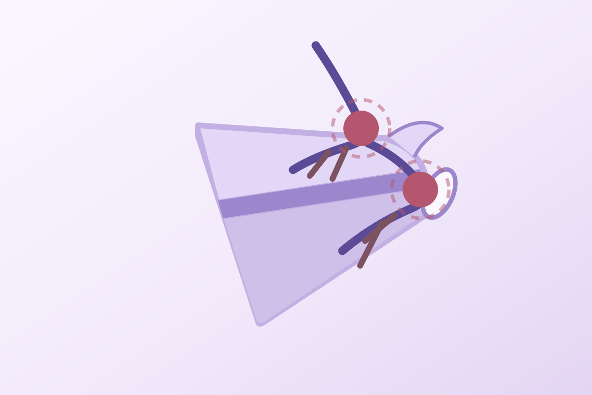
The diagram below shows the nerve's course on a posterior view of the right scapula, with the two compression sites numbered. It is the map to keep in mind throughout the examination : it is enough to turn an observed hollow into a topographical hypothesis.
Course of the suprascapular nerve and its two compression sites
Posterior view of the right scapula. The site affected decides which muscles are involved, and therefore what you see
Explanatory diagram. Anatomical data : Ticker JB et al., J Shoulder Elbow Surg 1998 (PMID 9814925) ; Piasecki DP et al., J Am Acad Orthop Surg 2009 (PMID 19880677) ; Clavert P, Thomazeau H, Orthop Traumatol Surg Res 2014 (PMID 25454727).
The literature gathers under one name two mechanisms that are not treated alike.
The first is compression : something takes up the space and crushes the nerve in a tunnel. Paralabral cyst, tumour, bony callus after a scapular fracture, osteophyte. These causes share one decisive feature : they are visible on imaging and most often reversible.
The second is traction : the nerve, short and fixed at both ends, is repeatedly stretched by extreme shoulder movements. It is the mechanism of the throwing athlete, described as early as 1987 by Ferretti in volleyball players, where tensioning occurs during the cocking and above all the follow-through of the serve4. Here there is nothing to see on imaging except the muscular consequences.
This distinction governs everything else : you decompress what compresses, you offload what tractions.
It is tempting to make the narrow notch or the ossified ligament the cause of the syndrome. The largest published series forbids it : across 1,063 shoulders examined on three-dimensional CT, including 53 with suprascapular nerve palsy, Honoki et al. found no difference in notch type or ossification between the palsied shoulders and the others5. These variants are plausible vulnerability factors, not explanations. And since 34.4 % of subjects have asymmetrical notches from one side to the other6, comparison with the sound side is not a sufficient argument either.
That is the elegance of this syndrome, and its most immediate practical value : the nerve gives its branches to the supraspinatus before crossing the second tunnel. Looking at which fossa is hollowed already places the lesion along the course.
The reasoning fits into one sentence. The branches to the supraspinatus arise in the supraspinous fossa, before the spinoglenoid notch. Therefore :
Isolated wasting of the infraspinatus, with a normal supraspinatus, places the lesion at the spinoglenoid tunnel before any investigation at all.
| What is compared | Suprascapular notch | Spinoglenoid notch |
|---|---|---|
| Muscles affected | Supraspinatus and infraspinatus | Infraspinatus alone |
| What you see | Both fossae hollow, on either side of the spine | An isolated hollow below the scapular spine |
| Functional deficit | Abduction and external rotation, often moderate (the deltoid compensates for abduction) | External rotation alone, sometimes marked |
| Pain | Usual, posterosuperior, dull and poorly localised | Often absent or minimal, especially in athletes |
| Dominant causes | Traction, tunnel variants, sequelae of scapular fracture, cyst (one third of cysts) | Paralabral cyst (two thirds of cysts), traction from the throwing movement |
| Yield of imaging | Variable : often normal apart from the muscular consequences | High : this is where MRI finds the cysts |
Distribution of cysts by site : 33 % at the suprascapular notch and 67 % at the spinoglenoid notch in the series of Feinberg et al. (9 patients, PMID 30291636). The other elements of the table synthesise Piasecki 2009 (PMID 19880677), Clavert 2014 (PMID 25454727) and Bozzi 2020 (PMID 32707860).
The clinical fact is consistent : in volleyball, throwing and tennis players, the wasting is confined to the infraspinatus, in an overwhelming proportion of cases. Two explanations complement each other.
The first is mechanical : during cocking and follow-through, the nerve is pressed and stretched against the lateral border of the scapular spine. Fabis-Strobin et al. modelled the compressive force exerted on the lateral trunk of the nerve by contraction of the infraspinatus, as a function of the angle the nerve makes with the scapular spine. Scapulae in which that angle is most closed undergo a force greater by 28 - 31 % than that of the others, which could explain why some players are affected and others not7. That work must be read for what it is : a simulation on dry anatomical specimens, not a clinical measurement, and its authors say so explicitly.
The second is topographical : the branch to the infraspinatus is terminal, so it is the only one to cross the second tunnel. Whatever happens at that level can reach only it.
Topography points the way, it does not decide alone. Meyer et al. report the case of a 40-year-old man who developed sudden left shoulder pain after lifting his child, with full range, painless Jobe and Patte tests but reduced strength, a sparse electromyogram and an MRI showing denervation oedema of both muscles. The picture therefore suggested compression at the suprascapular notch, and the indication for surgery seemed settled. MR neurography of the brachial plexus corrected the diagnosis : it was a traction injury, with no compression. The patient recovered without surgery, with a normal electromyogram at one year8.
There is no validated clinical test for this syndrome, and recent reviews say so bluntly. What remains is inspection, strength measurement, and a rule of proportion : when the deficit greatly exceeds the pain, think of the nerve.
Let us start with the admission the literature makes itself. In their 2025 review, Ayik et al. write that there is no definitive method for making this diagnosis, and that it is built by accumulating arguments : history, comparative examination of both shoulders, imaging, electrodiagnosis, sometimes an anaesthetic block9. No shoulder test does here what the Lachman test does at the knee.
The current concepts review of Bozzi et al., which retained 59 studies, puts the same thing differently : the diagnosis can be complex, it rests on appropriate instrumental assessment and imaging, and prompt diagnosis is decisive because chronic forms do worse than acute ones48. Time is therefore a variable of prognosis, not only of comfort.
Comparative inspection of both scapular fossae, with the patient stripped to the waist, arms by the side then in slight abduction, is the most profitable act of the examination. You look for a hollow, a sharper prominence of the scapular spine, a loss of contour below the spine. The important point is that this hollow is visible : in the large athlete series, wasting was recorded on that inspection criterion alone, with agreement between two independent examiners, a surgeon and a physiotherapist in Ellenbecker, two surgeons in Young1011.
Two practical traps. First, a well-developed trapezius can mask a supraspinous hollow : it is the infraspinous fossa that reads best. Second, comparison with the opposite side is essential, but it has its limits in the unilateral athlete, whose dominant side is normally more muscular: a dominant side that is simply equal to the non-dominant one is already suspicious.
Weakness in external rotation is the functional correlate of the wasting, and it can be measured. In 153 professional tennis players, visually observed wasting was significantly correlated with external rotation strength measured with the elbow at the side (p = 0.001) and at 90° of abduction (p = 0.009)10. In volleyball players, Ferretti measured as early as 1987 a loss of about 22 % of external rotation strength on isokinetic dynamometry4, and Lajtai found across 35 professional beach volleyball players an external rotation strength reduced to 90 % of that of the sound side12.
In practice, a hand-held dynamometer is enough to objectify a deficit and to follow it. Manual testing is too crude : Ellenbecker in fact notes the absence of correlation between wasting and instrumented manual testing of the supraspinatus, and the drop arm test says nothing about the infraspinatus.
This is the central reasoning of this article. A cuff tendinopathy hurts, limits through pain, and comes with strength that rises when the pain is eased. A nerve lesion makes muscle disappear, and the deficit persists with no relation to the pain level.
When weakness in external rotation is disproportionate to the pain, and a hollow can be seen in the fossa, it is not a tendinopathy.
That imbalance is all the more telling because suprascapular neuropathy is often painless. In the series by Mazza et al. covering 82 professional volleyball players, 9 % of the men and 12 % of the women had infraspinatus hypotrophy ; every one of them had weakness in external rotation ; none reported pain or a drop in performance13.
What to do faced with a hollowed scapular fossa
Triage tree : the three questions that separate emergency, investigation and monitoring
Author's synthesis, with no guideline equivalent. Elements taken from Bozzi F et al., J Clin Med 2020 (PMID 32707860) ; Ashton F et al., Indian J Orthop 2025 (PMID 40511344) ; Ayik G et al., Int Orthop 2025 (PMID 40082300).
The differential diagnosis of the wasted shoulder is the heart of this article. That is where the difference is decided between useful rehabilitation and months lost strengthening a cuff the nerve no longer commands.
A framing remark first : the entities that follow are not all mutually exclusive. A cuff tear can coexist with a cyst ; a patient can have both a tendinopathy and a neuropathy. The table below is not meant to pick a winner but to say, for each sign, what it points to.
| What is compared | Suprascapular nerve syndrome | Parsonage-Turner syndrome | Massive cuff tear | Cuff tendinopathy | Frozen shoulder | Cervical radiculopathy |
|---|---|---|---|---|---|---|
| Onset | Gradual, over weeks to months ; sometimes an incidental finding | Abrupt, often at night, within a few hours | Gradual, or abrupt after an injury | Gradual, load-related | Gradual, in three phases | Often subacute, initial neck pain |
| Initial pain | Moderate or absent, posterosuperior, poorly localised | Severe and constant, about 4 weeks | Moderate to severe, at night | Mechanical, on exertion and at end of range | Sharp, constant in phase 1 | Radicular, radiating into the limb |
| Wasting | Supraspinous and/or infraspinous fossa, depending on the site | Early, patchy, often several non-contiguous muscles | Supraspinous and infraspinous fossae, late, with fatty degeneration | Absent | Absent or from disuse, diffuse | Possible, in a myotome (deltoid, biceps) |
| Passive range | Preserved | Preserved (except secondary stiffness) | Preserved in most cases | Preserved | Restricted in all planes | Preserved |
| Involvement of other nerves | No, by definition | Common : long thoracic, spinal accessory, anterior interosseous | No | No | No | Radicular territory, not a nerve trunk |
| Sensory disturbance | No cutaneous territory | Present in 78 % of cases | None | None | None | In the dermatome concerned |
| Decisive investigation | MRI (cyst) and EMG | EMG and MRI or high-resolution ultrasound (hourglass constrictions) | Ultrasound or MRI | Clinical ; imaging of little help | Clinical | Cervical MRI, EMG |
| Dedicated page | This article | Dedicated article to come | Degenerative cuff tear | Cuff tendinopathy | Frozen shoulder | Cervical radiculopathy |
Parsonage-Turner syndrome data : van Alfen N, van Engelen BGM, Brain 2006, 246 cases (PMID 16371410). The other columns synthesise the corresponding articles on the site and the reviews cited in the bibliography.
It is the one that deserves most attention, for two reasons. First because the suprascapular nerve is precisely one of the nerves it most often affects : in a series of 355 patients investigated by electrodiagnosis, Seror counted 495 neuropathies, of which 129 of the suprascapular nerve, just behind the long thoracic nerve (138)14. Wasting of the supraspinatus and infraspinatus may therefore be the expression of neuralgic amyotrophy, and not of compression.
Second because the management is diametrically opposed : you do not decompress a Parsonage-Turner, and surgical wandering is a real risk there.
The key is the mode of onset. In the reference series of van Alfen and van Engelen, covering 246 cases, the pain runs through three successive phases, the first being severe, constant pain lasting on average four weeks. The paresis then appears, as the pain begins to settle. Involvement is patchy and preferentially affects the distribution of the upper and middle trunks, with the long thoracic nerve and/or the suprascapular nerve in 71.1 % of cases. Sensory involvement is found in 78.4 % of patients15.
The comparison is clear : abrupt and very painful onset on one side, gradual and often not very painful on the other. A patient who describes a particular night when the pain woke them, followed by weakness settling in over a few days, does not have chronic nerve trunk compression.
Three further elements help. Looking for a winged scapula or a deficit of another trunk, which does not belong to the picture of suprascapular compression. The context : a recent infection, vaccination, surgery or childbirth are described triggers16. And modern imaging : the hourglass constrictions of the nerve, visible on MRI or high-resolution ultrasound, have become an almost pathognomonic sign of neuralgic amyotrophy17.
A word about frequency, finally, because it changes the prior probability. Neuralgic amyotrophy was thought to be rare ; a prospective primary care cohort measured an incidence of 1 per 1,000 per year, that is 30 to 50 times more than was estimated18. Faced with a wasted shoulder, it is not an exceptional diagnosis you are raising, but a commonplace one that had stopped being seen.
You often read that Parsonage-Turner syndrome resolves spontaneously. Van Alfen's series says the opposite : among patients followed for three years or more, about two thirds still had pain or paresis15, and the 2020 review by Gstoettner et al. recalls that the majority do not achieve complete recovery17. Announcing spontaneous recovery to a patient is a promise the literature does not support.
The massive retracted tear is the second major source of wasting of both fossae, and its association with suprascapular neuropathy has been much debated. The hypothesis is mechanical : medial retraction of the supraspinatus and infraspinatus tractions the nerve, fixed at its tunnels.
Two older series strongly supported that link. Mallon et al. reported 8 patients with a massive tear, all with an abnormal electromyogram19. Costouros et al., across 26 massive tears, found peripheral nerve involvement in 54 % of patients, of whom 38 % had isolated suprascapular involvement, with electrophysiological recovery after arthroscopic repair20.
A prospective multicentre study by Collin et al. seriously tempered that result. Across 49 electromyograms performed in patients with retracted tears of the supraspinatus and infraspinatus, only 6 (12 %) had a neurological lesion, and a single one was a genuine suprascapular neuropathy. No correlation was found with the degree of fatty degeneration. The authors conclude that there is no argument for releasing the nerve routinely during a cuff repair21.
How to settle it for practice? The interesting subgroup is the one described by Kong et al. : when the infraspinatus degenerates more than the supraspinatus, which is atypical, retraction is greater and 23 % of those patients have an abnormal electrodiagnosis22. It is that dissociation, an infraspinatus more affected than the supraspinatus, that should raise the possibility of associated entrapment, and not the massive tear in itself.
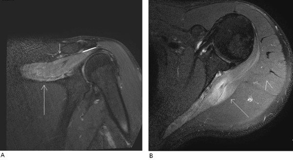
MRILeft shoulder MRI, coronal and axial T2 fat-saturated sections: diffuse hyperintensity of the supraspinatus and infraspinatus muscles suggesting denervation, with no visible lesion at the spinoglenoid notch.
Source : Economides et al., Journal of Medical Case Reports, 2011, figure 2 · CC BY
Unlike many painful shoulders, where imaging finds abnormalities just as often in people with no pain anywhere, MRI here genuinely changes decisions : it can find a cause we know how to treat.
MRI is the first-line investigation when a painful suprascapular neuropathy is suspected. It answers four questions at once.
First, is there a cyst, and where? That is the finding that changes management. Next, is there a superior labral tear, which is almost always associated with the cyst and is its source. Then, what is the state of the muscle : denervation oedema with high T2 signal marks recent and potentially reversible involvement, whereas fatty degeneration marks long-standing involvement. Finally, is there anything else : cuff tear, glenohumeral osteoarthritis, mass.
That fourth question is less incidental than it seems. Thomazeau et al. showed, across 20 patients with a spinoglenoid cyst, that 75 % had a posterior humeral subluxation index above 55 %, and that cysts accompanied by a cartilage lesion were associated with type B1, B2 or C glenoids. Their conclusion is directly useful to the physiotherapist : management must not be confined to the nerve compression, and the patient should be warned that posterior pain of articular origin may persist after the cyst is treated23.
Electrodiagnosis remains the reference for confirming nerve involvement, but with no consensus criterion. Boykin et al. report that, among the studies judged diagnostic in their practice, the mean motor latency was 2.90 ± 0.08 ms for the supraspinatus and 3.78 ± 0.14 ms for the infraspinatus, and that the commonest abnormality was not denervation, present in only 33 %, but abnormal motor unit potentials, present in 88 %24. Casazza et al. propose a side-to-side latency difference of 0.4 ms as an acceptable threshold, with recording by monopolar needle, more reproducible than a surface electrode25.
Two important reservations. The first comes from a recent review written for electromyographers : neuropathies in athletes do not follow the classic patterns of entrapment syndromes, and a routine protocol can miss them ; you have to explore beyond it26. The second comes from an imaging study : in 36 patients with a paralabral cyst investigated by both methods, Mun et al. found a discordance between electromyography and MRI in 10 of them : 5 had electrical neuropathy with a normal-looking muscle, and 5 the reverse27. Neither investigation is the arbiter of the other.
What each investigation brings, and what it misses
Measured figures, in different populations : to be read as orders of magnitude, not as comparable diagnostic performance
Sources : Boykin RE et al., J Shoulder Elbow Surg 2011 (PMID 21277806) ; Mun JW et al., Am J Sports Med 2024 (PMID 39439307).
Ultrasound has two uses here. It sees the spinoglenoid cyst well when it is large, and it allows infraspinatus thickness to be measured, which makes the wasting objective and trackable: a value highlighted in the clinical commentary of Lambrecht et al. for following tennis players28. It also serves to guide an aspiration or an injection.
MR neurography is the most recent addition. It is what shows the hourglass constrictions of neuralgic amyotrophy17, and it is what corrected the diagnosis in the case of Meyer et al. mentioned above, by distinguishing a traction injury from a compression8. It is not available everywhere, but it deserves to be requested when the question is precisely “should we operate?”.
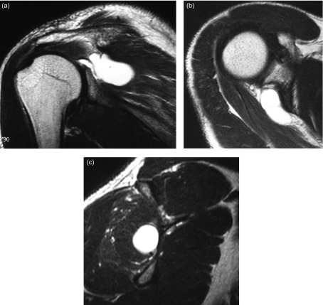
MRIT2-weighted left shoulder MRI, coronal, axial and sagittal sections: synovial cyst of the spinoglenoid notch, with oedema and atrophy of the infraspinatus muscle on the sagittal section.
Source : Hashiguchi et al., SICOT-J, 2016, figure 2 · CC BY
This is the part of the syndrome where we know best what to do. A cyst compresses the nerve, it comes from a breach in the labrum, and treating it makes the cyst disappear in nine cases out of ten.
The paralabral cyst is not a tumour : it is a pocket of synovial fluid that has escaped the joint. The dominant hypothesis is that of a one-way valve : a tear of the superior labrum, typically a SLAP lesion, lets the fluid escape backwards without being able to return, and the pocket grows until it presses on the nerve at the spinoglenoid notch.
That hypothesis has been validated indirectly, and in the best way possible : by showing that closing the breach is enough. Schroder et al. prospectively treated 42 patients with a posterosuperior labral tear and a cyst by simple labral repair, without touching the cyst. On follow-up MRI, the cyst had disappeared in 37 patients (88 %) and clearly shrunk in the other 5, who were all pain-free and satisfied. The Rowe score went from 61.5 to 9829.
The cyst is not the disease, it is its consequence : it is the breach in the labrum that has to be closed.
One contrast is worth noting. Across 79 cadaveric shoulders examined by Ticker et al., a single cyst was found, that is an incidence of 1 %1. In other words, the paralabral cyst is not a commonplace anatomical variant. But as soon as you look at the population of patients treated surgically for suprascapular neuropathy, it becomes the leading cause : in the systematic review of Memon et al. covering 259 patients, the spinoglenoid notch cyst accounted for 42 % of the aetiologies30. That is what Clavert and Thomazeau were already writing in 2014 in their French-language review : the commonest causes are paralabral cysts and microtrauma in the elite athlete31.
This is the only real therapeutic controversy on this subject, and it is instructive because both camps have data.
For repair alone. Besides Schroder and his 88 % disappearance, Kim et al. prospectively compared 28 patients split between SLAP repair alone and repair with decompression : the VAS, Rowe and Constant scores improved significantly in both groups, with no difference between them32. The systematic review of Schroeder et al., across 160 patients from 19 studies, likewise concludes that results are excellent in both arms and that the data show no advantage for decompression33.
For associated decompression. Pillai et al. compared the gain in external rotation strength : +40 % relative to the sound side with decompression and repair, against +10 % with repair alone. Their conclusion is that the gain in strength is mainly attributable to the decompression, which calls isolated repair into question in patients whose main problem is weakness34. The series comprises only 12 patients, however. A more recent systematic review, covering 206 patients, finds comparable resolution rates (95.5 % with decompression against 92.2 % without) but complication rates of 3.5 % against 11.4 % and revision surgery rates of 0 % against 5.3 % in favour of decompression35.
Labral repair alone or repair with cyst decompression
Four series, four partly divergent answers: none is a randomised trial
Sources : Schroder CP et al., J Bone Joint Surg Am 2008 (PMID 18310702) ; Pillai G et al., Clin Orthop Relat Res 2011 (PMID 21104358) ; Kim DH et al., Diagnostics 2023 (PMID 37510107).
A recent imaging study partly reconciles the two camps. Mun et al. compared 27 patients operated on with decompression and 16 without, with follow-up MRI at one year : the cyst had disappeared in 100 % of cases in both groups, the signs of infraspinatus denervation had resolved in all of them, and external rotation strength had improved in both groups with no significant difference27. The practical message, for the physiotherapist who receives the operated patient, is that neither technique should change their programme.
One finding runs through every series and deserves to be told to the patient before surgery : muscle volume does not always come back, and fatty degeneration never does.
Schroder et al. are the most explicit : the 3 patients whose wasting was not accompanied by fatty degeneration regained a normal-looking muscle, while the 7 who already had fatty change kept it29. That is what justifies not letting painful wasting drag on, and it is what Clavert and Thomazeau call the stake of early diagnosis31.
Conversely, recovery of the nerve is reliable once the compression is relieved. Feinberg et al. investigated 9 patients by electromyography before and after decompression, 3 cysts at the suprascapular notch, 6 at the spinoglenoid notch : all recovered completely electrophysiologically36.
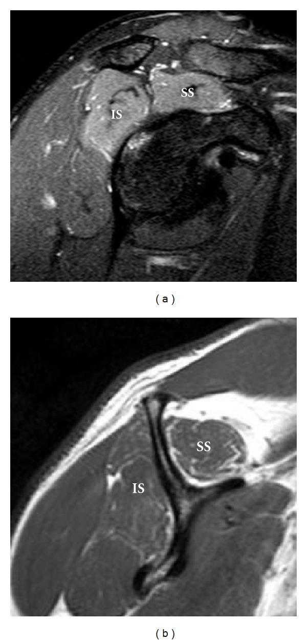
MRIShoulder MRI, oblique sagittal T2 fat-saturated and T1 sections: hyperintensity and atrophy with fatty infiltration of the supraspinatus and infraspinatus muscles, sequelae of suprascapular nerve injury after a swimming accident.
Source : Dong et al., Radiology Research and Practice, 2012, figure 2 · CC BY
The short answer is no, in most cases. This is probably the most useful message in this article, and the most counter-intuitive : in the overhead athlete, isolated wasting of the infraspinatus is common, generally painless, and it is not treated just because it can be seen.
The figures have been consistent for forty years, and they are high. Ferretti et al. examined 96 elite volleyball players at the 1985 European championships : 12 of them had isolated and asymptomatic palsy of the infraspinatus on the dominant side4. Holzgraefe et al. found nerve involvement in 33 % of 66 elite volleyball players, always on the hitting arm side, and latent in eight of the twelve tested electrically37. Lajtai et al., across 35 professional beach volleyball players examined at a tournament, observed visible wasting in 34 % : mild in 23 %, severe in 11 %12.
In tennis it is even commoner. In 125 professional women players examined during a pre-season assessment, Young et al. observed wasting of the dominant infraspinatus in 52 %, and none on the non-dominant side11. In 153 professional male players, Ellenbecker et al. found 60.1 % dominant-side wasting against 0.7 % on the non-dominant side10.
Prevalence of infraspinatus wasting in the overhead athlete
Seven series, from volleyball to tennis, with their sample sizes and their criteria: the criterion changes the measurement
Sources : Ferretti A et al., J Bone Joint Surg Am 1987 (PMID 3805088) ; Holzgraefe M et al., Br J Sports Med 1994 (PMID 8000816) ; Lajtai G et al., Am J Sports Med 2012 (PMID 22875791) ; Young SW et al., Am J Sports Med 2015 (PMID 26078449) ; Miura K et al., Prog Rehabil Med 2019 (PMID 32789249) ; Ellenbecker TS et al., Orthop J Sports Med 2020 (PMID 33195711) ; Mazza D et al., Phys Sportsmed 2021 (PMID 32372683).
The most decisive piece of data is a long-term follow-up published by Ferretti in 1998. Across 38 athletes with isolated infraspinatus wasting, all competitive volleyball players, 35 were pain-free and were treated with simple strengthening of the external rotators. Sixteen of them were reviewed at a mean follow-up of 5.5 years : thirteen were still playing volleyball, three had ended their career with no symptoms, and the wasting was unchanged in all of them. The three patients operated on had been operated on for posterior pain, and only one saw their wasting clearly resolve38.
Wasting that has not moved in five and a half years in players who go on playing is not a disease to treat : it is an adaptation to monitor.
Two recent series point the same way on the functional side. In Young et al., the wasting was associated with a better ranking, 58 % among players ranked in the world's top hundred against 40 % beyond, and was correlated neither with associated shoulder disease nor with a drop in performance. The authors conclude explicitly that team doctors can be reassured11. In Mazza et al., none of the players with hypotrophy complained of pain or of a drop in performance13.
One prevention finding deserves to be known, because it involves the coach directly. Mazza et al. put forward a simple hypothesis : the prevalence of the neuropathy has fallen in professional volleyball players since the 1980s and 1990s, and that fall coincides with the abandonment of the float serve in favour of the jump spike serve. Their measurement does indeed find 9 to 12 % against 12.5 to 33 % in the historical series, and they conclude that the jump spike serve appears safe for the nerve13. It is an ecological inference, not a demonstration of causation, but it gives a concrete lever when a player is symptomatic.
“Do not treat what does not trouble the patient” is the right rule, but the asymptomatic wasted shoulder is not a normal shoulder, and it would be dishonest to let people believe it is. The team of Contemori and Biscarini measured, in the same asymptomatic professional volleyball players, three abnormalities : an impaired sense of position of the shoulder in all three movements tested (p < 10⁻³)39 ; there is a reduction in static and dynamic stability of the shoulder, greater with the eyes closed40 ; and a reorganisation of muscle activity with more deltoid and trapezius, less serratus anterior, an upper trapezius recruited earlier and a serratus later41.
This work covers 24 players in total and does not demonstrate an increased risk of injury : it shows a plausible mechanism. The reasonable course is neither to treat everyone nor to say that all is well: it is to monitor and to strengthen preventively, which is also what Miura et al. recommend in view of the imbalance between external and internal rotation they measure in these players42.
It must be said plainly before going into detail : there is no randomised controlled trial of rehabilitation in this condition. The only conservative series published has fifteen patients and dates from 1997. Everything that follows is therefore mechanical reasoning supported by low-level series.
Martin et al. reviewed fifteen patients treated non-surgically for an electrically confirmed suprascapular neuropathy, at a mean follow-up of three years and eleven months. Treatment consisted of a physiotherapy programme aimed at restoring range and strengthening the shoulder muscles. The result was excellent in five patients and good in seven ; the other three were operated on for lack of improvement. Their conclusion, which remains the rule thirty years later : in the absence of a well-defined compressive lesion, a suprascapular neuropathy should be treated non-surgically43.
That rule is taken up by every recent review. Ayik et al. recommend trying non-operative treatment for at least six months in the absence of a space-occupying lesion9, and Ashton et al. recall that the majority of patients respond well to a multimodal approach44.
It is built around four axes, which follow from the mechanism more than from direct evidence.
The first step is not strengthening, it is removing a load. In athletes, that means temporarily suspending the offending movement (the serve, the spike, the throw), without stopping general activity. The clinical commentary of Lambrecht et al. proposes an explicit progression : first avoid the aggravating movements, then gradually reintroduce overhead work28. In workers, that means modifying tasks involving prolonged elevation.
Two targets here. Posterior capsular stiffness increases nerve tension during horizontal adduction and internal rotation : treating it is presented as a measure to reduce the risk of compression28. And neural mobilisation techniques (nerve gliding, rather than tensioning) aim to restore the excursion of the nerve trunk in its tunnels. Their level of evidence in this precise indication is nil : they are offered by analogy with other entrapment syndromes.
This is the core of the programme, and it is what Ferretti was already prescribing to his 35 pain-free volleyball players38. Two practical points. First, an affected infraspinatus does not respond like a healthy muscle : progression must be slower and guided by fatigability. Second, the teres minor, innervated by the axillary nerve, remains available and sometimes hypertrophies spontaneously: Mun et al. observed this in 5 patients out of 43, all with infraspinatus wasting27. External rotation work with the elbow at the side loads it preferentially.
The data of Contemori and Biscarini give precise and measurable targets here : reduce upper trapezius dominance, wake the serratus anterior, restore its anticipatory activation, and work on the sense of position, particularly under reduced vision since that is where the deficit is unmasked4041. The review by Leider et al. lists in the same spirit the muscles to target : cuff, trapezius, levator scapulae, rhomboids, serratus anterior and deltoid45.
Management modalities and level of evidence
Author's appraisal following GRADE logic : no modality in this table rests on a randomised trial
Sources by card : Schroder 2008 (PMID 18310702), Schroeder 2018 (PMID 29501216), Kim 2023 (PMID 37510107), Memon 2018 (PMID 28879607) ; Martin 1997 (PMID 9278075), Ayik 2025 (PMID 40082300) ; Ferretti 1998 (PMID 9850775), Miura 2019 (PMID 32789249) ; Lambrecht 2025 (PMID 40756798), Contemori 2018 (PMID 28605232), Leider 2021 (PMID 34745481).
Surgery gives good results on pain, less consistent results on strength, and frankly variable results on muscle volume. It is that hierarchy that must be announced to the patient, and it is that hierarchy that structures the expectations of postoperative rehabilitation.
A series of 87 cases collected over sixteen years in an electrodiagnostic centre gives the most honest distribution of causes, because it recruits upstream of surgery. Trauma comes top with 27 cases, followed by neuralgic amyotrophy with 21 cases. Fifty-seven patients had isolated suprascapular nerve involvement ; the others had associated involvement, most often of the axillary nerve (23 patients)46.
That figure has an important practical consequence : in an unselected population, nearly a quarter of “suprascapular neuropathies” are in fact neuralgic amyotrophies. It is not the same disease, and it is not the same management.
Causes of suprascapular neuropathies seen in electrodiagnosis
87 cases over sixteen years, before any surgical selection: the distribution changes completely depending on where you count
Sources : Memon AB et al., Muscle Nerve 2019, series of 87 cases (PMID 31294855) ; Memon M et al., Knee Surg Sports Traumatol Arthrosc 2018, systematic review of 259 patients (PMID 28879607).
They fall into three categories, with very different degrees of certainty.
Clear indication : the space-occupying lesion. A symptomatic paralabral cyst, a tumour, a bony callus compressing the nerve. Here surgery treats an identified cause. It is all the more justified because nerve recovery is reliable once the compression is relieved36.
Reasonable indication : failure of well-conducted conservative treatment. The reviews converge on an interval of at least six months9, with a common-sense qualification : if the wasting worsens, you do not wait for the interval to end.
Contested indication : releasing the nerve alongside a cuff repair. This is where caution is called for. Collin et al. find no argument for it routinely, with a single genuine neuropathy across 49 massive tears investigated21. Ashton et al. go further : decompression added to a cuff repair or a stabilisation was accompanied by a relatively high complication rate, which leads them to recommend a high threshold for the indication, reserved for proven neuropathies or at-risk anatomical variants44.
The largest systematic review covers 40 studies and 259 patients (261 shoulders) treated arthroscopically. The overall result is good : 97 % of patients report clear improvement or complete resolution of their symptoms (pain, strength and subjective function together), at a mean follow-up of 23.7 months, with an overall complication rate of 4 %. The authors are explicit about the limit : these are uncontrolled studies, level of evidence IV30.
A series devoted specifically to volleyball players usefully qualifies that figure. Brzoska et al. reviewed 10 players decompressed at both notches, at a mean follow-up of 78 months. The mean Constant score reached 89.9, but external rotation strength remained at 8 kg against 12.65 kg on the sound side (p < 0.01), and recovery of muscle volume was complete in 5 players, partial in 2 and nil in 347.
Surgery almost always relieves, often restores function, and gives the muscle back only half the time.
That hierarchy is not a recent discovery. Piasecki et al. were already writing it in 2009 in their review : open and arthroscopic approaches alike reliably provide pain relief and functional improvement, but the return of strength and muscle volume is less predictable50.
What can be promised after a decompression
Three outcomes, three levels of certainty, to be announced to the patient in this order
Sources : Memon M et al., Knee Surg Sports Traumatol Arthrosc 2018 (PMID 28879607) ; Feinberg JH et al., Muscle Nerve 2019 (PMID 30291636) ; Brzoska R et al., Orthop J Sports Med 2023 (PMID 36874055).
Six published cases, chosen for the reasoning error each corrects. They are all available online with their identifier.
Meyer JS et al., JSES International 2020: PMID 32939475.
Nair NMS et al., Journal of Orthopaedic Case Reports 2025: PMID 40092252.
Gomez DN et al., Malaysian Orthopaedic Journal 2022: PMID 3551952852.
Massel DH et al., Journal of Orthopaedic Case Reports 2022: PMID 3666016053.
Walker CR et al., Cureus 2021: PMID 3514108054.
Niemann AJ et al., Asian Journal of Sports Medicine 2013: PMID 23785580.
This chapter contains nothing new : it arranges what precedes in the order in which the questions arise in the consultation.
Faced with any painful or weak shoulder, have the patient strip to the waist and look at both scapular fossae from behind. It takes thirty seconds, and it is what separates correct management from useless cuff rehabilitation.
If a hollow is present, ask three questions in this order : was the onset abrupt and very painful? Is another nerve involved? Is the deficit disproportionate to the pain?
| What you observe | What you do | Why |
|---|---|---|
| Wasting of both fossae, abrupt and very painful onset, sometimes several nerves | Refer for a neurological opinion and electromyography, with no intensive rehabilitation for the time being | Parsonage-Turner syndrome accounts for nearly a quarter of suprascapular neuropathies in an unselected series46 |
| Isolated wasting of the infraspinatus, patient in pain, under 50 | Request an MRI before building a long programme | That is the typical presentation of the paralabral cyst, a curable and common cause30 |
| Isolated wasting of the infraspinatus, overhead athlete, painless, performance preserved | Do not investigate routinely. Measure external rotation strength, strengthen, review at six months | Prevalence of 34 to 60 % in these athletes, wasting stable at 5.5 years, with no demonstrated impact3811 |
| Wasting of both fossae in a patient over 60, with a known cuff tear | Do not conclude too quickly to a neuropathy : look at whether the infraspinatus is more affected than the supraspinatus | A single genuine neuropathy across 49 massive tears investigated ; it is the dissociation that alerts you2122 |
| Patient operated on for a cyst, referred for rehabilitation | The same programme whatever the technique used. Explain that strength comes back before volume | Comparable results with or without associated decompression ; volume recovered half the time2747 |
| No progress at six months of well-conducted rehabilitation | Refer again, do not prolong it | That is the interval used by the reviews to conclude that conservative treatment has failed9 |
Three sentences are enough, and they prevent most disappointments.
The first : this is not a tendon problem, it is a nerve problem, which explains why classic strengthening had given nothing, and why the muscle melted away.
The second : if a mechanical cause is found and relieved, the nerve almost always recovers ; that is what the follow-up electromyograms after decompression show36.
The third, the most important not to skip over : muscle volume comes back only half the time, and it never comes back where fat has taken its place2947. A patient who has been warned does not experience that persistence as a failure of their rehabilitation.
No. A massive retracted cuff tear melts the same muscles away by a different mechanism, and prolonged non-use produces diffuse wasting. What points to the nerve is that it is confined to one territory, the absence of a tendon explanation on imaging, and the disproportion between deficit and pain. See the article on degenerative cuff tear for the tendon side.
It can be strongly suspected, and that is even the rule in community practice. But electrodiagnosis remains the reference for confirmation, and it becomes essential as soon as a surgical decision is in prospect or a doubt exists with neuralgic amyotrophy. You should know that there is no consensus criterion, and that a quarter of patients have results discordant with MRI27.
No. It is the symptomatic cyst that is treated: pain, external rotation deficit, signs of denervation. An incidentally found cyst in a patient with no complaint does not call for a procedure. That said, when wasting is already present, time matters : established fatty degeneration does not recover29.
Rarely completely. In the athlete in pain, you suspend the offending movement (the serve, the spike, the throw), keeping general training, then reintroduce it gradually28. In the asymptomatic athlete with isolated wasting, there is no reason to stop : the longest follow-up series shows players continuing their career with stable wasting38.
For the pain, a few weeks to a few months. For the nerve, reinnervation takes several months, with follow-up electromyograms typically normalising towards six months to a year368. For return to throwing sport after surgery, the published cases give delays of five to six months5155. For muscle volume, count in years, and sometimes accept that it will not come back.
Two uses must be distinguished. As an analgesic in other shoulder conditions and in surgical anaesthesia, the suprascapular block is widely studied. As a diagnostic or therapeutic tool for the entrapment syndrome itself, it remains a possibility : the review of Ashton et al. mentions it among the developments to come, not among established practice44. Offering it is defensible ; presenting it as validated in this indication is not.
There is no occupational disease schedule specific to suprascapular neuropathy in the French general scheme, unlike cuff tendinopathies. A patient whose work involves repeated overhead movements would come under a claim for periarticular conditions instead, the outline of which is described in our file on occupational disease recognition.
Because both those pages are being written at the time this one is published. The differential with neuralgic amyotrophy and the link with superior labral lesions are therefore covered here independently, in the corresponding chapters. Cross-links will be added as soon as those pages exist.
Fifty-five references, all verified individually : identifier resolved on PubMed and abstract read before citation. The PMID links open the PubMed record, the DOI links the publisher.
Behind this article
An author who explains, a reviewer who checks.
How we write and check our content

Physiotherapist · co-founder of Physio Learning
Marked for life by his first four-hour lecture without a single image, he took a master’s in instructional design so that it would never happen to anyone again. He hunts down publication bias and unreadable slides with the same intransigence.

Head of scientific content
Physiotherapist specialising in neuro-musculoskeletal practice and holder of a master’s in public health. He checks the methodological rigour of every article: primary sources, levels of evidence, no exceptions.
