

















In brief
Degenerative lumbar spondylolisthesis (DLS) is an anterior vertebral slip, mainly at L4-L5, linked to facet joint osteoarthritis and disc degeneration, with no pars defect. Its cardinal symptom is neurogenic claudication: pain or heaviness in the lower limbs on walking and on standing, relieved by forward flexion (the shopping trolley sign); the diagnosis is clinical, with MRI confirming it but correlating weakly. Conservative treatment is the first line (a 3 to 6 month trial), lumbar segmental stabilisation exercise being the most effective; surgery is reserved for failures or for progressive neurological deficits. Prevalence is close to 11.5 % in adults, and up to 25 % in women over 65.
A clinical synthesis based on the NASS guideline, the SPORT trial, the RESTORE-CFT trial (Lancet 2023) and the most recent 2023-2025 meta-analyses.
Clinical synthesis
- DLS is an anterior vertebral slip (mainly L4-L5) linked to facet joint osteoarthritis and disc degeneration, with no pars defect. Prevalence is about 11.5 % in adults (up to 25 % in women > 65 years according to Wang 2013, the MsOS Hong Kong cohort).
- Firmly established risk factors: female sex (a major effect after the menopause), age > 50 years, high BMI, sagittal orientation of the facet joints (WMD −10.5° vs controls, Liu 2017) and increased lumbar lordosis.
- The natural history is slow: Matsunaga (1990, 10-year follow-up) shows that only 30 % of patients progress radiographically and that progression of the slip is NOT correlated with clinical worsening.
- The cardinal symptom is neurogenic claudication : pain or heaviness in the lower limbs brought on by walking or standing, relieved by forward flexion (the shopping trolley sign, sitting).
- The main differential diagnosis is vascular claudication, relieved by simply stopping the effort with no change of posture, so peripheral pulses must be palpated as a matter of routine.
- MRI is the gold standard (Kreiner 2013, NASS) but the clinical and radiological correlation is weak : treat the patient, not the image.
- The CARDS classification (Kepler 2015) describes DLS more finely than the Meyerding grade (disc height, translation, sagittal alignment, clinical radiculopathy), but it is validated on its inter-observer reliability, not on any superiority for guiding treatment.
- Conservative treatment is the 1st line (NASS 2009, Matz 2016), with a 3-6 month trial before surgery. Surgery is reserved for documented failures or for progressive neurological deficits.
- Lumbar segmental stabilisation exercise is the most effective intervention (Lin 2024, meta-analysis PMID 38514931). The programme must be individualised and progressive.
- Manual therapies are short-term adjuncts. Passive modalities (heat, electrotherapy, ultrasound) rest on a very low level of evidence.
- Patient education and the management of psychosocial factors (catastrophising, kinesiophobia) are crucial, and the RESTORE trial (Kent 2023, Lancet) shows the lasting efficacy of Cognitive Functional Therapy in chronic low back pain.
- The SPORT trial (Weinstein 2007 NEJM, 8-year follow-up Abdu 2017) shows a lasting surgical benefit but with 22 % reoperation at 8 years.
- The Försth trial (NEJM 2016) shows that adding fusion to decompression alone does not improve outcomes in DLS with stenosis, a strong recommendation to keep in mind for shared decision-making.
- The return to activity must be based on functional criteria (pain, strength, neuromuscular control) and not on a calendar or on follow-up imaging.
- DLS can mimic hip osteoarthritis: any hip or anterior thigh pain resistant to local treatment calls for a lumbar assessment.
- Red flags (Finucane 2020, JOSPT): cauda equina syndrome, progressive motor deficit, saddle anaesthesia, sphincter disturbance → urgent medical referral.
- Yellow flags (catastrophising, kinesiophobia, catastrophic beliefs about imaging) → psychological referral and a cognitive functional approach.
- Measuring outcomes with validated PROMs (ODI, ZCQ for stenosis, EQ-5D) is now the standard, beyond the VAS alone.
Contents
- What are the fundamentals to know about degenerative lumbar spondylolisthesis?
- How do you assess and diagnose degenerative lumbar spondylolisthesis with confidence?
- Which treatment strategies are the most effective for degenerative lumbar spondylolisthesis?
- Where do you start and what is the hierarchy of interventions?
- What place does exercise hold and is there a superior approach?
- Manual therapies and passive modalities: what real efficacy?
- Beyond the physical: how do you educate the patient and act on psychosocial factors?
- When should surgery be considered and what do the major trials say (SPORT, Försth)?
- How do you secure a lasting recovery and prevent recurrence in degenerative lumbar spondylolisthesis?
- What do real clinical cases teach us about degenerative lumbar spondylolisthesis?
- How do you apply these recommendations concretely in your practice?
What are the fundamentals to know about degenerative lumbar spondylolisthesis?
Degenerative lumbar spondylolisthesis (DLS) is a frequent cause of low back pain and of neurogenic claudication in adults over 50. It differs radically from the isthmic spondylolisthesis of the adolescent: here there is no stress fracture, but a cascade of facet joint osteoarthritis and disc degeneration that ends in an anterior vertebral slip. Understanding this mechanism, its true prevalence and its slow natural history is the essential precondition for any coherent management.
How is this condition defined, who is affected and what are the risk factors?
Degenerative lumbar spondylolisthesis is defined as an anterior slip of one lumbar vertebra over the one beneath it, in the absence of a defect of the pars interarticularis (hence without spondylolysis)¹,². This operational definition, adopted by the NASS guideline¹, is fundamental because it governs both prognosis and treatment strategy. The level most frequently affected is L4-L5, because of its considerable mobility and of the mechanical loads it carries, followed by L3-L4 and L5-S1².
Epidemiologically, DLS is far more frequent than people think. The Framingham community study (Kalichman 2009)³ finds a radiographic prevalence of degenerative spondylolisthesis of 19.1 % in men and 25.0 % in women aged 65 and over in the Hong Kong MsOS/MrOS cohort (Wang 2013)⁴. A systematic review focusing on age and sex (Wang 2017)⁵ confirms an overall prevalence of around 11-12 % in adults, rising linearly with age. 👩⚕️
The female predominance is one of the most firmly established risk factors. In the general population the F:M ratio is about 1.3:1; in clinical series (patients seeking care for symptoms) it can reach 3:1 to 6:1². This difference is attributed to post-menopausal hormonal factors (increased ligamentous laxity), to anatomical differences (facet orientation) and to multiparity⁴.
In summary, the risk factors firmly established by the recent literature are:
- Female sex : a major factor, above all after the menopause⁴,⁵.
- Age > 50-60 years : prevalence rises linearly with age².
- Sagittal facet orientation : the Liu 2017 meta-analysis⁶ finds a weighted mean difference (WMD) of −10.5° between DLS and controls, facets more parallel to the sagittal plane offering less resistance to anterior shear.
- High body mass index : obesity increases lumbar mechanical loads².
- Increased lumbar lordosis : raises shear forces at L4-L5⁷.
- Multiparity : a cumulative hormonal and biomechanical effect².
What happens in the body and how does DLS evolve naturally?
The pathophysiology of DLS is a four-step degenerative cascade, now well described in recent narrative reviews (Akkawi 2022⁸, Gagnet 2018⁹). ⚙️
- Degeneration of the intervertebral disc : loss of disc height and hydration, reduced shock absorption and reduced segmental stability⁸.
- Overloading of the facet joints : the loss of disc height transfers excessive load onto the posterior facets, leading to facet osteoarthritis (cartilage erosion, subchondral sclerosis, osteophytes)².
- Segmental instability and slip : osteoarthritis and facet remodelling, combined with laxity of the ligamentum flavum and of the capsule, allow the progressive anterior slip⁸.
- Canal or foraminal stenosis : facet hypertrophy, disc bulging and thickening of the ligamentum flavum reduce the space available for the neural structures, generating neurogenic claudication. It is this stenosis, more than the slip itself, that causes the neurological symptoms⁷.
📈 The natural history is slow. The seminal study by Matsunaga (Spine 1990)¹⁰, which followed 40 patients for 10 years, is the universally cited reference. It showed that:
- The slip progresses in only about 30 % of patients over 10 years, and usually remains modest¹⁰.
- Above all, worsening of the clinical symptoms is not correlated with an increase in the radiographic slip¹⁰,⁸.
- General joint laxity was observed in 65 % of patients, suggesting a constitutional component¹⁰.
This last point is fundamental in the consultation : a patient may see their slip stabilise while developing more severe symptoms (through worsening stenosis) or, conversely, see their slip progress slightly while remaining asymptomatic. This clinical and radiological dissociation is the pivot of an approach centred on function rather than on the image.
Criticism and controversy: the trap of the "radiological diagnosis"
One major controversy in DLS is the discordance between images and clinical picture. Many imaging studies in asymptomatic people show that radiographic DLS is very frequent in older people with no complaint. Brinjikji (AJNR 2015)¹¹ and the ACP/Chou guideline (Ann Intern Med 2011) call for caution in interpreting incidental findings. The risk of an anxiety-provoking incidental finding is real: a patient told "you have a slipped vertebra" may develop an iatrogenic kinesiophobia that complicates management.
Another controversy is the very definition of "symptomatic" DLS. Should there be a minimum radiographic threshold (≥ 3 mm, ≥ 5 mm)? The NASS guideline¹ imposes no threshold and gives priority to clinical correlation. The practical consequence is that imaging must never be requested "to look for a DLS" ; it confirms a prior clinical hypothesis of neurogenic claudication or of sciatica.
Key points
- DLS is an anterior vertebral slip due to facet joint osteoarthritis and disc degeneration, with no pars defect. The level of choice is L4-L5.
- Prevalence of about 11-25 % depending on age and sex; post-menopausal women are the most affected (Wang 2013/2017).
- Major risk factors: female sex, age > 50 years, sagittal facet orientation (WMD −10.5° vs controls, Liu 2017), high BMI, increased lordosis.
- The course is slow : 30 % of slips progress at 10 years (Matsunaga 1990), not correlated with clinical worsening.
- The symptoms depend essentially on the neurological stenosis, not on the slip itself.
Chapter 1 bibliography (Fundamentals)
- Watters WC 3rd, Bono CM, Gilbert TJ, et al. An evidence-based clinical guideline for the diagnosis and treatment of degenerative lumbar spondylolisthesis. Spine J. 2009;9(7):609-614. PMID 19447684.
- Matz PG, Meagher RJ, Lamer T, et al. Guideline summary review: an evidence-based clinical guideline for the diagnosis and treatment of degenerative lumbar spondylolisthesis. Spine J. 2016;16(3):439-448. PMID 26681351.
- Kalichman L, Kim DH, Li L, Guermazi A, Berkin V, Hunter DJ. Spondylolysis and spondylolisthesis: prevalence and association with low back pain in the adult community-based population. Spine. 2009;34(2):199-205. PMID 19139672.
- Wang YXJ, Káplár Z, Deng M, Leung JCS. Lumbar degenerative spondylolisthesis epidemiology — a systematic review with a focus on gender-specific and age-specific prevalence. J Orthop Translat. 2017;11:39-52. doi:10.1016/j.jot.2016.11.001.
- Wang Z, Parikh K, Liu C, et al. Prevalence and risk factors of lumbar spondylolisthesis in elderly Chinese men and women. Eur Radiol. 2014;24(2):441-448. PMID 24126641.
- Liu Z, Duan Y, Rong X, Wang B, Chen H, Liu H. Variation of facet joint orientation and tropism in lumbar degenerative spondylolisthesis and disc herniation at L4-L5: A systematic review and meta-analysis. Clin Neurol Neurosurg. 2017;161:41-47. PMID 28843706.
- Barrey C, Jund J, Noseda O, Roussouly P. Sagittal balance of the pelvis-spine complex and lumbar degenerative diseases. A comparative study about 85 cases. Eur Spine J. 2007;16(9):1459-1467. PMID 17211522.
- Akkawi I, Zmerly H. Degenerative Spondylolisthesis: A Narrative Review. Acta Biomed. 2022;92(6):e2021313. PMID 35075090.
- Gagnet P, Kern K, Andrews K, Elgafy H, Ebraheim N. Spondylolysis and spondylolisthesis: A review of the literature. J Orthop. 2018;15(2):404-407. PMID 29881164.
- Matsunaga S, Sakou T, Morizono Y, Masuda A, Nakahara S. Natural history of degenerative spondylolisthesis. Pathogenesis and natural course of the slippage. Spine. 1990;15(11):1204-1210. PMID 2267617.
- Brinjikji W, Luetmer PH, Comstock B, et al. Systematic literature review of imaging features of spinal degeneration in asymptomatic populations. AJNR Am J Neuroradiol. 2015;36(4):811-816. PMID 25430861.
How do you assess and diagnose degenerative lumbar spondylolisthesis with confidence?
The diagnosis of DLS is above all clinical. Imaging confirms it. The trap works the other way round: finding a spondylolisthesis on an MRI scan does not mean the patient is suffering from it. The reference diagnostic approach (NASS 2009/2016) rests on a rigorous triad: a history targeted on neurogenic claudication, a standardised physical examination with a walking test, and relevant imaging (dynamic radiographs and MRI), always correlated with the clinical picture.
Which questions should you ask to understand the patient and their history?
The history is the cornerstone of the diagnosis. 🔍 The most characteristic picture in symptomatic DLS is intermittent neurogenic claudication, whose features must be explored systematically¹,².
- Description of the symptoms : pain, heaviness, cramping, paraesthesia or weakness, most often bilateral but sometimes asymmetrical, radiating into the buttocks, the thighs or the legs³. These symptoms are classically worsened by walking or by prolonged standing, postures that place the spine in extension and reduce the diameter of the spinal canal².
- Relieving factors : neurogenic claudication is relieved by lumbar flexion. The patient reports relief on sitting down, on leaning forward or on leaning on a trolley (the "shopping trolley sign")⁴. This flexed posture increases the space available for the neural structures.
- Walking distance : quantifying it (distance, time to the onset of symptoms) is the most useful objective measure for tracking change. To be recorded in the notes in metres or in minutes¹.
- Associated mechanical low back pain : often present, worsened by activities in extension, relieved by rest or by flexion.
- Risk factors and comorbidities : age, sex, BMI, history of pregnancies, osteoporosis, diabetes (which can produce a peripheral polyneuropathy mimicking claudication)⁵.
One crucial question, often neglected, is that of the patient's beliefs and representations. What is their interpretation of the condition? Have they seen their MRI scan? What were they told? The word "instability" can be anxiety-provoking and generate an iatrogenic kinesiophobia⁶. Anticipating this educational work starts with the history itself.
Which clinical tests should you perform and which other conditions should you rule out?
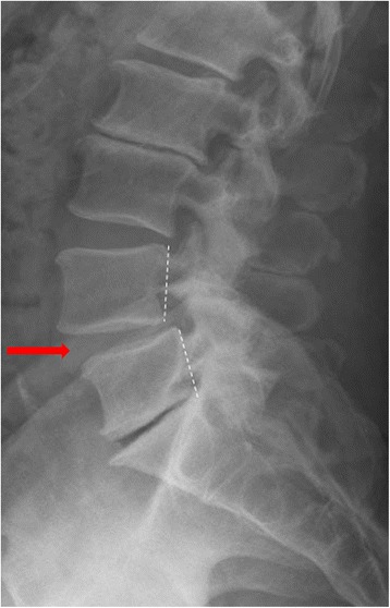
X-rayStanding lateral lumbar spine radiograph: degenerative spondylolisthesis at L4-L5, arrow and dotted markers showing the anterior slip of L4 on L5.
Source : Samartzis et al., Scoliosis and Spinal Disorders, 2016, figure 1 · CC BY
The physical examination aims to confirm the hypothesis raised by the history, to assess any neurological involvement and to rule out the differential diagnoses. 🚶♂️
- Postural inspection : look for a loss of lordosis and an antalgic posture in flexion (trunk slightly bent, hips and knees flexed).
- Full neurological examination : motor strength testing (in particular tibialis anterior L4, extensor hallucis longus L5, triceps surae S1), dermatomal sensation, tendon reflexes (knee L4, ankle S1), straight leg raise (often negative because the compression is central or foraminal, not discal)⁷.
- Two-stage treadmill walking test (Fritz 1997) : the patient first walks on the level until symptoms appear, then, after a rest, on an inclined treadmill (which favours flexion). A significant improvement in walking time on the inclined treadmill is highly suggestive of neurogenic claudication, with a sensitivity of about 50 % and a specificity of ≈ 92 %⁸. A simple tool to build into physiotherapy practice.
- Functional assessment : actual walking distance, standing tolerance, Schober test (lumbar mobility), single leg squat to assess neuromuscular control.
Red flags to screen for systematically (Finucane 2020, JOSPT)
- Cauda equina syndrome : saddle anaesthesia (S2-S5), sphincter disturbance (urinary retention or incontinence, faecal disturbance), loss of anal tone, acute bilateral motor deficit → surgical emergency, MRI without delay⁹.
- Progressive motor deficit : rapid worsening of a weakness (foot drop, giving way) → prompt neurosurgical opinion.
- Signs of associated cervical myelopathy : loss of fine dexterity, balance disturbance, hyperreflexia, Babinski sign → cervical MRI.
- Fever, unexplained weight loss, history of cancer, immunosuppression → suspected spondylodiscitis or metastasis; MRI with gadolinium, blood tests.
- Severe, non-mechanical, night pain at rest → suspected malignancy or inflammatory disease.
- Significant recent trauma in an osteoporotic patient → vertebral fracture.
The essential differential diagnosis is vascular claudication (peripheral arterial disease of the lower limbs). The distinction is fundamental and has a direct impact on management:
| Criterion | Neurogenic claudication (DLS/stenosis) | Vascular claudication (peripheral arterial disease) |
|---|---|---|
| Trigger | Walking, prolonged standing | Muscular effort (intensity-dependent) |
| Relief | Forward flexion (sitting down, leaning forward) | Simply stopping the effort, while standing |
| Shopping trolley sign | Present (walking improved) | Absent |
| Distribution | Diffuse, dermatomal (L4-S1) | Thigh, calf (arterial territory) |
| Peripheral pulses | Normal | Reduced or absent |
| Cycling | Well tolerated (spine in flexion) | Poorly tolerated (effort) |
| History | Low back pain, radicular pain | Smoking, diabetes, PAD |
| Further investigation | Lumbar MRI | Ankle-brachial index, arterial duplex ultrasound |
Other differential diagnoses to consider: hip osteoarthritis (pain referred to the groin), sacroiliac osteoarthritis, piriformis syndrome, peripheral diabetic neuropathy, meralgia paraesthetica (compression of the lateral femoral cutaneous nerve), trochanteric bursitis, gluteal tendinopathy.
As regards imaging, the NASS guideline¹,² and Kreiner's guideline on lumbar stenosis¹⁰ recommend:
- Weight-bearing dynamic radiographs (standing flexion-extension) : the first-line examination for visualising the slip and identifying any segmental instability (mobility > 3-4 mm in flexion and extension).
- Lumbar MRI : the gold standard for assessing central and foraminal stenosis, root compression, and the state of the discs and facets. Recommendation: to be requested only if the treatment decision depends on it (surgery under consideration, red flag)¹⁰.
- CT scan : useful in addition if MRI is contraindicated or to plan surgery (fine bone analysis).
Should DLS patients be classified, and for what benefit?
Yes, but by abandoning purely radiographic classifications in favour of clinically informative ones. 💡
The Meyerding classification (grade I: < 25 %, II: 25-50 %, III: 50-75 %, IV: 75-100 %, V: complete ptosis) is historically the most used. But in DLS almost every case is grade I or II, and the correlation between grade and symptoms is weak¹¹. It is therefore not enough to guide treatment.
The CARDS classification (Clinical And Radiographic Degenerative Spondylolisthesis, Kepler 2015)¹² is more modern and clinically relevant. It distinguishes 4 subtypes according to the combination of disc height, sagittal alignment and amount of translation, together with a clinical modifier for the absence of radiculopathy (0), unilateral radiculopathy (1) or bilateral radiculopathy (2):
- CARDS A (≈ 16 % of cases) : advanced disc collapse, without kyphosis.
- CARDS B (≈ 37 %) : disc height partly preserved, translation ≤ 5 mm.
- CARDS C (≈ 33 %) : disc height partly preserved, translation > 5 mm.
- CARDS D (≈ 14 %) : kyphotic segmental alignment.
Karamian (2023, Clin Spine Surg)¹³ showed that the CARDS subtypes predict functional outcomes (PROMs) after lumbar fusion at L4-L5, thereby validating the prognostic usefulness of this classification.
Criticism and controversy: MRI, overdiagnosis and the clinical threshold
The major controversy in DLS diagnosis remains the weak specificity of MRI on its own. Brinjikji (AJNR 2015) showed that a degenerative spondylolisthesis is seen in a substantial share of the completely asymptomatic older population¹⁴. Excessive requesting of MRI, above all in patients with no red flag, generates anxiety-provoking incidental findings and can paradoxically increase the risk of unnecessary surgery.
Another tension concerns the use of complex classifications in everyday practice. Kepler's CARDS¹² is validated in surgical centres but remains little used by community physiotherapists. For the first-line clinician, a simplified classification centred on the severity of the claudication (walking distance, ODI) is often more workable. The point is not to memorise a nomenclature but to stratify patients according to their functional impact.
Key points
- The diagnosis of DLS rests on the correlation between neurogenic claudication (lower limb pain on walking, relieved by flexion) and compatible imaging.
- The history must quantify the walking distance and identify the shopping trolley sign.
- Main differential diagnosis: vascular claudication (relieved by simply stopping, with no change of posture, and with reduced peripheral pulses).
- Red flags to screen for systematically (Finucane 2020): cauda equina, progressive deficit, fever, weight loss.
- Dynamic radiographs first; MRI only if the treatment decision depends on it (Kreiner 2013).
- The CARDS classification (Kepler 2015) describes DLS more finely than the Meyerding grade, but it is validated on its inter-observer reliability, not on any superiority for the surgical decision.
Chapter 2 bibliography (Diagnosis and classification)
- Watters WC 3rd, Bono CM, Gilbert TJ, et al. An evidence-based clinical guideline for the diagnosis and treatment of degenerative lumbar spondylolisthesis. Spine J. 2009;9(7):609-614. PMID 19447684.
- Matz PG, Meagher RJ, Lamer T, et al. Guideline summary review: an evidence-based clinical guideline for the diagnosis and treatment of degenerative lumbar spondylolisthesis. Spine J. 2016;16(3):439-448. PMID 26681351.
- Katz JN, Harris MB. Clinical practice. Lumbar spinal stenosis. N Engl J Med. 2008;358(8):818-825. PMID 18287604.
- Lurie J, Tomkins-Lane C. Management of lumbar spinal stenosis. BMJ. 2016;352:h6234. PMID 26727925.
- Akkawi I, Zmerly H. Degenerative Spondylolisthesis: A Narrative Review. Acta Biomed. 2022;92(6):e2021313. PMID 35075090.
- Bunzli S, Smith A, Schütze R, Lin I, O'Sullivan P. Making sense of low back pain and pain-related fear. J Orthop Sports Phys Ther. 2017;47(9):628-636. PMID 28704621.
- Genevay S, Atlas SJ. Lumbar spinal stenosis. Best Pract Res Clin Rheumatol. 2010;24(2):253-265. PMID 20227646.
- Fritz JM, Erhard RE, Delitto A, Welch WC, Nowakowski PE. Preliminary results of the use of a two-stage treadmill test as a clinical diagnostic tool in the differential diagnosis of lumbar spinal stenosis. J Spinal Disord. 1997;10(5):410-416. PMID 9355058.
- Finucane LM, Downie A, Mercer C, et al. International Framework for Red Flags for Potential Serious Spinal Pathologies. J Orthop Sports Phys Ther. 2020;50(7):350-372. PMID 32438853.
- Kreiner DS, Shaffer WO, Baisden JL, et al. An evidence-based clinical guideline for the diagnosis and treatment of degenerative lumbar spinal stenosis (update). Spine J. 2013;13(7):734-743. PMID 23830297.
- Kalichman L, Hunter DJ. Diagnosis and conservative management of degenerative lumbar spondylolisthesis. Eur Spine J. 2008;17(3):327-335. PMID 18026865.
- Kepler CK, Hilibrand AS, Sayadipour A, et al. Clinical and radiographic degenerative spondylolisthesis (CARDS) classification. Spine J. 2015;15(8):1804-1811. PMID 24704503.
- Karamian BA, Levy HA, DiMaria SL, et al. Effect of Clinical and Radiographic Degenerative Spondylolisthesis Classification on Patient-reported Outcomes and Spinopelvic Parameters for Patients With Single-level L4-L5 Degenerative Spondylolisthesis After Lumbar Fusion. Clin Spine Surg. 2023;36(8):E345-E352. PMID 37074794.
- Brinjikji W, Luetmer PH, Comstock B, et al. Systematic literature review of imaging features of spinal degeneration in asymptomatic populations. AJNR Am J Neuroradiol. 2015;36(4):811-816. PMID 25430861.
Which treatment strategies are the most effective for degenerative lumbar spondylolisthesis?
The management of DLS rests on a solid international consensus: conservative treatment is the first line (NASS), with a fair trial of at least 3 to 6 months before surgery is discussed. Lumbar stabilisation exercise is the most effective modality, completed by patient education and by the management of psychosocial factors. Surgery remains reserved for documented failures and for progressive neurological deficits.
Where do you start and what is the hierarchy of interventions?
The treatment hierarchy is codified by the NASS guideline (Watters 2009, updated by Matz 2016)¹,² and supported by a recent network meta-analysis (2024). The logic is "stepped care" : start with the least invasive interventions and step up the intensity if the response is insufficient.
- Patient education + adapted physical activity (weeks 1-4): deconstruct catastrophic beliefs, explain the pathophysiology, encourage the patient to keep up whatever physical activity is tolerated³.
- Supervised exercise programme (weeks 2-12): segmental stabilisation, motor control, functional exercises, the heart of the treatment⁴.
- Adjunct manual therapy if pain limits participation in the exercises (short term).
- Step 1-2 analgesics during a painful flare (paracetamol, a short course of NSAIDs if not contraindicated).
- Epidural injection : a modest and transient effect, a weak NASS recommendation; to be discussed case by case¹,².
- Surgery : after a fairly conducted conservative failure of ≥ 3-6 months OR in the presence of progressive neurological deficits or cauda equina⁵.
What place does exercise hold and is there a superior approach?
Therapeutic exercise is the most solid intervention in the conservative arsenal for DLS. 🎯 The meta-analysis by Lin (2024, Spine)⁶, which includes 6 RCTs on the efficacy of segmental stabilisation exercise in spondylolisthesis (isthmic and degenerative combined, about 350 patients), shows a significant improvement in ODI and in VAS compared with conventional exercise.
Beyond the type of exercise, what emerges from the recent analyses is this:
- Motor control and segmental stabilisation exercises (transversus abdominis, multifidus, pelvic floor) are superior to general exercise in DLS⁶,⁷.
- Individualisation and gradual progression matter more than the exact protocol.
- Long-term adherence is probably the factor that best predicts success, which argues for activities the patient enjoys (Pilates, yoga, Nordic walking, swimming, cycling)⁸.
- The flexion exercises (Williams) have been used historically and can relieve radicular symptoms by increasing the foraminal diameter; they are neither to be preferred nor to be ruled out systematically⁹.
- The extension exercises (McKenzie) are to be used with caution when neurogenic claudication predominates (they can worsen the symptoms).
| Modality | Effect on pain | Effect on function (ODI) | Level of evidence (GRADE) | Recommendation |
|---|---|---|---|---|
| Segmental stabilisation exercises | Moderate | Moderate to high | MODERATE-HIGH | Strongly recommended⁶ |
| Patient education / self-management | Moderate | Moderate | MODERATE | Recommended³ |
| Cognitive Functional Therapy (CFT) | High (RESTORE trial) | High (lasting at 1 year) | HIGH (chronic low back pain) | Recommended (Kent 2023)¹⁰ |
| Manual therapy (adjunct) | Low to moderate (short term) | Low | LOW | Optional, adjunct⁴ |
| Epidural injection | Moderate (short term) | Low (lasting) | LOW-MODERATE | Weak NASS recommendation¹,² |
| Step 1-2 analgesics | Variable | None in the long term | LOW | Symptomatic, short term |
| Passive modalities (heat, ultrasound, TENS) | Very low | None | VERY LOW | Not recommended on its own⁴ |
| Surgical decompression ± fusion | High (after conservative failure) | High (SPORT, 8 years) | HIGH | Conservative failure ≥ 6 months OR progressive deficit¹¹,¹² |
Manual therapies and passive modalities: what is their real efficacy?
Manual therapies (joint mobilisation, manipulation) play an adjunct and short-term role²: they can reduce pain and improve mobility, thereby opening a window for engaging the patient in an active programme. Their efficacy as monotherapy is very limited¹.
Flexion-distraction techniques (the Cox technique) have shown some benefit in reducing pain, always alongside an exercise programme, but the level of evidence remains modest¹³.
The passive physical modalities (electrotherapy, ultrasound, heat, ice, TENS) carry a very low to non-existent level of evidence in the medium and long term in DLS⁴. Their use, where it occurs, must remain occasional and always subordinate to setting up an active programme. 🧘
Beyond the physical: how do you educate the patient and act on psychosocial factors?
Patient education is a pillar as important as exercise¹⁴. It must cover:
- The nature of the condition in simple, non-catastrophic terms: a slip does not mean a spine that "is going to break".
- The clinical and radiological dissociation : the images are frequent in healthy older people (Brinjikji 2015).
- The importance of movement and of a graded return to activity: inactivity worsens chronicity.
- The pacing strategies (alternating activity and rest) and the relief techniques (flexion postures during a flare).
The psychosocial factors , catastrophising, fear of movement (kinesiophobia), anxiety, depression, are powerful predictors of chronicity, independently of radiographic severity¹⁵,⁵. To neglect them is to guarantee treatment failure despite the best manual technique or the best exercises. 💪
The RESTORE trial (Kent 2023, Lancet)¹⁰ is the major scientific contribution of the past decade on chronic low back pain. It randomised 492 adults with chronic disabling low back pain between Cognitive Functional Therapy (CFT, with or without biofeedback) and usual care. At 13 weeks the CFT arm achieved a greater reduction in disability (RMDQ) of −4.6 points (95% CI −5.9 to −3.4), with the effect maintained at 1 year and now validated at 3 years (Lancet Rheumatology 2025)¹⁶. CFT is now the most validated cognitive-behavioural approach for chronic disabling low back pain and is relevant to DLS with a marked psychosocial component.
When should surgery be considered and what do the major trials say (SPORT, Försth)?
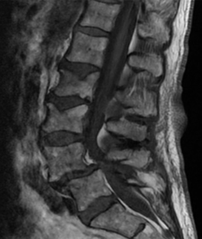
MRISagittal lumbar MRI: severe high-grade central canal stenosis with a subtle vertebral step at the level of the stenosis, suggesting a degenerative slip.
Source : Gabel et al., Cureus, 2015, figure 1 · CC BY
Surgery remains an effective option in DLS after a fairly conducted conservative failure or in the presence of progressive neurological deficits or cauda equina. Two randomised trials have transformed the decision:
- The SPORT trial (Weinstein 2007, NEJM)¹¹ compared surgery (decompression ± fusion) with conservative treatment in 304 DLS patients with stenosis. At 2 years the surgical arm had clearly better outcomes (pain, SF-36, ODI). The 8-year follow-up (Abdu 2017)¹² confirms the lasting benefit of surgery on pain and function, BUT with a reoperation rate of 22 % at 8 years (most often for recurrent stenosis or progression of the slip). Note that the SPORT patients were highly selected (failure of ≥ 12 weeks of conservative care).
- The Försth trial (NEJM 2016)¹⁷ randomised 247 patients (50-80 years) with lumbar stenosis ± DLS between decompression alone and decompression + fusion. Its major conclusion: adding fusion does not significantly change the outcomes at 2 years (Oswestry, EQ-5D, walking). This study revived the debate on the value of routine fusion in DLS and points towards decompression alone in the well-selected patient.
The surgical decision must therefore be shared with the patient, discussing explicitly the expected benefit for walking, the reoperation rate, the length of recovery, and the continuing alternative of conservative treatment (which remains effective in many).
Criticism and controversy: the paradoxes of "stabilisation"
Several paradoxes persist in the management of DLS.
1) The word "instability" is misleading: the vertebral slip is often chronic and stable, and insisting on the mechanics can generate an iatrogenic kinesiophobia. The benefits of "stabilisation" exercises may come less from mechanical re-stabilisation than from improved neuromuscular control, reduced central sensitisation and restored confidence in movement¹⁸.
2) The importance of exercise type remains debated. While recent RCTs tend to show a modest superiority of motor control exercise, several broad meta-analyses on chronic low back pain conclude that adherence to a regular programme matters more than the type of exercise¹⁹. This chimes with the CFT philosophy: individualise, give meaning, build adherence.
3) The surgical decision remains heterogeneous between countries and centres. Surgery rates for DLS vary considerably, a sign that indications are not standardised. The Försth trial calls for instrumental restraint (preferring decompression alone in the well-selected patient).
Key points
- The conservative approach is the first line, with a fairly conducted 3-6 month trial before surgery (NASS 2009/2016).
- The segmental stabilisation exercise programme is the most effective intervention (Lin 2024). Individualisation and adherence matter more than the exact protocol.
- The manual therapies and passive modalities are short-term adjuncts, never the heart of the treatment.
- The combination of patient education and CFT (the RESTORE trial, Kent 2023) is essential for DLS with a marked psychosocial component.
- The surgical option (decompression ± fusion) remains effective after conservative failure (SPORT, Weinstein 2007; Abdu 2017 at 8 years), but with 22 % reoperation at 8 years.
- The Försth trial (NEJM 2016): adding fusion to decompression does not improve the outcomes in DLS with stenosis. Shared decision-making is essential.
Chapter 3 bibliography (Treatment)
- Watters WC 3rd, Bono CM, Gilbert TJ, et al. An evidence-based clinical guideline for the diagnosis and treatment of degenerative lumbar spondylolisthesis. Spine J. 2009;9(7):609-614. PMID 19447684.
- Matz PG, Meagher RJ, Lamer T, et al. Guideline summary review: an evidence-based clinical guideline for the diagnosis and treatment of degenerative lumbar spondylolisthesis. Spine J. 2016;16(3):439-448. PMID 26681351.
- Engers A, Jellema P, Wensing M, van der Windt DA, Grol R, van Tulder MW. Individual patient education for low back pain. Cochrane Database Syst Rev. 2008;(1):CD004057. PMID 18254037.
- Saragiotto BT, Maher CG, Yamato TP, et al. Motor control exercise for chronic non-specific low-back pain. Cochrane Database Syst Rev. 2016;(1):CD012004. PMID 26742533.
- Resnick DK, Watters WC 3rd, Mummaneni PV, et al. Guideline update for the performance of fusion procedures for degenerative disease of the lumbar spine. Part 9: lumbar fusion for stenosis with spondylolisthesis. J Neurosurg Spine. 2014;21(1):54-61. PMID 24980587.
- Lin LH, Lin TY, Chang KV, et al. Effectiveness of Lumbar Segmental Stabilization Exercises in Managing Disability and Pain Intensity Among Patients With Lumbar Spondylolysis and Spondylolisthesis: A Systematic Review and Meta-analysis of Randomized Controlled Trials. Spine (Phila Pa 1976). 2024;49(21):1512-1520. PMID 38514931.
- Hides JA, Jull GA, Richardson CA. Long-term effects of specific stabilizing exercises for first-episode low back pain. Spine. 2001;26(11):E243-E248. PMID 11389408.
- Owen PJ, Miller CT, Mundell NL, et al. Which specific modes of exercise training are most effective for treating low back pain? Network meta-analysis. Br J Sports Med. 2020;54(21):1279-1287. PMID 31666220.
- Sinaki M, Lutness MP, Ilstrup DM, Chu CP, Gramse RR. Lumbar spondylolisthesis: retrospective comparison and three-year follow-up of two conservative treatment programs. Arch Phys Med Rehabil. 1989;70(8):594-598. PMID 2527488.
- Kent P, Haines T, O'Sullivan P, et al. Cognitive functional therapy with or without movement sensor biofeedback versus usual care for chronic, disabling low back pain (RESTORE): a randomised, controlled, three-arm, parallel group, phase 3, clinical trial. Lancet. 2023;401(10391):1866-1877. PMID 37146623.
- Weinstein JN, Lurie JD, Tosteson TD, et al. Surgical versus nonsurgical treatment for lumbar degenerative spondylolisthesis. N Engl J Med. 2007;356(22):2257-2270. PMID 17538085.
- Abdu WA, Sacks OA, Tosteson ANA, et al. Long-Term Results of Surgical and Nonsurgical Management of Lumbar Spinal Stenosis: 8-Year Results of the Spine Patient Outcomes Research Trial (SPORT). Spine. 2018;43(23):1619-1630. PMID 28399551.
- Gudavalli MR, Cambron JA, McGregor M, et al. A randomized clinical trial and subgroup analysis to compare flexion-distraction with active exercise for chronic low back pain. Eur Spine J. 2006;15(7):1070-1082. PMID 16341712.
- Louw A, Diener I, Butler DS, Puentedura EJ. The effect of neuroscience education on pain, disability, anxiety, and stress in chronic musculoskeletal pain. Arch Phys Med Rehabil. 2011;92(12):2041-2056. PMID 22133255.
- Wertli MM, Eugster R, Held U, Steurer J, Kofmehl R, Weiser S. Catastrophizing — a prognostic factor for outcome in patients with low back pain: a systematic review. Spine J. 2014;14(11):2639-2657. PMID 24607845.
- Kent P, Haines T, O'Sullivan P, et al. Cognitive functional therapy with or without movement sensor biofeedback versus usual care for chronic, disabling low back pain (RESTORE): 3-year follow-up of a randomised, controlled trial. Lancet Rheumatol. 2025;7(8):e547-e558. doi:10.1016/S2665-9913(25)00135-3.
- Försth P, Ólafsson G, Carlsson T, et al. A Randomized, Controlled Trial of Fusion Surgery for Lumbar Spinal Stenosis. N Engl J Med. 2016;374(15):1413-1423. PMID 27074066.
- O'Sullivan PB, Caneiro JP, O'Keeffe M, et al. Cognitive Functional Therapy: An Integrated Behavioral Approach for the Targeted Management of Disabling Low Back Pain. Phys Ther. 2018;98(5):408-423. PMID 29669082.
- Searle A, Spink M, Ho A, Chuter V. Exercise interventions for the treatment of chronic low back pain: a systematic review and meta-analysis of randomised controlled trials. Clin Rehabil. 2015;29(12):1155-1167. PMID 25681408.
How do you secure a lasting recovery and prevent recurrence in degenerative lumbar spondylolisthesis?
Resolving the acute symptoms is only one step: the real key to treatment success lies in the transition from passive management to active self-management. Since DLS is a chronic degenerative condition, the patient must become the main guarantor of their own spinal health. This chapter sets out how to build that autonomy and how to plan a return to activity based on functional criteria, not on imaging or on a calendar.
How do you make the patient an active player in their own recovery through self-management?
Empowering the patient is the cornerstone of preventing recurrence¹. Management limited to passive treatments, with no active involvement of the patient, is bound to fail in the long term². The aim is to provide the knowledge and the tools needed for independent management. 🧑🏫
The education of the patient (therapeutic patient education) is shown to be effective in improving pain and function in chronic low back pain³,⁴. It must cover:
- Understanding the condition : explain DLS simply, insist that the image does not define the suffering, since an asymptomatic slip is frequent (Brinjikji 2015)⁵. Demystify the fear attached to MRI.
- Pain management strategies : recognise the provocative activities, identify the relieving postures (forward flexion), understand the importance of movement even in the presence of moderate discomfort⁶.
- A home exercise programme : the transition to an independent programme is fundamental. Lumbopelvic stabilisation (transversus, multifidus, pelvic floor) is shown to be the most effective modality for reducing the ODI in DLS⁷.
- Lifestyle : sleep, stress management, regular physical activity (walking, cycling, swimming), weight control, all modifiable factors that influence chronicity.
Long-term adherence is the main challenge. To improve it:
- Co-build the programme with the patient (preferences, personal goals)⁸.
- Set progressive and realistic goals (the SMART technique).
- Plan spaced but regular follow-ups (at 1, 3, 6 and 12 months) to adjust.
- Build in health technologies (mobile reminder apps, exercise diaries) if the patient wants them⁹.
- Identify and treat early the psychosocial barriers (catastrophising, kinesiophobia), possibly through CFT (Kent 2023)¹⁰.
When and how do you plan a safe return to activity and sport?
The return to intense physical or sporting activity after a symptomatic episode of DLS must be an approach based on criteria, not on a rigid calendar¹¹. 🏃♀️ A return that is too early or poorly prepared increases the risk of symptomatic recurrence.
Progression is built around 4 phases:
- Symptom control (prerequisite): pain almost absent in activities of daily living, walking distance restored, no disabling claudication¹².
- Functional recovery : functional spinal mobility, adequate trunk and lower limb strength, good neuromuscular control. Tests: plank (isometric hold > 60 s), a good single leg squat, the ability to climb 2 flights of stairs without pain¹³.
- Progressive reintroduction of loading : gradual increase in volume, intensity and complexity. Start with low-impact activities (swimming, cycling, Nordic walking) before impact activities (running) or twisting ones (racket sports, golf)¹¹. The "10 % per week" rule for progressing intensity.
- Validation by performance criteria : the ability to perform the sport-specific movements without pain, without apprehension, with good movement quality¹⁴.
The physiotherapist's role is to guide this progression, making sure the patient keeps up the stabilisation programme alongside it. That programme becomes a lifelong preventive routine, not a one-off course of rehabilitation.
| Phase | Criteria to meet | Activities allowed | Indicative timescale |
|---|---|---|---|
| 1. Symptom control | VAS < 2/10 on walking, no disabling claudication | Normal ADLs, walking on level ground | 4-8 weeks |
| 2. Functional recovery | Isometric plank > 60 s, ODI < 20 % | Cycling, swimming, Nordic walking, progressive strengthening | 8-12 weeks |
| 3. Sporting reintroduction | No pain on progressive movements, neuromuscular control OK | Running on the flat (alternating), half-range sport-specific movements | 12-20 weeks |
| 4. Full return | Sporting movements without pain or apprehension, normal ODI | All activities, competition where applicable | ≥ 20 weeks (variable) |
Criticism and controversy: beyond protocols
Three tensions structure the recovery phase:
1) Which type of exercise matters most? Broad meta-analyses on chronic low back pain suggest that the specific type of exercise matters less than regular adherence¹⁵. The point is not to defend "the best protocol" but to find the activity the patient will keep up over time. Pilates, yoga, general strengthening, pure stabilisation, all have their place if they are actually practised.
2) Imaging must not guide the return to activity. The literature shows that the radiological parameters (Meyerding grade, dynamic instability) are weak predictors of functional outcomes in conservatively treated patients⁵,¹⁶. Basing activity restrictions on an image alone is a major source of iatrogenic kinesiophobia.
3) Sport is protective, not dangerous. Moderate-impact sports (running, racket sports), well prepared, are not contraindicated in stable DLS. On the contrary, prolonged sedentary behaviour is a major factor in chronicity. The message to pass on: "moving remains the best thing you can do for your back".
Key points
- ✅ Empowerment : patient education and self-management are the key to long-term success.
- 🏋️ Home programme : lumbopelvic stabilisation exercises practised regularly = the pillar of recurrence prevention.
- 📈 Progressive return : guided by functional criteria (pain, plank hold, neuromuscular control), not by a calendar.
- 🧠 Go beyond the image : the decision is made on the clinical picture and on function, not on radiological parameters.
- 🏃 The sport remains beneficial : sedentary behaviour is more dangerous than a well-prepared progressive return.
Chapter 4 bibliography (Lasting recovery)
- Du S, Hu L, Dong J, et al. Self-management program for chronic low back pain: a systematic review and meta-analysis. Patient Educ Couns. 2017;100(1):37-49. PMID 27554077.
- Macedo LG, Saragiotto BT, Yamato TP, et al. Motor control exercise for acute non-specific low back pain. Cochrane Database Syst Rev. 2016;(2):CD012085. PMID 26863390.
- Engers A, Jellema P, Wensing M, et al. Individual patient education for low back pain. Cochrane Database Syst Rev. 2008;(1):CD004057. PMID 18254037.
- Foster NE, Anema JR, Cherkin D, et al. Prevention and treatment of low back pain: evidence, challenges, and promising directions. Lancet. 2018;391(10137):2368-2383. PMID 29573872.
- Brinjikji W, Luetmer PH, Comstock B, et al. Systematic literature review of imaging features of spinal degeneration in asymptomatic populations. AJNR Am J Neuroradiol. 2015;36(4):811-816. PMID 25430861.
- Bunzli S, Smith A, Schütze R, Lin I, O'Sullivan P. Making sense of low back pain and pain-related fear. J Orthop Sports Phys Ther. 2017;47(9):628-636. PMID 28704621.
- Lin LH, Lin TY, Chang KV, et al. Effectiveness of Lumbar Segmental Stabilization Exercises in Managing Disability and Pain Intensity Among Patients With Lumbar Spondylolysis and Spondylolisthesis: A Systematic Review and Meta-analysis of Randomized Controlled Trials. Spine (Phila Pa 1976). 2024;49(21):1512-1520. PMID 38514931.
- Lin I, Wiles L, Waller R, et al. What does best practice care for musculoskeletal pain look like? Eleven consistent recommendations from high-quality clinical practice guidelines: systematic review. Br J Sports Med. 2020;54(2):79-86. PMID 30826805.
- Slade SC, Patel S, Underwood M, Keating JL. What are patient beliefs and perceptions about exercise for nonspecific chronic low back pain? A systematic review of qualitative studies. Clin J Pain. 2014;30(11):995-1005. PMID 24300225.
- Kent P, Haines T, O'Sullivan P, et al. Cognitive functional therapy with or without movement sensor biofeedback versus usual care for chronic, disabling low back pain (RESTORE): a randomised, controlled, three-arm, parallel group, phase 3, clinical trial. Lancet. 2023;401(10391):1866-1877. PMID 37146623.
- Ardern CL, Glasgow P, Schneiders A, et al. 2016 Consensus statement on return to sport from the First World Congress in Sports Physical Therapy, Bern. Br J Sports Med. 2016;50(14):853-864. PMID 27226389.
- O'Sullivan K, O'Sullivan PB, O'Keeffe M. The Lancet series on low back pain: reflections and clinical implications. Br J Sports Med. 2019;53(7):392-393. PMID 30170998.
- Cook G, Burton L, Hoogenboom B. Pre-participation screening: the use of fundamental movements as an assessment of function — part 1. N Am J Sports Phys Ther. 2006;1(2):62-72. PMID 21522216.
- Hodges PW, Smeets RJ. Interaction between pain, movement, and physical activity: short-term benefits, long-term consequences, and targets for treatment. Clin J Pain. 2015;31(2):97-107. PMID 24709625.
- Owen PJ, Miller CT, Mundell NL, et al. Which specific modes of exercise training are most effective for treating low back pain? Network meta-analysis. Br J Sports Med. 2020;54(21):1279-1287. PMID 31666220.
- Andersen T, Christensen FB, Høy KW, Helmig P, Niedermann B, Hansen ES, et al. The predictive value of pain drawings in lumbar spinal fusion surgery. Spine J. 2010;10(5):372-9. PMID 20421073.
What do real clinical cases teach us about degenerative lumbar spondylolisthesis?
Randomised trials and meta-analyses give the general framework; the published case reports illustrate the real complexity of practice. Three major lessons stand out: well-conducted conservative management often gives excellent results, DLS can present in deceptive guises (through referred hip pain in particular), and psychosocial factors can turn a "simple" file into a complex case. 🧐
Analysis of a "classic" case: from assessment to resolution
The published literature illustrates, through many randomised trials and patient series, the potential of a well-conducted conservative approach. The narrative review by Akkawi and Zmerly (2022, Acta Biomed)¹ recalls that a majority of patients with symptomatic DLS improved their pain and their function significantly with a structured rehabilitation programme, with no need for surgery.
A typical management pattern, validated by the NASS guidelines (Watters 2009 / Matz 2016)²,³ and supported by the Lin meta-analysis (2024)⁴, is built over 12 weeks:
- Weeks 1-2 : patient education (deconstructing catastrophic beliefs, explaining the clinical and radiological dissociation), pain management, relieving postures, introduction to lumbopelvic stabilisation (activation of transversus, multifidus).
- Weeks 3-6 : progression of the stabilisation programme, adding motor control exercises in functional positions (bridge, progressive plank, dead bug), adjunct manual therapy if a segment is restricted.
- Weeks 7-12 : functional exercises specific to the patient's goals (progressive walking, stairs, carrying light loads), reintroduction of the activities they enjoy (cycling, swimming, Nordic walking), transition to an independent home programme.
Recent RCTs on exercise in DLS typically report clinically significant reductions in the ODI (Oswestry Disability Index) of the order of 15 to 25 points and VAS reductions of 3 to 5 points⁴,⁵, well beyond the thresholds of clinically important difference (MCID: ODI ≥ 12 points, VAS ≥ 2 points). These results last when a home programme is kept up. The key is therefore less the specificity of the protocol than regularity and long-term adherence⁶.
The diagnostic challenge: when DLS mimics another condition
One of the most useful lessons drawn from clinical practice is the capacity of DLS to present in deceptive guises. 🎭 The foraminal or lateral stenosis that accompanies the slip can compress a root and cause pain referred to unexpected territories, in particular the groin, the anterior thigh or the greater trochanter.
Devin et al. (2012)⁷, in a detailed review of the "hip-spine syndrome", stress how difficult the differential diagnosis is between hip osteoarthritis and upper lumbar pathology (L2-L3, L3-L4). An L3 radiculopathy can produce pain exactly superimposable on hip osteoarthritis: groin + anterior thigh + medial side of the knee. The clinical risk is to focus the assessment and the treatment on the hip and to miss the lumbar cause, which explains the "failures" of total hip replacement in patients who in fact have an upper DLS.
Prather et al. (2017)⁸ prospectively examined 101 consecutive patients consulting for low back pain: 80 % had limited hip flexion and 75 % limited hip internal rotation, and those with a positive hip examination reported more intense pain and worse function. The clinical overlap between hip and spine is therefore frequent, and the rule that follows from it is plain: in any hip or anterior thigh pain that persists despite well-conducted local treatment, examine the lumbar spine systematically. Tests to build in: straight leg raise, femoral nerve stretch test, palpation of the posterior superior iliac spine, a search for neurogenic claudication, and lumbar imaging if the clinical findings agree.
Other "masks" of DLS are worth knowing:
- Piriformis syndrome : can be superimposed and mask a true neurogenic claudication.
- Meralgia paraesthetica (compression of the lateral femoral cutaneous nerve): numbness of the anterolateral thigh, to be distinguished from an L2-L3 radiculopathy.
- Trochanteric bursitis / gluteal tendinopathy : can coexist with a DLS in the older woman.
- Peripheral diabetic neuropathy : bilateral stocking-distribution numbness; frequently overlaps with DLS in older people.
A complex case with psychosocial factors
Complex cases remind us that a "simple" DLS on MRI can become extremely disabling when psychosocial factors dominate the picture. That is precisely the situation documented by the RESTORE programme of Kent et al. (Lancet 2023)⁹: patients with chronic disabling low back pain, often carrying modest radiological signs but a heavy psychosocial load (catastrophising, kinesiophobia, anxiety, mistaken beliefs).
The RESTORE trial (492 patients, 20 clinics in Australia) compared Cognitive Functional Therapy (CFT, with or without biofeedback) with usual care. At 13 weeks CFT achieved a reduction in disability (RMDQ) of −4.6 points (95% CI −5.9 to −3.4), with the effect maintained at 1 year and now at 3 years (Lancet Rheumatology 2025)¹⁰. CFT brings together:
- Shared understanding of the meaning of the pain (sense-making);
- Graded exposure to the avoided movements;
- Regaining confidence in the body through guided experiment;
- Work on lifestyle habits (sleep, activity, stress).
For DLS with a marked psychosocial component, building a cognitive functional approach in from the start of rehabilitation is now a recommendation drawn from the best available evidence¹¹,¹². This does not mean the physiotherapist must become a psychologist, but that they must know how to:
- Screen for yellow flags (brief questionnaires: STarT Back, ÖMPSQ-SF);
- Adapt their words so as not to reinforce catastrophic beliefs;
- Refer to a psychologist or to a CBT programme specialising in chronic pain when the psychosocial load exceeds their competence.
Criticism and controversy: the place of the case report in the evidence-based era
Three important points on the use of case reports:
1) A case report represents level 5 evidence (the lowest GRADE level, Oxford CEBM 5). It describes the experience of one individual and allows no generalisation. ⚠️
2) The main bias is publication bias : cases with spectacular results (resounding successes or failures) are over-represented, whereas the "average" cases, the majority, go unpublished.
3) Despite these limits, case reports remain indispensable as a teaching tool : they illustrate how the principles of SRs and MAs apply in the "disorder" of the real world (comorbidities, preferences, life context), they generate hypotheses (atypical cases), and they engrave in the clinician's mind the need for a systematic differential assessment (for example DLS mimicking hip osteoarthritis).
In short, case reports do not tell us "what works" in general, but they show us "what is possible" in a particular situation and they train us in clinical reasoning. They are the bridge between the rigour of meta-analyses and the humanity of each patient.
Key points
- Well-conducted conservative programmes (education + segmental stabilisation + adjunct manual therapy) typically achieve ODI reductions of 15-25 points and VAS reductions of 3-5 points, lasting if self-management follows (Lin 2024 MA).
- DLS can mimic hip osteoarthritis (groin or anterior thigh pain): in hip pain resistant to local treatment, assess the lumbar spine systematically (hip-spine syndrome).
- The psychosocial factors (catastrophising, kinesiophobia) can turn a "simple" DLS into a complex case. The CFT (the RESTORE trial, Kent 2023, Lancet) is now the reference for chronic low back pain with a marked psychosocial component.
- Case reports are valuable teaching tools but they carry the lowest level of evidence, to be used for reasoning, not for generalisation.
Chapter 5 bibliography (Clinical cases and lessons)
- Akkawi I, Zmerly H. Degenerative Spondylolisthesis: A Narrative Review. Acta Biomed. 2022;92(6):e2021313. PMID 35075090.
- Watters WC 3rd, Bono CM, Gilbert TJ, et al. An evidence-based clinical guideline for the diagnosis and treatment of degenerative lumbar spondylolisthesis. Spine J. 2009;9(7):609-614. PMID 19447684.
- Matz PG, Meagher RJ, Lamer T, et al. Guideline summary review: an evidence-based clinical guideline for the diagnosis and treatment of degenerative lumbar spondylolisthesis. Spine J. 2016;16(3):439-448. PMID 26681351.
- Lin LH, Lin TY, Chang KV, et al. Effectiveness of Lumbar Segmental Stabilization Exercises in Managing Disability and Pain Intensity Among Patients With Lumbar Spondylolysis and Spondylolisthesis: A Systematic Review and Meta-analysis of Randomized Controlled Trials. Spine (Phila Pa 1976). 2024;49(21):1512-1520. PMID 38514931.
- Saragiotto BT, Maher CG, Yamato TP, et al. Motor control exercise for chronic non-specific low-back pain. Cochrane Database Syst Rev. 2016;(1):CD012004. PMID 26742533.
- Owen PJ, Miller CT, Mundell NL, et al. Which specific modes of exercise training are most effective for treating low back pain? Network meta-analysis. Br J Sports Med. 2020;54(21):1279-1287. PMID 31666220.
- Devin CJ, McCullough KA, Morris BJ, Yates AJ, Kang JD. Hip-spine syndrome. J Am Acad Orthop Surg. 2012;20(7):434-442. PMID 22751162.
- Prather H, Cheng A, Steger-May K, Maheshwari V, Van Dillen L. Hip and Lumbar Spine Physical Examination Findings in People Presenting With Low Back Pain, With or Without Lower Extremity Pain. J Orthop Sports Phys Ther. 2017;47(3):163-172. PMID 28158964.
- Kent P, Haines T, O'Sullivan P, et al. Cognitive functional therapy with or without movement sensor biofeedback versus usual care for chronic, disabling low back pain (RESTORE): a randomised, controlled, three-arm, parallel group, phase 3, clinical trial. Lancet. 2023;401(10391):1866-1877. PMID 37146623.
- Kent P, Haines T, O'Sullivan P, et al. Cognitive functional therapy with or without movement sensor biofeedback versus usual care for chronic, disabling low back pain (RESTORE): 3-year follow-up of a randomised, controlled trial. Lancet Rheumatol. 2025;7(8):e547-e558. doi:10.1016/S2665-9913(25)00135-3.
- O'Sullivan PB, Caneiro JP, O'Keeffe M, et al. Cognitive Functional Therapy: An Integrated Behavioral Approach for the Targeted Management of Disabling Low Back Pain. Phys Ther. 2018;98(5):408-423. PMID 29669082.
- Hill JC, Dunn KM, Lewis M, et al. A primary care back pain screening tool: identifying patient subgroups for initial treatment. Arthritis Rheum. 2008;59(5):632-641. PMID 18438893.
How do you apply these recommendations concretely in your practice?
Applying the evidence in clinical practice is the bridge between science and care. For DLS it translates into three concrete requirements: knowing how to refer (red flags, yellow flags), knowing how to measure (validated PROMs) and knowing how to overcome the barriers to implementation. This chapter provides the practical tools for structuring those three axes in a practice or in a rehabilitation centre. 🧑⚕️
When, and to which other health professionals, should you refer?
The physiotherapist is often the first-line professional, which gives them a major responsibility for triage and referral. Identifying the situations that lie beyond their scope of competence is an essential component of safe care.
The detection of red flags is the non-negotiable step. The international reference framework is the International Framework for Red Flags for Potential Serious Spinal Pathologies (Finucane 2020, JOSPT)¹, a consensus tool that structures screening to identify infections, fractures, tumours and cauda equina syndromes. When a red flag is confirmed, medical referral is immediate (prompt medical consultation or emergency department depending on severity)².
Beyond the emergencies, assessment of the psychosocial factors (yellow flags) is just as fundamental. Catastrophising, kinesiophobia and avoidance beliefs are powerful predictors of chronicity³,⁴. Simple tools:
- STarT Back Tool (Hill 2008)⁵: 9 questions, stratification into low / medium / high risk.
- ÖMPSQ-SF (Örebro Musculoskeletal Pain Screening Questionnaire): the short 10-item version.
- FABQ (Fear-Avoidance Beliefs Questionnaire): 16 items, a score > 14 on the "Work" subscale predicts chronicity.
- PCS (Pain Catastrophizing Scale): 13 items, a score > 30 = marked catastrophising.
If the score is high, referral to a psychologist specialising in chronic pain or to a CBT programme is indicated. The physiotherapist can open the discussion as part of shared decision-making⁶.
Modern management of DLS is part of integrated multidisciplinary care models. The physiotherapist can refer to:
- General practitioner / rheumatologist : if progress stalls, for medication adjustment, epidural injection, or an opinion on whether an MRI is warranted.
- Orthopaedic spine surgeon : conservative failure ≥ 6 months OR a progressive neurological deficit. The shared discussion must include the SPORT figures (Weinstein 2007, Abdu 2017) and the Försth NEJM 2016 trial on the absence of benefit from routine fusion⁷,⁸,⁹.
- Pain physician : if a neuropathic component dominates or the chronic pain is refractory to standard treatment.
- Psychologist / psychotherapist : high yellow flags (catastrophising, kinesiophobia, associated depression), for CBT or ACT.
- Occupational therapist : adapting the workstation, the home, the vehicle.
- Sports physician : return to regular sporting practice, planning the resumption.
Referral ideally fits into a stepped care model ("stepped care"), where the intensity of the treatment and the number of professionals involved are adjusted to the patient's response and to the complexity of their condition¹⁰. The pathway physiotherapist → GP → specialist → surgeon is not a linear route but a modular network.
How do you measure outcomes and overcome the barriers to implementation?
Applying the recommendations is effective only if its results are measured. 📈 The PROMs (patient-reported outcome measures) have become the standard for judging the efficacy of a treatment from the patient's point of view¹¹. For DLS, the validated PROMs are:
- Oswestry Disability Index (ODI) : 10 items, score 0-100 %. The reference in low back pain. MCID estimated at 12 points¹².
- Zurich Claudication Questionnaire (ZCQ) or Swiss Spinal Stenosis Questionnaire: specific to lumbar stenosis (including DLS). Three domains: symptom severity, physical function, satisfaction. MCID estimated at 0.5 point per domain¹³.
- Roland-Morris Disability Questionnaire (RMDQ) : 24 items, simpler than the ODI; used in the RESTORE trial¹⁴.
- EQ-5D : generic quality of life, used in the Försth NEJM 2016 trial⁹.
- NPRS (Numeric Pain Rating Scale) or VAS: pain intensity.
- Walking distance : a simple functional measure, very useful in everyday practice.
Implementing PROMs requires standardised procedures, clinician training, and ideally integration into the electronic patient record for longitudinal follow-up¹¹. Failing that, a simple paper record at 4 key points (baseline, 6 weeks, 3 months, 6 months) is already a major advance on measuring nothing at all.
Several barriers hold back the adoption of evidence-based practice (EBP). The barriers most frequently cited by physiotherapists¹⁵:
- Lack of time : critical reading and putting it into practice demand unpaid time.
- Lack of research skills : initial training is sometimes insufficient in methodology.
- Lack of resources : access to the scientific databases is limited outside the university.
- Patient beliefs : expectations of passive modalities, requests for imaging, fear of moving¹⁶.
- Tension between guidelines and the patient-centred approach : the need to adapt the recommendations to the singularity of each case.
To overcome these barriers, a multifaceted approach is needed¹⁷:
- At clinician level : continuing education in EBP, subscribing to 1-2 newsletters from key journals (BJSM, JOSPT, Spine), taking part in a peer group.
- At organisation level : the organisational support is a key factor¹⁵. Allocate dedicated time for training, provide access to the databases, promote a culture in which EBP is valued.
- At system level : discourage low-value care (routine imaging, prolonged rest), promote evidence-based models in initial and continuing training¹⁸.
Criticism and controversy: the tensions of modern practice
Applying the recommendations rigorously raises several debates:
1) The paradox of the practice guideline : guidelines are essential for standardising, but they risk promoting a "cookbook medicine" that neglects individual complexity. The tension between applying a protocol validated for a population and adapting it to a unique individual is a permanent challenge.
2) The persistent "implementation gap" : despite solid evidence, low-value practices (excessive imaging, prolonged rest) persist, while high-value interventions (education, exercise) are under-used¹⁸. The gap is not down to the clinician alone: it rests on systemic factors, reimbursement, patient expectations, and a lack of time for in-depth patient education.
3) The utopia of interprofessional collaboration : while everyone agrees on its benefits, the reality often remains one of silos. Waiting lists, difficult communication between professionals, records that are not shared, the structural obstacles limit the efficacy of the collaborative model.
4) The burden of measurement : integrating PROMs is an advance, but it represents an extra administrative load for clinicians who are already overloaded. Without smooth technological integration and recognition, financial and in time, of the time spent, PROMs risk becoming a "box-ticking" exercise rather than a true improvement tool.
Physiotherapy for DLS must therefore navigate between scientific rigour and human flexibility, while campaigning for the systemic changes that support high-quality practice.
Key points
- Red flags (Finucane 2020): urgent medical referral. Yellow flags (catastrophising, kinesiophobia): psychological referral and CFT.
- Stepped care model ("stepped care"): adjust intensity and multidisciplinarity to the response and the complexity.
- Validated PROMs for DLS : ODI (MCID 12 points), ZCQ (stenosis), RMDQ, EQ-5D. To be measured at the key points (baseline, 6 weeks, 3 months, 6 months).
- Barriers to EBP: time, skills, resources, patient beliefs, the guideline-individualisation tension. A multifaceted approach is needed (clinician + organisation + system).
- Referral to the surgeon must rest on shared decision-making (the SPORT benefits, but 22 % reoperation at 8 years; no benefit from routine fusion according to Försth NEJM 2016).
Chapter 6 bibliography (Practical application)
- Finucane LM, Downie A, Mercer C, et al. International Framework for Red Flags for Potential Serious Spinal Pathologies. J Orthop Sports Phys Ther. 2020;50(7):350-372. PMID 32438853.
- Underwood M, Buchbinder R. Red flags for back pain. BMJ. 2013;347:f7432. PMID 24336004.
- Wertli MM, Eugster R, Held U, Steurer J, Kofmehl R, Weiser S. Catastrophizing — a prognostic factor for outcome in patients with low back pain: a systematic review. Spine J. 2014;14(11):2639-2657. PMID 24607845.
- Bunzli S, Smith A, Schütze R, Lin I, O'Sullivan P. Making sense of low back pain and pain-related fear. J Orthop Sports Phys Ther. 2017;47(9):628-636. PMID 28704621.
- Hill JC, Dunn KM, Lewis M, et al. A primary care back pain screening tool: identifying patient subgroups for initial treatment. Arthritis Rheum. 2008;59(5):632-641. PMID 18438893.
- Hoffmann TC, Légaré F, Simmons MB, et al. Shared decision making: what do clinicians need to know and why should they bother? Med J Aust. 2014;201(1):35-39. PMID 24999896.
- Weinstein JN, Lurie JD, Tosteson TD, et al. Surgical versus nonsurgical treatment for lumbar degenerative spondylolisthesis. N Engl J Med. 2007;356(22):2257-2270. PMID 17538085.
- Abdu WA, Sacks OA, Tosteson ANA, et al. Long-Term Results of Surgical and Nonsurgical Management of Lumbar Spinal Stenosis: 8-Year Results of the Spine Patient Outcomes Research Trial (SPORT). Spine. 2018;43(23):1619-1630. PMID 28399551.
- Försth P, Ólafsson G, Carlsson T, et al. A Randomized, Controlled Trial of Fusion Surgery for Lumbar Spinal Stenosis. N Engl J Med. 2016;374(15):1413-1423. PMID 27074066.
- Foster NE, Anema JR, Cherkin D, et al. Prevention and treatment of low back pain: evidence, challenges, and promising directions. Lancet. 2018;391(10137):2368-2383. PMID 29573872.
- Black N. Patient reported outcome measures could help transform healthcare. BMJ. 2013;346:f167. PMID 23358487.
- Copay AG, Glassman SD, Subach BR, Berven S, Schuler TC, Carreon LY. Minimum clinically important difference in lumbar spine surgery patients: a choice of methods using the Oswestry Disability Index, Medical Outcomes Study questionnaire Short Form 36, and pain scales. Spine J. 2008;8(6):968-974. PMID 18201937.
- Stucki G, Daltroy L, Liang MH, Lipson SJ, Fossel AH, Katz JN. Measurement properties of a self-administered outcome measure in lumbar spinal stenosis. Spine. 1996;21(7):796-803. PMID 8779009.
- Kent P, Haines T, O'Sullivan P, et al. Cognitive functional therapy with or without movement sensor biofeedback versus usual care for chronic, disabling low back pain (RESTORE): a randomised, controlled, three-arm, parallel group, phase 3, clinical trial. Lancet. 2023;401(10391):1866-1877. PMID 37146623.
- Da Silva T, Mills K, Brown BT, Pocovi N, de Campos T, Maher C, Hancock MJ. Recurrence of low back pain is common: a prospective inception cohort study. J Physiother. 2019;65(3):159-165. PMID 31208917.
- Slade SC, Patel S, Underwood M, Keating JL. What are patient beliefs and perceptions about exercise for nonspecific chronic low back pain? A systematic review of qualitative studies. Clin J Pain. 2014;30(11):995-1005. PMID 24300225.
- Greenhalgh T, Howick J, Maskrey N. Evidence based medicine: a movement in crisis? BMJ. 2014;348:g3725. PMID 24927763.
- Lin I, Wiles L, Waller R, et al. What does best practice care for musculoskeletal pain look like? Eleven consistent recommendations from high-quality clinical practice guidelines: systematic review. Br J Sports Med. 2020;54(2):79-86. PMID 30826805.
What next after reading this?
This article is part of a collection of evidence-based clinical syntheses. A question, a comment, a correction to suggest? Contact us directly through the WhatsApp button at the bottom right of the screen.

