















In brief
Pes anserine pain syndrome (PAPS), the term now preferred to “anserine bursitis”, denotes pain on the anteromedial aspect of the knee which most often corresponds to an insertional tendinopathy of the pes anserinus tendons rather than to a true bursitis. It is characterised by exquisite tenderness on palpation 5 to 7 cm below the medial joint line, worse on stairs and when lying on the side with the knees together; the at-risk populations are overweight middle-aged women, people with knee osteoarthritis and people with type 2 diabetes. Management is progressive: education, load management and targeted exercise, with injections reserved for failures.
Clinical synthesis based on the most recent narrative review (Aicale 2024), the 2025 systematic review of the knee bursae (Hasan 2025) and the 2023-2024 multicentre RCTs (Babaei-Ghazani, Gouda), for an evidence-based approach to medial knee pain.
Clinical summary
- The term “pes anserine pain syndrome” (PAPS) is more accurate than “anserine bursitis”: imaging shows that in the majority of cases this is an insertional tendinopathy of the sartorius, gracilis and semitendinosus tendons rather than an isolated bursal effusion (Aicale 2024; Hasan 2025).
- The diagnosis is clinical : exquisite tenderness on palpation 5-7 cm below the medial joint line, worse on stairs, on sit-to-stand and when lying on the side with the knees together (Pompan 2016; Mohseni StatPearls 2024).
- The dominant at-risk populations are: overweight middle-aged women (Pompan 2016, “underdiagnosed cause of knee pain in overweight women”), patients with medial knee osteoarthritis (44-71 % with PAPS on ultrasound depending on the cohort), and people with type 2 diabetes (24-34 % of those with knee pain; Aicale 2024 review).
- Ultrasound is the investigation of choice: it visualises the bursa (often barely distended) and an insertional tendinopathy, or rules out a stress fracture, a meniscal cyst or dominant knee osteoarthritis (Aicale 2024).
- Differential diagnoses to exclude: stress fracture of the medial tibial plateau, a meniscal cyst mimicking PAPS, isolated medial knee osteoarthritis, an MCL lesion, a localised Baker's cyst, and bursitis secondary to a tibial osteochondroma (PMC11231497, 2024).
- Management is progressive and tiered : 1) education and load management; 2) targeted exercise (hip and quadriceps strengthening, stretching); 3) adjunctive modalities (ESWT, HILT, taping); 4) injections (corticosteroids, PRP, prolotherapy, oxygen-ozone) where these fail.
- The strengthening of the hip stabilisers (abductors and external rotators) is central to correcting dynamic valgus, the main source of mechanical overload on the medial region of the knee (extrapolated from the patellofemoral data, Neal 2016).
- The multicentre RCT Babaei-Ghazani 2024 (Am J Phys Med Rehabil) compares prolotherapy, oxygen-ozone and corticosteroid under ultrasound guidance: all three are effective in the short term, with a longer-lasting effect for prolotherapy.
- The use of corticosteroid injections gives rapid relief but is associated with a recurrence rate of 20-40 % according to Hasan's 2025 SR; PRP and prolotherapy are promising alternatives where symptoms recur.
- Both shockwave therapy (ESWT) and high-intensity laser therapy (HILT) are the physical modalities with the most robust evidence; physiotherapy alone is non-inferior to steroid injection at 6-12 months (Sarifakioglu 2016, Gouda 2023 RCT).
- A load progression of ≤ 10 % per week (Gabbett 2016) prevents recurrence in runners; return to sport should be guided by objective functional criteria (strength ≥ 90 % of the healthy side, hop tests, Y-Balance).
- Medical referral is required where red flags are present (Finucane 2020): intense night pain, fever, unexplained weight loss, a painless palpable mass, or a recent injury with loss of function, to rule out fracture, infection or malignancy.
- The yellow flags (kinesiophobia, catastrophising, fear-avoidance, on Vlaeyen's model) are strong predictors of chronicity and justify a cognitive-behavioural approach and collaboration with a psychologist.
- The effectiveness of management is measured with validated PROMs : VAS, LEFS (Lower Extremity Functional Scale), KOOS for patients with associated osteoarthritis, and the Global Rating of Change.
- Current limits of the evidence: no SR or meta-analysis dedicated to PAPS exists yet . The recommendations rest on modestly sized RCTs, a general SR of the knee bursae (Hasan 2025), and extrapolation from peripheral tendinopathies.
Contents
- What are the fundamentals to know about pes anserine pain syndrome (PAPS)?
- How do you assess and diagnose PAPS with certainty?
- Which vulnerable subgroups call for specific clinical stratification?
- Which treatment strategies are the most effective?
- How do you secure lasting recovery and prevent recurrence?
- What do real clinical cases teach us about PAPS?
- How do you apply these recommendations concretely in your practice?
What are the fundamentals to know about pes anserine pain syndrome (PAPS)?
How is this condition defined, and why do we now speak of a “syndrome” rather than a “bursitis”?
The historical terminology of “anserine bursitis” is reductive and often inaccurate. 🧐 Modern imaging studies, high-frequency ultrasound and MRI, converge in showing that most patients clinically labelled with “bursitis” in fact have insertional tendon thickening (enthesopathy), a hypoechoic tendinopathy or infiltration of the periarticular soft tissues, without any significant fluid distension of the bursa.¹,² This is why the scientific community has gradually adopted the generic term “pes anserine pain syndrome” (PAPS), covering both true bursitis (rare) and insertional tendinopathy (by far the more common).¹,³- In the general population the estimate is below 1 % of isolated presenting complaints; some North American military administrative databases find the diagnostic code in about 1.4 % of patients with non-specific knee pain.²
- In the population with symptomatic knee osteoarthritis, systematic ultrasound finds anserine signs (tendon thickening and/or bursal effusion) in 44 - 71 % of patients depending on the series; clinical PAPS is therefore a very frequent “satellite comorbidity” of medial knee osteoarthritis.¹
- In the population with type 2 diabetes and knee pain, ultrasound cohorts report anserine tendinobursitis in 24 to 34 % of patients, with a strong female predominance.¹,⁴
📊 PAPS prevalence by subgroup
Anserine signs (clinical + ultrasound) by clinical context
* Grey-literature estimate from specialist sports medicine reviews, to be interpreted with caution (no dedicated SR or meta-analysis). Main sources: Aicale 2024 (EJMD); Hasan 2025 (Cartilage); Mohseni StatPearls 2024.
What happens in the body, and how does PAPS evolve naturally?
The pathophysiology of PAPS rests mainly on repetitive mechanical stress and on friction between the anserine tendons and the underlying structures (MCL, medial tibial condyle).¹,⁵ In acute overuse presentations the bursa can become inflamed (bursal synovitis) with hyperaemia and slight fluid distension, visible on ultrasound as a well-defined hypoechoic collection.² In chronic presentations, which make up most consultations, the dominant lesion is an insertional tendinopathy, characterised by tendon thickening, loss of the fibrillar echo-structure and sometimes neovascularisation on colour Doppler.¹,⁵ The typical pain is:- Localised 5-7 cm below the medial joint line, over the tibial insertion of the three tendons²;
- Described as a dull ache or tenderness on palpation, sometimes a burning or pulling sensation;⁶
- Worse on going up and down stairs, on sit-to-stand, on running (particularly on slopes), and when lying on the side with the knees touching (direct compression at night);²,⁶
- Most often unilateral, but bilateral in patients with T2DM or knee osteoarthritis.¹
Key points
- The term “pes anserine pain syndrome” (PAPS) has replaced “anserine bursitis”, because imaging shows that most cases are insertional tendinopathies, not fluid bursitis.
- Prevalence is very low in the general population (< 1 %) but rises sharply in people with medial knee osteoarthritis (44-71 %) and in people with type 2 diabetes and knee pain (24-34 %).
- Exquisite tenderness on palpation 5-7 cm below the medial joint line, worse on stairs and when lying on the side.
- The course is favourable with appropriate management of the underlying factors, but the risk of recurrence is 20-40 % after steroid alone without biomechanical correction.
Chapter 1 bibliography
- Aicale R, et al. Comprehensive Review of Pes Anserinus Syndrome: Etiology, Diagnosis, and Management. Eur J Musculoskelet Dis. 2024;13(3):60-69. Biolife Publisher (open access).
- Mohseni M, Mabrouk A, Li D. Pes Anserine Bursitis. In: StatPearls [Internet]. Treasure Island (FL): StatPearls Publishing; 2024. NBK532941.
- Hasan M, Berkovich Y, Khatib M, Steinfeld Y, Sleiman A, Ben Zvi L, Abu Alhija A, Ginesin E, Yonai Y. Knee Bursae: A Comprehensive Review of Clinical Evaluation, Imaging Differentiation, and the Expanding Role of Biologic Therapies. Cartilage. 2025. PMID 41239892. doi:10.1177/19476035251362434.
- Alvarez-Nemegyei J. Risk factors for pes anserinus tendinitis/bursitis syndrome: a case control study. J Clin Rheumatol. 2007;13(2):63-65. PMID 17414530.
- Hubbard MJ, Hildebrand BA, Battafarano MM, Battafarano DF. Common Soft Tissue Musculoskeletal Pain Disorders. Prim Care. 2018;45(2):289-303. PMID 29759125.
- Pompan DC. Pes Anserine Bursitis: An Underdiagnosed Cause of Knee Pain in Overweight Women. Am Fam Physician. 2016;93(3):170. PMID 26926606.
How do you assess and diagnose PAPS with certainty?
Which questions should you ask to understand the patient and their history?
History-taking should explore five main dimensions:- Location and description of the pain : anteromedial aspect of the knee, 5-7 cm below the joint line (much lower than meniscal pain, which is on the joint line); a dull ache, sometimes burning; intensity varying with activity.¹,⁴
- Aggravating factors : key questions to ask systematically. “Does your pain increase when you go up or down stairs? When you get up from a low chair? When you sleep on your side with your knees touching?” Their combination is highly suggestive.¹,²
- Relevant past history : weight and BMI, type 2 diabetes (duration, glycaemic control, micro- and macrovascular complications), knee osteoarthritis already diagnosed (side, Kellgren-Lawrence severity), previous knee surgery (total arthroplasty; PAPS after TKA is a described and sometimes painful entity).⁵
- Sporting and occupational history : for runners, recent changes in weekly volume (> 10 % per week is a key risk factor, Gabbett 2016), a change of shoes or surface, more elevation gain; for standing occupations, prolonged static postures and hard floors.⁶
- Treatments already tried : rest, NSAIDs, previous injection (steroid? when?), physiotherapy (modalities, duration, response). These are essential to deciding the next strategy.²
Which clinical tests should you perform, and which other conditions should you rule out?
The physical examination aims to reproduce the patient's familiar pain and to rule out the differential diagnoses. 🩺 The reference clinical test is direct, precise palpation of the anserine insertion site: patient supine, hip in abduction and external rotation, knee at 30° of flexion. The thumb finds the medial joint line, then moves 5 to 7 cm down the anteromedial aspect of the tibia. The exquisite reproduction of the patient's usual pain at that precise point, with no equivalent pain over the joint line or along the MCL, is the most reliable clinical sign.¹,²| Diagnosis to rule out | Pain location | Orienting test | Confirmatory investigation |
|---|---|---|---|
| Medial knee osteoarthritis | Over the medial joint line, diffuse | Morning stiffness, varus, crepitus | Weight-bearing radiograph (Kellgren-Lawrence) |
| Medial meniscal lesion | Over the posteromedial joint line | McMurray, Thessaly, locking | MRI |
| MCL lesion | Along the ligament (joint line ↑) | Valgus stress at 0° and 30° | Ultrasound / MRI |
| Tibial plateau stress fracture | Medial, bony, exquisite | Pain on single-leg stance, hop test impossible | MRI / bone scan |
| Medial meniscal cyst | Swelling over the joint line | Reducible palpable mass | Ultrasound / MRI |
| Posteromedial Baker's cyst | Popliteal fossa, medial | Posterior mass, pain on forced flexion | Ultrasound |
| Septic bursitis | Local, with inflammatory signs | Redness, heat, fever | Aspiration + culture (emergency) |
🚩 Red flags specific to medial knee pain
- Severe night pain waking the patient + unexplained weight loss → work-up for malignancy (primary bone tumour or tibial metastasis)
- Exquisite bony pain + inability to weight-bear of sudden onset in a runner → stress fracture of the medial tibial plateau ; MRI is essential before any return to sport
- Hot, red, painful swelling + fever → suspected septic bursitis (rare but documented, notably after injection); urgent aspiration and antibiotics
- Painless, firm, progressively enlarging palpable mass → suspected tumour (synovial chondromatosis, osteochondroma, giant cell tumour; cases reported)
- Recent injury with haematoma, instability or inability to extend fully → suspected MCL rupture, plateau fracture or acute meniscal lesion
⚠️ Any red flag → prompt medical referral (GP, emergency department, orthopaedic surgeon) before physiotherapy management.
What is the place of ultrasound and MRI in confirming the diagnosis?
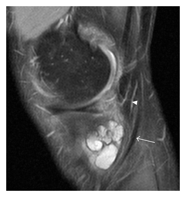
MRISagittal knee MRI, fat-saturated proton density sequence: fluid collection in the pes anserine bursa, arrow on the semitendinosus tendon.
Source : Rytter et al., International journal of rheumatology, 2012, figure 2 · CC BY
- The anserine bursa : normal thickness < 2 mm; a well-defined hypoechoic collection > 2 mm indicates a distended bursitis (rare);²
- The sartorius, gracilis and semitendinosus tendons at their tibial insertion: thickening, loss of the fibrillar echo-structure and focal hypoechogenicity indicate an insertional tendinopathy (by far the more common);¹,²
- The neovascularisation on colour Doppler, a marker of inflammatory activity and a prognostic factor;¹
- The neighbouring structures : joint effusion, an overflowing meniscal cyst, a Baker's cyst, signs of knee osteoarthritis (osteophytes, cartilage degeneration).¹
🔍 Performance of the diagnostic tools for PAPS
Hierarchy according to the consolidated 2024-2025 evidence
Source: synthesis of Aicale 2024 (EJMD), Hasan 2025 (Cartilage), Mohseni StatPearls 2024.
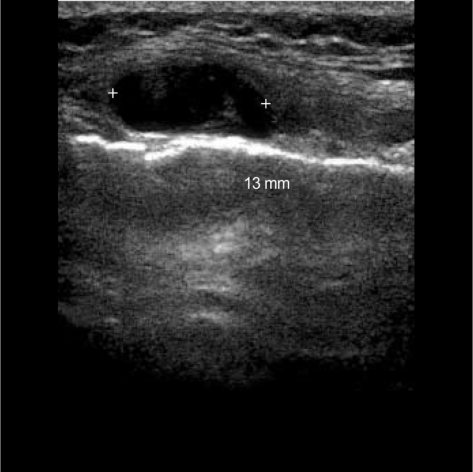
UltrasoundUltrasound of the medial aspect of the knee: anechoic fluid collection measuring 13 mm within the pes anserine bursa, measured by cursors.
Source : Yoon et al., Journal of Korean medical science, 2005, figure 2 · CC BY-NC
Key points
- The diagnosis is clinical : exquisite, reproducible tenderness on palpation 5-7 cm below the medial joint line, worse on stairs and when lying on the side.
- Always rule out the differential diagnoses : medial knee osteoarthritis, meniscal lesion, MCL, stress fracture, Baker's cyst, septic bursitis.
- Ultrasound is the investigation of choice: it shows insertional tendinopathy (frequent), bursal effusion (rare) and Doppler neovascularisation.
- MRI is reserved for atypical cases or where a red flag is present; it rarely confirms a “true bursitis” but it does rule out fracture, tumour and ligament injury.
Chapter 2 bibliography
- Aicale R, et al. Comprehensive Review of Pes Anserinus Syndrome. Eur J Musculoskelet Dis. 2024;13(3):60-69. Biolife Publisher.
- Mohseni M, Mabrouk A, Li D. Pes Anserine Bursitis. StatPearls; 2024. NBK532941.
- Hasan M, et al. Knee Bursae: A Comprehensive Review. Cartilage. 2025. PMID 41239892.
- Pompan DC. Pes Anserine Bursitis: An Underdiagnosed Cause of Knee Pain in Overweight Women. Am Fam Physician. 2016;93(3):170. PMID 26926606.
- Allen MF, Allen DE. Pes Anserinus Bursitis: A Case Report. Cureus. 2022;14(10):e30650. PMID 36415475. PMC 9674038.
- Gabbett TJ. The training-injury prevention paradox: should athletes be training smarter and harder? Br J Sports Med. 2016;50(5):273-280. PMID 26758673.
Which vulnerable subgroups call for specific clinical stratification?
The “overweight middle-aged woman” profile: why is it dominant?
The landmark publication by Pompan 2016 in American Family Physician is explicit from its title: “Pes Anserine Bursitis: An Underdiagnosed Cause of Knee Pain in Overweight Women ”.¹ This female over-representation is confirmed in every published series:- In the case-control series of Alvarez-Nemegyei 2007, the 22 consecutive patients were all women (mean age 62), with a marked excess risk in overweight patients and those with osteoarthritis.²
- In the ultrasound cohorts of knee osteoarthritis, women account for more than 80 % of the cases of anserine tendinobursitis detected.³
- Pelvic anatomy : a wider pelvis → an increased quadriceps Q angle → a tendency to dynamic knee valgus under load → greater medial stress.²
- Body composition and hormones : abdominal and periarticular adiposity, perimenopausal hormonal changes (falling oestrogen affecting tendon quality).¹
- Associated comorbidities : knee osteoarthritis, obesity and type 2 diabetes are more frequent in women over 50.¹,²
Knee osteoarthritis, type 2 diabetes: how do these comorbidities change management?
Together, medial knee osteoarthritis and PAPS form a clinical pair: osteoarthritis alters knee biomechanics (varus, extension deficit, quadriceps weakness), increases friction at the pes anserinus and produces chronic tendon overload.³ Conversely, PAPS can be the dominant source of the pain a patient with knee osteoarthritis feels, sometimes more so than the arthritis itself, which is why it is worth systematically assessing the anserine area in every patient complaining of medial knee pain, even where knee osteoarthritis is already known.¹,³ Type 2 diabetes 🩸 is an independent and powerful risk factor: the non-enzymatic glycation of collagen alters the mechanical properties of tendons (increased stiffness, cumulative micro-damage), and diabetic microangiopathy compromises tendon healing.² Ultrasound cohorts find a PAPS prevalence of 24 to 34 % in patients with T2DM and knee pain.¹,² Clinical implications:- Optimise glycaemic control in partnership with the endocrinologist;
- Anticipate slower tendon healing when planning the rehabilitation programme (longer duration, more cautious load progression);
- Use corticosteroid injections sparingly: the risk of post-injection glycaemic disturbance is well documented in T2DM.
⚖️ Relative risk factors for PAPS: synthesis of the available data
A qualitative estimate of impact, to be interpreted with caution in the absence of a dedicated meta-analysis
A qualitative synthesis of converging data (Alvarez-Nemegyei 2007, Pompan 2016, Aicale 2024, Hasan 2025); no dedicated meta-analysis quantifies these effects as OR or RR for PAPS.
| Profile | Clinical features | Treatment priority | Specifics |
|---|---|---|---|
| Woman > 50 + overweight | Bilateral pain, associated knee osteoarthritis frequent | Weight loss, quadriceps and hip abductor strengthening | Systematic ultrasound |
| Medial knee osteoarthritis | Diffuse pain + exquisite anserine tenderness | Global management of the osteoarthritis (KOOS, supervised exercise) | Medial offloading, insoles? |
| T2 diabetes + knee pain | PAPS often bilateral, little inflammation | Glycaemic control, strengthening, longer physiotherapy | Steroids sparingly |
| Endurance runner | Acute pain after a rise in mileage | Load reduction, biomechanical correction, gradual return | Podiatric assessment |
Key points
- Four dominant profiles: overweight middle-aged woman, medial knee osteoarthritis, type 2 diabetes, endurance runner.
- Stratifying the profile guides the strategy: weight loss is central for profiles 1-2,glycaemic balance for profile 3, and training load management for profile 4.
- Use corticosteroid injections sparingly in T2DM (risk of post-injection glycaemic disturbance).
- No dedicated meta-analysis quantifies the ORs or RRs; the recommendations rest on case-control series and the Aicale 2024 synthesis.
Chapter 3 bibliography
- Pompan DC. Pes Anserine Bursitis: An Underdiagnosed Cause of Knee Pain in Overweight Women. Am Fam Physician. 2016;93(3):170. PMID 26926606.
- Alvarez-Nemegyei J. Risk factors for pes anserinus tendinitis/bursitis syndrome: a case control study. J Clin Rheumatol. 2007;13(2):63-65. PMID 17414530.
- Aicale R, et al. Comprehensive Review of Pes Anserinus Syndrome. Eur J Musculoskelet Dis. 2024;13(3):60-69. Biolife Publisher.
- Hubbard MJ, Hildebrand BA, Battafarano MM, Battafarano DF. Common Soft Tissue Musculoskeletal Pain Disorders. Prim Care. 2018;45(2):289-303. PMID 29759125.
Which treatment strategies are the most effective for PAPS?
Where do you start? What is the recommended hierarchy of interventions?
- Tier 1: education + load management : explaining the condition (insertional tendinopathy more often than true bursitis), defusing the fear, temporarily modifying the triggering activities, ice in the acute phase, topical or short-course oral NSAIDs if needed.¹
- Tier 2: targeted exercise programme : the cornerstone of long-term treatment. It includes strengthening (quadriceps, hamstrings, hip abductors and external rotators) and stretching (hamstrings, adductors).³,⁴
- Tier 3: adjunctive modalities where pain persists despite tiers 1-2: shockwave therapy (ESWT), high-intensity laser therapy (HILT), kinesio taping, therapeutic ultrasound (weaker evidence), manual therapy.⁴,⁵
- Tier 4: ultrasound-guided injections where pain is disabling or the previous tiers have failed: a choice between corticosteroids (rapid effect but 20-40 % recurrence), PRP, prolotherapy (hypertonic dextrose), oxygen-ozone . The Babaei-Ghazani 2024 multicentre RCT shows comparable short-term efficacy, with a longer-lasting effect for prolotherapy.⁶,⁷
What is the place of exercise, and is there a superior approach?
Therapeutic exercise is a fundamental and non-negotiable element of PAPS management, particularly in the patient with associated knee osteoarthritis. 💪 Several findings converge:- The reference RCT by Sarifakioglu 2016 (J Phys Ther Sci, n = 60) compared a physiotherapy programme (ultrasound + isometric quadriceps exercise + stretching) with a corticosteroid injection. In the short term the injection was slightly better for pain, but at 3 months and beyond the two groups were comparable, with an advantage for the physiotherapy group in terms of freedom from recurrence.³
- Strengthening should target first and foremost the hip stabilisers (gluteus medius, external rotators) to correct dynamic knee valgus, a source of mechanical overload on the pes anserinus.⁸,⁹
- Meanwhile, hamstring and adductor stretching reduces insertional tension over the anserine area and is recommended systematically (Aicale 2024).¹
| Muscle group | Type of exercise | Functional goal | Typical progression |
|---|---|---|---|
| Hip abductors (gluteus medius) | Clamshell, side-lying abduction, monster walk | Control dynamic valgus | 3 × 12 → progress with bands |
| Hip external rotators | Side-lying external rotation | Stabilise the knee under load | 3 × 10 → single-leg loading |
| Quadriceps | Isometric → partial squat → full squat | Absorb flexion-extension stress | Progressive over 6-8 weeks |
| Hamstrings | Active stretching + Nordic hamstring if tolerated | Reduce tendon tension at the insertion | Daily stretching, 3 × 30 s |
| Adductors | Frog stretch, lateral lunge | Reduce tension on gracilis (a pes anserinus component) | Daily stretching |
Manual therapies, shockwave, laser, injections: what does the evidence say?
Extracorporeal shockwave therapy (ESWT) . Several RCTs have evaluated ESWT specifically in PAPS:- The RCT by Gouda 2023 (Advances in Orthopedics, n = 180 patients) compares steroid vs PRP vs ESWT: all three are effective at 1 month, with PRP superior at 3-6 months and good overall tolerance of ESWT.⁷
- Another RCT (Tu 2023, Open-Label RCT) confirms the non-inferiority of ESWT versus steroid, with a better safety profile (no risk of glycaemic disturbance, no skin atrophy).⁹
- The systematic review by Korakakis 2018 (Br J Sports Med) on ESWT in lower-limb conditions finds a low to moderate level of evidence overall, with encouraging but heterogeneous results.¹⁰
- Prolotherapy (hypertonic dextrose): efficacy comparable to steroid in the short term, with a longer-lasting effect ;
- Oxygen-ozone : an emerging alternative with a favourable safety profile;
- Corticosteroids : rapid effect but 20-40 % recurrence (Hasan 2025).⁵,⁶,⁷
⏱️ Time course of the effect: corticosteroids vs PRP vs prolotherapy vs ESWT
Qualitative synthesis of the 2023-2024 data
How to read this: corticosteroids act quickly but decline over 3-6 months (20-40 % recurrence, Hasan 2025); PRP and prolotherapy act more slowly but last longer (Babaei-Ghazani 2024, Gouda 2023). ESWT sits in between.
| Intervention | Short-term effect (≤ 3 months) | Long-term effect (> 6 months) | Level of evidence (GRADE) |
|---|---|---|---|
| Education + load management | Moderate | Good (if adherence) | Moderate |
| Targeted exercise (strengthening + stretching) | Good | Good | Moderate |
| ESWT | Good | Moderate | Moderate |
| High-intensity laser (HILT) | Good (short term) | Not documented | Low |
| Kinesio taping | Slight (adjunct) | Not demonstrated | Low |
| Steroid injection | Very good | Poor (20-40 % recurrence) | Moderate |
| PRP injection | Good (delayed effect) | Good | Moderate |
| Prolotherapy (dextrose) | Moderate (delayed effect) | Good (Babaei-Ghazani 2024) | Moderate |
| Oxygen-ozone | Good (emerging data) | To be documented | Low |
| Therapeutic ultrasound alone | Marginal | Not demonstrated | Very low |
Beyond the physical: how do you educate the patient and address psychological factors?
Therapeutic education 🧠 is a major determinant of success. It should cover:- The nature of the condition : this is most often an overload condition of tendons, not a “serious injury”. That reassuring message reduces kinesiophobia.¹¹
- Load management : teaching the principle of progressing by ≤ 10 % per week (Gabbett 2016); the concept of “optimal stress” for tendon tissue; the use of a diary to identify personal thresholds.¹²,¹³
- Adherence to the exercises : the benefits are cumulative; a programme followed 3-5 times a week for 8-12 weeks is needed for lasting effects.³,¹¹
- Self-assessment of pain : the “24-hour rule” (pain acceptable up to 3/10 during exercise, and back to baseline within 24 hours), adapted from Silbernagel's model.¹³
- Kinesiophobia (Vlaeyen's 2007 fear-avoidance model): fear of movement → avoidance → deconditioning → worsening pain.¹¹
- Catastrophising (“I'll never walk normally again”): an independent predictor of a poor prognosis.
- Limiting beliefs (“running is bad for my knees”): to be explored and addressed through education and graded exposure.
Key points
- A four-tier hierarchy: 1) education + load management → 2) targeted exercise → 3) adjunctive modalities → 4) injections.
- Exercise (hip and quadriceps strengthening, stretching) is the non-negotiable mainstay; Sarifakioglu 2016 shows PT to be non-inferior to steroid at 3 months and beyond.
- The Babaei-Ghazani 2024 multicentre RCT : prolotherapy ≈ oxygen-ozone ≈ steroid in the short term, with a more durable effect for prolotherapy.
- ESWT : moderate evidence, a useful alternative to steroid (particularly in T2DM).
- Education and addressing psychosocial factors (kinesiophobia) determine long-term success.
Chapter 4 bibliography
- Aicale R, et al. Comprehensive Review of Pes Anserinus Syndrome. Eur J Musculoskelet Dis. 2024;13(3):60-69. Biolife Publisher.
- Mohseni M, Mabrouk A, Li D. Pes Anserine Bursitis. StatPearls; 2024. NBK532941.
- Sarifakioglu B, Afsar SI, Yalbuzdag SA, Ustaömer K, Bayramoğlu M. Comparison of the efficacy of physical therapy and corticosteroid injection in the treatment of pes anserine tendino-bursitis. J Phys Ther Sci. 2016;28(7):1993-1997. PMID 27512249.
- Hubbard MJ, Hildebrand BA, Battafarano MM, Battafarano DF. Common Soft Tissue Musculoskeletal Pain Disorders. Prim Care. 2018;45(2):289-303. PMID 29759125.
- Homayouni K, Foruzi S, Kalhori F. Effects of kinesiotaping versus non-steroidal anti-inflammatory drugs and physical therapy for treatment of pes anserinus tendino-bursitis: A randomized comparative clinical trial. Phys Sportsmed. 2016;44(3):252-256. PMID 27276165.
- Babaei-Ghazani A, Eftekharsadat B, Soleymanzadeh H, ZoghAli M. Ultrasound-Guided Pes Anserine Bursitis Injection Choices: Prolotherapy or Oxygen-Ozone or Corticosteroid: A Randomized Multicenter Clinical Trial. Am J Phys Med Rehabil. 2024;103(4):310-317. PMID 37752656.
- Gouda AM, et al. Comparing the Efficacy of Local Corticosteroid Injection, Platelet-Rich Plasma, and Extracorporeal Shockwave Therapy in the Treatment of Pes Anserine Bursitis: A Prospective, Randomized, Comparative Study. Adv Orthop. 2023;2023:5545520. PMC 10565108. doi:10.1155/2023/5545520.
- Khosrawi S, Taheri P, Ketabi M. Investigating the Effect of Extracorporeal Shock Wave Therapy on Reducing Chronic Pain in Patients with Pes Anserine Bursitis: A Randomized, Clinical- Controlled Trial. Adv Biomed Res. 2017;6:70. PMID 28626745.
- Hasan M, et al. Knee Bursae: A Comprehensive Review. Cartilage. 2025. PMID 41239892.
- Korakakis V, Whiteley R, Tzavara A, Malliaropoulos N. The effectiveness of extracorporeal shockwave therapy in common lower limb conditions: a systematic review including quantification of patient-rated pain reduction. Br J Sports Med. 2018;52(6):387-407. PMID 28954794.
- Louw A, Zimney K, Puentedura EJ, Diener I. The efficacy of pain neuroscience education on musculoskeletal pain: A systematic review of the literature. Physiother Theory Pract. 2016;32(5):332-355. PMID 27351541.
- Gabbett TJ. The training-injury prevention paradox: should athletes be training smarter and harder? Br J Sports Med. 2016;50(5):273-280. PMID 26758673.
- Silbernagel KG, Thomeé R, Eriksson BI, Karlsson J. Continued sports activity, using a pain-monitoring model, during rehabilitation in patients with Achilles tendinopathy: a randomized controlled study. Am J Sports Med. 2007;35(6):897-906. PMID 17307888.
How do you secure lasting recovery and prevent recurrence?
How do you make the patient an active participant in their recovery through self-management?
Patient autonomy (self-management) is the cornerstone of preventing recurrence. 🎓 A systematic review by Babatunde 2017 (PLoS One) on musculoskeletal conditions in primary care confirms that self-management interventions combined with exercise are among the most effective for pain control and long-term functional improvement.¹ Key components:- Self-monitoring of symptoms : recognising the early signs (morning tenderness, pain after exercise); keeping a diary to correlate them with activity.
- Load management : progressing by ≤ 10 % per week in volume, intensity and frequency (Gabbett 2016).²
- A home programme : targeted exercises, 3-5 times a week, for at least 8-12 weeks, then maintenance twice a week.
- Optimising systemic factors : weight loss (5-10 % of the starting BMI in overweight women over 50), glycaemic control in T2DM, and management of associated knee osteoarthritis according to the OARSI recommendations.³
When and how should a safe return to sport and to activity be planned?
The decision to return to sport (RTS) must rest on objective functional criteria, not on a fixed calendar.⁴ The Burgi 2019 review (Br J Sports Med) of post-ACL RTS criteria, which can be extrapolated to other knee conditions, recommends a combination of:- Fundamental prerequisites : no pain in daily activities; full and symmetrical range of motion; strength ≥ 90 % of the healthy side (quadriceps, hamstrings, hip abductors).⁴
- Functional tests : Y-Balance Test (dynamic balance), hop tests (single hop, triple hop, crossover hop), step-down test.
- Progressive reintegration : resuming in a straight line (jogging) → changes of direction → acceleration and deceleration → jumping → opposition → competition.
- Pain tolerance : Silbernagel's model, adapted. Pain acceptable at ≤ 3/10 during exercise, back to baseline within 24 h, with no progressive increase session after session.⁵
🏃 Return-to-sport algorithm after PAPS
A criteria-based approach, inspired by Burgi 2019 and adapted to PAPS
Adapted from Burgi 2019 (Br J Sports Med, post-ACL scoping review); the principles transfer to PAPS.
Key points
- Self-management is essential: education, home exercises, load progression ≤ 10 % per week, a diary.
- The Return to sport must be guided by objective functional criteria (strength ≥ 90 %, Y-Balance and hop tests) and not by a calendar.
- The 24-hour rule (Silbernagel): pain acceptable at ≤ 3/10, back to baseline within 24 h after exercise.
- The risk of recurrence is greatly reduced where the systemic determinants (weight, blood glucose, biomechanics) are treated alongside the rehabilitation.
Chapter 5 bibliography
- Babatunde OO, Jordan JL, Van der Windt DA, Hill JC, Foster NE, Protheroe J. Effective treatment options for musculoskeletal pain in primary care: A systematic overview of current evidence. PLoS One. 2017;12(6):e0178621. PMID 28640822.
- Gabbett TJ. The training-injury prevention paradox: should athletes be training smarter and harder? Br J Sports Med. 2016;50(5):273-280. PMID 26758673.
- Bannuru RR, Osani MC, Vaysbrot EE, et al. OARSI guidelines for the non-surgical management of knee, hip, and polyarticular osteoarthritis. Osteoarthritis Cartilage. 2019;27(11):1578-1589. PMID 31278997.
- Burgi CR, Peters S, Ardern CL, Magill JR, Gomez CD, Sylvain J, et al. Which criteria are used to clear patients to return to sport after primary ACL reconstruction? A scoping review. Br J Sports Med. 2019;53(18):1154-1161. PMID 30712009.
- Silbernagel KG, Thomeé R, Eriksson BI, Karlsson J. Continued sports activity, using a pain-monitoring model, during rehabilitation in patients with Achilles tendinopathy. Am J Sports Med. 2007;35(6):897-906. PMID 17307888.
- Bennell KL. Osteoarthritis year in review 2022: rehabilitation. Osteoarthritis Cartilage. 2023;31(2):177-186. OAC fulltext.
What do real clinical cases teach us about PAPS? 📚
Analysis of a classic case (Allen & Allen 2022): from assessment to resolution
The case report by Allen & Allen 2022 published in Cureus (PMC9674038, PMID 36415475) illustrates the typical PAPS picture perfectly. The patient presents with medial knee pain of several months' duration, worse on stairs and when running.¹ Clinical examination finds exquisite tenderness on palpation of the anserine insertion site, with no involvement of the joint line or the MCL. The authors stress one major teaching point: “Without proper physical examination and thorough history taking, the diagnosis of pes anserinus may be delayed”.¹ Resolution was achieved with a multimodal conservative approach (activity modification + targeted exercise + short-course NSAIDs), illustrating the value of an early diagnosis and a well-conducted evidence-based strategy.The diagnostic challenge: when PAPS mimics another condition
The Corominas 2021 case report in Reumatología Clínica (PMID 34301386) describes an atypical and rare presentation: a giant anserine bursitis appearing as a palpable mass on the medial aspect of the knee.² The authors (Hospital Universitari de Sant Pau, Barcelona) stress that anserine bursae, “clinically frequent, are not always visible on ultrasound or MRI”, which is why histopathological examination has diagnostic value in unusual presentations.² This case illustrates the phenotypic variability of PAPS and the need to consider differential diagnoses even for a typical location:- Medial meniscal cyst : can be confused with a palpable anserine bursitis; ultrasound settles it.
- Baker's cyst that has migrated towards the medial aspect.
- Bursitis secondary to an osteochondroma (see the complex case below).
- Stress fracture of the medial tibial plateau, particularly in the endurance runner, a major red flag.
Complex cases: osteochondroma, giant anserine bursitis, septic bursitis
The 2024 case report (PMC11231497) describes a case of chronic PAPS in a 25-year-old patient with recurrent medial knee pain resistant to every conservative approach.³ Investigation revealed a proximal tibial osteochondroma (a benign exostosis) in mechanical conflict with the pes anserinus tendons. Surgical resection of the exostosis brought complete and definitive resolution of the symptoms.³ Similar cases in the literature:- A paediatric series of 5 patients (PMC5447900, mean age 13) with tibial osteochondromas causing resistant PAPS: 4 of the 5 recovered after surgical excision.
- Isolated cases of septic anserine bursitis in immunocompromised patients or after injection, requiring antibiotics and drainage.
- Giant cell tumour of the anserine tendon sheath (very rare, PMC3114461).
🚩 When “resistant” PAPS should prompt a search for something else
- Complete failure of 3-6 months of well-conducted conservative treatment (tiers 1-3 of chapter 4)
- The appearance of a palpable mass that is firm, painless and growing → MRI
- Exquisite bony pain beyond the tendon area → MRI (stress fracture, tumour)
- Local inflammatory signs (redness, heat, fever) → urgent aspiration (septic bursitis)
- A young patient (< 30) with no obvious risk factor → consider an exostosis or an anatomical anomaly
Key points
- The Allen & Allen 2022 case is a reminder that the clinical examination and precise palpation come first.
- The Corominas 2021 case illustrates an atypical presentation (a palpable giant bursitis) that widens the differential diagnosis.
- The complex cases (tibial osteochondroma, septic bursitis), rare though they are, justify thorough investigation where standard treatment fails.
- Any PAPS “resistant at 3-6 months” deserves a diagnostic reassessment (MRI, sometimes aspiration).
Chapter 6 bibliography
- Allen MF, Allen DE. Pes Anserinus Bursitis: A Case Report. Cureus. 2022;14(10):e30650. PMID 36415475. PMC 9674038.
- Corominas H, Balius R, Estrada-Alarcón P, Reina D, Moya P, Videla M. Giant pes anserinus bursitis: A rare soft tissue mass of the medial knee. Reumatol Clin (Engl Ed). 2022;18(8):488-490. PMID 34301386.
- Pes anserine bursitis as a complication of tibial osteochondroma. Arthroscopy Techniques / J Orthop Case Reports. 2024. PMC 11231497.
- Lin H, et al. Pes Anserinus Syndrome Caused by Osteochondroma in Paediatrics: A Case Series Study. PMID 28603571. PMC 5447900.
- Mohseni M, Mabrouk A, Li D. Pes Anserine Bursitis. StatPearls; 2024. NBK532941.
- Aicale R, et al. Comprehensive Review of Pes Anserinus Syndrome. Eur J Musculoskelet Dis. 2024;13(3):60-69. Biolife Publisher.
How do you apply these recommendations concretely in your practice?
When and to which other health professionals should you refer?
Red flags 🚩 . The international IFOMPT framework (Finucane 2020) recommends immediate medical referral in the presence of:- Exquisite bony pain with inability to weight-bear → stress fracture
- Hot, red swelling + fever → septic bursitis
- A painless, growing palpable mass → tumour
- Severe night pain + unexplained weight loss → work-up for malignancy
- Recent injury with instability → fracture / ligament lesion
- Marked kinesiophobia (Tampa Scale > 37)
- Catastrophising (“I'll never walk normally again”)
- Limiting beliefs (“running is bad for my knees”)
- Associated anxiety and depressive symptoms
- GP / rheumatologist : confirming the diagnosis, imaging work-up, ultrasound-guided injection, pharmacological optimisation.
- Endocrinologist / diabetologist : optimising glycaemic control in T2DM.
- Dietitian / nutritionist : a structured weight-loss programme in the overweight patient.
- Podiatrist : analysis of pronation, foot orthoses where there are static problems.
- Orthopaedic surgeon : where symptoms resist beyond 6 months or an anatomical cause is suspected (osteochondroma, partial meniscectomy).
- Psychologist / psychiatrist : marked yellow flags, chronic pain with psychosocial comorbidity.
How do you measure outcomes and overcome barriers to implementation?
The systematic use of validated PROMs is an evidence-based standard of practice 📊 :- VAS (Visual Analogue Scale) : pain 0-10, to be measured at every session.
- LEFS (Lower Extremity Functional Scale) : 20 items, score 0-80, with a validated French version; MCID ≈ 9 points.
- KOOS (Knee injury and Osteoarthritis Outcome Score) : essential where knee osteoarthritis is associated (5 subscales); the “Symptoms” and “Sport” subscales are the most relevant to PAPS.
- Global Rating of Change (GROC) : “Overall, since treatment began, do you feel… better / the same / worse?” on an 11-point scale.
| PROM | Use | Measurement frequency | MCID (minimal relevant change) |
|---|---|---|---|
| VAS pain | Tracking pain | Every session | 2 points out of 10 |
| LEFS | Lower-limb function | Baseline, M1, M3, M6 | ≈ 9 points |
| KOOS | If knee osteoarthritis is associated | Baseline, M3, M6 | 8-10 points per subscale |
| GROC | Overall perception of change | M1, M3, M6 | ≥ +2 = clinically important improvement |
- Lack of time for reading and critical appraisal.
- Lack of research and biostatistics skills .
- Limited access to the scientific literature (paywalls).
- Patient resistance to change (expectations of spectacular “passive techniques”).
- Insufficient organisational support.
- Condensed clinical syntheses (such as this article) rather than reading RCTs in full.
- Journal clubs with peers, monthly or every two months.
- Targeted continuing education on the most frequent conditions.
- Digital PROMs built into the practice software (a time saver).
- Active communication with patients about the available evidence and the expected benefits.
Key points
- Medical referral is required where a red flag is present (Finucane 2020); never hesitate when in doubt.
- The yellow flags (kinesiophobia, catastrophising) justify collaboration with a psychologist and pain neuroscience education.
- The Interprofessional collaboration (doctor, rheumatologist, endocrinologist, dietitian, podiatrist) optimises overall management.
- The validated PROMs (VAS, LEFS, KOOS, GROC) are indispensable for quantifying progress and adapting treatment.
- Overcoming the barriers to EBP requires multi-level strategies: syntheses, continuing education, journal clubs, digital PROMs.
Chapter 7 bibliography
- Finucane LM, Downie A, Mercer C, et al. International Framework for Red Flags for Potential Serious Spinal Pathologies. J Orthop Sports Phys Ther. 2020;50(7):350-372. PMID 32438853. doi:10.2519/jospt.2020.9971.
- Vlaeyen JW, Linton SJ. Fear-avoidance and its consequences in chronic musculoskeletal pain: a state of the art. Pain. 2000;85(3):317-332. (The original fear-avoidance model). Update: Vlaeyen JW, Linton SJ. Pain. 2012;153(6):1144-1147. PMID 22321917.
- Louw A, Zimney K, Puentedura EJ, Diener I. The efficacy of pain neuroscience education on musculoskeletal pain: A systematic review of the literature. Physiother Theory Pract. 2016;32(5):332-355. PMID 27351541.
- Babatunde OO, Jordan JL, Van der Windt DA, Hill JC, Foster NE, Protheroe J. Effective treatment options for musculoskeletal pain in primary care: A systematic overview of current evidence. PLoS One. 2017;12(6):e0178621. PMID 28640822.
- Coombes BK, Bisset L, Vicenzino B. Efficacy and safety of corticosteroid injections and other injections for management of tendinopathy: a systematic review of randomised controlled trials. Lancet. 2010;376(9754):1751-1767. PMID 20970844.
- Bennell KL. Osteoarthritis year in review 2022: rehabilitation. Osteoarthritis Cartilage. 2023;31(2):177-186. OAC fulltext.
And after this article?
This article is part of a collection of evidence-based clinical syntheses. A question, a comment, a correction to suggest? Contact us directly through the WhatsApp button at the bottom right of the screen.

