


















In brief
An ankle that has twisted, that swells and no longer bears weight: three decisions are about to follow one another, and two of them at last have randomised trials. Is a radiograph needed? Which category does the fracture line belong to? And when should the foot go back to the ground? This article deals with fracture of the ankle. For the same injury without fracture, the dedicated article is the lateral ankle sprain ; for the ankle that gives way months later, chronic instability ; and for the long-term osteoarthritic sequela, post-traumatic talocrural osteoarthritis.
Clinical review based on the WAX trial in the Lancet (2024) on weight-bearing at two weeks, the Cochrane review of rehabilitation after ankle fracture (2024), the Beckenkamp meta-analysis (BJSM 2017) on the Ottawa rules, the Finnish Kortekangas trial (BMJ 2019) on immobilisation duration and Willett's AIM trial (JAMA 2016) in patients over 60: 59 references checked one by one on PubMed.
Clinical summary
We have to start with the statistic that goes against custom. The Ottawa rules are taught as a test that says whether there is a fracture. They do nothing of the sort. Across 66 studies, their pooled sensitivity reaches 99.4 % and their specificity 35.3 % (PMID 27884861). In other words: when they are negative there is almost never a fracture ; when they are positive, two times out of three there is none either. These are rules of exclusion, and treating them as a confirmation test is asking of them what they have never been able to do.
The second decision is a matter of level, not of apparent severity. The Weber classification measures neither pain, nor swelling, nor displacement: it locates the fibular fracture line relative to the syndesmosis, below it, at its level or above it. It is also the most reproducible of the three classifications in circulation. Measured under time pressure on 80 radiographs, it yields an interobserver kappa of 0.67, versus 0.38 to 0.50 for Lauge-Hansen and 0.13 to 0.49 for the AO/OTA (PMID 36690511). The simplest classification is here the most reliable, and that is no accident: it asks a single question of a single image.
The Ottawa rules rule out a fracture. Weber locates it. Neither of them says whether the ankle is stable, and yet it is stability that decides treatment.
The third decision is the only one that truly belongs to the rehabilitation clinician, and it is the one that has moved the most. The WAX trial randomised 561 patients operated on for an unstable fracture in 23 British hospitals between weight-bearing at two weeks and weight-bearing at six. At four months, the mean OMAS is 65.9 versus 61.2, an adjusted difference of 4.47 points in favour of early weight-bearing (PMID 38848738). Complications do not differ: 16 % versus 14 %, with an adjusted odds ratio of 1.18 whose confidence interval contains 1. The trial was built to show non-inferiority ; it concludes to a modest superiority, and to a probability above 80 % that the early strategy is also the least costly one.
The 2024 Cochrane review tempers this, and usefully so. The comparison brings together 12 studies and 1,403 participants ; across the 5 studies and 890 participants that report function, weight-bearing within three weeks improves it by 3.56 points (confidence interval 1.35 to 5.78), moderate-certainty evidence, but the authors note explicitly that this difference does not reach the threshold of clinical relevance (PMID 39312389). Early weight-bearing is therefore not a treatment : it is a restriction that can be lifted with no measurable risk, and the patient draws real comfort from it rather than faster healing.
On immobilisation, the gap between habit and evidence is wider still. In 247 patients with an isolated Weber B and a congruent mortise, three weeks of cast or of a simple orthosis are not inferior to six weeks of cast: at one year, the OMAS is 91.7 for three weeks of cast, 89.8 for three weeks of orthosis and 87.6 for six weeks of cast (PMID 30674451). The non-inferiority margin was −8.8 points ; no interval reaches it. Three weeks less immobilisation, with no measured trade-off.
And in older patients, surgery is not the obvious choice it is thought to be. The AIM trial compared, in 620 adults over 60 with a frankly unstable fracture, internal fixation and a close contact cast applied in theatre. At six months, function is equivalent: OMAS 66.0 versus 64.5, a difference of −0.6 points. But infection and wound breakdown affect 10 % of the operated patients versus 1 % of the casted patients (odds ratio 7.3), while radiological malunion strikes 15 % of the casted patients versus 3 % of the operated patients (odds ratio 6.0) (PMID 27727383). This is not a draw : it is an exchange of risks, and the exchange is discussed patient by patient.
What supervised rehabilitation adds, on the other hand, remains indefensible as a routine. The EXACT trial randomised 214 patients on the day the immobilisation was removed between a supervised exercise programme with advice and advice alone. At three months, the effect on activity limitation is 0.4 points on a scale of 80, with an interval from −3.3 to 4.1: nothing (PMID 26441182). The honest reading is not « physiotherapy is useless » : the trial dealt with fractures that were isolated and uncomplicated, and that is exactly the population for whom well-delivered advice is enough. What remains is to recognise the population for whom it is not, and that is the subject of this article.
- The Ottawa rules rule out a fracture (sensitivity 99.4 %) and do not confirm it (specificity 35.3 %). A positive rule is not a diagnosis, it is a request for a radiograph.
- Weber is the most reproducible of the three classifications (kappa 0.67), and the only one whose reading is within reach of a standard radiograph.
- After internal fixation of an unstable fracture, weight-bearing at two weeks gains 4.5 OMAS points at four months without increasing complications.
- For a stable Weber B, three weeks of immobilisation are worth six, and a simple orthosis is worth a cast.
- After 60, close contact casting and internal fixation give the same function at six months: a skin risk is exchanged for a malunion risk.
- At one year, only 52 % of operated patients under 65 have regained their previous activity level, and 72 % still complain of stiffness.
A swollen ankle: severe sprain or fracture?
The patient arrives with the same history in both cases: the foot twisted, they heard or felt something, the ankle has doubled in size and they no longer dare put it down. This chapter sets out what really separates the two, how often each is met, and why the tibiofibular mortise is the joint that least forgives approximation.
The ankle is a joint with a low geometric tolerance. The talus is wedged between two bony jaws, the medial malleolus of the tibia and the lateral malleolus of the fibula, which the syndesmosis holds tight against one another. What is called the mortise is not a picture: it is a fit whose width governs the contact area of the cartilage. A widening of a few millimetres is enough to shift the load onto a smaller zone, and it is this mechanism that links a poorly reduced fracture to osteoarthritis twenty years later.
How many fractures, in whom, and by what mechanism
The reference series is Danish: 9,767 ankle fractures treated over ten years in a complete population. The mean incidence there is 168.7 per 100,000 per year, the mean age 41.4 years, and the isolated lateral malleolus fracture accounts for 55 % of the whole, all ages taken together (PMID 29413771). A Korean national survey covering 735,073 fractures finds a similar order of magnitude, 171.37 per 100,000 in 2018 (PMID 36193640). Two health systems, two continents, the same frequency: this is an ordinary clinic fracture, not a hospital rarity.
The age and sex profile deserves a pause, because it governs the rest of the management. Incidence peaks in adolescence in both sexes, with a male predominance ; it then declines in men with age, whereas it rises in women (PMID 29413771). The same crossover appears in the Korean series, where fractures in children and adolescents occur mainly in spring and autumn, whereas those in older people cluster in winter (PMID 36193640). These are not two versions of the same injury: on one side a sporting trauma on solid bone, on the other a fall from standing height on bone that is no longer solid.
What 9,767 ankle fractures say
Injury mechanism and incidence by sex, complete Danish population over ten years
Source: Elsoe 2018, Foot Ankle Surg, 9,767 consecutive fractures (PMID 29413771). The « fall » and « sport » shares are those of the publication ; the « remainder » is obtained by subtraction and gathers the mechanisms not detailed by the authors.
What clinically separates a sprain from a fracture, and what does not
Three beliefs hold up badly against the data. The first is that an ankle that bears weight is not broken: immediate weight-bearing appears in the Ottawa rules precisely because it discriminates poorly on its own, and it is of use there only in combination with four other points. The second is that a large haematoma signals a fracture: nothing in the diagnostic accuracy data gives it any value of its own. The third is more costly: believing that a normal ankle radiograph rules out the injury. It rules out only what it frames, and that is the whole problem of the Maisonneuve fracture, whose fracture line sits at the neck of the fibula, out of field (PMID 3592337).
What really separates the two pictures is the location of bone tenderness on palpation. Not the diffuse pain all around, not the spot that makes the patient jump as soon as it is touched: the pain reproduced over the posterior edge or the tip of the malleolus, over six centimetres. It is this anatomical precision that gives the Ottawa rules all their value, and it is also the first thing lost when they are applied from memory.
Red flags in a traumatic ankle
- Obvious deformity, or taut, white skin over a bony prominence : associated dislocation. Emergency reduction, before any imaging, to save the skin.
- Tenderness on palpation of the proximal third of the leg in a patient whose ankle has twisted: suspected Maisonneuve fracture. Radiographs are needed of the whole leg, not of the ankle.
- A skin breach, even a pinpoint one : open fracture until proven otherwise, antibiotic prophylaxis and theatre.
- Distal sensory or motor deficit, absent dorsalis pedis pulse : neurovascular injury.
- Pain out of proportion, compartment tightness, pain on passive stretch of the toes : compartment syndrome, a surgical emergency.
- Diabetic patient with neuropathy : pain is a poor indicator, the threshold for imaging and monitoring must be lowered (PMID 35097368).
- Ankle fracture is frequent and unremarkable: about 170 per 100,000 per year, the same value in Denmark and in Korea.
- One fracture in two is an isolated lateral malleolus, that is, the form most often manageable without surgery.
- Two populations coexist under one name: the sporting adolescent and the older woman who falls, whose incidence rises with age.
- Neither weight-bearing nor the amount of swelling sorts a sprain from a fracture. Only localised bone palpation does.
Do the Ottawa rules really say whether a radiograph is needed?
They are taught everywhere, applied from memory almost everywhere, and read backwards very often. This chapter goes back over their exact statement, what thirty years of meta-analyses say about them, and why a positive rule permits no conclusion.
The Ottawa rules consist of two blocks that apply separately, one to the ankle, the other to the midfoot. Each combines a painful zone and a condition : a radiograph is indicated only if the pain sits in the zone concerned and if at least one of the three conditions is met. It is this « and » then « or » structure that gets lost when the rule is applied from memory, and with it a good part of the tool's already weak specificity.
| Block | Painful zone required | Radiograph if at least one condition |
|---|---|---|
| Ankle | Malleolar zone | Bone tenderness over the distal 6 cm of the posterior edge or the tip of the lateral malleolus ; or over the distal 6 cm of the posterior edge or the tip of the medial malleolus ; or inability to take four steps, at the scene and on examination |
| Midfoot | Midfoot zone | Bone tenderness at the base of the fifth metatarsal ; or over the navicular ; or inability to take four steps, at the scene and on examination |
The four palpation points, and the two zones that govern them
Schematic lateral view. The zone decides which block applies, the points decide whether a radiograph is indicated.
Diagram after the original statement of the rules, as taken up by the systematic reviews cited in this chapter (PMID 12595378, PMID 27884861). The proportions are schematic and claim no anatomical accuracy.
What thirty years of meta-analyses have measured
The first synthesis still carries authority for one reason: it is the only one to give the figure that matters to a clinician, the negative likelihood ratio. Across 27 studies and 15,581 patients, it is 0.08 for the ankle as for the midfoot. Applied to a fracture prevalence of 15 %, this brings the probability of fracture after a negative rule below 1.4 %, and systematic use of the rule should cut the number of radiographs by 30 to 40 % (PMID 12595378).
Fourteen years later, Beckenkamp's meta-analysis takes up 66 studies and at last separates the two sides. Pooled sensitivity of the ankle rules: 99.4 %, interval 97.9 to 99.8. Specificity: 35.3 %, interval 28.8 to 42.3 (PMID 27884861). The authors add two observations that count as much as the figures. First, sensitivity is higher in adults than in children. Second, and this is the most honest methodological admission in the whole file, studies at low risk of bias and those in which every patient had a radiograph give lower estimates : real performance is therefore probably somewhat below the average published performance.
Sensitivity and specificity of the Ottawa rules, three meta-analyses
Sensitivity is stable and high. Specificity is low in all three, and it is specificity that decides the number of useless radiographs.
Sources: Beckenkamp 2017, Br J Sports Med (PMID 27884861) ; Gomes 2022, BMC Musculoskelet Disord (PMID 36151550) ; Sharifi Razavi 2026, Arch Acad Emerg Med (PMID 42524327). The three populations and the three pooling methods differ : the gap in sensitivity between 99.4 and 91 comes from those choices, not from any change in the rule.
The two most recent syntheses confirm the shape and lower the level a little. Gomes, in 2022, across 15 studies and 8,560 patients from 13 countries, finds a sensitivity of 0.91 and a specificity of 0.25 (PMID 36151550). The 2026 update gives 0.92 and 0.35, a negative likelihood ratio of 0.13 and a diagnostic odds ratio of 16.21 (PMID 42524327). Jonckheer's review, which set itself the task of looking for something better than the Ottawa rules ever since they were published, sums up the state of play in one sentence: sensitivity 92 to 100 %, specificity 16 to 51 % (PMID 26691309).
A negative Ottawa rule rules out the fracture. A positive rule says only that it cannot be ruled out. These are two very different statements, and only one of them is a diagnosis.
The alternatives, and why none of them has caught on
Since the weakness is specificity, several teams have proposed more restrictive rules. The most complete comparison sets six tools against one another. The negative likelihood ratio there is 0.12 for the Ottawa rules, 0.14 for the ankle-and-foot version, 0.23 for the malleolar zone algorithm and 0.39 for the Bernese rules (PMID 28764972). The Bernese rules gain in specificity what they lose in power to exclude, and that is a bad trade in the emergency department: missing a fracture costs more than one radiograph too many. Jonckheer's review also mentions the tuning fork and ultrasound as filters downstream of a positive rule, without any of them reaching the level of evidence needed for a recommendation (PMID 26691309).
In children: the rule holds, with one reservation
The paediatric meta-analysis gathers 12 studies and 3,130 children, in whom the prevalence of fracture reaches 21.4 %. Pooled sensitivity is 98.5 %, interval 97.3 to 99.2: the rule can therefore rule out a fracture in children too (PMID 19187397). The reservation lies in the detail of the missed fractures: of the ten undetected fractures, four have their nature described, and they are a Salter-Harris type I, a Salter-Harris type IV and two fractures judged non-significant, avulsions of less than 3 mm. In other words, what escapes is almost always what would not have changed treatment, except for the Salter-Harris type IV, which remains the reason to stay cautious in front of an open growth plate.
- The Ottawa rules are read in two steps: a painful zone, then at least one of the three conditions. Applying them without the zone distorts them.
- Negative likelihood ratio 0.08–0.13 according to the syntheses: after a negative rule, the probability of fracture falls below 1.4 % for a prevalence of 15 %.
- The specificity, at 25–35 %, is the accepted price: two thirds of the radiographs requested will come back normal, and that is the tool working as expected.
- No competing rule has done better without sacrificing the power to exclude.
- In children, sensitivity of 98.5 %. Caution applies to physeal separations, not to the rule itself.
What exactly does the Weber classification decide?
It is invoked to justify a course of action, but it describes only one thing: where the fibular fracture line crosses the syndesmosis. This chapter shows what that single landmark really predicts, what it does not, and why it nonetheless remains the most reliable of the three systems in use.
The Danis and Weber classification asks one question and one only: is the fibular fracture line below the distal tibiofibular syndesmosis, at its level, or above it? Three answers, three types. This minimalism is its strength. It asks neither for the mechanism to be reconstructed nor for the fragments to be counted: it reads a height on an anteroposterior radiograph.
The mechanical reasoning behind it is simple to state. The higher the fracture line, the more likely it is to have crossed the ligaments that hold the mortise closed. A fracture below the syndesmosis leaves the ligament complex intact ; a fracture above it has necessarily crossed it. Between the two, the Weber B is the zone of uncertainty, and that is precisely where the most frequent therapeutic decision is played out.
The three Weber types in relation to the syndesmosis
A single landmark: the height of the fibular fracture line relative to the tibial plafond and the inferior tibiofibular ligament.
Schematic diagram. The distribution observed across 373 operated malleolar fractures is 9 % Weber A, 58 % Weber B and 33 % Weber C (PMID 22249843).
| Type | Level of the fibular fracture line | Syndesmosis | What to check before concluding |
|---|---|---|---|
| Weber A | Below the tibial plafond | Intact | Absence of an associated medial injury. Stability is the rule, and so is functional management. |
| Weber B | At the level of the tibial plafond | Injury inconstant | Congruence of the mortise, medial clear space, medial pain. This is where the essentials are decided. |
| Weber C | Above the tibial plafond | Crossed | Exact height of the fracture line, up to the neck of the fibula. A very high Weber C is a Maisonneuve fracture. |
The simplest is the most reproducible, and that is no accident
Three classifications compete for use: Weber, which reads a height ; Lauge-Hansen, which reconstructs the mechanism in two steps, foot position then direction of the force ; and the AO/OTA, which codes the injury exhaustively. The only study that compares all three on the same radiographs, with and without time pressure, comes down clearly in favour of the one poorest in information: across 80 malleolar fractures, the interobserver kappa is 0.67 for Weber, 0.38 to 0.50 for Lauge-Hansen and 0.13 to 0.49 for the AO/OTA. For intraobserver agreement, Weber reaches 0.78 to 0.85 (PMID 36690511). A constraint of 25 seconds per radiograph significantly degrades none of the three systems.
An independent series of 50 radiographs re-read by five observers of varying experience confirms the order of magnitude for Weber: mean kappa of 0.61 for interobserver agreement, 0.74 for intraobserver agreement, with 78 % and 85 % observed agreement (PMID 16943473). The result that contradicts this must nonetheless be cited: a Chinese study of 56 patients finds the AO better than Lauge-Hansen, with mean kappas of 0.708 and 0.608 versus 0.402 and 0.398 (PMID 26186326). What is constant from one study to the next is therefore not the full ranking: it is that Lauge-Hansen stays in the moderate agreement zone everywhere, and that its complexity buys no additional reliability.
Two teams drew the logical conclusion and proposed giving up mechanism-based classifications in favour of purely descriptive systems that list what is seen: number of malleoli involved, type of fracture line, congruence of the joint. The first version, tested on 20 radiographs by three groups of observers of differing experience, was explicitly aimed at improving agreement (PMID 30795890). A more recent alphanumeric version, evaluated in 90 operated patients, reports kappas of 0.935 to 0.954 for medial malleolus fractures and 0.873 to 0.891 for syndesmotic injuries, far above the three classical systems (PMID 39901191). Neither has entered routine use, and there is therefore no reason to use them in a report : their interest lies elsewhere, in what they say about the price paid for wanting to code the mechanism.
Interobserver agreement of the three classifications
Cohen's kappa, read on the Landis and Koch scale. The bars represent the reported range, the circles a single value.
Sources: Glen 2023, J Foot Ankle Surg, 80 radiographs (PMID 36690511) ; Malek 2006, J Bone Joint Surg Br, 50 radiographs and 5 observers (PMID 16943473) ; Yin 2016, Orthopedics, 56 patients and 5 observers (PMID 26186326). Reading bands after Landis and Koch. The Glen 2023 ranges plotted here are those of the reading WITHOUT time pressure ; the text gives the full ranges. The two studies do not rank the AO/OTA at the same level, and that is reported as it stands.
A technical remark follows, useful when re-reading a report: agreement does not improve with the number of radiographs. An old but well-conducted study of 99 series compared classification with three radiographic views and with two: the kappas are comparable in both systems, for Weber as for Lauge-Hansen (PMID 9728704). What does improve agreement is CT, and only for Lauge-Hansen: adding a CT scan raises interobserver agreement and changes the surgical plan in a non-negligible proportion of cases (PMID 41044862).
What Weber does not say, and what has to be looked for
The classification locates the fracture line ; it says nothing about syndesmotic injury in a given Weber B. Yet that is precisely the question that decides fixation. A series of 287 operated 44-B2.1 fractures gives the most useful published answer on this point, by dividing the fibula into three zones according to the height of extension of the proximal fragment: syndesmotic injury in 17 % of zone 1 fractures, below the plafond ; 42 % of zone 2 fractures, between the residual growth plate and the plafond ; and 74 % of zone 3 fractures, above it (PMID 33395177). The height of the fracture line, within type B itself, therefore multiplies the risk of syndesmotic instability by four.
Nor does it say anything about bony ligament avulsion, which is frequent and rarely looked for. Across 1,770 ankle fractures analysed on radiographs and CT, an avulsion at one of the four insertions of the tibiofibular ligaments is present in 26.3 % of cases, the Volkmann fracture being the most frequent on its own, 19.9 % (PMID 39395996). A fracture that looks « simple » on the anteroposterior radiograph can therefore carry off a posterior fragment that only an axial slice will show.
- Weber answers a single question: where the fibular fracture line crosses the syndesmosis. It is this bareness that makes it reproducible.
- Interobserver kappa of 0.61–0.67 : substantial agreement, better than Lauge-Hansen in every published series.
- It predicts neither stability nor syndesmotic injury: within type B, the syndesmotic risk goes from 17–74 % depending on the height of the fracture line.
- A bony ligament avulsion is present in 26 % of ankle fractures, and only CT shows it.
- A very high Weber C calls for palpation and imaging of the whole fibula, up to the neck.
How can you tell whether a Weber B fracture is stable?
It is the most frequent and the most poorly equipped question in all ankle trauma. An isolated lateral malleolus, a mortise that looks normal, and a binary decision: cast and walk, or operating theatre. This chapter shows what settles it, what misleads, and why the weight-bearing radiograph has dethroned the stress radiograph.
The stability of an isolated lateral malleolus fracture does not depend on the fibula. It depends on the deltoid ligament, the medial tension band that stops the talus sliding laterally when the lateral buttress is broken. If the deltoid is competent, the mortise stays closed under load and the fracture unites in a good position. If it is torn, the talus translates, the medial clear space widens, and the contact area collapses. The whole assessment therefore comes down to answering a question that has nothing to do with bone: is that ligament holding?
What misleads: almost every clinical sign taken on its own
The systematic review that catalogued the ways of assessing the deltoid in these fractures is explicit. Across nine studies and 423 fractures, swelling, bruising, medial pain, the initial radiograph and the Lauge-Hansen classification are all poor predictors of ligament integrity. The external rotation stress radiograph, manual or by gravity, is designated there as the reference, with a widening of the medial clear space of at least 5 mm as the most reliable threshold (PMID 18953550). The authors add the sentence that people always forget to quote: the indication for surgery must not rest on the absolute value of a single parameter.
A more recent piece of work qualifies this in the opposite direction, and it is interesting precisely because it covers few patients. Across 27 fractures with ultrasound of the deltoid, the negative predictive value of medial swelling is 93 % and that of medial pain 100 % (PMID 37252532). The two results do not contradict each other: an absent medial sign makes complete rupture improbable, but a present medial sign does not prove it. This is the asymmetric reading, the same one as for the Ottawa rules, and it is worth saying to the patient: « you have no pain on the inner side, and that is what reassures me most ». The systematic review that compared the different stress radiograph modalities, across ten retained articles, reaches the same finding of dispersion: thresholds and techniques vary from one study to another, and none stands out (PMID 35097353).
What settles it: load, not stress
The reversal of the last ten years is here. The stress radiograph looks for ligament rupture ; the weight-bearing radiograph looks for what really counts, the congruence of the mortise when the patient puts their weight on it. Across 104 supination-external rotation fractures followed prospectively, 44 patients showed borderline instability on gravity stress but a stable mortise under load. All were treated without surgery. At a mean follow-up of 23 months, their AOFAS score is 92 points, versus 93 in the other group, and every fracture united with anatomical congruence. The authors' conclusion is unambiguous: the gravity stress radiograph overestimates the need for surgery (PMID 28511569).
The protocol that made this reasoning operational is five years older. Thirty-eight lateral malleolus fractures with medial signs and a positive stress test were put in a walking boot, then reviewed at seven days with a weight-bearing radiograph. If the mortise was congruent, non-operative treatment continued. Result: three patients out of 38, that is 8 %, were finally operated on, and the mean medial clear space under load was 2.9 mm. The final AOFAS score reaches 92 (PMID 22381339). With one reservation the authors state themselves: two of the three failures were mechanisms in pronation external rotation, and this protocol is not recommended for them.
The barest version of the same principle comes from a Korean series of 2026: the weight-bearing test, plain and simple. If the patient can stand up and take at least four steps unaided, the fracture is considered stable and treated without surgery. Across 68 patients followed for more than six months, every fracture united while keeping its congruence, with a mean pain of 1.03 out of 10 and a mean AOFAS of 87.3 (PMID 42226793). The attentive reader will have recognised the four steps of the Ottawa rules, put to use at the other end of the decision chain: first to know whether to image, then to know whether to operate.
Deciding in front of an isolated lateral malleolus fracture with a congruent mortise
The pathway that avoids the greatest number of operations with no measured functional loss
Built from van den Bekerom 2009 (PMID 18953550), Hoshino 2012 (PMID 22381339), Seidel 2017 (PMID 28511569), Zeni 2023 (PMID 37252532) and Seo 2026 (PMID 42226793). No randomised trial compares these strategies with one another : this pathway summarises concordant prospective series, not level 1 evidence.
The syndesmosis: to be fixed less often than was thought
The syndesmosis is injured in a substantial proportion of Weber B fractures, up to 40 % in surgical series. The systematic review devoted to fixing it in this precise type retained eight studies and 292 fractures, with heterogeneity such that no pooling was possible. The narrative finding is nonetheless consistent: functional, radiological and quality-of-life outcomes, as well as the incidence of post-traumatic osteoarthritis, do not differ between patients whose syndesmosis was screwed and the others (PMID 38857233). The more general review of syndesmotic fractures points the same way on a point that worries patients a great deal: neither the number of screws, nor their position, nor their postoperative breakage has an adverse effect on outcome (PMID 28708780).
The debate between rigid screw fixation and dynamic suture button fixation plays out at the same level of nuance. Eight studies and 673 patients show no functional difference at final follow-up, the announced advantage of dynamic fixation being mainly that it spares implant removal (PMID 38282112). A review of level 1 meta-analyses adds the methodological argument that was missing: the median fragility index of the significant results in this file is 3.5, in other words three or four extra events would be enough for the significance to vanish (PMID 34455448). For the rehabilitation clinician, the practical consequence is modest and reassuring: the type of syndesmotic fixation should not change the programme.
- The stability of an isolated lateral malleolus fracture is a ligament question, not a bony one: everything depends on the deltoid.
- Taken in isolation, swelling, bruising and medial pain are poor predictors of rupture. Their value is negative: their absence is reassuring.
- The weight-bearing radiograph has dethroned the stress radiograph: the latter overestimates the need for surgery, and only 8 % of « stress positive » patients end up operated on.
- A mechanism in pronation external rotation falls outside this reasoning and is treated as unstable from the outset.
- Whether or not the syndesmosis is fixed, by screw or by suture button, changes neither the measured functional outcome nor the rehabilitation programme.
Should we operate, and what does the patient's age change?
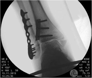
X-rayMortise view ankle X-ray after osteosynthesis: antiglide plate on the posterior malleolus and screwed plate on the lateral malleolus.
Source : Braunstein et al., BMC Musculoskeletal Disorders, 2016, figure 3 · CC BY
In a young adult with an unstable fracture, the question hardly arises. After sixty, it arises differently, and a British trial of 620 patients gave an answer that few units apply. This chapter sets out its figures, because it is the kind of result you need to be able to quote from memory in front of an anxious patient.
The AIM trial randomised 620 adults over 60 carrying a frankly unstable ankle fracture, in 24 British centres, between conventional internal fixation and a close contact cast, that is, a short leg cast with minimal padding, applied in the operating theatre under anaesthesia by a surgeon trained in the technique. The mean age was 71 years and 74 % of the participants were women (PMID 27727383).
| Outcome at six months | Internal fixation | Close contact cast | Reading |
|---|---|---|---|
| OMAS score, out of 100 | 66.0 (63.6–68.5) | 64.5 (61.8–67.2) | Equivalence demonstrated, difference −0.6 points |
| Infection or wound breakdown | 29 out of 298, that is 10 % | 4 out of 275, that is 1 % | Odds ratio 7.3 against surgery |
| Additional surgical procedure | 18 out of 298, that is 6 % | 3 out of 275, that is 1 % | Odds ratio 5.8 against surgery |
| Radiological malunion | 8 out of 274, that is 3 % | 38 out of 249, that is 15 % | Odds ratio 6.0 against casting |
| Secondary conversion to surgery | not applicable | 52 out of 275, that is 19 % | Anticipated by the protocol, in case of loss of reduction |
| Theatre time per patient | reference | −54 minutes | Saving of operating resource |
This table does not read as a draw. It describes an exchange: a skin and infection risk is traded for a risk of union in a poor position. In an 82-year-old woman, diabetic, on corticosteroids, whose skin is thin and whose walking is already limited, the exchange is clearly in favour of the cast. In a 62-year-old woman who is independent and active, whose functional demand is high and whose skin is healthy, it is far less so. Quality of life, pain, ankle mobility and satisfaction differed significantly in neither direction.
The authors themselves came back three years later to the obstacles to adopting this technique: close contact casting requires training, a trip to theatre and close monitoring, and these three requirements explain why a result of equivalence has not changed practice (PMID 31787004). The full health technology assessment report that accompanies the trial adds the economic analysis and the qualitative study of patients' experiences (PMID 27735787).
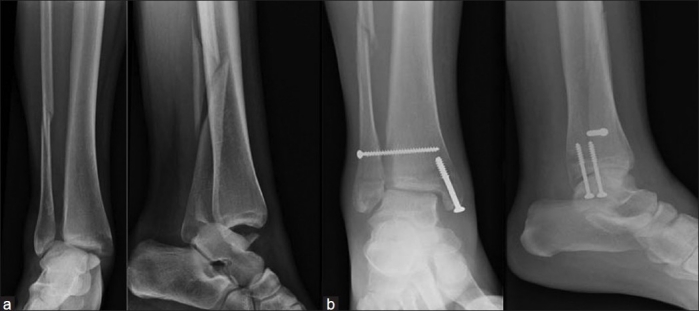
X-rayAnkle X-rays: displaced bimalleolar fracture (a), followed by syndesmosis-only screw fixation without fibular osteosynthesis, showing at 7 weeks an imperfectly reduced syndesmosis and an unhealed fibular fracture (b).
Source : Mohammed et al., Indian Journal of Orthopaedics, 2011, figure 3 · CC BY-NC-SA
- After 60, close contact casting and internal fixation give the same function at six months.
- Complications do not cancel out, they change in nature : skin and infection on one side, malunion on the other.
- One casted patient in five will finally be operated on, and that is anticipated by the protocol, not a failure.
- The physiotherapist who receives an older patient casted in theatre is not dealing with undertreatment: it is an evaluated strategy.
How long should a stable fracture be immobilised?
Six weeks of cast is the default answer in many units. A Finnish non-inferiority trial tested three, and it did not find the difference that was expected. This chapter draws the practical consequences, including for the choice of device.
Two hundred and forty-seven patients carrying an isolated Weber B fracture with a congruent mortise were randomised into three arms: six weeks of cast, three weeks of cast, or three weeks of a simple orthosis. The primary outcome was the OMAS at twelve months, with a non-inferiority margin set at −8.8 points. Result: 87.6 for six weeks of cast, 91.7 for three weeks of cast, 89.8 for three weeks of orthosis. The difference between three weeks of cast and six is 3.6 points in favour of the short arm, with an interval from −1.9 to 9.1 ; that of the orthosis against six weeks of cast is 1.7 points, interval from −4.0 to 7.3. No interval crosses the margin (PMID 30674451).
The only significant differences concerned secondary outcomes and both leaned the same way: better plantar flexion and fewer deep vein thromboses in the three-week orthosis arm than in the six-week cast arm. In other words, three weeks less immobilisation costs nothing measurable and brings a little.
The 2024 Cochrane review confirms the second part, that of the device. The comparison between removable and non-removable support brings together 25 studies and 2,206 participants ; across the 6 studies and 677 participants that report function, the use of a removable support after surgery may improve it by 6.39 points, interval 1.69 to 11.09, low-certainty evidence whose interval contains both clinically important differences and negligible ones (PMID 39312389). The cautious reading is the right one: the removable device is not superior to the cast, it is at least equivalent, and it allows removal for hygiene and mobilisation. That last point appears in no score.
An American retrospective cohort of 119 operated patients, of whom 47 were immobilised for less than six weeks and 68 for six weeks or more, finds no difference in PROMIS score between the two groups, including after adjustment for age, body mass index, polyarthritis, smoking and diabetes, and no difference in fixation complications either. Its conclusion is framed in terms of safety rather than benefit: early mobilisation in a walking boot is a safe alternative to the cast in the non-neuropathic patient (PMID 36741682). The level of evidence is low, and that is exactly how it should be cited.
What forbids shortening immobilisation
- A non-congruent mortise on the follow-up radiograph: the fracture is not stable, the Finnish trial does not apply.
- Bimalleolar or trimalleolar fracture : outside the scope of the trial, which covered only isolated Weber B fractures.
- Peripheral neuropathy, complicated diabetes : skin surveillance takes precedence over the timetable.
- Loss of reduction on a follow-up radiograph : surgical reassessment, not passive prolongation of the cast.
- Pain that increases under the cast : think of compartment syndrome, of pressure from a cast ridge, of venous thrombosis.
Early weight-bearing: what the trials actually showed
This is the only point in this article where the rehabilitation clinician really decides, and it is also the one that has moved the most since 2016. Five randomised trials and six syntheses later, the question is no longer « can we? » but « how much does it bring? ». This chapter gives both answers, including the disappointing one.
The starting point is a Canadian trial from 2016. One hundred and ten patients operated on for an unstable fracture were randomised between weight-bearing and motion at two weeks, and no weight-bearing with a cast for six weeks. The primary outcome, the time to return to work, does not differ. But at six weeks, the early group has a range of motion of 41 degrees versus 29, an OMAS of 45 versus 32 and better physical and mental scores on the SF-36 (PMID 27045369). A detail that counts, and that runs against intuition: implant removals for plate irritation were more frequent in the late group, 19 % versus 2 %.
The WAX trial, and why its result is solid
Eight years later, WAX took the question to the scale that allows a conclusion: 561 adults operated on for an unstable fracture in 23 hospitals of the British public health service, randomised between weight-bearing at two weeks and weight-bearing at six. The primary outcome was the OMAS at four months, in a per-protocol analysis, with a non-inferiority margin of −6 points and a superiority test planned in case of non-inferiority. Primary outcome data were collected in 86 % of the participants.
At four months, the mean OMAS is 65.9 in the early group versus 61.2 in the late group, an adjusted difference of 4.47 points, interval 0.58 to 8.37, and the superiority test is positive. Complications concern 46 patients out of 281 in the early group, that is 16 %, versus 39 out of 280, that is 14 %, an adjusted odds ratio of 1.18 whose interval from 0.80 to 1.75 contains 1. Costs for the health system are 725 pounds versus 785, and the probability that the early strategy is cost-effective exceeds 80 % (PMID 38848738).
Two exclusions matter when transposing. WAX excluded patients treated with a retrograde tibial nail and, above all, those who had no protective sensation at the ankle, that is, peripheral neuropathies. The most recent meta-analysis says it even more clearly: several trials excluded a body mass index above 30, trimalleolar fractures and syndesmotic injuries, and most participants were under 65. Its results apply first of all to low-risk patients (PMID 41472367).
What early weight-bearing gains, in OMAS points
Mean differences in favour of early weight-bearing, with their 95 % confidence interval
Sources: Bretherton 2024, Lancet (PMID 38848738) ; Lewis 2024, Cochrane (PMID 39312389) ; Llombart-Blanco 2025, Life (PMID 40003723) ; Egu 2026, Foot Ankle Int (PMID 41472367). The scales are not all the same OMAS measured at the same time : the figure compares orders of magnitude, not interchangeable values.
The useful disappointment: it is small, and that has to be said
The 2024 Cochrane review is the most rigorous of the set and the least enthusiastic. The comparison brings together 12 studies and 1,403 participants ; across the 5 studies and 890 participants that report function, weight-bearing within three weeks of surgery probably improves it by 3.56 points, interval 1.35 to 5.78, at moderate certainty ; but the authors specify that this difference does not include a clinically significant difference. On quality of life, the effect is null or negligible, a standardised difference of 0.15, interval from −0.01 to 0.30. On revision surgery, there is probably no difference, a relative risk of 0.50 with an interval from 0.09 to 2.68 (PMID 39312389).
Three other syntheses point the same way from different angles. Tong's shows that the functional superiority of early weight-bearing is seen at twelve weeks but has disappeared at one year, and that early mobilisation, for its part, comes with an increased risk of postoperative complications (PMID 37561102). Chen's concludes to a benefit on scores and on the time to return to work (PMID 38635458), as does the 2025 review covering 1,847 participants (PMID 40841661).
Early weight-bearing does not make healing faster. It makes life better while the bone heals, at no measured cost. That is reason enough, and it should be presented as such.
The distinction that Tong's review imposes is the one most often forgotten in the clinic: early weight-bearing and early mobilisation are not the same thing. Putting weight on a foot inside a locked walking boot is not mobilising the tibiotalar joint. The favourable data concern weight-bearing ; the unfavourable signal, where it exists, concerns free mobilisation. A reasonable protocol therefore allows weight-bearing early, in a device that protects range of motion, and frees the range afterwards.
- WAX, 561 operated patients: weight-bearing at two weeks gains 4.47 OMAS points at four months, with no excess of complications, and more than 80 % probability of also being the least costly strategy.
- The Cochrane review confirms the direction and cools the size: 3.56 points, below the threshold of clinical relevance.
- At one year, the gap between early and late weight-bearing has disappeared. The advantage is early and transient, and that is already a great deal.
- Weight-bearing and mobilisation are not to be confused: it is weight-bearing that is validated.
- The trials broadly excluded neuropathies, obesity, trimalleolar fractures and the very old. Outside that frame, caution remains in order.
Which rehabilitation after immobilisation, and does supervision change anything?
The honest answer is uncomfortable, and it is better to know it before a patient reads it elsewhere: for an isolated and uncomplicated fracture, a supervised programme did no better than well-delivered advice. This chapter sets out the trial that showed it, its limits, and what it leaves intact of the job.
The EXACT trial recruited, in seven Australian hospitals, patients carrying an isolated ankle fracture, randomised on the day the immobilisation was removed into two arms: a supervised exercise programme, individualised, prescribed, monitored and progressed, accompanied by self-management advice ; or the advice alone. Both were delivered by a physiotherapist. Of 571 eligible patients, 357 declined to take part and 214 were randomised.
The two groups started at 30.1 and 30.2 points of activity limitation on the lower extremity functional scale, graded out of 80. At three months, both are at 64.3. The difference in effect is 0.4 points, interval from −3.3 to 4.1. On quality of life, it is −0.01, interval from −0.06 to 0.04. Neither fracture severity, nor age, nor sex moderated the effect (PMID 26441182).
These results do not support the routine use of a supervised programme after an isolated and uncomplicated ankle fracture. They say nothing about the others.
Three limits must accompany this result every time it is cited. First, recruitment was interrupted before term for lack of funding: 214 patients instead of the 342 planned. Second, two eligible patients out of three declined to take part, which strongly selects the population analysed. Finally and above all, the trial explicitly covered fractures that were isolated and uncomplicated. It says nothing about the trimalleolar fracture operated on in a 78-year-old woman, about the fracture-dislocation, about the neuropathic diabetic patient, or about the patient who, six weeks after cast removal, still cannot manage a single-leg stance.
The 2024 Cochrane review confirms, moreover, the general poverty of the rehabilitation file: across 53 studies and 4,489 adults, every study is unavoidably exposed to a risk of performance and detection bias, and certainty was downgraded for this reason in all the comparisons (PMID 39312389).
The missing trial is under way, and it targets the right population
The British AFTER trial targets precisely the gap left by EXACT: adults aged 50 and over. It compares supervised rehabilitation, four to six individual sessions of tailored advice and home exercise over three months, with self-directed rehabilitation based on a booklet and a progressive programme, in 344 participants recruited in at least twenty hospitals. Randomisation takes place when the cast or the boot is removed and the weight-bearing and mobility restrictions are lifted (PMID 38898823). The detail of how the two interventions were built, from two British guidelines, a survey of 59 physiotherapists and a pilot trial of 61 participants, has been published separately (PMID 40250162). This is the trial to watch.
What we know, and to what degree of certainty
The six decisions in this file, ranked by the strength of the evidence that supports them
Levels taken from the syntheses cited, in their authors' own terms: Kortekangas 2019 (PMID 30674451), Lewis 2024 (PMID 39312389), Willett 2016 (PMID 27727383), Moseley 2015 (PMID 26441182) and Lim 2024 (PMID 38857233). The GRADE qualifiers are explicit only in the Cochrane review ; elsewhere they reflect the design and the size of the study.
- For an isolated and uncomplicated fracture, a supervised programme did no better than structured advice: 0.4 points apart out of 80.
- The trial was stopped early and two eligible patients out of three refused to take part. It is evidence, not a verdict.
- It covers neither the older patient, nor the complex fracture, nor the at-risk profile, which are precisely what we see in the clinic.
- The AFTER trial tests exactly this population, in the 50 and over.
- What the trial validates by implication: well-delivered advice is a therapeutic act, and it deserves the time given to it.
On what criteria should walking, work and sport be resumed?
A Canadian cohort followed 142 operated patients under 65 for one year and measured, rather than assumed, what they had recovered. Its figures are the best available markers for speaking frankly to a patient, and for choosing what to measure.
Participants were assessed at six weeks, six months and one year on three planes: functional dorsiflexion with the weight-bearing lunge test, measured as the difference between the two sides ; patient-reported function on the OMAS ; and return to the previous activity and work level. Everything improves significantly, and that is the only reassuring point.
At one year after surgery, the dorsiflexion difference between the two ankles remains 3.22 cm on average, standard deviation 2.68 ; 72 % of patients still report ankle stiffness ; and only 52 % have regained their previous activity level. On the other hand, among those who were working, 97 % had returned to work at one year. The only two factors significantly associated with return to the previous activity level are the OMAS and the weight-bearing lunge difference (PMID 35464787).
These four figures are read together, and they draw a precise clinical message. Returning to work is not the problem: it almost always happens. The problem is the return to sport and to demanding activities, which fails one time in two, and its best measurable correlate is a test that can be done in the clinic in one minute, with no equipment. A weight-bearing lunge deficit that does not narrow is the earliest signal that the patient will not come back to their level, and it is also a direct training target.
| Phase | Dominant objective | Criterion for moving on, measured | What should raise the alarm |
|---|---|---|---|
| Protection immobilisation | Union, swelling, mobility of the rest of the limb | End of the prescribed period, congruent follow-up radiograph | Increasing pain under the cast, calf swelling, fever |
| Return to weight-bearing according to the protocol | Progressive weight-bearing, walking without an antalgic limp | Painless full weight-bearing, gait cycle symmetrical to the eye | Focal bone pain on weight-bearing, giving way |
| Range of motion | Functional dorsiflexion | Weight-bearing lunge difference below 2 cm, narrowing from one session to the next | Dorsiflexion plateau over three weeks |
| Strength and control | Triceps surae, peroneals, single-leg control | Single-leg heel raises in series, stable single-leg stance with eyes open then closed | Marked asymmetric fatigability, apprehension |
| Return to sport | Impact, changes of direction | Symmetrical single-leg hops, no pain the next day | Swelling the day after exertion |
This progression does not come from a trial: no trial has validated it, and that must be said as such. It assembles markers measured in the Ramadi cohort, the inclusion criteria of the early weight-bearing trials, and the logic common to all lower limb trauma. Its only virtue, but it is a real one, is to replace dates with measures that can be repeated at every session.
- At one year, only one patient in two has regained their previous activity level, whereas 97 % have returned to work.
- The stiffness persists in 72 % of operated patients, and this is a fact to announce up front rather than discover in the third month.
- The weight-bearing lunge test, compared with the uninjured side, is one of only two predictors of return to previous activity.
- Progress by measured criteria, not by calendar. No calendar has been validated.
Which patient profiles change the prognosis?
The early weight-bearing trials excluded obesity, neuropathy and complex fractures. This chapter deals with what is left, that is, a good share of the real caseload, and with the long-term outcome that nobody announces at the time of casting.
Diabetes, and why the figure to ask for is glycated haemoglobin
The ankle fracture of a diabetic patient is not the same disease. A retrospective cohort of diabetic patients operated on with a documented glycated haemoglobin shows that radiological union is significantly more frequent in those whose HbA1c is below 8 % (PMID 39157192). The sample is small, 44 patients, and association does not prove causation ; but the direction is consistent with everything we know about bone and skin healing in this population.
The far end of the spectrum gives the measure of the risk. A series of 13 diabetic patients who had a primary ankle arthrodesis, for a traumatic fracture with severe diabetic complications, reports an overall complication rate above 75 % : 38.5 % revisions, 38.5 % infections, 53.8 % wound healing complications and 23.1 % secondary Charcot arthropathy (PMID 35097368). This is not the usual situation in the clinic, but it is a reminder of why skin surveillance takes precedence over the timetable when protective sensation is missing.
Fragile bone, and why the ankle is not a hip
The ankle fracture of the older patient occupies an ambiguous place among fragility fractures. The review devoted to it puts it this way: these fractures are among the most frequent in older people, their number is set to grow, and osteoporosis should enter the conversation with the patient at the time of diagnosis, even when the functional result of fixation remains good (PMID 23544826). Postoperative complications there are more frequent in diabetic patients, in those with peripheral arterial disease and in smokers, which overlaps exactly with the profiles mentioned above. For the physiotherapist, the practical consequence is simple and often forgotten: an ankle fracture from a fall from standing height after 65 deserves the same question as a vertebral compression fracture, that of a bone work-up and prevention of the next fall.
Obesity: more medical complications than mechanical ones
The largest series available on this point analyses 160,415 ankle fixations with a minimum two-year follow-up. Obese patients there show higher rates of 90-day readmission, urinary tract infections, venous thromboses and pulmonary emboli, pneumonia, and post-traumatic osteoarthritis (PMID 39150303). A matched study of 632 patients, in which each obese patient was matched to a non-obese one on age, sex, ethnicity, diabetes and fracture type, usefully tempers this: the two groups did not differ in the frequency of open fractures, and the non-obese were more often smokers, 63.3 % versus 40.2 % (PMID 32534816). Obesity and smoking therefore do not always add up the way one imagines, and each deserves to be explored for itself.
Post-traumatic osteoarthritis: the outcome nobody talks about
It is the sequela that is least often announced and the one that weighs heaviest at twenty years. A Geneva cohort reviewed 102 patients operated on for a malleolar fracture between 1988 and 1997, with a mean follow-up of 17.9 years. Radiographic osteoarthritis rated advanced, Kellgren and Lawrence grades 3 or 4, is present in 37 patients, that is 36.3 %. The significant risk factors are the fracture type Weber C, the associated medial malleolus fracture, the fracture-dislocation, rising body mass index, an age of 30 years or over at surgery, and how long ago the surgery was. In patients combining three factors or more, the probability reaches 60 to 70 % (PMID 22249843).
Advanced radiographic osteoarthritis, 12 to 22 years after a surgically treated malleolar fracture
The average hides a sorting: adding up the factors nearly doubles the risk.
Source: Lübbeke 2012, Int Orthop, 373 fractures operated on between 1988 and 1997, 102 patients reviewed at a mean of 17.9 years (PMID 22249843). The 60 to 70 % range is the one given by the authors, without a point estimate.
Two more recent series refine the picture. A follow-up of 69 trimalleolar fractures at 11.3 years compares fixed and unfixed posterior fragments, without fixation emerging as decisive for functional scores (PMID 40531350). And the study of fracture-dislocations recalls that they account for 21 to 36 % of all ankle fractures and that they come with more extensive bone and soft tissue damage, with malreduction, chronic pain and post-traumatic osteoarthritis all more frequent (PMID 38407969). The inflammatory component of this degradation is the subject of active work, with no therapeutic translation to date (PMID 38892089).
- In the diabetic patient, union is linked to glycaemic control : aiming for an HbA1c below 8 % is an objective shared with the general practitioner.
- Obesity mainly increases medical complications and readmission, more than mechanical failure.
- 36.3 % advanced osteoarthritis at a mean of 18 years ; 60–70 % if three risk factors coexist.
- The three heaviest factors are the ones read on the first radiograph: Weber C, associated medial injury, dislocation.
- This prognosis is not stated to alarm, but to justify maintaining activity and a controlled body weight over years, not over six weeks.
What do concrete clinical cases teach us?
One and the same injury, the Maisonneuve fracture, comes back in five case reports published over forty years, and each time for the same reason. These cases are real and referenced: they are worth more than a textbook case, because their authors agreed to publish what had been missed.
Case 1. The normal radiograph that did not frame the fracture
A patient presents with pain of the medial ankle. The ankle radiographs are normal, and the proximal fibula is not examined. He comes back four days later, in more pain ; examination then finds tenderness over the proximal fibula and over the medial ankle, and the new radiographs show a widening of the medial joint space and an oblique fracture of the proximal fibula. Fixation of the syndesmosis and repair of the deltoid, uneventful recovery (PMID 3592337).
What this case teaches: medial ankle pain with no visible medial fracture must lead to palpating the whole leg. The article's conclusion, published in 1987, is still the best formulation of the principle: examination of an ankle injury must include the proximal fibula.
Case 2. The same error, thirty-two years later
A 35-year-old servicewoman presents with acute pain of the ankle and of the leg , both on the left, after misjudging a step. The diagnosis retained combines a high fibular fracture, a medial malleolus fracture and a syndesmotic rupture, by external rotation and pronation of a fixed foot. The authors note that this injury, well known in sports medicine and orthopaedics, is frequently missed in primary care, and recall the general rule it illustrates: examine the joints above and below the painful site, and obtain orthogonal views (PMID 29415223).
Case 3. The injury seen, but the instability missed
A 16-year-old patient presents with a fracture of the left medial malleolus ; the associated tibiofibular instability goes unnoticed. After fixation of the malleolus, further imaging reveals a syndesmotic diastasis with rupture of the interosseous membrane. A second operation places two transsyndesmotic screws, removed at twelve weeks ; at six months, range of motion is complete (PMID 34046477). The authors propose regarding any complete syndesmotic injury with rupture of the interosseous membrane as an injury of the Maisonneuve type, even without a fibular fracture.
Case 4. The mechanism that does not fit the box
A 31-year-old woman twists her right ankle while walking, five days before her admission, after a first missed diagnosis elsewhere. Full-length leg radiographs show a proximal fibular fracture, an inferior tibiofibular separation and a medial malleolus fracture including the posterior malleolus ; MRI confirms rupture of the anterior inferior tibiofibular ligament and of the anterior talofibular ligament. The authors stress that this picture combines the features of two Lauge-Hansen mechanisms that should not coexist (PMID 35663082).
Case 5. And a conservative treatment that held for 41 months
A woman in her twenties twists her left ankle while running. The work-up finds a Maisonneuve fracture with a high fibular fracture, a fracture of the Volkmann tubercle and slight syndesmotic separation. She refuses the recommended surgery and even refuses immobilisation of the knee ; she is therefore treated with a short leg cast for eight weeks. At 41 months, the radiograph shows complete union of the proximal fibular fracture, with no post-traumatic osteoarthritis, no chronic pain and no instability (PMID 38728515).
- Four cases out of five were first missed, and always by the same mechanism: the radiograph did not frame the site of the fracture line.
- The common signal is medial pain with no explained medial injury, or associated leg pain.
- The fifth case is a reminder of modesty: an injury reputed to be surgical did well without surgery in a young woman. One case is not a series, and it justifies no change of practice.
- For the physiotherapist, the operational rule fits in one sentence: faced with a traumatic ankle that is not progressing, palpate the fibula along its whole length.
How is this applied concretely in the clinic?
Here is the article reduced to what gets decided, in the order in which it gets decided, with the errors that cost the most and the reasons for referral that are not up for discussion.
Seven decisions, in order
- Decide whether to image. Painful zone, then the three conditions. A negative rule rules out the fracture ; a positive rule concludes nothing.
- Read the level of the fracture line. Weber A, B or C. Faced with a high C or with leg pain, require radiographs of the whole leg.
- Look for the medial signs. Their absence is strongly reassuring ; their presence calls for a weight-bearing radiograph at seven days rather than immediate theatre.
- Adapt the duration of immobilisation. Three weeks are enough for a stable Weber B, in a cast as in an orthosis.
- Allow weight-bearing early. Two weeks after internal fixation, in a device that protects range of motion, except in neuropathy, a fragile construct or a contrary surgical instruction.
- Measure rather than date. Weight-bearing lunge compared with the uninjured side, single-leg heel raises, single-leg stance, hops at the end of the pathway.
- Announce the long term. Stiffness frequent at one year, return to sport one time in two, osteoarthritis at twenty years in one third. These three facts are said early and calmly.
Six frequent errors
- Applying the Ottawa rules without the painful zone. The structure of the rule is lost, and with it the little specificity that remains.
- Taking a positive rule for a diagnosis. Two radiographs out of three will come back normal, and that is how the tool is meant to work.
- Palpating only the ankle. Four of the five published cases in this file were missed for this reason.
- Confusing weight-bearing and mobilisation. The favourable data concern loading, not freedom of range.
- Prolonging immobilisation « to be safe ». Three more weeks have shown no gain and cost plantar flexion.
- Treating the casted older patient as undertreated. Close contact casting is an evaluated strategy, equivalent at six months.
When to refer on, and to whom
| Situation | To whom | Time frame |
|---|---|---|
| Deformity, taut skin, skin breach, neurovascular deficit | Emergency department | Immediate |
| Increasing pain under immobilisation, compartment tightness, painful calf | Emergency department or referring surgeon | Same day |
| Tenderness on palpation of the proximal third of the fibula | Surgeon, for full-length leg radiographs | Within 48 hours |
| Loss of reduction or widened medial clear space on a follow-up radiograph | Referring surgeon | Within one week |
| Dorsiflexion at a plateau for three weeks, or repeated giving way | Prescribing doctor, surgical opinion depending on the context | Scheduled reassessment |
| Uncontrolled diabetes, neuropathy, indolent wound | General practitioner and podiatrist, diabetology opinion | Without waiting for the end of rehabilitation |
Frequently asked questions
Does a positive Ottawa rule mean the ankle is broken?
No, and this is the most common misreading. The specificity of the rules lies between 25 and 35 % depending on the meta-analyses (PMID 36151550, PMID 42524327). A positive rule means that the fracture cannot be ruled out without imaging, nothing more. The strength of the tool is on the other side: with a negative likelihood ratio of 0.08 to 0.13, a negative rule brings the probability of fracture below 1.4 % for a prevalence of 15 % (PMID 12595378).
Can you walk straight away after ankle fixation surgery?
Not straight away, but far sooner than six weeks. The WAX trial compared weight-bearing at two weeks with weight-bearing at six in 561 operated patients: function is better at four months, by 4.47 OMAS points, and complications do not differ (PMID 38848738). Two reservations apply: the trial excluded patients without protective sensation at the ankle, and the surgical construct remains the surgeon's decision. The timetable is agreed with them, it is not decided against them.
Do you need six weeks of cast for a lateral malleolus fracture?
For an isolated Weber B with a congruent mortise, no. Three weeks of cast, or three weeks of a simple orthosis, are not inferior to six weeks of cast on the OMAS at one year, with better plantar flexion and fewer venous thromboses in the orthosis arm into the bargain (PMID 30674451). This does not hold for bimalleolar or trimalleolar fractures, nor for a non-congruent mortise.
My surgeon did not fix the syndesmosis. Is that serious?
Nothing in the data suggests so. In Weber B fractures, the review of the eight available studies finds no difference in functional or radiological outcome or quality of life between patients whose syndesmosis was screwed and the others, nor any difference in post-traumatic osteoarthritis (PMID 38857233). The number of screws, their position and even their secondary breakage have no demonstrated adverse effect (PMID 28708780).
My 78-year-old mother was put in a cast without surgery. Was she given lesser care?
No. A British randomised trial of 620 patients over 60 carrying an unstable fracture shows equivalent function at six months between close contact casting and internal fixation, with ten times fewer infections and wound breakdowns on the casting side (PMID 27727383). In exchange, radiological malunion is five times more frequent, and about one casted patient in five ends up operated on, which the protocol anticipates. It is a reasoned choice, not a giving up.
Do I really need physiotherapy sessions after a simple ankle fracture?
For an isolated and uncomplicated fracture, the EXACT trial found no difference between a supervised programme and well-delivered advice: 0.4 points apart on a scale of 80 (PMID 26441182). That trial covers neither complex fractures, nor older patients, nor at-risk profiles, nor patients who plateau. The right question is therefore not « are sessions needed » but « what, in this patient, falls outside the scope of the trial ».
How long before I can run again?
No trial has validated a return-to-sport timetable after ankle fracture, and it is better to say so than to invent a date. What we do know is a worrying order of magnitude: at one year, only 52 % of operated patients under 65 have regained their previous activity level, and the best measurable correlate of that return is the weight-bearing dorsiflexion deficit compared with the uninjured side (PMID 35464787). Progress is therefore made by criteria, not by dates.
Will my ankle stay stiff?
Often, partly, and this is announced from the outset. In the Canadian cohort, 72 % of patients still report stiffness one year after internal fixation, with a weight-bearing dorsiflexion deficit of 3.2 cm on average compared with the uninjured side (PMID 35464787). This stiffness is compatible with a normal life and with work, since 97 % of those who were working had returned. It mainly limits activities that demand full dorsiflexion, squatting and going downhill.
Will I get osteoarthritis?
The risk exists and depends on what the first radiograph showed. Twelve to twenty-two years after a surgically treated malleolar fracture, advanced radiographic osteoarthritis is present in 36.3 % of the patients reviewed, and in 60–70 % of those who combined three or more risk factors, namely a Weber C fracture, an associated medial injury, a fracture-dislocation, a high body mass index and an age of 30 years or over (PMID 22249843). Radiographic osteoarthritis is not painful osteoarthritis: it is a reason to maintain activity and body weight over the long run, not to stop.
References
59 references, resolved one by one through the PubMed E-utilities API on 16 August 2026. For each of them, the abstract was read and the figures quoted in the article were checked at source. The metadata below, authors, journal, year, pagination and DOI, are extracted from the PubMed XML by script, not copied by hand.
Epidemiology, natural history and post-traumatic osteoarthritis (7)
- Elsoe R, Ostgaard SE, Larsen P. Population-based epidemiology of 9767 ankle fractures. Foot Ankle Surg. 2018;24(1):34-39. PMID 29413771. DOI 10.1016/j.fas.2016.11.002.
- Kang HJ, Lee JW, Kwon YM, et al. Epidemiology of Ankle Fractures in Korea: A Nationwide Population-Based Study. J Korean Med Sci. 2022;37(38):e288. PMID 36193640. DOI 10.3346/jkms.2022.37.e288.
- Olsen JR, Hunter J, Baumhauer JF. Osteoporotic ankle fractures. Orthop Clin North Am. 2013;44(2):225-41. PMID 23544826. DOI 10.1016/j.ocl.2013.01.010.
- Lübbeke A, Salvo D, Stern R, et al. Risk factors for post-traumatic osteoarthritis of the ankle: an eighteen year follow-up study. Int Orthop. 2012;36(7):1403-10. PMID 22249843. DOI 10.1007/s00264-011-1472-7.
- Chalidapong P, Vaseenon T, Chattipakorn N, et al. Potential Roles of Inflammation on Post-Traumatic Osteoarthritis of the Ankle. Int J Mol Sci. 2024;25(11). PMID 38892089. DOI 10.3390/ijms25115903.
- Pehlivanoglu T, Demirel M, Koyuncu D, et al. Long-Term Clinical and Radiographic Outcomes After Surgical Treatment for Ankle Fracture-Dislocations. J Am Podiatr Med Assoc. 2025;115(3). PMID 38407969. DOI 10.7547/22-225.
- Chidda A, Soares S, Tannast M, et al. Long-term outcomes after a trimalleolar fracture involving the posterior malleolar fragment: an 11-year follow-up. Arch Orthop Trauma Surg. 2025;145(1):346. PMID 40531350. DOI 10.1007/s00402-025-05959-w.
The Ottawa rules: diagnostic accuracy (7)
- Bachmann LM, Kolb E, Koller MT, et al. Accuracy of Ottawa ankle rules to exclude fractures of the ankle and mid-foot: systematic review. BMJ. 2003;326(7386):417. PMID 12595378.
- Beckenkamp PR, Lin CC, Macaskill P, et al. Diagnostic accuracy of the Ottawa Ankle and Midfoot Rules: a systematic review with meta-analysis. Br J Sports Med. 2017;51(6):504-510. PMID 27884861. DOI 10.1136/bjsports-2016-096858.
- Gomes YE, Chau M, Banwell HA, et al. Diagnostic accuracy of the Ottawa ankle rule to exclude fractures in acute ankle injuries in adults: a systematic review and meta-analysis. BMC Musculoskelet Disord. 2022;23(1):885. PMID 36151550. DOI 10.1186/s12891-022-05831-7.
- Dowling S, Spooner CH, Liang Y, et al. Accuracy of Ottawa Ankle Rules to exclude fractures of the ankle and midfoot in children: a meta-analysis. Acad Emerg Med. 2009;16(4):277-87. PMID 19187397. DOI 10.1111/j.1553-2712.2008.00333.x.
- Barelds I, Krijnen WP, van de Leur JP, et al. Diagnostic Accuracy of Clinical Decision Rules to Exclude Fractures in Acute Ankle Injuries: Systematic Review and Meta-analysis. J Emerg Med. 2018;53(3):353-368. PMID 28764972. DOI 10.1016/j.jemermed.2017.04.035.
- Jonckheer P, Willems T, De Ridder R, et al. Evaluating fracture risk in acute ankle sprains: Any news since the Ottawa Ankle Rules? A systematic review. Eur J Gen Pract. 2017;22(1):31-41. PMID 26691309. DOI 10.3109/13814788.2015.1102881.
- Sharifi Razavi D, Seiri M, Farrokhi M. Revisiting the Diagnostic Accuracy of Ottawa Ankle Rules for Ankle Fractures: A Systematic Review and Meta-Analysis. Arch Acad Emerg Med. 2026;14(1):e24. PMID 42524327. DOI 10.22037/aaem.v14i1.3051.
Weber, Lauge-Hansen and AO/OTA classifications (7)
- Glen LZQ, Wong JYS, Tay WX, et al. Weber Ankle Fracture Classification System Yields Greatest Interobserver and Intraobserver Reliability Over AO/OTA and Lauge-Hansen Classification Systems Under Time Constraints in an Asian Population. J Foot Ankle Surg. 2023;62(3):505-510. PMID 36690511. DOI 10.1053/j.jfas.2022.12.004.
- Malek IA, Machani B, Mevcha AM, et al. Inter-observer reliability and intra-observer reproducibility of the Weber classification of ankle fractures. J Bone Joint Surg Br. 2006;88(9):1204-6. PMID 16943473.
- Yin MC, Yuan XF, Ma JM, et al. Evaluating the Reliability and Reproducibility of the AO and Lauge-Hansen Classification Systems for Ankle Injuries. Orthopedics. 2016;38(7):e626-30. PMID 26186326. DOI 10.3928/01477447-20150701-62.
- Briet JP, Hietbrink F, Smeeing DP, et al. Ankle Fracture Classification: An Innovative System for Describing Ankle Fractures. J Foot Ankle Surg. 2019;58(3):492-496. PMID 30795890. DOI 10.1053/j.jfas.2018.09.028.
- Sahin M, Ergisi Y, Dasar U, et al. Computed Tomography Improves Interobserver Agreement and Influences Surgical Planning in Lauge-Hansen Classification of Ankle Fractures. Foot Ankle Int. 2025;46(11):1294-1301. PMID 41044862. DOI 10.1177/10711007251372146.
- Brage ME, Rockett M, Vraney R, et al. Ankle fracture classification: a comparison of reliability of three X-ray views versus two. Foot Ankle Int. 1998;19(8):555-62. PMID 9728704.
- Aktan C, Kose O. A novel alphanumeric classification system for ankle fractures: clinical applications and evaluation. J Orthop Surg Res. 2025;20(1):130. PMID 39901191. DOI 10.1186/s13018-025-05539-z.
Stability of a Weber B: demonstrating it rather than assuming it (6)
- van den Bekerom MP, Mutsaerts EL, van Dijk CN. Evaluation of the integrity of the deltoid ligament in supination external rotation ankle fractures: a systematic review of the literature. Arch Orthop Trauma Surg. 2009;129(2):227-35. PMID 18953550. DOI 10.1007/s00402-008-0768-6.
- Zeni F, Cavazos DR, Bouffard JA, et al. Indications and Interpretation of Stress Radiographs in Supination External Rotation Ankle Fractures. Cureus. 2023;15(4):e38092. PMID 37252532. DOI 10.7759/cureus.38092.
- Seidel A, Krause F, Weber M. Weightbearing vs Gravity Stress Radiographs for Stability Evaluation of Supination-External Rotation Fractures of the Ankle. Foot Ankle Int. 2017;38(7):736-744. PMID 28511569. DOI 10.1177/1071100717702589.
- Hoshino CM, Nomoto EK, Norheim EP, et al. Correlation of weightbearing radiographs and stability of stress positive ankle fractures. Foot Ankle Int. 2012;33(2):92-8. PMID 22381339. DOI 10.3113/FAI.2012.0092.
- Seo M, Kwon M, Sung KS. Weight-Bearing Test: A Straightforward and Reliable Indicator of Stability in Supination-External Rotation Type, Weber B Ankle Fractures. Clin Orthop Surg. 2026;18(3):543-547. PMID 42226793. DOI 10.4055/cios25349.
- Yousaf S, Saleh A, Ahluwalia A, et al. Systematic Review of Stress Radiographic Modalities Stability Assessment in Supination External Rotation Ankle Fractures. Foot Ankle Orthop. 2022;4(4):2473011419890861. PMID 35097353. DOI 10.1177/2473011419890861.
Syndesmosis: detection, fixation and outcome (6)
- Kellam PJ, Dekeyser GJ, Haller JM, et al. Predicting Syndesmotic Injury in OTA/AO 44-B2.1 (Danis-Weber B) Fractures. J Orthop Trauma. 2021;35(9):473-478. PMID 33395177. DOI 10.1097/BOT.0000000000002052.
- Lim B, Shaalan M, O'hEireamhoin S, et al. Syndesmotic fixation in Weber B ankle fractures: A systematic review. PLoS One. 2024;19(6):e0304148. PMID 38857233. DOI 10.1371/journal.pone.0304148.
- Murphy SC, Murphy B, O'Loughlin P. Syndesmotic injury with ankle fracture: A systematic review of screw vs dynamic fixation. Ir J Med Sci. 2024;193(3):1323-1330. PMID 38282112. DOI 10.1007/s11845-024-03619-3.
- Michelson JD, Wright M, Blankstein M. Syndesmotic Ankle Fractures. J Orthop Trauma. 2018;32(1):10-14. PMID 28708780. DOI 10.1097/BOT.0000000000000937.
- Marasco D, Russo J, Izzo A, et al. Static versus dynamic fixation of distal tibiofibular syndesmosis: a systematic review of overlapping meta-analyses. Knee Surg Sports Traumatol Arthrosc. 2021;29(11):3534-3542. PMID 34455448. DOI 10.1007/s00167-021-06721-6.
- Yao X, Wang C, Pan W, et al. Ankle syndesmotic ligaments avulsion fractures: incidence in adult population. J Orthop Surg Res. 2024;19(1):642. PMID 39395996. DOI 10.1186/s13018-024-05156-2.
Early weight-bearing after internal fixation: the trials and their syntheses (8)
- Bretherton CP, Achten J, Jogarah V, et al. Early versus delayed weight-bearing following operatively treated ankle fracture (WAX): a non-inferiority, multicentre, randomised controlled trial. Lancet. 2024;403(10446):2787-2797. PMID 38848738. DOI 10.1016/S0140-6736(24)00710-4.
- Lewis SR, Pritchard MW, Parker R, et al. Rehabilitation for ankle fractures in adults. Cochrane Database Syst Rev. 2024;9(9):CD005595. PMID 39312389. DOI 10.1002/14651858.CD005595.pub4.
- Dehghan N, McKee MD, Jenkinson RJ, et al. Early Weightbearing and Range of Motion Versus Non-Weightbearing and Immobilization After Open Reduction and Internal Fixation of Unstable Ankle Fractures: A Randomized Controlled Trial. J Orthop Trauma. 2017;30(7):345-52. PMID 27045369. DOI 10.1097/BOT.0000000000000572.
- Egu C, Akil H, Hakim RA, et al. Early vs Late Weight Bearing After Ankle Fracture Fixation: A Meta-analysis of Randomized Controlled Trials. Foot Ankle Int. 2026;47(2):174-183. PMID 41472367. DOI 10.1177/10711007251392223.
- Chen B, Ye Z, Wu J, et al. The effect of early weight-bearing and later weight-bearing rehabilitation interventions on outcomes after ankle fracture surgery: A systematic review and meta-analysis of randomised controlled trials. J Foot Ankle Res. 2024;17(2):e12011. PMID 38635458. DOI 10.1002/jfa2.12011.
- Tong J, Ajrawat P, Chahal J, et al. Early Versus Delayed Weight Bearing and Mobilization After Ankle Fracture Fixation Surgery: A Systematic Review and Meta-analysis of Randomized Controlled Trials. Orthopedics. 2024;47(2):71-78. PMID 37561102. DOI 10.3928/01477447-20230804-08.
- Wang C, Li C. Early weight-bearing after ankle fracture surgery: a systematic review and meta-analysis of functional outcomes and safety. J Orthop Surg Res. 2025;20(1):785. PMID 40841661. DOI 10.1186/s13018-025-06216-x.
- Llombart-Blanco R, Mariscal G, Khalil I, et al. Weight-Bearing Versus Non-Weight-Bearing After Ankle Fracture: A Systematic Review and Meta-Analysis of Patient-Reported Outcome. Life (Basel). 2025;15(2). PMID 40003723. DOI 10.3390/life15020314.
Immobilisation, orthosis and non-operative treatment (5)
- Kortekangas T, Haapasalo H, Flinkkilä T, et al. Three week versus six week immobilisation for stable Weber B type ankle fractures: randomised, multicentre, non-inferiority clinical trial. BMJ. 2019;364:k5432. PMID 30674451. DOI 10.1136/bmj.k5432.
- Willett K, Keene DJ, Mistry D, et al. Close Contact Casting vs Surgery for Initial Treatment of Unstable Ankle Fractures in Older Adults: A Randomized Clinical Trial. JAMA. 2016;316(14):1455-1463. PMID 27727383. DOI 10.1001/jama.2016.14719.
- Keene DJ, Willett K. Implications of the Ankle Injury Management (AIM) trial: close contact casting or surgery for older adults with an unstable ankle fracture? Bone Joint J. 2019;101-B(12):1472-1475. PMID 31787004. DOI 10.1302/0301-620X.101B12.BJJ-2019-0171.R2.
- Keene DJ, Mistry D, Nam J, et al. The Ankle Injury Management (AIM) trial: a pragmatic, multicentre, equivalence randomised controlled trial and economic evaluation comparing close contact casting with open surgical reduction and internal fixation in the treatment of unstable ankle fractures in patients aged over 60 years. Health Technol Assess. 2018;20(75):1-158. PMID 27735787.
- Carney DD, Vyas PS, Hicks JJ, et al. Effect of Postoperative Immobilization Time on PROMIS Scores and Clinical outcomes in Ankle Fracture Patients. Foot Ankle Orthop. 2023;8(1):24730114221151080. PMID 36741682. DOI 10.1177/24730114221151080.
Supervised rehabilitation and functional recovery (4)
- Moseley AM, Beckenkamp PR, Haas M, et al. Rehabilitation After Immobilization for Ankle Fracture: The EXACT Randomized Clinical Trial. JAMA. 2015;314(13):1376-85. PMID 26441182. DOI 10.1001/jama.2015.12180.
- Keene DJ, Achten J, Forde C, et al. Effectiveness of supervised versus self-directed rehabilitation for adults aged 50 years and over with ankle fractures: protocol for the AFTER trial. Bone Jt Open. 2025;5(6):499-513. PMID 38898823. DOI 10.1302/2633-1462.56.BJO-2023-0183.
- Forde CP, Costa ML, Achten J, et al. Development and delivery of the rehabilitation interventions for older adults with an ankle fracture in the AFTER (Ankle Fracture Treatment Enhancing Rehabilitation) trial. Physiotherapy. 2025;128:101789. PMID 40250162. DOI 10.1016/j.physio.2025.101789.
- Ramadi A, Beaupre LA, Heinrichs L, et al. Recovery and Return to Activity 1 Year After Ankle Fracture Managed With Open Reduction and Internal Fixation: A Prospective Longitudinal Cohort Study. Foot Ankle Orthop. 2022;7(2):24730114221091806. PMID 35464787. DOI 10.1177/24730114221091806.
At-risk profiles: diabetes, obesity, fragile bone (4)
- Alsheikh KA, Alrehaili HH, Alsabr AA, et al. Outcomes of surgical management of ankle fractures in patients with diabetes. J Taibah Univ Med Sci. 2024;19(4):800-805. PMID 39157192. DOI 10.1016/j.jtumed.2024.07.003.
- Benedick A, Audet MA, Vallier HA. The effect of obesity on post-operative complications and functional outcomes after surgical treatment of torsional ankle fracture: A matched cohort study. Injury. 2021;51(8):1893-1898. PMID 32534816. DOI 10.1016/j.injury.2020.05.006.
- Janghala A, Niknam K, Freshman R, et al. Effect of Obesity on Short- and Long-Term Complications After Ankle Fracture Fixation. J Orthop Trauma. 2024;38(9):e312-e317. PMID 39150303. DOI 10.1097/BOT.0000000000002849.
- Grote CW, Tucker W, Stumpff K, et al. Primary Arthrodesis for Diabetic Ankle Fractures. Foot Ankle Orthop. 2022;5(1):2473011420908841. PMID 35097368. DOI 10.1177/2473011420908841.
Published clinical cases: the Maisonneuve fracture (5)
- Lock TR, Schaffer JJ, Manoli A. Maisonneuve fracture: case report of a missed diagnosis. Ann Emerg Med. 1987;16(7):805-7. PMID 3592337.
- Richmond RR, Henebry AD. A Maisonneuve Fracture in an Active Duty Sailor: A Case Report. Mil Med. 2019;183(5-6):e278-e280. PMID 29415223. DOI 10.1093/milmed/usx080.
- Wang C, Dong S, Li X, et al. Maisonneuve fracture treated with short leg cast: A case report with 41-month follow-up. Medicine (Baltimore). 2024;103(19):e38105. PMID 38728515. DOI 10.1097/MD.0000000000038105.
- Liu GP, Li JG, Gong X, et al. Maisonneuve injury with no fibula fracture: A case report. World J Clin Cases. 2021;9(15):3733-3740. PMID 34046477. DOI 10.12998/wjcc.v9.i15.3733.
- Zhao B, Li N, Cao HB, et al. Rare pattern of Maisonneuve fracture: A case report. World J Clin Cases. 2023;10(14):4684-4690. PMID 35663082. DOI 10.12998/wjcc.v10.i14.4684.
Behind this article
An author who explains, a reviewer who checks.
How we write and check our content

Physiotherapist · co-founder of Physio Learning
Marked for life by his first four-hour lecture without a single image, he took a master’s in instructional design so that it would never happen to anyone again. He hunts down publication bias and unreadable slides with the same intransigence.

Head of scientific content
Physiotherapist specialising in neuro-musculoskeletal practice and holder of a master’s in public health. He checks the methodological rigour of every article: primary sources, levels of evidence, no exceptions.