















Clinical synthesis on acute unilateral vestibulopathy: recognising the presentation, ruling out stroke at the bedside, and conducting vestibular rehabilitation. Every reference has been individually verified on PubMed.
📝 In brief: clinical synthesis
- Definition and criteria. Vestibular neuritis (acute unilateral vestibulopathy, AUVP) combines spinning or non-spinning vertigo of acute or subacute onset, of moderate to severe intensity, lasting at least 24 hours, a spontaneous peripheral nystagmus of fixed direction increased by suppression of visual fixation (usually horizontal-torsional), and unambiguous reduction of the vestibulo-ocular reflex on the side opposite the fast phase, with no acute central or audiological signs 1. This prolonged duration immediately sets it apart from BPPV, whose vertigo is brief and positional.
- Epidemiology and mechanism. Its reported annual incidence is 3.5 to 15.5 per 100,000 people; it is the sixth most common cause of vertigo/dizziness and the third most common cause of peripheral vestibular disorder, usually occurring between the ages of 30 and 60 1. It is most often a partial lesion, preferentially affecting the superior division of the vestibular nerve, whose bony canal averages 2.30 mm compared with 0.59 mm for the singular nerve, a long, narrow passage that favours entrapment and ischaemia 6. The leading hypothesis is reactivation of a latent HSV-1 infection, viral DNA and RNA having been detected in human vestibular ganglia 3.
- Rule out stroke before any rehabilitation. Faced with an acute vestibular syndrome, the bedside HINTS examination (Head-Impulse, Nystagmus, Test-of-Skew) was, in 101 high-risk patients, 100% sensitive and 96% specific for stroke, whereas early diffusion-weighted MRI was falsely negative in 12% of cases, all within 48 h of symptom onset 8. A head impulse test that is normal in a patient with acute vestibular syndrome is a central warning sign, not a reassuring one 1. Caution: this performance is operator-dependent: sensitivity 96.7% and specificity 94.8% in the hands of neurologists, but 83% and 44% in mixed cohorts including emergency physicians 9.
- The cardinal clinical sign. The head impulse test (Halmagyi manoeuvre) can be performed in the clinic without equipment: a rapid horizontal head rotation towards the affected side triggers a large corrective refixation saccade, or several small ones, in the opposite direction; the sign was validated with magnetic search coils in 12 patients who had undergone unilateral vestibular neurectomy 7. Instrumentally, the vHIT distinguishes neuritis from other acute vertigo conditions with a sensitivity of 87.9% and a specificity of 94.3% 10 ; the Bárány thresholds require a VOR gain below 0.70 with a between-side difference greater than 0.30, bearing in mind that no definitive test of AUVP exists 1.
- Corticosteroids: debated benefit, which does not govern rehabilitation. The landmark 2×2 randomised trial (141 patients) showed, at 12 months, an improvement in vestibular function of 62.4 ± 16.9 percentage points on methylprednisolone versus 39.6 ± 28.1 on placebo (P<0,001), le valaciclovir étant inefficace (P=0,43) 4. But the Cochrane review (4 trials, 149 participants) finds an effect only on complete caloric recovery at 1 month (RR 2.81; 95% CI 1.32–6.00), an effect that had disappeared by 12 months (RR 1.58; P=0.48) and with no symptomatic translation 5 ; the most recent randomised trial (69 patients, 3 centres) finds no benefit on caloric recovery, vHIT gain or subjective experience 15.
- Vestibular rehabilitation = the reference treatment, and as early as possible. The Cochrane review (39 trials, 2,441 participants) concludes that there is moderate to strong evidence in favour of safe and effective management, with an odds ratio of 2.67 (95% CI 1.85–3.86) for vertigo frequency and a marked improvement in the Dizziness Handicap Inventory (SMD −0.83; 95% CI −1.02 to −0.64), with no adverse effects reported 11. The APTA recommendation is strong (level I): offer vestibular physiotherapy from the acute/subacute phase onwards, 3 times a day for at least 12 minutes daily, then 3 to 5 times a day for at least 20 minutes over 4 to 6 weeks in the chronic phase, and never use saccades or smooth pursuit in isolation, without head movement 19. In practice, early rehabilitation reduces perceived vertigo at 3 months (p=0.007) and 12 months (p=0.001) 18, and improves vestibulospinal compensation: sway path 3.2 ± 1.9 m/min versus 16.9 ± 6.1 m/min without exercises 17. Corollary: vestibular suppressants must be tapered quickly, because they reduce the tonic imbalance that is the main driver of central compensation 16.
- Prognosis cannot be read from vestibular tests. It is the anxiety following the episode, dependent personality traits, autonomic activation, bodily hypervigilance and visual dependence, and not the severity of the structural vestibular deficit or compensation status, that best predict chronic dizziness and PPPD 12 ; visual dependence and autonomic activation measured in the acute phase predict outcome 13. Screening thresholds at 10 weeks have been established: Vestibular Activities Avoidance Instrument 23/54, Visual Vertigo Analogue Scale 33.5/100, HADS-Anxiety 7.5/21 22. Hence rehabilitation oriented towards early exposure, countering avoidance and restoring confidence.
- Two complications to screen for at follow-up. Secondary BPPV of the posterior canal of the affected ear occurs in 5/51 patients (9.8%) over 4 to 6 years of follow-up, whereas recurrence of the neuritis itself is rare (1/51, 2.0%) 2 : the reappearance of brief positional vertigo calls for a Dix-Hallpike test, not a verdict of failed compensation, and BPPV is treated with repositioning manoeuvres, which are superior to exercises in the short term (OR 0.19; 95% CI 0.07–0.49) 11. Second pitfall: AUVP is the third most common trigger of secondary functional dizziness, after BPPV and vestibular migraine 1, PPPD combining dizziness, unsteadiness or non-spinning vertigo on most days for 3 months or more, worsened by upright posture, movement and complex visual stimuli 23.
🌀 What are the fundamentals to know about vestibular neuritis?
🦴 Why it is almost always the superior vestibular nerve
The answer is mechanical: its bony canal is nearly four times longer. A long, narrow passage, hence a nerve trapped as soon as oedema develops.
Mean length of the bony course, measured on 40 temporal bones. The passage is also relatively narrower for the nerve and its arteriole, which exposes the superior division to entrapment and ischaemia. Source: Goebel et al., 2001 (PMID 11449110).
Vestibular neuritis is one of the few presentations in which the clinician can, from the very first consultation, do three decisive things: recognise a precise nosological entity, rule out a life-threatening emergency, and start rehabilitation. That does require mastering the exact definition, which is neither "a case of vertigo" nor a total vestibular loss.
Definition: acute unilateral vestibulopathy (AUVP)
The consensus document of the Bárány Society defines acute unilateral vestibulopathy (AUVP, the international term covering "vestibular neuritis") as an acute vestibular syndrome due to acute unilateral loss of peripheral vestibular function, with no evidence of acute central or audiological symptoms or signs 1. Three words carry the whole clinical meaning:
- Acute : acute or subacute onset, not a complaint that develops over months.
- Unilateral : one side impaired, the other intact ; it is this tonic imbalance that produces the entire picture.
- Peripheral : the deficit lies in the vestibular nerve; the absence of central signs is a condition of the diagnosis, not an option.
The cardinal sign is a spontaneous peripheral vestibular nystagmus, of fixed direction, increased by suppression of visual fixation, generally horizontal-torsional in trajectory 1. This sign alone immediately separates neuritis from BPPV, where the nystagmus is positional, brief, and where there is no spontaneous nystagmus.
Epidemiology: common, but not the first hypothesis
3.5 to 15.5 / 100,000 people per year: this is the reported annual incidence of AUVP 1. This figure places it 6th among the causes of vertigo/dizziness from all causes combined, and 3rd among peripheral vestibular disorders 1. Translation for practice: faced with an untriaged patient with vertigo, five diagnoses are statistically more likely, hence the importance of not slapping the "neuritis" label on every case of acute vertigo.
The usual age at onset lies between 30 and 60 years, with a plateau in the distribution between 40 and 50 1. This is therefore typically an active patient, in the midst of working life, for whom the stakes of functional recovery are high.
Two follow-up findings usefully complete this epidemiological framing Moderate evidence : in a prospective cohort of 51 patients followed for 4 to 6 years (mean 4.9 years), recurrence of neuritis is rare (1/51; 2.0%), whereas secondary BPPV occurs in 9.8% (5/51), typically affecting the posterior canal of the previously affected ear, more frequent in cases of incomplete initial recovery and more resistant to treatment than idiopathic BPPV 2. A patient who "relapses" after neuritis far more often has BPPV than a second episode of neuritis.
Pathophysiology: the viral/inflammatory hypothesis
The leading aetiological hypothesis is reactivation of a latent herpes simplex virus type 1 (HSV-1) infection, considered the most likely cause; the main argument is direct:HSV-1 DNA and RNA have been detected in human vestibular ganglia 3. The lesional mechanism adopted by the Bárány consensus is then inflammatory : the virus replicates, inducing inflammation and oedema of the vestibular ganglion and, through this, a secondary cellular damage 1.
This is where pharmacology has served as a pathophysiological experiment. The landmark double-blind, placebo-controlled randomised trial, with a 2×2 factorial design in 141 patients, compared methylprednisolone, valaciclovir, their combination and placebo High-level evidence. At 12 months, the mean improvement in peripheral vestibular function (caloric testing) was:
| Group 4 | Improvement at 12 months (percentage points) | Effect |
|---|---|---|
| Placebo | 39.6 ± 28.1 | — |
| Methylprednisolone | 62.4 ± 16.9 | Significant (P < 0.001) |
| Valaciclovir | 36.0 ± 26.7 | Not significant (P = 0.43) |
| Methylprednisolone + valaciclovir | 59.2 ± 24.1 | Not superior to the corticosteroid alone |
Pathophysiological reading: the antiviral changes nothing, the anti-inflammatory works. This strengthens the hypothesis of an inflammatory mechanism rather than persistent active viral replication 4. Care must be taken, however, not to turn this mechanistic argument into therapeutic certainty: the clinical benefit of corticosteroids is debated: the Cochrane review (4 trials, 149 participants) concludes that the evidence is insufficient, the effect on complete caloric recovery present at 1 month (RR 2.81; 95% CI 1.32–6.00) disappearing by 12 months, with no difference in symptomatic recovery 5. The question is the prescriber's remit and does not govern rehabilitation management.
Why the superior division of the vestibular nerve?
This is the least well known point, and the most useful. Contrary to a widespread idea, vestibular neuritis is not, in most cases, a total vestibular deafferentation : it is most often a partial lesion, preferentially affecting the superior division alone of the vestibular nerve 1. This division innervates:
- the horizontal semicircular canal (hence the positive HIT in the horizontal plane and the horizontal-torsional nystagmus);
- the anterior semicircular canal ;
- the utricle.
The explanation is mechanical, and it has been quantified. In 40 temporal bones studied, the bony canal of the superior vestibular nerve averaged 2.30 mm in length compared with 0.59 mm for the canal of the singular nerve (the nerve of the posterior canal, arising from the inferior division), that is, a bony course nearly four times longer (p < 0.001), and a relatively narrower passage for the nerve and its arteriole (bony spine/canal width ratio 0.34 vs 0.30 mm, p < 0.05; arteriolar ratio 0.54 vs 0.45 mm, p < 0.05). This long, narrow bony passage makes the superior division more vulnerable to entrapment and to possible labyrinthine ischaemic changes when inflammatory oedema occurs 6.
Direct clinical corollary : the inferior vestibular nerve, and hence the posterior canal, is usually spared. It is precisely this sparing that explains the "post-infectious" BPPV of the posterior canal described after AUVP 1. A physiotherapist who sees brief positional vertigo reappear in a patient undergoing rehabilitation should not conclude that compensation has failed: they should retest with Dix-Hallpike 2.
Typical clinical picture: the six Bárány criteria
The diagnosis rests on six criteria, two of which are exclusion criteria International consensus 1 :
- A. Spinning or non-spinning vertigo, of acute or subacute onset, sustained, of moderate to severe intensity, lasting at least 24 hours.
- B. Spontaneous peripheral vestibular nystagmus, of fixed direction, increased by suppression of visual fixation, with a trajectory matching the canal afferents involved (generally horizontal-torsional).
- C. Evidence unambiguous of a reduction in the vestibulo-ocular reflex (VOR) on the side opposite the fast phase of the nystagmus.
- D. Absence of acute central neurological symptoms and of cochlear (hearing loss, tinnitus) or otological (ear pain) symptoms.
- E. Absence of central signs: no central oculomotor or vestibular sign, in particular no skew deviation, no gaze-evoked nystagmus, and no acute audiological sign.
- F. Presentation not better explained by another condition.
Criterion A (≥ 24 hours) immediately distinguishes neuritis from BPPV (brief vertigo, triggered by positional changes); criterion D distinguishes it from labyrinthitis and Ménière's disease (associated audiological involvement). Neuritis is a diagnosis of exclusion : any departure from this framework, cochlear or central sign, should prompt referral of the patient, not entry into rehabilitation.
The sign that makes the diagnosis… and the one that should raise the alarm
The head impulse test (Halmagyi manoeuvre) is the cardinal sign of unilateral canal paresis, usable in the clinic without equipment: a rapid, passive horizontal head rotation towards the affected side triggers a large corrective refixation saccade, or several small ones, in the opposite direction. The authors describe it as a simple, reliable sign of unilateral loss of horizontal semicircular canal function, validated with magnetic search coils in 12 patients who had undergone unilateral vestibular neurectomy 7.
The reversal is crucial: in an acute vestibular syndrome, a normal HIT is not reassuring, it is a central red flag. The Bárány consensus is explicit: a normal head impulse test in an acute vestibular syndrome with nystagmus is not compatible with a peripheral deficit 1. In 101 high-risk patients (76 central lesions including 69 ischaemic strokes, 25 peripheral), the combination of "horizontal HIT normal, direction-changing nystagmus on eccentric gaze, or skew deviation" was 100% sensitive and 96% specific for stroke, whereas initial diffusion-weighted MRI was falsely negative in 12% of cases, all performed within the first 48 hours 8 High-level evidence. A normal early MRI therefore does not rule out a posterior fossa stroke.
An essential caveat before relying on HINTS: its performance is strongly operator-dependent. The meta-analysis of 5 prospective studies (617 patients) finds, in the hands of neurologists, a sensitivity of 96.7% (95% CI 93.1–98.5) and a specificity of 94.8% (95% CI 91–97.1); but in mixed cohorts including emergency physicians, sensitivity falls to 83% (95% CI 63–95) and specificity to 44% (95% CI 36–51): used in isolation by a clinician without specific training, HINTS is not enough to rule out a stroke 9 Moderate evidence.
Instrumental assessment: thresholds, not a verdict
| Test | Threshold adopted 1 |
|---|---|
| vHIT | VOR gain < 0.70 and difference between the two sides > 0.30 (there is no gold standard for calculating gains) |
| Caloric testing | Canal paresis usually defined by an asymmetry > 25% between the two ears, or an absolute value < 6°/s |
The committee strongly recommends the vHIT because of its high diagnostic value 1. The performance data point the same way: across 33 cases of neuritis, 96 other acute vertigo conditions and 50 healthy controls, the vHIT distinguished neuritis from normal subjects and from other acute vertigo conditions with a sensitivity of 87.9% and a specificity of 94.3% 10 Moderate evidence. But the consensus sets a limit that no test crosses: there is no definitive test for AUVP: the diagnosis rests on the history, the bedside examination and, if necessary, instrumental assessment 1.
Key points
- Definition : acute unilateral vestibulopathy = acute vestibular syndrome due to acute unilateral loss of peripheral vestibular function, without acute central or audiological signs 1.
- Epidemiology : 3.5–15.5/100,000/year; 6th most common cause of vertigo, 3rd most common cause of peripheral vestibular disorder; age 30–60 years (plateau 40–50) 1.
- Pathophysiology : reactivation of latent HSV-1 (DNA/RNA detected in human vestibular ganglia) → inflammation and oedema of the ganglion → secondary cellular damage 31. The ineffectiveness of valaciclovir against the effectiveness of methylprednisolone on caloric recovery at 12 months argues for an inflammatory mechanism 4.
- Partial lesion, not total : preferential involvement of the superior division (horizontal and anterior canals, utricle); anatomical explanation = bony canal 2.30 mm vs 0.59 mm for the singular nerve, narrower passage → entrapment and ischaemia 6.
- Clinical picture : sustained vertigo ≥ 24 h + spontaneous nystagmus of fixed direction increased by suppression of fixation (Frenzel) + reduced VOR on the side opposite the fast phase + HIT positive towards the affected side 7 + no cochlear or central signs.
- Red flag : HIT normal, direction-changing nystagmus or skew deviation = 100% sensitive / 96% specific for stroke; diffusion-weighted MRI falsely negative in 12% of cases < 48 h 8. HINTS is operator-dependent 9.
- Expected course : secondary BPPV of the posterior canal in 9.8% of cases, recurrence of neuritis rare (2.0%) over 4–6 years 2.
🔍 How is vestibular neuritis diagnosed?
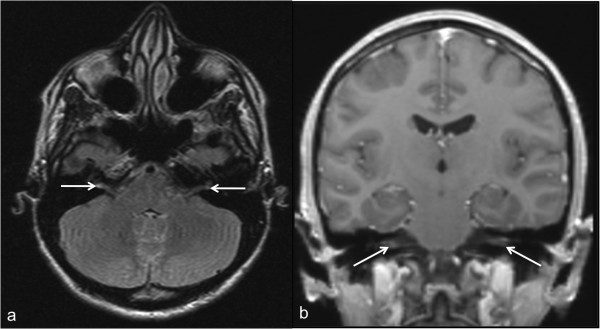
MRIContrast-enhanced brain MRI: bilateral enhancement of the vestibular nerves, arrows on the two inflamed nerves.
Source : Farshad-Amacker et al., BMC research notes, 2013, figure 2 · CC BY
🎥 What the vHIT is worth for confirming neuritis
The video head impulse test distinguishes neuritis from other acute vertigo conditions and from healthy subjects, but it does not replace red-flag triage.
Study of 33 cases of vestibular neuritis, 96 other acute vertigo conditions and 50 healthy controls, based on vestibulo-ocular reflex gain asymmetries. Source: Guan et al., 2017 (PMID 28862109).
The diagnosis of vestibular neuritis, now acute unilateral vestibulopathy (AUV / AUVP) in international nomenclature, rests on no single test. The Bárány Society consensus is explicit: "there is no definitive test for AUVP" 1. It is built on the history, the bedside oculomotor examination and, if necessary, instrumental assessment, and remains, in its very principle, a diagnosis of exclusion.
For the physiotherapist, there is a twofold challenge: recognising the typical presentation in order to begin rehabilitation whose effectiveness is established 11, and above all spotting what is not neuritis: first and foremost vertebrobasilar stroke, which shares the same initial presentation.
The Bárány Society diagnostic criteria (2022)
The international consensus defines acute unilateral vestibulopathy by six criteria, all required 1 :
| Criterion | Content | Clinical translation |
|---|---|---|
| A: Symptom | Spinning or non-spinning vertigo, of acute or subacute onset, sustained, of moderate to severe intensity, lasting at least 24 hours | The duration criterion immediately separates it from BPPV (brief, positional vertigo) |
| B: Nystagmus | Spontaneous peripheral vestibular nystagmus, of fixed direction, increased by suppression of visual fixation, generally horizontal-torsional in trajectory | Cardinal sign; best observed under Frenzel goggles |
| C: VOR | Evidence unambiguous of a reduction in the vestibulo-ocular reflex on the side opposite the fast phase of the nystagmus | Bedside head impulse test, vHIT or caloric testing |
| D: No warning symptom | No acute central neurological symptom, no audiological symptom (hearing loss, tinnitus) or otological symptom (ear pain) | A cochlear sign takes the case outside the framework |
| E: No central sign | No central oculomotor or vestibular sign: no skew deviation, no gaze-evoked nystagmus | This is exactly what HINTS tests |
| F: Exclusion | Presentation not better explained by another condition | Acknowledged diagnosis of exclusion |
Level of evidence: international consensus document of the Bárány Society Classification Committee 1.
The epidemiological context helps to weigh things up: reported annual incidence of 3.5 to 15.5 per 100,000 people, sixth most common cause of vertigo/dizziness and third most common cause of peripheral vestibular disorder, usual age at onset between 30 and 60 years with a plateau between 40 and 50 1.
Spontaneous unidirectional nystagmus: the cardinal sign
The nystagmus of neuritis follows a highly recognisable signature. It is spontaneous (present without a triggering manoeuvre, unlike BPPV), of fixed direction (it always beats towards the same side, whatever the direction of gaze), with a horizontal-torsional trajectory, which corresponds to the canal afferents involved 1.
The most discriminating point is how the nystagmus behaves with respect to visual fixation. The consensus is categorical:
"A nystagmus whose intensity is not reduced by visual fixation is not a spontaneous peripheral nystagmus." 1
In concrete terms: fixating a target attenuates peripheral nystagmus; suppressing that fixation (Frenzel goggles or an equivalent device) increases it typically 1. An examination carried out in full light, without occlusion of fixation, may therefore wrongly conclude that there is no nystagmus: reason enough, for a clinic that sees patients with vertigo, to have Frenzel goggles or videonystagmoscopy available. Conversely, a nystagmus changing direction on eccentric gaze or not attenuated by fixation constitutes a central red flag 81.
The head impulse test: the cardinal sign, without equipment
Described by Halmagyi and Curthoys in 1988, the head impulse test (HIT) remains the most rewarding manoeuvre in the clinic, requiring no equipment at all. The principle: a rapid, passive horizontal head rotation towards the affected side triggers "a large corrective refixation saccade, or several small ones, in the opposite direction", a sign of loss of horizontal semicircular canal function. The authors validated it with magnetic search coils in 12 patients who had undergone unilateral vestibular neurectomy, in whom caloric testing produced only a minimal nystagmic response 7.
Practical translation: a HIT that is positive on the side opposite the fast phase of the nystagmus documents canal paresis and satisfies criterion C. A HIT normal, by contrast, is not reassuring: see below.
vHIT and caloric testing: the consensus thresholds
When instrumental assessment is available, the Bárány consensus adopts numerical benchmarks 1 :
| Test | Threshold for a significant deficit | Consensus comment |
|---|---|---|
| vHIT (video head impulse test) | VOR gain < 0.70 and difference between the two sides > 0.30 | There is no gold standard for calculating gains; the committee strongly recommends the vHIT, because of its high diagnostic value |
| Caloric testing | Canal paresis usually defined by an asymmetry > 25% between the two ears, or an absolute value < 6°/s | Explores mainly low frequencies; historically the reference test in therapeutic trials 4 |
The vHIT performs well in this indication: in a series of 33 cases of vestibular neuritis, 96 patients with other acute vertigo conditions and 50 healthy controls, the vHIT distinguished neuritis from normal subjects and from other acute vertigo conditions with a sensitivity of 87.9% and a specificity of 94.3%, based on VOR gain asymmetries (significant differences between neuritis and other acute vertigo conditions, p < 0.01) 10.
Level of evidence: single-centre diagnostic study with controls and a clinical comparison group 10, supplemented by expert consensus for the thresholds 1.
An anatomical nuance sheds light on some seemingly discordant assessments: in most cases neuritis is not a total vestibular deafferentation, but a partial lesion preferentially affecting the superior division of the vestibular nerve alone: horizontal and anterior canals, utricle 1. The explanation is mechanical: across 40 temporal bones studied, the bony canal of the superior vestibular nerve averaged 2.30 mm compared with 0.59 mm for the canal of the singular nerve (the nerve of the posterior canal, arising from the inferior division): a bony passage nearly four times longer and relatively narrower for the nerve and its arteriole, hence more exposed to entrapment and labyrinthine ischaemia when oedema occurs 6.
Rule out stroke first: HINTS
Faced with an acute vestibular syndrome, the question is not "which rehabilitation?" but "is this really peripheral?". The three-step bedside oculomotor examination HINTS (Head Impulse, Nystagmus, Test of Skew) answers this question better than early imaging.
In the landmark study of 101 high-risk patients with acute vestibular syndrome (25 peripheral lesions, 76 central including 69 ischaemic strokes), the presence of any one of the following three elements (NORMAL horizontal head impulse test, direction-changing nystagmus on eccentric gaze, or skew deviation) was 100% sensitive and 96% specific for stroke. Skew deviation, present in 17% of cases, was associated with brainstem lesions. A major finding:initial diffusion-weighted MRI was falsely negative in 12% of cases, all within 48 hours of symptom onset: the three-step bedside examination "appears more sensitive for stroke than early MRI" 8.
Level of evidence: prospective study in a high-risk cohort 8, reinforced by international consensus 1.
The Bárány consensus states the counter-intuitive rule to remember: "a normal head impulse test in an acute vestibular syndrome with nystagmus is not compatible with a peripheral deficit". The combination of history-taking and physical examination (HINTS and HINTS-plus protocols, designed to rule out central signs) distinguishes AUV from an acute central vestibular syndrome with a sensitivity and specificity of around 90% 1.
Two major caveats frame this tool. First, HINTS applies only to acute vestibular syndrome (sustained vertigo, with nystagmus), not to brief positional vertigo. Second, its performance depends heavily on the operator : a meta-analysis of 5 prospective studies (617 patients) finds, in the hands of neurologists, a sensitivity of 96.7 % (95% CI 93.1–98.5) and a specificity of 94.8 % (95% CI 91–97.1); but in mixed cohorts including emergency physicians, sensitivity falls to 83 % (95% CI 63–95) and specificity to 44 % (95% CI 36–51). Used in isolation by a clinician without specific training, HINTS is not enough to rule out a stroke 9.
What the physiotherapist must know how to test and recognise
1. Recognise the presentation (criteria A–C). Sustained vertigo ≥ 24 h, spontaneous nystagmus of fixed direction increased by suppression of fixation (Frenzel), HIT positive on the side opposite the fast phase 71.
2. Recognise what is not neuritis. Any cochlear sign (hearing loss, tinnitus) or otological sign (ear pain), any central sign (skew deviation, direction-changing nystagmus, normal HIT) takes the case outside the framework and calls for medical referral, not rehabilitation 18.
3. Do not confuse it with BPPV: the costliest error. BPPV produces vertigo that is brief, positional, with no spontaneous nystagmus 1. The distinction is not academic: the Cochrane review (39 trials, 2,441 participants) establishes moderate to strong evidence in favour of exercises in unilateral vestibular hypofunction (OR 2.67; 95% CI 1.85–3.86; 4 studies, 565 participants), but shows that in BPPV specifically, it is repositioning manoeuvres that are superior to exercises in the short term (OR 0.19; 95% CI 0.07–0.49) 11. Exercises treat neuritis, manoeuvres treat BPPV.
4. Retest throughout follow-up. A secondary "post-infectious" BPPV of the posterior canal is an expected complication of neuritis 1 : across 51 patients followed for 4 to 6 years (mean 4.9 years), 5/51 (9.8 %) developed BPPV (typically of the posterior canal of the affected ear, more frequent in cases of incomplete recovery and more resistant to treatment than idiopathic BPPV), whereas recurrence of the neuritis is rare (1/51, 2.0 %) 2. Brief positional vertigo during rehabilitation calls for a Dix-Hallpike test, not a verdict of failed compensation.
5. Widen the assessment beyond canal tests. Prognosis cannot be read from VOR gain. A systematic review (13 studies) shows that it is the anxiety following the vestibular episode, dependent personality traits, autonomic activation, bodily hypervigilance and visual dependence (and not the severity of the initial or subsequent structural deficit, nor compensation status), that best predict chronic dizziness and PPPD 12. A prospective cohort of 40 patients assessed acutely (median 2 days) and then 32 during recovery (median 10 weeks) confirms this: visual dependence and autonomic activation measured acutely predict outcome, not the vestibular variables 13.
Key points
- No definitive test. The diagnosis of AUV/neuritis rests on the history, the bedside examination and, if needed, instrumental assessment, and remains a diagnosis of exclusion 1.
- Three positive criteria: sustained vertigo ≥ 24 h; spontaneous nystagmus of fixed direction, horizontal-torsional, increased by suppression of fixation; unambiguous reduction of the VOR on the side opposite the fast phase 1.
- A nystagmus not attenuated by fixation is not peripheral, hence the value of Frenzel goggles 1.
- Head impulse test 7 : refixation saccade on rapid rotation towards the affected side. vHIT: gain < 0.70 and between-side difference > 0.30; caloric: asymmetry > 25% or < 6°/s 1. vHIT: Se 87.9% / Sp 94.3% 10.
- A NORMAL HIT in an acute vestibular syndrome is a central red flag, not a reassuring sign 81. HINTS: Se 100%, Sp 96% for stroke; early diffusion-weighted MRI falsely negative in 12% of cases (< 48 h) 8. But performance is operator-dependent: Se 83%, Sp 44% in non-specialist cohorts 9.
- Neuritis ≠ BPPV: exercises for unilateral hypofunction, manoeuvres for BPPV 11: think of secondary BPPV (9.8%) when brief positional vertigo appears during follow-up 2.
- The prognostic assessment is not vestibular: screen for visual dependence and anxiety from the acute phase onwards 1312.
🚩 How to avoid missing a stroke (red flags)?
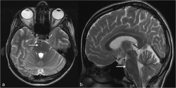
MRIT2-weighted brain MRI, axial and sagittal views: hyperintensity of the brainstem at the level of the pons, arrows, in the same patient with vestibular neuritis due to neuroborreliosis.
Source : Farshad-Amacker et al., BMC research notes, 2013, figure 1 · CC BY
🚩 HINTS is only as good as the hand performing it
The same examination, in different hands, does not protect in the same way. This is the most important piece of data in this section.
Meta-analysis of 5 prospective studies (617 patients with acute vestibular syndrome). Specificity collapses from 94.8% to 44% outside the hands of trained specialists: used in isolation by a clinician without specific training, HINTS is not enough to rule out a stroke. Source: Ohle et al., 2020 (PMID 32167642).
A patient arrives with spinning vertigo that has been present since the day before, nausea, marked unsteadiness and nystagmus. This presentation, the acute vestibular syndrome, has two major causes that look deceptively alike at first glance: vestibular neuritis, which is benign, and vertebrobasilar stroke, which is life-threatening. The Bárány Society consensus is unambiguous about the nature of the diagnosis of acute unilateral vestibulopathy (AUV): it is a diagnosis of exclusion, requiring the absence of acute central neurological signs, the absence of cochlear signs, and that the presentation be not better explained by another condition 1. In other words: before rehabilitating neuritis, stroke must have been ruled out.
Faced with prolonged acute vertigo, the question is not "is this neuritis?" but "is this not a stroke?".
HINTS: three bedside manoeuvres, more sensitive than early MRI
The three-step oculomotor examination HINTS (Head-Impulse, Nystagmus, Test-of-Skew) is the reference tool. In the landmark study by Kattah et al. 8, 101 high-risk patients with acute vestibular syndrome were investigated (25 peripheral lesions, 76 central lesions including 69 ischaemic strokes). The presence of a normal horizontal head impulse test, direction-changing nystagmus on eccentric gaze, or skew deviation was 100% sensitive and 96% specific for stroke. Skew deviation was present in 17% of cases and associated with brainstem lesions.
- H: Head Impulse Test. Rapid, passive horizontal head rotation. The cardinal sign described by Halmagyi and Curthoys 7, validated with magnetic search coils in 12 patients after neurectomy, is a large corrective refixation saccade, or several small ones, in the opposite direction, triggered by rotation towards the affected side. This sign reflects loss of horizontal semicircular canal function: it is expected in neuritis.
- N: Nystagmus. In neuritis, the nystagmus is spontaneous, of fixed direction, horizontal-torsional, and increased by suppression of visual fixation 1. A nystagmus that changes direction according to the position of eccentric gaze is a central red flag.
- TS: Test of Skew. The alternate cover test looks for a vertical ocular misalignment (skew deviation). Its presence is a formal exclusion criterion for AUV according to the Bárány Society 1 and a marker of a brainstem lesion 8.
The counter-intuitive pitfall: a NORMAL head impulse test is alarming
This is the point most often reversed in practice. The natural reflex is to feel reassured when the examination looks "clean". Exactly the opposite is required. The Bárány consensus states it bluntly: "a normal head impulse test in an acute vestibular syndrome with nystagmus is not compatible with a peripheral deficit" 1: it points towards a central cause. Kattah et al. 8 make it one of the three items in the triad with 100% sensitivity.
The logic is mechanical: neuritis destroys the vestibulo-ocular reflex on one side, so the HIT must be abnormal. If the patient has had violent vertigo for 24 hours, with nystagmus, and their VOR works normally, then the vertigo does not come from the labyrinth. A normal HIT in a patient with acute vertigo is a warning signal, not a reassuring sign.
Key points: the HIT paradox
Abnormal HIT (refixation saccade) = rather reassuring → consistent with peripheral involvement.
Normal HIT in a patient with acute vestibular syndrome = central red flag → suspected stroke until proven otherwise 81.
Three other signs that immediately exclude neuritis: skew deviation, direction-changing nystagmus on eccentric gaze, any acute cochlear sign (hearing loss, tinnitus) or otological sign (ear pain): criteria D and E of the Bárány Society 1.
HINTS is operator-dependent: do not overestimate your own performance
The figure of 100% sensitivity does not transfer as such to the clinic. The meta-analysis by Ohle et al. 9, which pools 5 prospective studies and 617 patients with acute vestibular syndrome, shows performance that is strongly dependent on the examiner :
| Context in which HINTS is used | Sensitivity for stroke | Specificity |
|---|---|---|
| Neurologists (dedicated cohorts) | 96.7% (95% CI 93.1–98.5) | 94.8% (95% CI 91–97.1) |
| Mixed cohorts including emergency physicians | 83 % (95% CI 63–95) | 44 % (95% CI 36–51) |
The authors explicitly conclude that "the HINTS examination, used in isolation by emergency physicians, has not demonstrated sufficient accuracy to rule out stroke in patients presenting with acute vestibular syndrome" 9. The reading for the physiotherapist is direct: HINTS serves to spot a red flag and to refer, never to self-authorise rehabilitation. The Bárány consensus, moreover, places the performance of the combination of history-taking and physical examination (HINTS and HINTS-plus) at around 90% sensitivity and specificity for distinguishing AUV from an acute central vestibular syndrome 1: excellent, but not infallible.
The limits of early MRI: normal imaging is not reassuring
This is the second frequent misreading. In the study by Kattah et al. 8, initial diffusion-weighted MRI was falsely negative in 12% of cases, and all these falsely negative MRIs had been performed within the first 48 hours of symptom onset. The authors' conclusion is blunt: the three-step bedside examination "appears more sensitive for stroke than early MRI in acute vestibular syndrome".
Practical consequence: "the MRI was normal" does not rule out a posterior fossa stroke if it was performed early. A patient referred to the emergency department at H+12, discharged with a normal MRI and a prescription for anti-vertigo drugs, but who has a normal HIT or a skew deviation, remains an at-risk patient. The imaging report does not replace the oculomotor examination: it complements it.
Distinguishing neuritis, BPPV, Ménière's disease and labyrinthitis
Triage rests first on three axes: duration, trigger, the presence of auditory signs. The Bárány Society criteria 1 define AUV by spinning or non-spinning vertigo, of acute or subacute onset, sustained, of moderate to severe intensity, lasting at least 24 hours, with a spontaneous peripheral nystagmus of fixed direction, increased by suppression of visual fixation, and unambiguous evidence of a reduced VOR on the side opposite the fast phase.
| Vestibular neuritis (AUV) | BPPV | Ménière's disease / labyrinthitis | Vertebrobasilar stroke | |
|---|---|---|---|---|
| Duration of vertigo | Sustained, ≥ 24 h | Brief, paroxysmal | Episodic or acute | Sustained (acute vestibular syndrome) |
| Trigger | Spontaneous | Positional (changes of position) | Variable | Spontaneous |
| Spontaneous nystagmus | Present, fixed direction, horizontal-torsional, increased without fixation | Absent (positional nystagmus only) | — | May change direction on eccentric gaze |
| Auditory signs | None (exclusion criterion) | None | Associated audiological involvement | — |
| Head impulse test | Abnormal (saccade) | — | — | Often normal = red flag |
| Skew deviation | Absent (exclusion criterion) | Absent | — | Present in 17% 8 |
BPPV. The distinction is settled straight away by the clinical picture: brief, positional vertigo, triggered by head movements, with no spontaneous nystagmus, in contrast to neuritis, whose cardinal criterion is precisely that spontaneous nystagmus of fixed direction 1. This distinction is not academic: it dictates treatment. The Cochrane review by McDonnell and Hillier 11 shows that in BPPV it is repositioning manoeuvres that are superior to exercises in the short term (OR 0.19; 95% CI 0.07–0.49), whereas exercise-based rehabilitation is aimed at unilateral vestibular hypofunction: neuritis (OR 2.67; 95% CI 1.85–3.86 in favour of vestibular rehabilitation).
Ménière's disease and labyrinthitis. The discriminator is auditory. Criterion D of the Bárány Society excludes AUV in the presence of "acute audiological symptoms such as hearing loss or tinnitus, or other otological symptoms such as ear pain" 1. This is precisely what separates neuritis from labyrinthitis or Ménière's disease, in which there is associated audiological involvement. Acute vertigo with sudden hearing loss is not neuritis: it does not warrant direct entry into rehabilitation.
What instrumental assessment can, and cannot, do
The Bárány consensus sets an essential limit: "there is no definitive test for AUV", the diagnosis rests on the history, the bedside examination and, if necessary, instrumental assessment 1. The thresholds adopted: on vHIT, a VOR gain below 0.70 with a between-side difference greater than 0.30 (the document specifying that there is no gold standard for calculating gains); on caloric testing, an asymmetry greater than 25% between the two ears, or an absolute value below 6°/s. The committee strongly recommends the vHIT because of its high diagnostic value. Guan et al. 10, across 33 cases of neuritis, 96 other acute vertigo conditions and 50 controls, report a sensitivity of 87.9% and a specificity of 94.3% for the vHIT in distinguishing neuritis from other acute vertigo conditions.
But none of these figures rules out a stroke: they confirm peripheral involvement once central causes have been clinically eliminated. The order of reasoning matters.
When to refer urgently
Any departure from the Bárány Society diagnostic framework should prompt referral of the patient, and not entry into rehabilitation 1. In practice, refer without delay in the presence of:
- A normal head impulse test in a patient with acute vestibular syndrome and nystagmus 81 ;
- A direction-changing nystagmus on eccentric gaze (gaze-evoked nystagmus = exclusion criterion E);
- A skew deviation (vertical ocular misalignment);
- A nystagmus not attenuated by visual fixation : the consensus recalls that "a nystagmus that is not reduced in intensity by visual fixation is not a spontaneous peripheral vestibular nystagmus", Frenzel goggles typically increase its intensity in neuritis 1 ;
- Any acute cochlear or otological sign : hearing loss, tinnitus, ear pain;
- Any acute central neurological sign or symptom.
A final contextual reminder: neuritis is common: reported annual incidence of 3.5 to 15.5 per 100,000 people, sixth most common cause of vertigo from all causes combined, third most common cause of peripheral vestibular disorder, usual age at onset between 30 and 60 years with a plateau between 40 and 50 1. Its very frequency is the trap: it is because we see it often that we risk labelling as "neuritis" the case that is not. Central triage happens with every patient, not once in ten.
🎯 Which vestibular rehabilitation to offer?
⏱️ The recommended dosage of gaze stabilisation exercises
The Academy of Neurologic Physical Therapy recommendation puts precise figures on what should be prescribed, and it is a daily minimum, not an average.
In the chronic phase, these durations should be maintained for 4 to 6 weeks. Source: updated clinical practice guideline, Hall et al., 2022 (PMID 34864777).
Once the diagnosis has been made (sustained vertigo lasting more than 24 hours, spontaneous nystagmus of fixed direction increased by suppression of fixation, VOR reduced on the side opposite the fast phase, and above all absence of central signs 1), the question becomes therapeutic. And it is quickly settled: this is the only area of management where the level of evidence is solid. Where corticosteroid therapy remains debated 4 and where vestibular suppressants actively slow recovery 16, rehabilitation has a coherent body of randomised evidence.
Evidence for vestibular rehabilitation: what Cochrane says
The Cochrane review by McDonnell and Hillier 11 remains the reference: 39 randomised controlled trials, 2,441 participants with unilateral peripheral vestibular dysfunction. Its conclusion is explicit: there is moderate to strong evidence that vestibular rehabilitation is a safe and effective management approach for unilateral peripheral vestibular deficit, on the basis of several high-quality RCTs.
Two trials structure our understanding of the mechanism. Strupp et al. 17 compared 19 patients receiving specific vestibular exercises with 20 control patients without exercises after neuritis: at day 30, the postural sway path was 3.2 ± 1.9 m/min in the physiotherapy group versus 16.9 ± 6.1 m/min in the control group (ANOVA, p < 0.001). A crucial and often misread point: ocular torsion and the subjective visual vertical normalised comparably in both groups. Rehabilitation acts on central vestibulospinal compensation, not on recovery of peripheral function itself.
Tokle et al. 18 then tested the addition of a rehabilitation programme started early after diagnosis in 65 patients with neuritis confirmed by videonystagmography (27 in the intervention group), in addition to standard care (prednisolone for 10 days + advice). Result: a significant reduction in perceived vertigo at 3 months (p = 0.007) and at 12 months (p = 0.001), with, at 12 months, improvement in the DHI (p = 0.049), the HADS (p = 0.039) and the VAS-C (p = 0.012). By contrast, no difference in standing balance or walking speed: the gain is seen in the patient's experience and daily functioning, not in raw postural performance.
"There is moderate to strong evidence that vestibular rehabilitation is a safe and effective management for unilateral peripheral vestibular dysfunction.", McDonnell & Hillier, Cochrane 2015
The line never to cross: neuritis ≠ BPPV
The same Cochrane review carries the most important exception for everyday practice. In the specific diagnostic subgroup of BPPV, it is physical repositioning manoeuvres that are superior to movement-based rehabilitation exercises in short-term cure rate (OR 0.19; 95% CI 0.07–0.49), even though combining the two is useful for longer-term functional recovery 11.
| Vestibular neuritis (AUV) | BPPV | |
|---|---|---|
| Vertigo | Sustained, ≥ 24 h, moderate to severe 1 | Brief, positional, triggered by changes of position |
| Nystagmus | Spontaneous, fixed direction, increased by suppression of fixation, horizontal-torsional | Positional, no spontaneous nystagmus |
| Treatment, 1st line | Exercises vestibular rehabilitation strong evidence | Manoeuvres repositioning (OR 0.19 in favour of manoeuvres vs exercises) strong evidence |
In other words: exercises are the treatment for neuritis, manoeuvres the treatment for BPPV. Transposing the protocol of one to the other means doing worse than doing nothing specific.
Gaze stabilisation: the core of the programme
The cardinal functional deficit in neuritis is the loss of vestibulo-ocular reflex gain on the affected side. Its clinical signature has been known since the article by Halmagyi and Curthoys 7 : a rapid horizontal head rotation towards the affected side triggers a large corrective refixation saccade, or several small ones, in the opposite direction, a sign validated with magnetic search coils in 12 patients after neurectomy. It is exactly this deficit that gaze stabilisation exercises aim to have compensated.
The clinical practice guideline of the Academy of Neurologic Physical Therapy 19 is unambiguous here, and it contains an explicit prohibition:
"Clinicians should not include voluntary saccadic or smooth-pursuit eye movements in isolation (ie, without head movement) to promote gaze stability.", Hall et al. 2022 strong evidence, recommendation against
Isolated eye movements (voluntary saccades, smooth pursuit), do not stabilise gaze. A gaze stabilisation exercise is only a gaze stabilisation exercise if it engages the VOR through a head movement. This is the most rewarding sorting criterion for auditing an exercise programme: if there is no head movement, there is no vestibular stimulus.
Habituation: provoke the error signal, do not switch it off
The rationale for habituation is documented pharmacologically in reverse, by what should not be done. Sousa et al. 16 state it in one sentence: symptomatic treatment reduces the vestibular tonic imbalance, which acts precisely as the main driver of central compensation, which in all likelihood has a negative impact on compensation and long-term outcomes; prolonged use of these drugs prolongs, or even prevents, central compensation. The rehabilitative logic is exactly the reverse: using vestibular exercises, in particular head movements, to increase this tonic imbalance, enhance central compensation and allow an adequate return to a fully functional state. To this is added the argument of Dyhrfjeld-Johnsen and Attali 20 : sedation, the main and limiting adverse effect of vestibular suppressants, is counterproductive for normal functioning and for the natural recovery process through central compensation.
A patient still on anti-vertigo medication beyond the acute phase is therefore a patient whose compensation is being pharmacologically held back: information to obtain at assessment, and to pass on to the prescriber.
Substitution: aim for compensation, not restoration
A realistic goal must be set, and stated to the patient. In the double-blind randomised trial by Strupp et al. 4 in 141 patients, the mean improvement in peripheral vestibular function (caloric paresis) at 12 months was only 39.6 ± 28.1 percentage points on placebo and 62.4 ± 16.9 on methylprednisolone. Even on corticosteroids, a residual canal deficit very often persists at one year. Peripheral restoration is not the achievable goal; central compensation is, and it is central compensation that Strupp et al. 17 showed to be modifiable by exercises along the vestibulospinal pathway (sway path 3.2 vs 16.9 m/min).
This dissociation is the foundation of the substitution strategy: the canal is not repaired, functional performance is rebuilt with the remaining inputs. This is why the APTA guidelines systematically pair gaze work with static and dynamic balance exercises, at least 20 minutes a day for at least 4 to 6 weeks 19.
Timing and dosage: the figures to apply
The APTA recommendation places rehabilitation as first-line treatment from the acute phase onwards: "Clinicians should offer vestibular physical therapy to individuals with acute or subacute unilateral vestibular hypofunction", evidence quality I, strong recommendation 19. This is consistent with Tokle et al. 18, whose benefit at 3 and 12 months is obtained with a programme started early after diagnosis.
Key points: dosage 19
- Acute/subacute unilateral hypofunction: gaze stabilisation: 3 times a day, for a total of at least 12 minutes daily evidence quality II, weak recommendation
- Chronic unilateral hypofunction: gaze stabilisation: 3 to 5 times a day, for a total of at least 20 minutes daily over 4 to 6 weeks moderate to weak evidence
- Static and dynamic balance : at least 20 minutes a day for at least 4 to 6 weeks
- Prohibited : voluntary saccades or smooth pursuit in isolation, without head movement, to improve gaze stability strong evidence against
- Pharmacological corollary : taper vestibular suppressants as quickly as possible, they suppress the driver of compensation 1620
Progression: what really governs prognosis
Progression is not set by vestibular tests. Arshad et al. 21 are categorical: after neuritis, long-term prognosis does not depend on the extent of the residual peripheral deficit, measured by caloric testing or vHIT; recovery is determined by a combination of visuo-vestibular (visual dependence), psychological (anxiety) and perceptual factors. The systematic review by Trinidade et al. 12 converges: it is anxiety following the vestibular episode, dependent personality traits, autonomic activation, bodily hypervigilance and visual dependence, and not the severity of the structural deficit or compensation status, that best predict chronic dizziness.
Cousins et al. 13 showed this prospectively (40 patients in the acute phase, median 2 days; 32 during recovery, median 10 weeks): visual dependence and autonomic activation measured in the acute phase predicted outcome, poorer recovery being associated with visual dependence, autonomic activation, anxiety/depression and fear of bodily sensations, but not with vestibular variables. Van Laer et al. 22, across 103 participants followed at 4 and 10 weeks, quantified usable thresholds at 10 weeks for identifying at-risk patients: Vestibular Activities Avoidance Instrument 23/54, Visual Vertigo Analogue Scale 33.5/100, HADS-Anxiety 7.5/21 ; their models explained up to 64.0% of the variability in dizziness at 6 months.
Direct consequence: progression is built on early exposure, countering avoidance and restoring confidence 12: visual dependence and anxiety are screened for at the first session, not in the third month.
What should prompt reassessment during rehabilitation
- Brief positional vertigo that appears : look for secondary BPPV, do not conclude that compensation has failed. Mandalà et al. 2, across 51 patients followed for 4 to 6 years (mean 4.9 years): 5/51 (9.8%) developed BPPV: typically of the posterior canal of the affected ear, more frequent in cases of incomplete recovery and more resistant than idiopathic BPPV. Recurrence of the neuritis itself is rare (1/51, 2.0%). The Bárány consensus describes this "post-infectious" BPPV of the posterior canal 1. And BPPV predicts long-term dizziness handicap (r = 0.626, p = 0.001), as do migraine (r = 0.640, p = 0.001) and acute visual dependence (r = 0.667, p < 0.001): Arshad et al. 21.
- Symptoms that become chronic beyond 3 months : consider PPPD, dizziness, unsteadiness or non-spinning vertigo present on most days for 3 months or more, worsened by upright posture, active or passive movement and exposure to moving or complex visual stimuli 23. This is a chronic functional vestibular disorder: neither structural nor psychiatric. AUV is its 3rd most common trigger, after BPPV and vestibular migraine 1.
- Any cochlear or central sign : refer, do not rehabilitate. A head impulse test that is normal in a patient with acute vestibular syndrome and nystagmus is not compatible with a peripheral deficit 1: it is a red flag, not a reassuring sign 8.
In one sentence
Exercises with head movement, early, every day, at the APTA dosage, in a patient who has not been sedated, and follow-up that tracks visual dependence, avoidance and anxiety as much as VOR gain, because it is these, and not the residual canal deficit, that decide the outcome at one year.
💊 Corticosteroids and anti-vertigo drugs: what does the evidence say?
⚖️ Twenty years on, the controversy is not settled
Successive syntheses do not say the same thing. Here is the actual state of the debate: worth knowing before asserting anything to a patient.
What the physiotherapist should take from this: the symptomatic benefit of corticosteroids has never been demonstrated, and the only intervention whose effectiveness is solidly established remains vestibular rehabilitation. Sources: Strupp 2004 (PMID 15269315); Fishman 2011 (PMID 21563170); Leong 2021 (PMID 33525978); Bogdanova 2022 (PMID 36029039); Sjögren 2025 (PMID 39973595).
💊 The trial that launched corticosteroid therapy
Recovery of peripheral vestibular function at 12 months, double-blind randomised trial, 2×2 factorial design, 141 patients.
Mean improvement in percentage points (± 16.9 / 28.1 / 26.7). Methylprednisolone comes out on top (p < 0.001); valaciclovir adds nothing, and the combination does no better than the corticosteroid alone. Source: Strupp et al., NEJM 2004 (PMID 15269315).
The physiotherapist does not prescribe the medical treatment of vestibular neuritis, but does have to deal with its direct consequences. A patient still on a vestibular suppressant at three weeks is a patient whose central compensation is being pharmacologically held back. A patient who arrives for rehabilitation convinced that "steroids will fix everything" is a patient whose expectations are going to be disappointed. Understanding what pharmacology does, and above all what it does not do, in this condition is therefore a clinical prerequisite, not academic decoration.
This section takes stock of three questions: do corticosteroids have a demonstrated benefit? Do antivirals still have a place? And why should anti-vertigo drugs be limited in time, even though they relieve the patient?
Corticosteroids: from the landmark 2004 trial to the current controversy
The whole discussion starts from a trial that has become a classic. Strupp et al. 4, in the New England Journal of Medicine, conducted a double-blind, placebo-controlled randomised trial with a 2×2 factorial design in 141 patients with acute vestibular neuritis. The four arms: placebo (38 patients), methylprednisolone (35), valaciclovir (33), combination (35). The endpoint was peripheral vestibular function measured by caloric testing (vestibular paresis formula), within 3 days and then at 12 months.
The results at 12 months are clear-cut:
Strupp et al. 2004, NEJM
This trial produced two messages, only one of which has really held up. The first, antivirals are of no use, has never been challenged since (see below). The second, corticosteroids improve peripheral recovery, is today the subject of open controversy.
Why the controversy?
Because the 2004 demonstration concerns a laboratory test, not the patient's experience. The endpoint was the percentage of caloric paresis: a number. And the later systematic reviews, which looked at whether that number translated into clinical benefit, did not find that it did.
The Cochrane review by Fishman et al. 5 pooled 4 placebo-controlled randomised trials, totalling 149 participants. Its verdict is instructive in detail:
- Significant effect on complete caloric recovery at 1 month : RR 2.81 (95% CI 1.32–6.00; P=0.007).
- Effect gone at 12 months : RR 1.58 (95% CI 0.45–5.62; P=0.48).
- No difference in symptomatic recovery : neither for vertigo at 24 h (RR 0.39; 95% CI 0.04–3.57; P=0.40), nor for the Dizziness Handicap Inventory at 1, 3, 6 and 12 months.
The authors' conclusion: the evidence is insufficient to recommend corticosteroid therapy in idiopathic acute vestibular dysfunction. In other words: a transient biological benefit on a test, with no translation for the patient.
The systematic review with meta-analysis by Leong et al. 14, covering 8 studies of which 6 were meta-analysed (data 2010–2019), reaches the same conclusion on a more recent basis: no significant difference between corticosteroids and comparators (placebo, vestibular exercises alone, or the combination) in the proportion of patients with complete recovery at 1, 6 and 12 months. Corticosteroids have only a short-term benefit on canal paresis, with no long-term benefit, either on canal paresis or on symptomatic recovery.
The controversy is not closed for all that. The updated meta-analysis by Bogdanova et al. 24, which brings together 15 trials and 852 participants, by contrast finds odds ratios favouring corticosteroids: OR 3.1 for a good outcome in the acute phase, OR 2.4 for restoration of vestibular function at follow-up. But the authors themselves temper their result: the quality of the evidence remains limited (heterogeneity of studies, risk of bias), and above all adverse effects are markedly increased with corticosteroids, OR 10.9. Rigorous randomised trials are still needed.
Those rigorous trials are beginning to arrive. The double-blind, placebo-controlled randomised trial by Sjögren et al. 15, conducted in 69 patients across three Swedish centres, compared three arms: placebo, corticosteroids for 3 days, corticosteroids for 10 days. Result: all groups improved significantly over time in caloric function (p=0.002), with no difference between groups: mean difference −8.34 (95% CI −25.93 to 9.26; p=0.347) for the long regimen, −6.61 (−24.67 to 11.45; p=0.467) for the short regimen. The conclusion is unambiguous: corticosteroid therapy improves neither caloric recovery, nor vHIT gain, nor subjective experience in acute unilateral vestibulopathy. It is the most recent and methodologically most solid trial since 2004.
| Study | Design / sample size | Result | Level |
|---|---|---|---|
| Strupp 2004 (NEJM) | Double-blind RCT, 2×2 factorial, 141 patients | Methylprednisolone: +62.4 vs +39.6 pts (placebo) on caloric testing at 12 months, P<0.001 | Strong (but paraclinical endpoint) |
| Fishman 2011 (Cochrane) | Meta-analysis, 4 RCTs, 149 participants | Effect at 1 month (RR 2.81) but gone at 12 months (RR 1.58, P=0.48); no effect on DHI or vertigo | Strong: insufficient evidence |
| Leong 2021 | Systematic review + meta-analysis, 8 studies (6 meta-analysed) | No difference in complete recovery at 1, 6 and 12 months | Moderate: insufficient evidence |
| Bogdanova 2022 | Meta-analysis, 15 trials, 852 participants | OR 3.1 (acute phase) and 2.4 (follow-up) in favour, but limited quality and adverse effects OR 10.9 | Weak: heterogeneity, bias |
| Sjögren 2025 | Double-blind placebo-controlled RCT, 69 patients, 3 centres, 3 arms | No benefit: neither caloric, nor vHIT, nor subjective experience | Strong: negative |
Twenty years after the landmark trial, corticosteroid therapy for vestibular neuritis remains a widespread practice whose clinical benefit is not demonstrated, and whose adverse effects are.
Antivirals: the question is settled
Here, at least, the literature is simple. The dominant pathophysiological hypothesis is viral: according to Strupp and Brandt 3, reactivation of a latent herpes simplex virus type 1 (HSV-1) infection is the most likely cause, with a major supporting argument: HSV-1 DNA and RNA have been detected in human vestibular ganglia. The Bárány consensus 1 specifies the mechanism: HSV-1 replicates, inducing inflammation and oedema of the vestibular ganglion, responsible for secondary cellular damage.
A viral aetiology logically called for antiviral treatment. But the trial by Strupp et al. 4 definitively ruled out this avenue: valaciclovir gives +36.0 ± 26.7 percentage points at 12 months: a result comparable to placebo (+39.6 ± 28.1), a non-significant effect (P=0.43). And the methylprednisolone + valaciclovir combination adds nothing to the corticosteroid alone.
This negative result has strong explanatory value: it suggests that what damages the nerve is not persistent active viral replication that an antiviral might curb, but the inflammatory response and the resulting oedema. The lesional mechanism is as much mechanical as inflammatory, as confirmed by the anatomy described by Goebel et al. 6 : across 40 temporal bones studied, the bony canal of the superior vestibular nerve averaged 2.30 mm in length compared with 0.59 mm for the canal of the singular nerve (p<0.001), that is, a bony course nearly four times longer, with a relatively narrower passage for the nerve and its arteriole. This long bony passage makes the superior division more vulnerable to entrapment and to labyrinthine ischaemia as soon as inflammatory oedema occurs, which incidentally explains why neuritis is most often a partial lesion, preferentially affecting the superior division alone 1.
Vestibular suppressants: the trap of relief
This is the point most directly actionable for the physiotherapist, and the most counter-intuitive for the patient.
Vestibular suppressants (anti-vertigo drugs, antihistamines such as dimenhydrinate, benzodiazepines) work: they reduce subjective vertigo and the nystagmus. That is precisely the problem. As Sousa et al. 16 put it in the Journal of Audiology & Otology : symptomatic treatment reduces the vestibular tonic imbalance, which is precisely the main driver of central compensation. By attenuating this imbalance, the error signal that the central nervous system needs in order to recalibrate is removed. The authors are explicit: this "probably has a negative impact on central compensation and long-term outcomes", and "prolonged use of these drugs prolongs, or even prevents, central compensation of the vestibular tonic imbalance". Hence the accepted rule: all symptomatic therapies should be tapered rapidly, in order to avoid impairing central compensatory mechanisms.
A second harmful effect is added to this, highlighted by Dyhrfjeld-Johnsen and Attali 20 : the main and limiting adverse effect of anti-vertigo drugs, sedation, is counterproductive for normal functioning and for the natural recovery process that runs through central compensation. A sedated patient moves less, exposes themselves less, and feeds recalibration less.
The logic of rehabilitation is the exact opposite of that of the suppressant. Sousa et al. 16 say so in as many words: the aim is to use vestibular exercises, in particular head movements, to increase the vestibular tonic imbalance, enhance central compensation and allow an adequate return to a fully functional state. The physiotherapist seeks to provoke the signal that the drug seeks to switch off.
Key points, what the physiotherapist should know about medical treatment
- Corticosteroids: debated benefit, does not govern rehabilitation. Strupp 2004 shows a gain on caloric testing at 12 months (+62.4 vs +39.6 pts, P<0.001), but Cochrane 5 finds no symptomatic effect (DHI at 1, 3, 6, 12 months), Leong 2021 no difference in complete recovery, and the RCT by Sjögren 2025 no benefit (caloric, vHIT, subjective experience). Bogdanova 2022 remains favourable (OR 3.1 / 2.4) but with limited-quality evidence and adverse effects OR 10.9. Discordant evidence
- Antivirals: ineffective, question settled. Valaciclovir = placebo (P=0.43), the combination adds nothing to the corticosteroid alone 4. The HSV-1 hypothesis 3 remains valid, but it is inflammation and oedema, not persistent active replication, that damage the nerve in its narrow bony passage 6. Strong evidence
- Vestibular suppressants: the shortest possible acute phase. They switch off the tonic imbalance that is the very driver of central compensation 16, and their sedation is counterproductive for natural recovery 20. A patient still on anti-vertigo medication beyond the acute phase is a patient whose compensation is pharmacologically held back : this is information to collect at assessment and to pass on to the prescriber. Moderate evidence, robust mechanism
- The treatment whose effectiveness is best established is yours. Cochrane 11 concludes that there is moderate to strong evidence in favour of vestibular rehabilitation, OR 2.67 for vertigo frequency (95% CI 1.85–3.86), SMD −0.83 on the DHI (95% CI −1.02 to −0.64), no adverse effects reported. The APTA 19 recommends strongly (level I) offering vestibular rehabilitation from the acute/subacute phase onwards. Strong evidence
The practical consequence: mobilise early rather than sedate
The contrast is striking. On one side, a pharmacopoeia whose benefit is either nil (antivirals), or debated and burdened with adverse effects (corticosteroids), or frankly counterproductive beyond the acute phase (suppressants). On the other, an intervention whose evidence is moderate to strong, with no adverse effects reported 11.
The clinical practice guideline of the Academy of Neurologic Physical Therapy 19 is unambiguous: " Clinicians should offer vestibular physical therapy to individuals with acute or subacute unilateral vestibular hypofunction ", strong recommendation, level of evidence I. The dosage proposed in the acute/subacute phase is 3 sessions a day for a total of at least 12 minutes daily (level II, weak recommendation), to be distinguished from the chronic phase (3 to 5 times/day, at least 20 min/day, 4 to 6 weeks). This framing is perfectly consistent with limiting anti-vertigo drugs: mobilise early rather than sedate the system.
The clinical demonstration exists, moreover, in the exact context that concerns us. The randomised trial by Tokle et al. 18 included 65 patients with vestibular neuritis confirmed by videonystagmography; all received standard care (prednisolone for 10 days + advice), 27 of them additionally receiving vestibular rehabilitation started early after diagnosis. Result: a significant reduction in the perception of vertigo at 3 months (p=0.007) and at 12 months (p=0.001), with, at 12 months, significant improvement in the DHI (p=0.049) and the HADS anxiety-depression score (p=0.039). In other words, against a background of standard corticosteroid therapy, it is the addition of early rehabilitation that makes the difference to the patient's experience.
Finally, an expectation-setting message to give the patient. Even on corticosteroids, peripheral recovery remains partial: in the Strupp trial 4, the mean improvement in caloric paresis at 12 months plateaus at 62.4 ± 16.9 percentage points on methylprednisolone. A residual canal deficit very often persists at one year. Peripheral restoration is not the realistic goal of management: central compensation is. And that is just as well, since long-term prognosis precisely does not depend on the measured residual vestibular deficit: according to Arshad et al. 21, recovery is determined by a combination of visuo-vestibular (visual dependence), psychological (anxiety) and perceptual factors, not by the magnitude of remaining peripheral function on caloric testing or vHIT.
In other words: what the drug has not repaired, compensation can compensate for: provided it has not been put to sleep.
🔄 Recovery, prognosis and complications
🔄 Secondary BPPV is common, recurrence of neuritis is rare
A patient who "relapses" a few months later most often has BPPV of the posterior canal on the affected side, not a new episode of neuritis. To be screened for systematically at follow-up.
Prospective cohort of 51 patients followed for 4 to 6 years (mean 4.9 years). Secondary BPPV typically affects the posterior canal of the previously affected ear, occurs more often in cases of incomplete initial recovery and is more resistant to treatment than idiopathic BPPV. Source: Mandalà et al., 2010 (PMID 19883173).
🔮 What predicts recovery is not what is measured at the ear
The severity of the vestibular deficit does not predict outcome. It is psychological burden, visual dependence and static balance that do.
Share of the variability in dizziness at 6 months explained by the models, prospective cohort of 103 patients assessed at 3 weeks, 4 and 10 weeks. The vestibular deficit itself does not feature in them. Source: Van Laer et al., 2025 (PMID 39224036).
Vestibular neuritis is one of the few vestibular presentations in which prognosis cannot be read from the instrumental assessment. The patient who is doing well at one year is not the one whose caloric testing has normalised, and the patient who remains disabled is not the one whose vestibulo-ocular reflex (VOR) gain has stayed lowest. Understanding this dissociation (between peripheral recovery, central compensation and functional outcome) is probably the most structuring point of rehabilitative management.
Central compensation: the engine of recovery
The initial lesion creates a vestibular tonic imbalance between the two nuclei. It is this imbalance, and not its absence, that drives central reorganisation. The therapeutic logic follows directly: symptomatic treatment reduces the vestibular tonic imbalance, which acts precisely as the main driver of central compensation, which in all likelihood has a negative impact on compensation and long-term outcomes; prolonged use of these drugs prolongs, or even prevents, central compensation 16. Conversely, vestibular exercises, in particular through head movements, increase this tonic imbalance, reinforcing central compensation and an adequate return to a fully functional state 16. The main and limiting adverse effect of vestibular suppressants, sedation, is likewise counterproductive for normal functioning and for the natural recovery process that runs through central compensation 20.
The experimental demonstration of this compensation is long-standing and clear-cut. In the landmark study by Strupp 17, 19 patients receiving specific vestibular exercises were compared with 20 control patients without exercises: at day 30, the postural sway path (sway path) was 3.2 ± 1.9 m/min in the physiotherapy group versus 16.9 ± 6.1 m/min in the control group (ANOVA, p < 0.001). The crucial detail lies elsewhere: ocular torsion and the subjective visual vertical normalised comparably in both groups. In other words, rehabilitation acts on the vestibulospinal pathway and not on recovery of peripheral function itself.
Canal recovery: partial, slow, and weakly informative
The idea of a complete peripheral restoration must be abandoned. In the double-blind randomised trial by Strupp 4, the mean improvement in peripheral vestibular function measured by caloric testing at 12 months was 39.6 ± 28.1 percentage points on placebo and 62.4 ± 16.9 on methylprednisolone (significant effect of methylprednisolone P < 0.001; valaciclovir not significant P = 0.43; combination not superior to corticosteroid therapy alone). Even in the most favourable arm, improvement remains incomplete: a residual canal deficit very often persists at one year.
This observation is consistent with the anatomy of the lesion. Contrary to a widespread idea, in most cases neuritis is not a total vestibular deafferentation : it is most often a partial lesion, preferentially affecting the superior division of the vestibular nerve alone, horizontal and anterior semicircular canals, utricle 1. The explanation is mechanical: across 40 temporal bones studied, the bony canal of the superior vestibular nerve averaged 2.30 mm in length compared with 0.59 mm for the canal of the singular nerve, with a relatively narrower passage for the nerve and its arteriole, making the superior division more vulnerable to entrapment and labyrinthine ischaemia in the event of inflammatory oedema 6.
Let us recall the reference thresholds for documenting this residual deficit 1 : on vHIT, a significant deficit corresponds to a VOR gain < 0.70 with a difference between the two sides > 0.30 (the consensus specifying that there is no gold standard for calculating gains); on caloric testing, canal paresis is usually defined by an asymmetry > 25% between the two ears or an absolute value < 6°/s.
Key points: the fundamental dissociation
- Peripheral function recovers poorly : +39.6 points on placebo at 12 months, +62.4 on corticosteroids 4. A residual deficit is the rule.
- Prognosis does not depend on this residual deficit, measured on caloric testing or vHIT: recovery is determined by a combination of visuo-vestibular (visual dependence), psychological (anxiety) and vestibulo-perceptual factors 21.
- The lever is central compensation, sustained by head movement and curbed by vestibular suppressants 16.
- Practical consequence : normalising tests is not the goal. Restoring function, exposure and confidence is.
Residual symptoms and incomplete recovery
A compensated patient is not an asymptomatic patient. The benefit of rehabilitation is real but concerns specific dimensions: the Cochrane review (39 randomised trials, 2,441 participants) finds a significant effect on vertigo frequency (OR 2.67; 95% CI 1.85–3.86 ; 4 studies, 565 participants) and on activity and participation measures such as the Dizziness Handicap Inventory (SMD −0.83; 95% CI −1.02 to −0.64), with no adverse effects reported at all, and a moderate to strong level of evidence 11.
How early management starts weighs on these residual symptoms. In the randomised trial by Tokle 18 covering 65 patients with neuritis confirmed by videonystagmography (27 in the rehabilitation group), a programme started early after diagnosis, in addition to standard care (prednisolone for 10 days + advice), significantly reduced the perception of vertigo at 3 months (p = 0.007) and at 12 months (p = 0.001), with, at 12 months, significant improvement in the DHI (p = 0.049), the HADS anxiety-depression score (p = 0.039) and the VAS-C (p = 0.012). By contrast, no difference was found in standing balance or walking speed: the gains concern the symptom and perceived handicap, not raw motor performance.
Secondary BPPV: the complication to screen for systematically
This is the most frequent pitfall in follow-up. The Bárány consensus notes that patients with acute unilateral vestibulopathy may develop a secondary "post-infectious" BPPV of the posterior semicircular canal, probably because the inferior vestibular nerve is spared 1: consistent with preferential involvement of the superior division.
The figures come from the prospective cohort of Mandalà 2, 51 patients followed for 4 to 6 years (mean 4.9 years): 5/51 patients (9.8%) developed BPPV, a rate higher than in the general population, affecting the posterior canal of the previously affected ear, more frequent in cases of incomplete initial recovery and more resistant to treatment than idiopathic BPPV. Conversely, recurrence of the neuritis itself is rare: 1/51 patients (2.0%).
The rehabilitative consequence is direct: the reappearance of brief positional vertigo in a patient undergoing post-neuritis rehabilitation should prompt a repeat Dix-Hallpike test and a search for BPPV, not a conclusion that compensation has failed. The treatment is not the same, and this is the major exception in the Cochrane literature: in BPPV, physical repositioning manoeuvres are superior to rehabilitation exercises in the short term (OR 0.19; 95% CI 0.07–0.49), even though combining the two is useful for longer-term functional recovery 11.
| Criterion | Secondary BPPV | Incomplete compensation / PPPD |
|---|---|---|
| Symptom | Brief, positional vertigo, triggered by a change of position | Dizziness, unsteadiness or non-spinning vertigo present on most days ≥ 3 months |
| Trigger | Tipping of the head (lying down, sitting up) | Upright posture, active or passive movement, moving or complex visual stimuli |
| Frequency | 9.8% at 4–6 years 2 | AUV = 3rd most common trigger of secondary functional dizziness, after BPPV and vestibular migraine 1 |
| Response | Repositioning manoeuvres 11 | Graded exposure, countering avoidance, visual dependence 12 |
PPPD: the real long-term complication
The persistent postural-perceptual dizziness (PPPD) combines one or more symptoms of dizziness, unsteadiness or non-spinning vertigo present on most days for three months or more, worsened by upright posture, active or passive movement, and exposure to moving or complex visual stimuli. It may be precipitated by conditions that disturb balance, including peripheral vestibular disorders. An essential framing point: it is classified as a chronic functional vestibular disorder: neither structural nor psychiatric 23. The Bárány consensus places acute unilateral vestibulopathy as the third most common trigger of secondary functional dizziness, after BPPV and vestibular migraine 1.
Prognostic factors: look somewhere other than the tests
The systematic review by Trinidade 12 is unambiguous: it is anxiety following the vestibular episode, dependent personality traits, autonomic activation, bodily hypervigilance and visual dependence (and not the severity of the initial or subsequent structural vestibular deficit, nor compensation status), that best predict chronic dizziness. Psychological and behavioural responses and cerebral maladaptation are the most likely predictors of PPPD, rather than the severity of abnormalities on vestibular testing.
Solid evidence This conclusion converges with the longitudinal data. In 40 patients assessed in the acute phase (median 2 days) and then 32 in the recovery phase (median 10 weeks), poorer recovery (DHI score) was associated with a combination of increased visual dependence, autonomic activation, anxiety/depression and fear of bodily sensations, but not with vestibular variables ; visual dependence and autonomic activation measured in the acute phase predicted the course 13.
Neuro-otological comorbidities also weigh in. Long-term dizziness handicap is significantly predicted by migraine (r = 0.640; p = 0.001), the occurrence of BPPV (r = 0.626; p = 0.001) and visual dependence measured in the acute phase (r = 0.667; p < 0.001) 21: one more argument for screening for secondary BPPV, which is not merely an intercurrent nuisance but a documented brake on symptomatic recovery.
Follow-up: what to measure, when, with which thresholds
The longitudinal prospective cohort of Van Laer 22, 103 participants with acute unilateral vestibulopathy (mean age 55.2 years; 49 women) assessed within the first 3 weeks and then at 4 and 10 weeks, provides the timetable and the thresholds. A higher psychological burden, greater visual dependence and poorer static balance performance are associated with chronic dizziness, and not the severity of the vestibular deficit. The models explained 33.0%, 47.6% and 64.0% of the variability in outcome at 6 months depending on the time of assessment: the later the assessment within these 10 weeks, the more reliable the prediction.
Thresholds at 10 weeks for identifying a patient at risk of chronicity 22
- Vestibular Activities Avoidance Instrument: 23/54
- Visual Vertigo Analogue Scale: 33.5/100
- HADS-Anxiety: 7.5/21
These three targets (avoidance, visual dependence, anxiety), fall directly within the physiotherapist's remit. They steer rehabilitation towards early exposure, countering avoidance and restoring confidence, rather than towards test normalisation alone 12. In practical terms, the Academy of Neurologic Physical Therapy guideline 19 recommends offering vestibular rehabilitation to patients with acute or subacute unilateral vestibular hypofunction (level of evidence I, strong recommendation), with gaze stabilisation exercises 3 times a day for at least 12 minutes daily in the acute/subacute phase, and 3 to 5 times a day for at least 20 minutes daily over 4 to 6 weeks in the chronic phase, supplemented by static and dynamic balance exercises for at least 20 minutes a day over at least 4 to 6 weeks. Moderate to weak evidence on dosage
Strong evidence against One error remains common in practice: clinicians should not use isolated voluntary saccadic or smooth-pursuit eye movements (that is, without head movement), to improve gaze stability 19. The exercise is effective only if it engages the VOR through a head movement, which matches exactly the mechanism described by Alves de Sousa 16 : it is the head movement that recreates the tonic imbalance driving compensation.
In summary, useful follow-up of vestibular neuritis comes down to three questions: is the patient moving their head and exposing themselves, or avoiding? Are they still on a vestibular suppressant beyond the acute phase, in which case their compensation is being pharmacologically held back 16? And faced with any new brief positional vertigo, has a Dix-Hallpike test been repeated 2? The vHIT gain, for its part, will say where the nerve stands, not where the patient will stand.
📋 What do concrete clinical cases teach us?
The two vignettes that follow are entirely fictional : no real patient is described in them, no personal data appears in them. They were built from scratch using only the validated data presented above, for a teaching purpose: to make the reasoning visible (red-flag triage, bedside examination, rehabilitation decisions), rather than reciting a list of criteria. They replace neither a medical examination nor specialist advice.
Case 1: "Mrs A., 47": the typical presentation, and the triage that must precede it
Fictional vignette. Mrs A. describes spinning vertigo of sudden onset on waking, intense, with vomiting, inability to stand without support, worsening with every head movement. Thirty-six hours later, the vertigo has never let up: it is continuous, attenuated but permanent. No hearing loss, no tinnitus, no ear pain, no unusual headache. The age fits: vestibular neuritis usually occurs between 30 and 60 years, with a plateau between 40 and 50 1.
First question, before any other: is it peripheral? The first triage is not diagnostic, it is life-saving. Faced with any acute vestibular syndrome, the challenge is to rule out a vertebrobasilar stroke before settling on neuritis. That is the role of the three-step bedside oculomotor examination HINTS: Head-Impulse, Nystagmus, Test-of-Skew 8.
| Step of the examination | What Mrs A. shows (vignette) | What would have required immediate referral |
|---|---|---|
| Head impulse test 7 | Rapid head rotation to the right: clearly visible corrective refixation saccade → right canal paresis | Normal HIT (no saccade): incompatible with a peripheral deficit in acute vestibular syndrome with nystagmus → central red flag 18 |
| Nystagmus | Spontaneous, horizontal-torsional, beating to the left, of fixed direction in all gaze positions, increased under Frenzel goggles (suppression of fixation) | Nystagmus changing direction on eccentric gaze; or nystagmus not attenuated by visual fixation, which is then not a peripheral nystagmus 1 |
| Test of skew | Alternate cover test: no vertical corrective movement | Skew deviation: present in 17% of cases in the landmark series and associated with brainstem lesions 8 |
High-level evidence Two counter-intuitive corollaries to anchor. First: a normal head impulse test in a patient with acute vertigo is a warning signal, not a reassuring sign. Second: a normal early MRI does not rule out a posterior fossa stroke. A third nuance, decisive for not over-interpreting one's own examination: in the hands of neurologists, HINTS reaches 96.7% sensitivity and 94.8% specificity, but in mixed cohorts including emergency physicians, sensitivity falls to 83% and specificity to 44% 9. Used in isolation by a clinician without specific training, HINTS is not enough to rule out a stroke. In the vignette, the physiotherapist's reasoning is therefore not "I have ruled out stroke" but "nothing here contradicts a peripheral origin, and medical triage has been carried out".
Second question: does the presentation tick the criteria? The Bárány Society criteria require (A) acute or subacute vertigo, sustained, moderate to severe, lasting at least 24 hours ; (B) a spontaneous peripheral nystagmus of fixed direction increased by suppression of fixation; (C) unambiguous reduction of the vestibulo-ocular reflex on the side opposite the fast phase; (D) no acute central, otological or audiological symptoms; (E) no central signs; (F) not better explained by something else 1. Mrs A. meets them: nystagmus beating to the left, VOR deficit on the right. The vHIT confirms it (right gain < 0,70, différence entre côtés > 0.30); in practice the vHIT distinguishes neuritis from other acute vertigo conditions with a sensitivity of 87.9% and a specificity of 94.3% 10. But the consensus is categorical: there is no definitive test for acute unilateral vestibulopathy: the diagnosis rests on the history, the bedside examination and, if necessary, instrumental assessment 1.
Note what duration settles straight away: continuous vertigo for 36 hours with spontaneous nystagmus is not BPPV, whose vertigo is brief, positional, without spontaneous nystagmus 1. The lesion is most often partial, preferentially affecting the superior division of the vestibular nerve alone: a vulnerability with a mechanical explanation: the bony canal of the superior vestibular nerve averages 2.30 mm compared with 0.59 mm for the canal of the singular nerve, that is, a passage nearly four times longer and relatively narrower for the nerve and its arteriole, conducive to entrapment and labyrinthine ischaemia in the event of inflammatory oedema 6. The leading hypothesis remains reactivation of a latent HSV-1 infection, viral DNA and RNA having been detected in human vestibular ganglia 3.
Third question: what is done, and when? Mrs A. leaves the emergency department with an anti-vertigo drug. That is where the point of vigilance lies. Moderate evidence Vestibular suppressants reduce subjective vertigo and the nystagmus, but by attenuating the vestibular tonic imbalance, they suppress precisely the main driver of central compensation; their prolonged use delays, or even prevents, this compensation 16. Sedation, the main and limiting adverse effect, is likewise counterproductive for normal functioning and for the natural recovery process 20. The message to the patient is therefore explicit: symptomatic treatment acute and short, then get moving.
Progression of rehabilitation (vignette, weeks 1 to 6).
- Week 1 (acute/subacute): Gaze stabilisation × 1 (fixed target, active head movements) split up: 3 times a day, at least 12 minutes in total 19. Major technical safeguard: high-level evidence clinicians should not use isolated voluntary saccades or smooth pursuit, that is, without head movement, to improve gaze stability 19: the exercise is effective only if it engages the VOR through a head movement.
- Weeks 2–3: Adding static then dynamic balance, walking with head turns, progressive reduction of visual and foot support. The rationale is physiological: vestibular exercises increase the tonic imbalance, enhance central compensation and allow a return to a fully functional state 16.
- Weeks 4–6: Real-world settings: supermarket, crowds, uneven ground, head in motion. Systematic screening for avoidance and visual dependence (see case 2).
High-level evidence The foundation: the Cochrane review of 39 randomised trials and 2,441 participants concludes that there is moderate to strong evidence in favour of safe and effective vestibular rehabilitation in unilateral peripheral vestibular deficit: a significant effect on vertigo frequency (OR 2.67; 95% CI 1.85–3.86; 4 studies, 565 participants) and on the Dizziness Handicap Inventory (SMD −0.83; 95% CI −1.02 to −0.64), with no adverse effects reported at all 11. The APTA strongly recommends (level I evidence) offering rehabilitation from the acute or subacute phase onwards 19. And starting early pays off: in a randomised trial of 65 patients with confirmed neuritis, rehabilitation added to standard care significantly reduced perceived vertigo at 3 months (p = 0.007) and at 12 months (p = 0.001), with improvement in the DHI (p = 0.049) and the HADS (p = 0.039) at one year 18. The landmark study had already shown where rehabilitation acts: at day 30, the sway path was 3.2 ± 1.9 m/min with physiotherapy versus 16.9 ± 6.1 m/min without exercises (p < 0.001), whereas ocular torsion and the subjective visual vertical normalised comparably in both groups 17. In other words: we rehabilitate the vestibulospinal pathway, we do not repair the nerve.
Case 2: "Mr B., 55": when the tests improve and the patient does not
Fictional vignette. Mr B. had a typical right-sided vestibular neuritis ten weeks ago. His vHIT has improved. Yet he no longer drives, avoids supermarket aisles, and describes permanent blurring and unsteadiness worsened when standing and in visually busy environments. He still takes an anti-vertigo drug "as needed", almost every day. His DHI is poor.
It is neither the severity of the initial vestibular deficit nor compensation status that best predicts chronic dizziness.
Moderate evidence The systematic review of 13 studies on predictors of PPPD is explicit: it is anxiety following the vestibular episode, dependent personality traits, autonomic activation, bodily hypervigilance and visual dependence (and not the severity of the initial or subsequent structural deficit, nor compensation status), that best predict chronic dizziness; psychological and behavioural responses and cerebral maladaptation are its most likely predictors 12. In a prospective cohort of 40 patients assessed in the acute phase (median 2 days) and then 32 at recovery (median 10 weeks), poorer recovery was associated with a combination of visual dependence, autonomic activation, anxiety/depression and fear of bodily sensations, but not with vestibular variables ; visual dependence and autonomic activation measured acutely predicted the course 13. The same conclusion applies in the long term: prognosis does not depend on residual function measured by caloric testing or vHIT, but on visuo-vestibular, psychological and perceptual factors 21.
The assessment Mr B. should have had sooner. A prospective cohort of 103 patients (mean age 55.2 years) assessed within the first 3 weeks and then at 4 and 10 weeks proposes thresholds usable at 10 weeks for identifying patients at risk of chronicity: Vestibular Activities Avoidance Instrument ≥ 23/54, Visual Vertigo Analogue Scale ≥ 33.5/100, HADS-Anxiety ≥ 7.5/21; the models explained up to 64% of the variability in dizziness at 6 months 22. Avoidance, visual dependence, anxiety: three targets that fall directly within the physiotherapist's remit. Mr B.'s presentation corresponds to PPPD (dizziness, unsteadiness or non-spinning vertigo present on most days for 3 months or more, worsened by upright posture, movement and moving or complex visual stimuli), classified as a chronic functional vestibular disorder, neither structural nor psychiatric 23. Acute unilateral vestibulopathy is its third most common trigger, after BPPV and vestibular migraine 1.
The twist at week 14. Mr B. reports brief episodes of vertigo, triggered by turning over in bed. The reflex to have: this is not a failure of compensation, it is a diagnosis to redo. A "post-infectious" BPPV of the posterior semicircular canal may occur secondary to neuritis 1 ; in a cohort of 51 patients followed for 4 to 6 years (mean 4.9 years), 5 patients (9.8%) developed BPPV, against a single recurrence of neuritis (2.0%): typically the posterior canal of the previously affected ear, more frequent in cases of incomplete recovery and more resistant to treatment than idiopathic BPPV 2. Now, migraine, BPPV and acute visual dependence predict long-term dizziness handicap (migraine r = 0.640; BPPV r = 0.626; acute visual dependence r = 0.667; all p ≤ 0.001) 21. A Dix-Hallpike test is called for. High-level evidence And if BPPV is confirmed, the tool changes: in this specific diagnostic group, repositioning manoeuvres are superior to exercises in the short term (OR 0.19; 95% CI 0.07–0.49) 11. Exercises treat neuritis, manoeuvres treat BPPV.
Resuming the plan (vignette). Withdrawal of the vestibular suppressant in agreement with the prescriber 16 ; repositioning manoeuvre for the BPPV; then "chronic" dosage according to the APTA: gaze stabilisation 3 to 5 times a day, at least 20 minutes in total, over 4 to 6 weeks, and static and dynamic balance exercises for at least 20 minutes a day over at least 4 to 6 weeks 19 ; graded exposure to visually rich environments, explicit work against avoidance, restoration of confidence, rather than test normalisation alone 12.
And the drug debate, in all this?
It runs through both vignettes without deciding them. The landmark double-blind randomised trial, 2×2 factorial, in 141 patients, showed an improvement in peripheral vestibular function at 12 months of 62.4 ± 16.9 percentage points on methylprednisolone versus 39.6 ± 28.1 on placebo and 36.0 ± 26.7 on valaciclovir (methylprednisolone P < 0.001; valaciclovir P = 0.43), with the combination adding nothing to the corticosteroid alone 4, which has lastingly ruled out antivirals. But weak / discordant evidence the Cochrane review of 4 trials (149 participants) finds an effect on complete caloric recovery at 1 month (RR 2.81; 95% CI 1.32–6.00; P = 0.007) that disappears by 12 months (RR 1.58; P = 0.48), and no difference in symptomatic recovery or in the DHI at 1, 3, 6 and 12 months 5 ; the meta-analysis of 8 studies by Leong et al. 14 confirms the absence of any difference in complete recovery at 1, 6 and 12 months; the most recent randomised controlled trial (69 patients, placebo vs corticosteroids for 3 days vs 10 days) concludes that corticosteroids improve neither caloric recovery, nor vHIT gain, nor subjective experience 15. Conversely, the meta-analysis of 15 trials and 852 participants by Bogdanova et al. 24 finds favourable ORs (3.1 in the acute phase; 2.4 for restoration of function at follow-up), but with markedly increased adverse effects (OR 10.9) and limited evidence quality. Above all, for the vignette, let us remember: even on corticosteroids, a residual canal deficit very often persists at one year 4. Peripheral restoration is not the realistic goal; central compensation is.
Key points from these two fictional vignettes
- Triage before diagnosis. Faced with an acute vestibular syndrome, HINTS first: normal HIT, direction-changing nystagmus on eccentric gaze or skew deviation = 100% sensitivity and 96% specificity for stroke, better than early MRI (falsely negative in 12% of cases < 48 h) 8. A normal HIT alarms, it does not reassure 1.
- Know the limits of your own examination. Sensitivity 96.7% among neurologists, 83% (specificity 44%) in mixed cohorts: HINTS alone is not enough to rule out a stroke for a clinician without specific training 9.
- Duration separates everything. ≥ 24 h of sustained vertigo with spontaneous nystagmus of fixed direction increased by suppression of fixation = neuritis; brief positional vertigo without spontaneous nystagmus = BPPV 1. Any cochlear or central sign takes the case outside the framework and calls for referral, not entry into rehabilitation.
- Move early, sedate little. Vestibular suppressants erase the tonic imbalance that drives compensation 1620. Rehabilitation is recommended from the acute/subacute phase onwards 19, with benefit at 3 and 12 months 18.
- Dose precisely, and never eyes alone. Acute/subacute: ≥ 12 min/day in 3 sessions; chronic: 3 to 5 times/day, ≥ 20 min/day, 4 to 6 weeks, + balance ≥ 20 min/day. No isolated saccades or smooth pursuit without head movement 19.
- Prognosis is not in the vHIT. Visual dependence, anxiety, autonomic activation and avoidance predict chronic dizziness, not the residual deficit 121321. Screen early, with thresholds usable at 10 weeks (VAAI 23/54; VVAS 33.5/100; HADS-A 7.5/21) 22.
- Brief positional vertigo appearing during follow-up = secondary BPPV (9.8% at 4–6 years), not a failure of compensation 2, and it calls for manoeuvres, not exercises 11.
🧭 How to apply this concretely in practice?
Vestibular neuritis, acute unilateral vestibulopathy (AUV/AUVP) in the Bárány Society nomenclature, is a situation in which the physiotherapist intervenes on an already-labelled presentation, but where clinical reasoning remains entirely their responsibility: confirming that the diagnostic framework holds, ruling out what has no place there, dosing the rehabilitation, and above all screening early for the patients who will become chronic. Here is the operational translation of the data presented above.
1. The algorithm: four questions in order
The order is not negotiable. Each step determines the next.
Question 1: Is this an acute vestibular syndrome, and is it central?
Faced with prolonged vertigo with nausea, unsteadiness and nystagmus, the first question is never "which canal?" but "is this a vertebrobasilar stroke?". In 101 high-risk patients (76 central lesions including 69 ischaemic strokes, 25 peripheral lesions), the combination of "horizontal head impulse test normal, direction-changing nystagmus on eccentric gaze, or skew deviation" was 100% sensitive and 96% specific for stroke 8. The counter-intuitive point to internalise once and for all: a normal head impulse test in a patient with acute vestibular syndrome is a red flag, not a reassuring sign: it is incompatible with a peripheral deficit 1. And imaging does not make up for the clinical examination: initial diffusion-weighted MRI was falsely negative in 12% of cases, all performed within the first 48 hours 8.
An essential caveat on use: the performance of HINTS depends on the operator. A meta-analysis of 5 prospective studies (617 patients) finds a sensitivity of 96.7% (95% CI 93.1–98.5) and a specificity of 94.8% in the hands of neurologists, but a sensitivity falling to 83% (95% CI 63–95) and a specificity to 44% in mixed cohorts including emergency physicians 9. Used in isolation by a clinician without specific training, HINTS is not enough to rule out a stroke. For the physiotherapist, HINTS is therefore not a tool for clearing a patient: it is a tool for raising the alarm and referring on.
Question 2: Is the Bárány framework satisfied?
Six criteria 1 : (A) spinning or non-spinning vertigo, acute or subacute, sustained, of moderate to severe intensity, lasting at least 24 hours ; (B) spontaneous peripheral vestibular nystagmus, of fixed direction, increased by suppression of visual fixation, generally horizontal-rotational in trajectory; (C) unambiguous evidence of a reduced VOR on the side opposite the fast phase; (D) absence of acute central, otological or audiological symptoms; (E) absence of central signs, no skew deviation, no gaze-evoked nystagmus; (F) presentation not better explained by another condition. It is a diagnosis of exclusion.
Question 3: Which side and what deficit?
The bedside head impulse test 7 remains the cardinal tool, usable without equipment: a rapid horizontal head rotation towards the affected side triggers a large corrective refixation saccade, or several small ones, in the opposite direction. The sign was validated with magnetic search coils in 12 patients after unilateral neurectomy. If a vHIT is available, the consensus strongly recommends it for its diagnostic value: VOR gain < 0.70 with a difference between the two sides > 0.30 1 ; sensitivity 87.9%, specificity 94.3% for distinguishing neuritis from other acute vertigo conditions and from normal subjects 10. On caloric testing, canal paresis is usually defined by an asymmetry > 25% or an absolute value < 6°/s 1. A methodological reminder: there is no definitive test for AUV: history, bedside examination, then instrumental assessment if necessary 1.
Question 4: What is the risk of chronicity?
This is the question that gets forgotten, and it is the one that decides the outcome at one year. See below.
2. The assessment that counts: moving beyond canal tests
The most useful message from the recent literature is unsettling: prognosis cannot be read from vestibular tests. The systematic review of 13 studies on predictors of PPPD shows that it is anxiety following the vestibular episode, dependent personality traits, autonomic activation, bodily hypervigilance and visual dependence (and not the severity of the initial or subsequent structural vestibular deficit, nor compensation status), that best predict chronic dizziness 12. The same conclusion in a prospective cohort: in 40 patients assessed in the acute phase (median 2 days) and then 32 during recovery (median 10 weeks), poorer recovery was associated with a combination of increased visual dependence, autonomic activation, anxiety/depression and fear of bodily sensations, but not with vestibular variables ; visual dependence and autonomic activation measured in the acute phase predicted the course 13. After neuritis, recovery is determined by a combination of visuo-vestibular, psychological and perceptual factors, not by residual function on caloric testing or vHIT 21.
This study provides thresholds directly usable at the 10th week for identifying at-risk patients: Vestibular Activities Avoidance Instrument 23/54; Visual Vertigo Analogue Scale 33.5/100; HADS-Anxiety 7.5/21 22. These three targets (avoidance, visual dependence, anxiety), fall fully within the physiotherapist's remit. Also to be factored in: migraine and the occurrence of BPPV interfere with symptomatic recovery, and predict the long-term Dizziness Handicap Inventory (migraine r = 0.640, p = 0.001; BPPV r = 0.626, p = 0.001; acute visual dependence r = 0.667, p < 0.001) 21.
3. Treatment: mobilise early, dose correctly
Vestibular rehabilitation is the treatment whose effectiveness is best established: the Cochrane review (39 randomised trials, 2,441 participants) concludes that there is moderate to strong evidence in favour of safe and effective management of unilateral peripheral vestibular dysfunction, with a significant effect on vertigo frequency (OR 2.67; 95% CI 1.85–3.86; 4 studies, 565 participants) and on activity/participation measures such as the DHI (SMD −0.83; 95% CI −1.02 to −0.64), with no adverse effects reported at all 11. The Academy of Neurologic Physical Therapy recommendation is explicit and strong, level of evidence I : offer vestibular physiotherapy to people with acute or subacute unilateral vestibular hypofunction 19.
The benefit is demonstrated from the early phase onwards. In a randomised trial of 65 patients with neuritis confirmed by videonystagmography, rehabilitation started early in addition to standard care significantly reduced the perception of vertigo at 3 months (p = 0.007) and at 12 months (p = 0.001), with, at 12 months, improvement in the DHI (p = 0.049) and the HADS score (p = 0.039) 18. The landmark study by Strupp 17 had already shown the effect on the vestibulospinal pathway: at day 30, the sway path was 3.2 ± 1.9 m/min with physiotherapy (19 patients) versus 16.9 ± 6.1 m/min without exercises (20 patients; ANOVA p < 0.001), by contrast, ocular torsion and the subjective visual vertical normalised comparably in both groups. In other words: rehabilitation acts on central compensation, not on repair of the periphery.
| Phase | Gaze stabilisation | Balance |
|---|---|---|
| Acute / subacute | 3 ×/day, total ≥ 12 min/day (moderate to weak evidence) | — |
| Chronic (unilateral hypofunction) | 3 to 5 ×/day, total ≥ 20 min/day over 4 to 6 weeks | Static and dynamic, ≥ 20 min/day over at least 4 to 6 weeks |
Technical contraindication to know (strong evidence): clinicians should not use voluntary saccadic or smooth-pursuit eye movements in isolation, that is, without head movement, to improve gaze stability 19. The exercise is effective only if it engages the VOR through a head movement.
4. Key messages to deliver to the patient
- "This is not a stroke, and we have checked." Set the framework explicitly: a patient left in diagnostic uncertainty feeds exactly the anxiety and bodily hypervigilance that predict chronicity 12.
- "The tests will not say how this will end." A residual canal deficit very often persists at one year: even on methylprednisolone, the mean improvement in peripheral function at 12 months was only 62.4 ± 16.9 percentage points (39.6 ± 28.1 on placebo) 4. The realistic goal is not peripheral restoration, it is central compensation.
- "Movement-provoked vertigo is the treatment, not the symptom to run from." Vestibular exercises, in particular through head movements, increase the vestibular tonic imbalance, which enhances central compensation and the return to a fully functional state 16.
- "Anti-vertigo drugs, for as short a time as possible." All symptomatic therapies should be tapered rapidly so as not to impair central compensatory mechanisms: they reduce the tonic imbalance that is precisely the main driver of compensation, with a probable negative impact on long-term outcome; their prolonged use delays or even prevents compensation 16. Their main and limiting adverse effect, sedation, is counterproductive for normal functioning and for the natural recovery process 20.
- "Avoiding visually busy situations will cost you more than it relieves you." Avoidance, visual dependence and anxiety are the prognostic targets 22.
5. Common errors
| Error | What the data say |
|---|---|
| Feeling reassured by a normal head impulse test | It is a central warning sign: HINTS 100% sensitive / 96% specific for stroke 8 ; "a normal HIT in an acute vestibular syndrome with nystagmus is not compatible with a peripheral deficit" 1 |
| Confusing neuritis and BPPV | Neuritis = sustained vertigo ≥ 24 h, spontaneous nystagmus of fixed direction increased by suppression of fixation. BPPV = brief, positional vertigo, without spontaneous nystagmus 1 |
| Treating neuritis with manoeuvres, or BPPV with exercises | In BPPV, repositioning manoeuvres are superior to exercises in the short term (OR 0.19; 95% CI 0.07–0.49); exercises are aimed at unilateral hypofunction 11 |
| Prescribing isolated saccades or pursuit | Strong recommendation against : no eye movements without head movement 19 |
| Waiting for tests to normalise before signing off | Neither the severity of the deficit nor compensation status predicts chronic dizziness 1221 |
| Interpreting brief post-neuritis positional vertigo as a failure of compensation | It is a secondary BPPV: 5/51 patients (9.8%) over 4 to 6 years of follow-up, posterior canal of the affected ear, more frequent in cases of incomplete recovery and more resistant than idiopathic BPPV 2. Retest with Dix-Hallpike |
| Believing in a total deafferentation | AUV is most often a partial lesion, preferentially affecting the superior division of the vestibular nerve alone 1: mechanical explanation: bony canal of 2.30 mm compared with 0.59 mm for the singular nerve, relatively narrower passage, hence entrapment and ischaemia 6 |
On corticosteroid therapy, the physiotherapist should know that the debate is not settled and that this does not change their management. The landmark trial showed a benefit of methylprednisolone at 12 months (P < 0.001), with no effect of valaciclovir (P = 0.43) 4. But Cochrane (4 trials, 149 participants) concludes that the evidence is insufficient: an effect on complete caloric recovery at 1 month (RR 2.81; 95% CI 1.32–6.00; P = 0.007) but no longer at 12 months (RR 1.58; P = 0.48), and no difference in the DHI at 1, 3, 6 and 12 months 5. Leong 14 confirms the absence of any difference in complete recovery at 1, 6 and 12 months. Bogdanova 24 conversely finds favourable ORs (3.1 in the acute phase; 2.4 at follow-up) but with markedly increased adverse effects (OR 10.9). Finally, the most recent trial (69 patients, placebo vs 3 days vs 10 days) finds no benefit at all: all groups improve (p = 0.002) with no difference between them 15.
6. When to refer on, without delay
- Normal head impulse test, direction-changing nystagmus on eccentric gaze, or skew deviation → emergency, suspected stroke 8. A normal early MRI does not rule out the diagnosis: 12% false negatives, all < 48 h 8.
- Any cochlear or otological sign: hearing loss, tinnitus, ear pain: criterion D excludes AUV 1 → ENT.
- Any central oculomotor or vestibular sign (criterion E) → neurology.
- Brief positional vertigo appearing during rehabilitation → screen for a secondary BPPV of the posterior canal, treat with a manoeuvre 21.
- Symptoms ≥ 3 months, present on most days, worsened by upright posture, active or passive movement and moving or complex visual stimuli → consider PPPD, a chronic functional vestibular disorder: neither structural nor psychiatric 23. AUV is its 3rd most common trigger, after BPPV and vestibular migraine 1.
- Patient still on anti-vertigo medication beyond the acute phase → discuss with the prescriber: their compensation is being pharmacologically held back 16.
Key points
- Rule out stroke first : HINTS is 100% sensitive and 96% specific for stroke in 101 high-risk patients, more sensitive than early MRI (12% false negatives < 48 h), but its performance collapses outside trained hands (sensitivity 83%, specificity 44% in mixed cohorts). A normal HIT alerts, it does not reassure 891.
- The Bárány framework is a diagnosis of exclusion : sustained vertigo ≥ 24 h, spontaneous nystagmus of fixed direction increased by suppression of fixation, VOR reduced on the side opposite the fast phase, no central or cochlear signs. No test is definitive 1.
- Rehabilitate, early : moderate to strong evidence, OR 2.67 for vertigo frequency, no adverse effects reported 11 ; strong recommendation, level I, from the acute phase onwards 19 ; benefit maintained at 12 months when started early 18.
- Dosage : acute/subacute 3 ×/day, ≥ 12 min/day; chronic 3 to 5 ×/day, ≥ 20 min/day over 4 to 6 weeks, plus 20 min/day of balance work. Never isolated saccades or pursuit without head movement 19.
- Do not sedate the system : vestibular suppressants remove the tonic imbalance that drives central compensation; taper them quickly 1620.
- Prognosis is not in the tests : visual dependence, anxiety, autonomic activation, avoidance and bodily hypervigilance predict chronic dizziness, not the severity of the deficit 121321. Thresholds at 10 weeks: VAAI 23/54, VVAS 33.5/100, HADS-A 7.5/21 22.
- Anticipate two complications : secondary BPPV of the posterior canal (9.8% at 4–6 years, to be treated with a manoeuvre) and PPPD 223. Recurrence of neuritis, for its part, is rare (2.0%).
Bibliography
Each reference individually verified on PubMed (clickable PMID). 24 sources. Click a superscript note marker in the text: the bibliography opens and highlights the source.
- Strupp M, Bisdorff A, Furman J, Hornibrook J, Jahn K, Maire R, Newman-Toker D, Magnusson M (2022). Journal of Vestibular Research. PMID 35723133. doi:10.3233/VES-220201.
- Mandalà M, Santoro GP, Awrey J, Nuti D (2010). Acta Oto-Laryngologica. PMID 19883173. doi:10.3109/00016480903311278.
- Strupp M, Brandt T (2009). Seminars in Neurology. PMID 19834862. doi:10.1055/s-0029-1241040.
- Strupp M, Zingler VC, Arbusow V, Niklas D, Maag KP, Dieterich M, Bense S, Theil D, Jahn K, Brandt T (2004). New England Journal of Medicine. PMID 15269315. doi:10.1056/NEJMoa033280.
- Fishman JM, Burgess C, Waddell A (2011). Cochrane Database of Systematic Reviews. PMID 21563170. doi:10.1002/14651858.CD008607.pub2.
- Goebel JA, O'Mara W, Gianoli G (2001). Otology & Neurotology. PMID 11449110. doi:10.1097/00129492-200107000-00018.
- Halmagyi GM, Curthoys IS (1988). Archives of Neurology. PMID 3390028. doi:10.1001/archneur.1988.00520310043015.
- Kattah JC, Talkad AV, Wang DZ, Hsieh YH, Newman-Toker DE (2009). Stroke. PMID 19762709. doi:10.1161/STROKEAHA.109.551234.
- Ohle R, Montpellier RA, Marchadier V, Wharton A, McIsaac S, Anderson M, Savage D (2020). Academic Emergency Medicine. PMID 32167642. doi:10.1111/acem.13960.
- Guan Q, Zhang L, Hong W, Yang Y, Chen Z, Lu P, Zhang D, Hu X (2017). Canadian Journal of Neurological Sciences. PMID 28862109. doi:10.1017/cjn.2017.202.
- McDonnell MN, Hillier SL (2015). Cochrane Database of Systematic Reviews. PMID 25581507. doi:10.1002/14651858.CD005397.pub4.
- Trinidade A, Cabreira V, Goebel JA, Staab JP, Kaski D, Stone J (2023). Journal of Neurology, Neurosurgery & Psychiatry. PMID 36941047. doi:10.1136/jnnp-2022-330196.
- Cousins S, Kaski D, Cutfield N, Arshad Q, Ahmad H, Gresty MA, Seemungal BM, Golding J, Bronstein AM (2017). Annals of Clinical and Translational Neurology. PMID 28491901. doi:10.1002/acn3.386.
- Leong KJ, Lau T, Stewart V, Canetti EFD (2021). Otolaryngology–Head and Neck Surgery. PMID 33525978. doi:10.1177/0194599820982910.
- Sjögren J, Fransson PA, Magnusson M, Karlberg M, Tjernström F (2025). Journal of Vestibular Research. PMID 39973595. doi:10.1177/09574271241307649.
- Alves de Sousa F, Serdoura Alves C, Nóbrega Pinto A, Meireles L, Reis Rego (2024). Journal of Audiology & Otology. PMID 37953517. doi:10.7874/jao.2023.00066.
- Strupp M, Arbusow V, Maag KP, Gall C, Brandt T (1998). Neurology. PMID 9748036. doi:10.1212/wnl.51.3.838.
- Tokle G, Mørkved S, Bråthen G, Goplen FK, Salvesen Ø, Arnesen H, Holmeslet B, Nordahl SHG, Wilhelmsen KT (2020). Otology & Neurotology. PMID 31789800. doi:10.1097/MAO.0000000000002443.
- Hall CD, Herdman SJ, Whitney SL, Anson ER, Carender WJ, Hoppes CW, Cass SP, Christy JB, Cohen HS, Fife TD, Furman JM, Shepard NT, Clendaniel RA, Dishman JD, Goebel JA, Meldrum D, Ryan C, Wallace RL, Woodward NJ (2022). Journal of Neurologic Physical Therapy. PMID 34864777. doi:10.1097/NPT.0000000000000382.
- Dyhrfjeld-Johnsen J, Attali P (2019). British Journal of Clinical Pharmacology. PMID 31269270. doi:10.1111/bcp.14046.
- Arshad Q, Cousins S, Golding JF, Bronstein AM (2023). Journal of the Neurological Sciences. PMID 36807973. doi:10.1016/j.jns.2023.120579.
- Van Laer L, Hallemans A, De Somer C, Janssens de Varebeke S, Fransen E, Schubert M, Van Rompaey V, Vereeck L (2025). Otolaryngology–Head and Neck Surgery. PMID 39224036. doi:10.1002/ohn.964.
- Staab JP, Eckhardt-Henn A, Horii A, Jacob R, Strupp M, Brandt T, Bronstein A (2017). Journal of Vestibular Research. PMID 29036855. doi:10.3233/VES-170622.
- Bogdanova A, Dlugaiczyk J, Heckmann JG, Schwab S (2022). Acta Neurologica Scandinavica. PMID 36029039. doi:10.1111/ane.13676.
❓ Frequently asked questions
How do you distinguish vestibular neuritis from BPPV?
It is duration and the spontaneous character that settle it. The Bárány Society criteria define vestibular neuritis (acute unilateral vestibulopathy) by spinning or non-spinning vertigo, acute or subacute, sustained, of moderate to severe intensity, whose symptoms last at least 24 hours, combined with a spontaneous peripheral nystagmus of fixed direction increased by suppression of visual fixation, generally horizontal-torsional, and with an unambiguous reduction of the vestibulo-ocular reflex on the side opposite the fast phase 1. BPPV, by contrast, presents with brief, positional vertigo, triggered by changes of position, without spontaneous nystagmus. This distinction dictates treatment: in BPPV, it is physical repositioning manoeuvres that are superior to exercises in the short term (OR 0.19; 95% CI 0.07–0.49), whereas exercise-based rehabilitation is aimed at unilateral vestibular hypofunction 11.
Why is a normal head impulse test worrying in a patient with acute vertigo?
Because it points towards a central rather than a peripheral cause. The Bárány consensus is explicit: a normal head impulse test in an acute vestibular syndrome with nystagmus is not compatible with a peripheral deficit 1. The landmark study showed, in 101 high-risk patients (76 central lesions including 69 ischaemic strokes, 25 peripheral lesions), that the combination of "normal horizontal head impulse test, direction-changing nystagmus on eccentric gaze, or skew deviation" was 100% sensitive and 96% specific for stroke; skew deviation was present in 17% of cases and associated with brainstem lesions 8. In other words, the absence of a refixation saccade in a patient with prolonged acute vertigo is a red flag, and not a reassuring sign.
Is a normal MRI enough to rule out a stroke in a patient with acute vertigo?
No. In the reference study, initial diffusion-weighted MRI was falsely negative in 12% of cases, all occurring within 48 hours of symptom onset: the three-step bedside oculomotor examination proved more sensitive for stroke than early MRI in acute vestibular syndrome 8. A normal early MRI therefore does not rule out a posterior fossa stroke. One caution, however: the performance of HINTS depends heavily on the operator. A meta-analysis of 5 prospective studies (617 patients) finds a sensitivity of 96.7% (95% CI 93.1–98.5) and a specificity of 94.8% (95% CI 91–97.1) in the hands of neurologists, but a sensitivity falling to 83% (95% CI 63–95) and a specificity to 44% (95% CI 36–51) in mixed cohorts including emergency physicians: used in isolation by a clinician without specific training, HINTS is not enough to rule out a stroke 9.
Are corticosteroids useful in vestibular neuritis?
The debate is not settled, and it does not govern rehabilitation. The landmark double-blind randomised trial, with a 2×2 factorial design in 141 patients, showed at 12 months an improvement in peripheral vestibular function of 62.4 ± 16.9 percentage points on methylprednisolone versus 39.6 ± 28.1 on placebo (significant effect, P<0,001), le valaciclovir étant sans bénéfice (36,0 ± 26,7 points ; P=0,43) et l'association n'étant pas supérieure à la corticothérapie seule 4. But the Cochrane review (4 trials, 149 participants) finds a significant effect only on complete caloric recovery at 1 month (RR 2.81; 95% CI 1.32–6.00; P=0.007), an effect that disappears by 12 months (RR 1.58; 95% CI 0.45–5.62; P=0.48), with no difference at all in symptomatic recovery or in the Dizziness Handicap Inventory at 1, 3, 6 and 12 months 5. The systematic review by Leong et al. (8 studies) confirms the absence of any difference in complete recovery at 1, 6 and 12 months 14, and the most recent placebo-controlled randomised trial (69 patients, three Swedish centres) concludes that corticosteroid therapy improves neither caloric recovery, nor vHIT gain, nor subjective experience 15. Conversely, an updated meta-analysis of 15 trials and 852 participants finds favourable odds ratios (3.1 in the acute phase, 2.4 for restoration of function at follow-up), but with markedly increased adverse effects (OR 10.9) and limited evidence quality 24.
What is the recommended dosage of gaze stabilisation exercises?
The clinical practice guideline of the Academy of Neurologic Physical Therapy (APTA) places vestibular physiotherapy as first-line treatment from the acute phase onwards, with a strong level I recommendation: offer vestibular rehabilitation to people with acute or subacute unilateral vestibular hypofunction. The dosage is 3 times a day for a total of at least 12 minutes daily in the acute/subacute phase, and 3 to 5 times a day for a total of at least 20 minutes daily over 4 to 6 weeks in the chronic phase, with static and dynamic balance exercises for at least 20 minutes a day over at least 4 to 6 weeks 19. A crucial point, with a strong level of evidence against: clinicians should not use isolated saccadic or smooth-pursuit eye movements, that is, without associated head movement, to improve gaze stability 19. Pharmacological corollary: vestibular suppressants must be tapered quickly, because they reduce the vestibular tonic imbalance that is precisely the main driver of central compensation, which has a negative impact on compensation and long-term outcome 16.
What predicts that dizziness will become chronic after neuritis?
Not the residual vestibular deficit. The systematic review of 13 studies shows that it is anxiety following the vestibular episode, dependent personality traits, autonomic activation, bodily hypervigilance and visual dependence (and not the severity of the initial or subsequent structural vestibular deficit, nor compensation status), that best predict chronic dizziness; psychological and behavioural responses and cerebral maladaptation are the most likely predictors of PPPD, rather than the severity of abnormalities on vestibular testing 12. A prospective cohort of 40 patients assessed in the acute phase (median 2 days) and then 32 in the recovery phase (median 10 weeks) confirms that visual dependence and autonomic activation measured in the acute phase predict the course, poorer recovery being associated with visual dependence, autonomic activation, anxiety/depression and fear of bodily sensations, but not with vestibular variables 13. A cohort of 103 participants established screening thresholds at 10 weeks: Vestibular Activities Avoidance Instrument 23/54, Visual Vertigo Analogue Scale 33.5/100, HADS-Anxiety 7.5/21 22. Finally, migraine and BPPV interfere with symptomatic recovery (long term: migraine r = 0.640, p = 0.001; BPPV r = 0.626, p = 0.001) 21.
Also worth reading in the review

