















Clinical synthesis on peripheral facial palsy (Bell's palsy): recognising the presentation, ruling out a central cause, protecting the eye, and delivering rehabilitation that prevents synkinesis. Every reference has been individually verified on PubMed.
📝 In brief: clinical synthesis
- Bell's palsy (idiopathic peripheral facial palsy, or "a frigore" palsy) is the most common acute mononeuropathy; its annual incidence is estimated at between 11.5 and 53.3 cases per 100,000 people, with reactivation of herpes simplex virus type 1 around the geniculate ganglion as the dominant hypothesis 2.
- The spontaneous prognosis is favourable: more than two thirds of patients recover completely without treatment, and up to 90 % in children and pregnant women; recovery begins within 3 weeks in 85 % of patients 41.
- First-line treatment is early oral corticosteroid therapy (prednisolone within 72 h): 83.0 % complete facial recovery at 3 months versus 63.6 % without prednisolone, and 94.4 % versus 81.6 % at 9 months 10. Ten patients need to be treated to prevent one incomplete recovery 11.
- Antivirals alone are ineffective and should not be prescribed as monotherapy; combined with corticosteroids they reduce long-term sequelae (synkinesis, crocodile tears: RR 0.56) but do not improve the rate of incomplete recovery 12.
- In rehabilitation, low-quality evidence supports tailored facial exercises, particularly in moderate palsies and chronic cases; they significantly reduce the occurrence of synkinesis (RR 0.24), whereas electrical stimulation remains debated and is not recommended in routine practice 13.
- Mime therapy (massage, relaxation, inhibition of synkinesis, coordination and expression) improves facial symmetry in long-standing paresis: a gain of 20.4 points on the Sunnybrook scale compared with the control group 8.
- Red flag: peripheral palsy affects the whole hemiface, including the forehead; involvement that spares the forehead (a patient who can still wrinkle the forehead) points towards a central cause (stroke). Any patient unable to close the eye must be given eye protection to prevent exposure keratitis 1. For established synkinesis, botulinum toxin type A is the reference treatment 3.
🙂 What are the fundamentals to know about facial palsy?
😊 The spontaneous prognosis: good news to give straight away
The largest published series (1,701 cases of Bell's palsy followed up) shows that most patients regain normal function, even without treatment.
Spontaneous outcome of Bell's palsy. Recovery begins within 3 weeks in 85 % of patients. Source: Peitersen, 2002 (PMID 12482166).
Before turning to rehabilitation, the physiotherapist must master a few simple but decisive landmarks: what peripheral facial palsy is, how often it will be encountered, what is believed about its mechanism, and above all what the facial nerve demands of us in order to assess, refer and protect the patient. These fundamentals govern both safety (recognising what is not Bell's palsy) and the relevance of the rehabilitation plan.
Definition: what are we talking about?
The Bell's palsy (also called idiopathic peripheral facial palsy, or "a frigore" palsy) is an acute, unilateral involvement of the facial nerve (7th cranial nerve) with no identified cause. It presents as weakness or paralysis of the hemiface that includes the forehead, in the absence of any other neurological abnormality 1. This last point is no semantic detail: it is the core of the diagnostic reasoning, to which we shall return.
The term "idiopathic" means that, by definition, Bell's palsy is a diagnosis of exclusion : it is confirmed when examination finds neither central signs nor any obvious locoregional cause. It is also by far the most common form of facial nerve involvement 2.
Epidemiology: a condition every physiotherapist will encounter
Bell's palsy is the most frequent diagnosis among facial nerve palsies and, more broadly, the most common acute mononeuropathy 2. Its annual incidence is estimated at between 11.5 and 53.3 cases per 100,000 people depending on the populations studied, across all ages and sexes 2 ; North American data more commonly cite a range of 20 to 30 per 100,000 per year 1.
These figures translate into a concrete clinical reality: peripheral facial palsy is not a curiosity, it is a situation the physiotherapist will encounter regularly, at various stages, from the acute phase referred early to the chronic patient carrying sequelae established for months.
Pathophysiology: the dominant viral hypothesis
The exact cause remains by definition unknown (hence "idiopathic"), but the dominant pathophysiological hypothesis is reactivation of herpes simplex virus type 1 (HSV-1) at the level of the geniculate ganglion of the facial nerve 2. This viral reactivation, centred on the geniculate ganglion, provides a coherent explanatory framework for the inflammatory involvement of the nerve, even though it does not account for every case.
For the clinician, this hypothesis has a twofold practical consequence. First, it sheds light on the, nuanced, place of antivirals, whose benefit remains marginal and never as monotherapy (see the treatment section). Second, it is a reminder that Bell's palsy is the result of acute nerve injury : it is the quality of the recovery and reinnervation that follow which will determine final function and any sequelae.
A favourable spontaneous prognosis… but not for everyone
Typical Bell's palsy carries a good prognosis. More than two thirds of patients recover completely and spontaneously, and this rate reaches up to 90 % in children and pregnant women 1. Most cases resolve within 4 to 6 months and almost always resolve completely within a year 2 ; between 66 % and 85 % of patients recover within the first three weeks 1.
The largest published longitudinal series refines this trajectory. Across 2,570 peripheral facial palsies followed for 25 years (including 1,701 cases of Bell's palsy), Peitersen reports that recovery begins within 3 weeks in 85 % of patients, and between 3 and 5 months in the remaining 15 %. A normal facial movement is found in 71 % of patients; mild sequelae persist in 12 %, moderate in 13 % and severe in 4 %. Contractures and associated movements (synkinesis) are observed in 17 % and 16 % of patients respectively 4.
In other words: while the majority recover without sequelae, close to one patient in three retains a sequela, synkinesis being the most frequent. It is precisely this minority (severe forms, incomplete recoveries, established sequelae), that constitutes the core target of rehabilitation. Knowing this prognosis avoids two symmetrical pitfalls: over-treating patients destined to recover on their own, and underestimating those who will retain a lasting deficit.
Bell's palsy most often resolves on its own; the physiotherapist's task is not to "cure" everyone, but to identify and support those who will retain a deficit or synkinesis.
The share of Bell's palsy among facial palsies
Bell's palsy dominates the landscape of facial nerve involvement, but it is not the only cause. It remains a diagnosis of exclusion : the role of the first professional to examine the patient, physiotherapist included, is to recognise what might not be Bell's palsy. The fundamental distinction, the one that governs emergency referral, sets peripheral involvement against central.
What the physiotherapist needs to understand about the facial nerve
The facial nerve is the motor nerve of facial expression. Three anatomo-clinical points are enough to structure the assessment and make management safe.
1. The peripheral / central red flag: the forehead is the key. The frontalis muscle receives bilateral cortical innervation. Direct consequence: a central lesion (supranuclear, stroke, tumour) paralyses the lower and middle hemiface but spares the forehead, whereas a peripheral lesion (Bell's palsy) affects the whole hemiface, including the forehead 5. A patient who can still wrinkle the forehead and close the eye tightly on the affected side must therefore be assessed for a central lesion and referred urgently 1. This simple test: "can the patient raise the eyebrow on the paralysed side?", is one of the most useful manoeuvres in the physiotherapist's repertoire.
| Criterion | PERIPHERAL palsy (Bell's) | CENTRAL lesion (stroke…) |
|---|---|---|
| Forehead / eyebrow | Affected (forehead smooth, does not wrinkle) | Spared (the patient wrinkles the forehead) |
| Eye closure | Impaired (lagophthalmos possible) | Most often preserved |
| Other neurological signs | Absent (by definition) | Often present (motor deficit, speech…) |
| Course of action | Assessment, early corticosteroid therapy, rehabilitation | Emergency: immediate neurological opinion |
2. The eye is the organ to protect first. The facial nerve innervates orbicularis oculi; its failure leads to lagophthalmos (inability to close the eye) and corneal exposure 6. Any patient unable to close the eye must be taught eye protection measures: artificial tears or lubricating gel, and taping the eyelid closed at night 1. Initial management rests on artificial tears and a lubricating ointment; severe exposure keratitis may be complicated by loss of corneal sensation leading to a neurotrophic corneal ulcer 6. Checking and protecting the eye is a non-negotiable reflex, whatever the stage of management.
3. Aberrant reinnervation explains synkinesis. After nerve injury, axonal regrowth may be "misdirected": fibres destined for one muscle reinnervate another. The result is synkinesis: involuntary co-contractions accompanying a voluntary movement, typically eye closure on smiling. These are the most frequent sequelae of peripheral facial palsy 4, and they are a major target of specialised facial neuromuscular rehabilitation. Understanding that synkinesis is not a "lack of strength" but a wiring error radically changes the logic of rehabilitation: the aim is to inhibit and re-coordinate, not to strengthen.
Grade in order to monitor. The severity of the involvement and its course are documented with standardised scales. The House-Brackmann scale, described in 1985 and adopted as the reference by the American Academy of Otolaryngology, grades facial nerve function into six grades, from grade I (normal function) to grade VI (complete palsy, no movement) 7. In rehabilitation, the Sunnybrook scale, which is more sensitive, is particularly useful for quantifying synkinesis and demonstrating progress.
Key points
- Definition: Bell's palsy is an acute, unilateral, idiopathic peripheral facial palsy that involves the forehead: it is a diagnosis of exclusion.
- Frequency: the most common facial palsy and the most common acute mononeuropathy; incidence ≈ 20-30/100,000/year 12.
- Mechanism: dominant hypothesis = reactivation of HSV-1 around the geniculate ganglion 2.
- Prognosis: favourable (more than 2/3 spontaneous recovery, up to 90 % in children and pregnant women), but close to one patient in three retains a sequela, mainly synkinesis 41.
- Red flag: forehead spared = suspect a central cause → emergency 15.
- Safety priority: protect the eye as soon as closure is impaired (tears, gel, night-time taping) 16.
🚩 Peripheral or central? The triage that saves
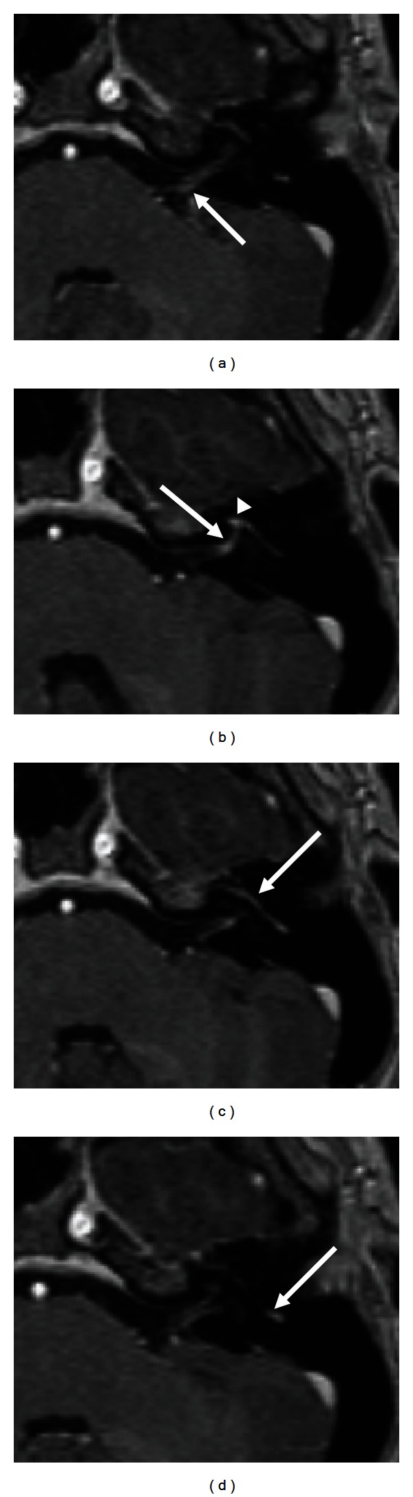
MRIT1-weighted MRI after gadolinium injection: abnormal enhancement of the facial nerve along its course, arrows, in Bell's palsy.
Source : Gupta et al., Radiology research and practice, 2013, figure 4 · CC BY
🚩 The triage that saves: forehead affected or spared?
This is the clinical sign that distinguishes Bell's palsy (benign) from a stroke (emergency). It rests on the anatomy of forehead innervation.
A patient who can still wrinkle the forehead and close the eye tightly on the affected side must be assessed for a central lesion. Source: Dalrymple et al., 2023 (PMID 37054419).
Faced with a drooping hemiface, the first decision is not a rehabilitation one: it is diagnostic. Not every acute facial palsy is Bell's palsy, and confusing peripheral involvement (the facial nerve itself) with central involvement (the upper motor neurone, upstream of the nucleus) can mean missing a life-threatening emergency. The physiotherapist, often on the front line or close behind it, must be able to recognise what falls outside the picture and redirect without delay.
Idiopathic peripheral facial palsy (Bell's palsy, or "a frigore" palsy) remains the most frequent cause. Its annual incidence is estimated at between 11.5 and 53.3 cases per 100,000 people depending on the population 2, a range that other sources narrow to around 20 to 30 per 100,000 1. It is the most common acute mononeuropathy, and its dominant pathophysiological hypothesis is reactivation of herpes simplex virus type 1 around the geniculate ganglion 2.
The forehead settles it: the sign that separates peripheral from central
The key to triage lies in one neuroanatomical detail. The frontalis muscle receives bilateral cortical innervation: the lower part of the facial nucleus, which controls the lower and middle hemiface, is supplied only by crossed fibres from a single hemisphere 5. Practical consequence: a central (supranuclear) lesion paralyses the lower face but spares the forehead, whereas a peripheral lesion affects the whole hemiface, forehead included.
The clinical rule is explicit: a patient able to close the eye tightly and wrinkle the forehead on the paralysed side must be assessed for a central lesion 1. Conversely, Bell's palsy is defined as acute unilateral facial weakness involving the forehead (inability to raise the eyebrow or close the eye), in the absence of any other neurological abnormality 1. This "forehead affected + isolated" pattern is the foundation of the positive diagnosis.
| Feature | Peripheral (e.g. Bell's) | Central (e.g. stroke) |
|---|---|---|
| Forehead / eyebrow | Affected (no longer wrinkles, no longer raises the eyebrow) | Spared (still wrinkles the forehead) |
| Eye closure | Impaired (lagophthalmos possible) | Preserved |
| Extent | The whole hemiface | Lower and middle face |
| Other neurological signs | Absent (by definition) | Often present |
| Course of action | Management of facial palsy | Urgent opinion (stroke pathway) |
Grading severity: House-Brackmann and Sunnybrook
Once peripheral involvement is confirmed, it must be quantified, to establish a prognosis, guide rehabilitation and monitor progress. The reference scale is the House-Brackmann scale, described in 1985 and adopted as the standard by the American Academy of Otolaryngology: it grades facial nerve function into six grades, from grade I (normal function) to grade VI (complete palsy, no movement) 7.
Simple and robust for a global assessment, House-Brackmann nonetheless shows its limits when it comes to demonstrating synkinesis, those involuntary movements accompanying a voluntary movement. This is where the Sunnybrook scale comes in (Sunnybrook Facial Grading System), finer and regional, widely used in rehabilitation to quantify symmetry, voluntary movement and synkinesis 78. It is on the Sunnybrook, moreover, that the gains of specialised facial rehabilitation are measured: mime therapy improves symmetry by 20.4 points (95 % CI 10.4-30.4) in long-standing paresis 8.
Red flags: when it is not (only) Bell's palsy
Bell's palsy is a diagnosis of clinical exclusion : it presupposes acute unilateral involvement, including the forehead, with no other neurological sign 1. Anything that goes beyond this definition should raise the alarm and prompt referral.
Key points: the signals that call for a specialist or emergency opinion
- Forehead spared (a patient who can still wrinkle the forehead and close the eye well on the affected side) → points towards a central lesion (stroke, tumour): urgent opinion 1.
- Other associated neurological abnormalities : by definition, their presence excludes typical Bell's palsy and calls for assessment 1.
- Eye that does not close : risk of exposure keratitis, immediate eye protection, and an ophthalmological opinion if there are signs of keratitis 1.
The most concrete short-term risk is ocular. Loss of orbicularis oculi function leads to lagophthalmos and corneal exposure 6. Any patient unable to close the eye must be taught protective measures: artificial tears or lubricating gel, and taping the eyelid closed at night 1. If neglected, severe exposure keratitis may be complicated by loss of corneal sensation leading to a neurotrophic corneal ulcer 6 : signs of keratitis warrant referral to an ophthalmologist 1.
This eye-protection reflex is, moreover, one of the strong clinical practice recommendations (AAO-HNS), on the same footing as early oral corticosteroid therapy 9. It depends on no uncertainty: it is a systematic safety measure as soon as eye closure is impaired.
Reassuring without trivialising
Triage does not mean dramatising. The spontaneous prognosis of typical Bell's palsy is broadly favourable: more than two thirds of patients recover completely without treatment, and this rate reaches up to 90 % in children and pregnant women 1. In the largest published longitudinal series (2,570 peripheral palsies followed over 25 years, including 1,701 cases of Bell's palsy), recovery begins within 3 weeks in 85 % of patients 4.
This favourable natural history has two virtues: it allows reassurance, and it is a reminder not to over-treat. But it must not mask the minority who retain sequelae: close to one patient in three in Peitersen's series, with contractures in 17 % and synkinesis in 16 % 4. It is precisely this fringe that will justify, later in management, targeted rehabilitation and early medical treatment.
Diagnostic triage also directly determines the therapeutic window. The medical mainstay, early oral corticosteroid therapy, has a demonstrated effect only when started within 72 hours 10. Confusing central involvement with Bell's palsy, or being slow to identify a genuine peripheral palsy, therefore means either delaying an emergency or missing the window in which treatment changes the prognosis. The right initial move is not to rehabilitate: it is to triage quickly and well.
📈 What prognosis? What can be said to the patient
📊 Confirmed at the highest level of evidence
Cochrane meta-analysis, 7 randomised trials, 895 participants. Proportion of patients with INCOMPLETE recovery at 6 months or more.
Relative risk 0.63 (95 % CI 0.50–0.80). Ten patients need to be treated with corticosteroids to prevent one incomplete recovery (NNT = 10). Source: Madhok et al., Cochrane review 2016 (PMID 27428352).
This is the patient's first question, often asked with the face still frozen and the fear of "staying like this" deep in their eyes. The honest answer comes in two parts: the prognosis of idiopathic peripheral facial palsy (Bell's palsy) is broadly favourable, but it is not universal. Our role is neither to promise full recovery nor to dramatise: it is to give reliable figures, credible timeframes, and to name the possible sequelae so that the patient knows what to expect, and so that rehabilitation finds its proper place.
A favourable spontaneous prognosis
Bell's palsy is the most common acute mononeuropathy, with an incidence estimated at between 11.5 and 53.3 cases per 100,000 people per year depending on the population 2. Its natural history is rather reassuring: more than two thirds of patients with a typical form recover completely, with no treatment at all 1. Most cases resolve within 4 to 6 months and almost always resolve completely within a year 2.
Some profiles do even better: in children and pregnant women, the rate of complete recovery reaches up to 90 % 1. This figure is important to know, not in order to trivialise, but to avoid over-treating cases destined to recover on their own and to reassure with solid data rather than empty phrases.
Timeframes: when recovery begins
The largest published longitudinal series is the reference here: 2,570 peripheral facial palsies followed over 25 years, including 1,701 cases of Bell's palsy 4. It provides a valuable timeline for guiding the patient:
- In 85 % of patients, recovery begins within the first 3 weeks ;
- In the remaining 15 %, it starts later, between 3 and 5 months.
This time to onset is itself a prognostic indicator: an early return of movement, within the first month, generally heralds a favourable course. Conversely, a face that remains completely frozen beyond three weeks calls for closer monitoring and for not promising a return to normal. In practical terms, you can tell the patient: "In the great majority of cases, the first signs of recovery appear within three weeks. If they are slow to come, it does not mean that nothing will move, but recovery will be longer and sometimes incomplete."
Factors that weigh on the prognosis
Several elements modulate the course, and it is useful to share them honestly with the patient rather than let them believe it is a lottery:
- Initial severity. It is graded with the House-Brackmann scale, the historical reference validated by the American Academy of Otolaryngology, which grades facial nerve function from I (normal function) to VI (complete palsy, no movement) 7. The more complete the initial involvement, the greater the risk of imperfect recovery; it is this initial grade that shapes follow-up.
- Patient characteristics. Young age and pregnancy are associated with better rates of complete recovery 1.
- How early medical treatment is started. Oral corticosteroid therapy started within 72 hours concretely alters the prognosis (see below).
- Time to return of movement, described above: an early return is a good sign.
One point is worth repeating to the anxious patient, because it governs safety: Bell's palsy affects the whole hemiface, forehead included. A patient who retains the ability to wrinkle the forehead and close the eye tightly on the affected side does not fit this diagnosis and must be assessed for a central lesion (stroke, tumour), which spares the forehead thanks to the bilateral cortical innervation of the frontalis muscles 15. This is not a prognostic factor for Bell's palsy, but a red flag that radically changes management.
Sequelae and synkinesis: close to one patient in three
This is the part one is sometimes tempted to leave unsaid, and which on the contrary must be stated clearly. In Peitersen's series 4, normal facial movement is found in 71 % of patients. In other words, close to one patient in three retains a sequela. In detail:
| Long-term outcome 4 | Proportion |
|---|---|
| Normal facial movement | 71 % |
| Mild sequelae | 12 % |
| Moderate sequelae | 13 % |
| Severe sequelae | 4 % |
| Contractures | 17 % |
| Associated movements (synkinesis) | 16 % |
The synkinesis is the emblematic sequela of peripheral facial palsy: an involuntary movement accompanying a voluntary one: the eye that closes when the patient smiles, the corner of the mouth that pulls when they blink. It results from aberrant reinnervation of the facial nerve during regrowth: regenerated fibres do not always find their original muscle again 3. It is precisely this late complication that rehabilitation seeks to prevent, then to correct once established. It can be explained simply to the patient: "The nerve will grow back, but it can take the wrong path. That is what produces these involuntary movements; we now know how to limit them."
What treatment changes about the prognosis
The prognosis is not fixed: early treatment shifts it. Oral corticosteroid therapy started within 72 hours is the recognised mainstay. In Sullivan's landmark double-blind randomised trial 10, complete recovery at 3 months was 83.0 % with prednisolone versus 63.6 % without, and 94.4 % versus 81.6 % at 9 months. The Cochrane review confirms this at the highest level of evidence: 17 % incomplete recovery with corticosteroids versus 28 % without (relative risk 0.63; 95 % CI 0.50–0.80), that is about 10 patients to treat to prevent one incomplete recovery 11.
A frequently forgotten fact, and one directly relevant to the physiotherapist: corticosteroids also reduce long-term motor synkinesis (RR 0.64; 95 % CI 0.45–0.91), that is the very sequela that rehabilitation will then seek to contain 11. Aciclovir alone provides no benefit 10, and antivirals can only be considered as an add-on to corticosteroids, where they might reduce long-term sequelae (synkinesis, "crocodile tears": RR 0.56; 95 % CI 0.36–0.87) without clearly improving the recovery rate 12.
On the rehabilitation side, there is a genuine role but the level of evidence must be stated without overselling it: the Cochrane review finds low-quality evidence that individualised ("tailored") facial exercises improve function, particularly in moderate palsies and chronic cases 13. A major point for prognosis: these targeted exercises significantly reduced the occurrence of synkinesis after an acute palsy (RR 0.24; 95 % CI 0.08–0.69) 13. In long-standing paresis, mime therapy (massage, relaxation, inhibition of synkinesis, coordination and expression) improves facial symmetry by 20.4 points on the Sunnybrook scale (95 % CI 10.4–30.4) 8. And once synkinesis is established, botulinum toxin type A is a reference treatment, alongside rehabilitation 314.
Key points, what can honestly be said to the patient
- "Most people recover" : more than 2 patients in 3 recover completely, up to 90 % in children and pregnant women 1.
- "It often starts quickly" : recovery begins within 3 weeks in 85 % of patients, later (3–5 months) in the rest 4.
- "But not everyone goes back to how they were" : 71 % regain a normal face; close to one in three retains a sequela, synkinesis being the most frequent 4.
- "Early treatment counts" : corticosteroids within 72 h markedly increase the chances of complete recovery and reduce synkinesis 1011.
- "Rehabilitation has a role, a targeted one" : individualised exercises and mime therapy for function and symmetry, prevention of synkinesis, with a modest level of evidence to be stated honestly 138.
💊 Corticosteroids and antivirals: what does the evidence say?
💊 Early prednisolone changes recovery: aciclovir does not
Landmark double-blind randomised trial. Treatment started within 72 hours. Rates of complete recovery of facial function.
Aciclovir, for its part, provides no significant benefit (71.2 % versus 75.7 % at 3 months; p = 0.50). Source: Sullivan et al., NEJM 2007 (PMID 17942873).
Bell's palsy is the most common acute mononeuropathy, with an annual incidence estimated at between 11.5 and 53.3 cases per 100,000 people depending on the population 2. Its spontaneous prognosis is broadly favourable, more than two thirds of patients recover completely without treatment, and this rate reaches 90 % in children and pregnant women 1. Yet this reassuring natural history must not obscure the fact that there is a narrow therapeutic window, and that the patient's fate, complete recovery or lifelong sequelae, is largely decided in the first 72 hours. The physiotherapist, often placed as an advanced observer along the care pathway, has a decisive role: recognising the urgency and directing the patient to medical prescription without delay.
Key points
- Early oral corticosteroids (< 72 h) = the mainstay of medical treatment, with benefit demonstrated at the highest level of evidence (NNT = 10).
- Antivirals alone : ineffective and not recommended. Added to corticosteroids, their effect is limited, mainly on sequelae.
- The physiotherapist is not a prescriber, but is on the front line to trigger urgent referral and to check eye protection.
- Dominant pathophysiology: reactivation of herpes simplex virus type 1 (HSV-1) around the geniculate ganglion 2.
Early corticosteroids: the demonstrated mainstay
Oral corticosteroid therapy started early is the first-line treatment. The landmark Scottish double-blind randomised trial 10compared prednisolone, aciclovir, the combination of both and placebo, in patients treated within 72 hours of symptom onset. The result is clear-cut: recovery of normal facial function at 3 months was 83.0 % with prednisolone versus 63.6 % without, and at 9 months 94.4 % versus 81.6 % (p < 0.001). In other words, treating early with corticosteroids shifts nearly one patient in five from residual weakness to complete recovery.
This efficacy is confirmed at the highest level of evidence by the Cochrane meta-analysis 11 : 17 % (79/452) incomplete recovery with corticosteroids versus 28 % (125/447) in the control group, a relative risk of 0.63 (95 % CI 0.50-0.80), with no excess of adverse effects. The number needed to treat to prevent one incomplete recovery is 10.
A crucial point for the rehabilitation practitioner: corticosteroids do not only prevent motor deficit, they also reduce long-term motor synkinesis at follow-up 11. And these involuntary co-contractions, frequent sequelae of aberrant reinnervation, are precisely the target that physiotherapy will subsequently try to limit. Well-conducted corticosteroid therapy upstream therefore means less desynkinesis work downstream. The reference clinical practice guideline 9 is explicit: clinicians should prescribe oral corticosteroids within 72 hours of symptom onset.
Antivirals: a debated and overestimated benefit
The pathophysiological rationale, herpes reactivation, long raised hopes of a benefit from antivirals. The evidence has tempered that hope. In Sullivan's trial 10, aciclovir alone provided no significant benefit : 71.2 % recovery versus 75.7 % without aciclovir (adjusted p = 0.50). Antiviral monotherapy is therefore probably inferior to the corticosteroid alone 12.
That leaves the question of the combination. The dedicated Cochrane review 12 shows that adding an antiviral to corticosteroids has little or no effect on the rate of incomplete recovery compared with corticosteroids alone (RR 0.81; 95 % CI 0.38-1.74; 3 trials, N = 766; low-certainty evidence). Here one must remain honest about the uncertainty: the wide confidence interval reflects inconclusive data, not proof of uselessness. There is, however, a signal on another outcome: antivirals plus corticosteroids reduce the proportion of patients developing long-term sequelae (synkinesis, gustatory lacrimation known as "crocodile tears"), compared with placebo plus corticosteroids 12.
The practical synthesis: the corticosteroid is the mainstay, the antiviral a possible add-on whose interest lies more in preventing sequelae than in motor recovery. The Baugh guideline 9 is clear on the point that matters most for triage: clinicians should not prescribe an antiviral alone.
| Treatment | Effect on recovery | Level of evidence |
|---|---|---|
| Early corticosteroids (< 72 h) | Clear benefit: 17 % vs 28 % incomplete recovery (RR 0.63), NNT 10 | High 11 |
| Antiviral alone | No demonstrated benefit (71.2 % vs 75.7 %), inferior to the corticosteroid | Low / not recommended 1012 |
| Antiviral + corticosteroid vs corticosteroid alone | Little/no effect on recovery (RR 0.81); reduction in late sequelae (RR 0.56) | Low to moderate 12 |
What the physiotherapist needs to know: the 72-hour urgency
The operational message fits in one sentence: medical treatment of Bell's palsy is a relative emergency. The 72-hour window is not an administrative convenience, it is the condition under which corticosteroids have demonstrated efficacy. A patient who consults their physiotherapist for a "drooping mouth" or an eye that no longer closes, which appeared the day before, must not be left waiting: they need a medical opinion the same day so that corticosteroid therapy can be started in time. Losing 48 hours "observing" means risking falling outside the window in which the benefit exists.
Two clinical reflexes frame this referral.
1. The peripheral/central red flag. Bell's palsy affects the whole hemiface, forehead included: the patient can neither raise the eyebrow nor close the eye on the affected side. The frontalis muscle receives bilateral cortical innervation: a patient able to wrinkle the forehead and close the eye tightly on the paralysed side does not have peripheral involvement but a central lesion (stroke, tumour), which calls for urgent management of a completely different nature 51. This simple test of the spared forehead must be systematic: it radically changes the referral pathway.
2. Eye protection. Any patient unable to close the eye must be taught protective measures without delay: artificial tears or lubricating gel during the day, taping the eyelid closed at night 19. Loss of orbicularis oculi function leads to lagophthalmos and corneal exposure; severe exposure keratitis may progress to a neurotrophic corneal ulcer through loss of sensation 6. This is a preventable complication, and the physiotherapist is often the first professional to see the patient regularly again: checking eye protection at every session is a safety measure, not a detail.
Placing rehabilitation within the care strategy
Understanding the medical treatment also means understanding what physiotherapy does not have to do. Since more than two thirds of typical Bell's palsies resolve spontaneously 1 and corticosteroids improve this prognosis further, rehabilitation does not aim to "bring about recovery" in cases destined to resolve on their own: it targets severe forms, chronic cases and the prevention of synkinesis. The Cochrane review on physiotherapy 13 finds no high-quality evidence, but low-quality evidence that tailored facial exercises improve function, and a notable signal on prevention: significantly fewer patients developed synkinesis after targeted exercises (RR 0.24; 95 % CI 0.08-0.69). The Baugh guideline 9 remains cautious: for lack of sufficient evidence, no formal recommendation can be made on physiotherapy. This should be told to the patient honestly, without overpromising.
Medical treatment and rehabilitation are therefore not in opposition: they take over from each other. Corticosteroids early to maximise recovery and reduce synkinesis; then, in forms that leave sequelae, specialised facial rehabilitation focused on symmetry and desynkinesis. Recognising the 72-hour urgency and referring quickly remains, for the physiotherapist, the most evidence-based contribution at this early stage.
🎭 Which facial rehabilitation should be offered?
🎭 Specialised facial rehabilitation: a real effect on symmetry
"Mime therapy" (massage, relaxation, inhibition of synkinesis, coordination and expression) in patients with long-standing paresis.
A gain of 20.4 points on the Sunnybrook facial symmetry scale (95 % CI 10.4–30.4) versus control. To be reserved for TAILORED, fine exercises, not for maximal or global analytical exercises, which promote synkinesis. Source: Beurskens et al., 2006 (PMID 16942452).
The question of rehabilitation in peripheral facial palsy must be framed clearly. The spontaneous prognosis is broadly favourable: more than two thirds of patients with typical Bell's palsy recover completely, and the rate reaches up to 90 % in children and pregnant women 1. In the largest published longitudinal series (2,570 palsies followed over 25 years), normal facial movement is found in 71 % of patients, with recovery beginning within 3 weeks in 85 % of them 4. In other words, a large share of palsies resolve on their own, and rehabilitation must never be presented as the driver of this natural recovery.
The fact remains that close to one patient in three retains a sequela: contractures (17 %), synkinesis (16 %), mild to severe residual paresis 4. This is precisely where facial physiotherapy has its place: less in the recovery of cases destined to resolve than in moderate to severe forms, chronic cases, and above all the prevention and treatment of sequelae.
What the evidence says: honest and modest
It must be stated plainly to the reader: facial rehabilitation has no high-level evidence behind it. The dedicated Cochrane review finds no high-quality evidence of benefit, or of harm, from physical therapies taken as a whole in idiopathic facial palsy 13. The American clinical practice guidelines point the same way: for lack of sufficiently good evidence, no formal recommendation can be made on the place of physiotherapy 9.
This caution does not mean "do nothing". The same Cochrane review finds low-quality evidence that tailored facial exercises improve facial function, particularly in moderate palsies and chronic cases, and reduce sequelae in acute cases 13. The key word is tailored : these are targeted, guided exercises, the opposite of maximal, forced or global analytical work, which has no basis and could maintain co-contractions.
The most interesting result of this review concerns prevention: significantly fewer patients developed motor synkinesis after a programme of targeted exercises (relative risk 0.24; 95 % CI 0.08–0.69) 13. The aim of rehabilitation is therefore as much the prevention of aberrant reinnervation as pure motor recovery.
Key points
- Most cases of Bell's palsy resolve spontaneously (complete recovery in more than two thirds of patients): do not over-treat, know how to reassure.
- The medical mainstay remains early oral corticosteroid therapy (within 72 h); antivirals alone are not recommended.
- Rehabilitation has only low-quality evidence: tailored facial exercises, not maximal global exercises.
- Two concrete targets: moderate/chronic forms and prevention/treatment of synkinesis.
- Absolute priority before any exercise: eye protection if eye closure is impaired.
Facial neuromuscular therapy (mime therapy)
The best-supported specialised facial rehabilitation is Beurskens' mime therapy : a neuromuscular approach combining massage, relaxation, inhibition of synkinesis, and exercises in coordination and emotional expression. In a randomised controlled trial in 50 patients with long-standing paresis (more than 9 months), three months of mime therapy improved facial symmetry by 20.4 points on the Sunnybrook Facial Grading System (95 % CI 10.4–30.4) and reduced severity by 0.6 grade on the House-Brackmann scale (95 % CI 0.1–1.1), independently of age, sex and duration of the paresis 8.
This is the evidence base for sequelae-oriented facial rehabilitation, and it concerns long-standing paresis, which usefully reframes the timeline: neuromuscular therapy retains value even long after the acute episode, in patients one might think of as "stable".
This approach fits into a now formalised therapeutic hierarchy. The recommendations of an international ENT scientific group place, for post-paralytic synkinesis, facial rehabilitation (biofeedback) as first line, followed by botulinum toxin, with surgery reserved for first-line failures 15. Physiotherapy is therefore the foundation, and toxin a targeted add-on, not the other way round.
Biofeedback and fine analytical work
The guiding principle of analytical work in facial rehabilitation is not strength but control. Faced with a muscle recovering in a disorganised way, the aim is to restore selective, symmetrical movements while inhibiting parasitic co-contractions. Biofeedback, visual (mirror) or electromyographic, supports this learning of dissociation: relearning how to move one region of the face without dragging another along with it.
This framework explains why mime therapy rests as much on inhibition of synkinesis and on relaxation as on strengthening: in a patient whose eye closes when smiling, asking for "more smile, harder" worsens the synkinesis. Fine work means breaking down, slowing down, dissociating: a logic consistent with the Cochrane signal favouring tailored exercises over maximal ones 13.
Progression: when and how?
Progression is reasoned according to phase and severity, graded with standardised scales. The House-Brackmann scale, the historical reference validated by the American Academy of Otolaryngology, grades facial nerve function from I (normal) to VI (complete palsy) 7. The Sunnybrook scale, more sensitive to synkinesis, is widely used in rehabilitation for fine-grained monitoring.
- Acute / flaccid phase : priority to eye protection and education; no forced analytical exercises. The aim is to avoid maintaining abnormal patterns while reinnervation is taking place.
- Recovery phase (moderate forms) : introduction of tailored, targeted facial exercises with feedback, to improve function and prevent synkinesis 13.
- Sequelae / chronic phase : neuromuscular therapy of the mime therapy type, centred on symmetry and inhibition of established synkinesis 8 ; recourse to botulinum toxin as an add-on if necessary.
Established sequelae: the place of botulinum toxin
Synkinesis that is already established (for example involuntary eye closure on smiling) results from aberrant reinnervation and responds poorly to exercises alone. Botulinum toxin type A is the reference treatment here, alongside rehabilitation. A systematic review with meta-analysis (34 studies, 1,314 patients) finds a significant improvement in synkinesis scores measured with the Synkinesis Assessment Questionnaire (mean difference 11.6; p < 0,01), les auteurs recommandant un traitement individualised rather than a standardised protocol 3. A clinical trial confirms this functional improvement, with a fall in the SAQ synkinesis score from 46.22 to 37.55 after injection (p = 0.001) and an improvement in quality of life 14.
Electrical stimulation: stating the uncertainty
This is the most disputed point, and it would be dishonest to settle it. The guidelines do not recommend electrical stimulation, and the historical Cochrane review found no benefit from it on incomplete recovery at six months compared with placebo 13. The classic fear is that, applied in the acute phase, it promotes synkinesis.
Recent data qualify this picture without overturning it. One meta-analysis finds that, added to usual care within 7 days, electrical stimulation reduces the risk of incomplete recovery (RR 0.65; 95 % CI 0.48–0.88; moderate certainty), while stressing that its efficacy and safety in the acute phase remain controversial and insufficiently studied 16. Other syntheses point towards a possible benefit on overall function without a significant excess risk of synkinesis, but with marked heterogeneity in the measures used 1718.
The honest conclusion for practice: the data are contradictory and the level of evidence insufficient to recommend electrical stimulation in routine practice. Where some teams see an encouraging signal with early, parameterised application, others find no difference in synkinesis. Caution, patient information, and no systematic use.
| Modality | What the evidence says | Level |
|---|---|---|
| Early corticosteroids (medical context) | Clear benefit on complete recovery 1011 | High |
| Mime therapy (long-standing paresis) | +20.4 pts Sunnybrook vs control 8 | Moderate |
| Tailored facial exercises | Improve function; reduce synkinesis (RR 0.24) 13 | Low |
| Botulinum toxin A (established synkinesis) | Significant reduction in synkinesis 314 | Moderate |
| Electrical stimulation | Contradictory data; not recommended in routine practice 1316 | Uncertain |
Never forget: the eye first, and the red flag
Before any exercise programme, one priority dominates: eye protection. Any patient unable to close the eye must be taught protective measures (artificial tears or lubricating gel, taping the eyelid closed at night), because loss of orbicularis function leads to lagophthalmos and corneal exposure, which may be complicated by exposure keratitis and then a neurotrophic corneal ulcer 16. No rehabilitation session takes precedence over this instruction.
Finally, the physiotherapist must keep the diagnostic red flag in mind. Peripheral facial palsy affects the whole hemiface, forehead included. A patient able to wrinkle the forehead and close the eye tightly on the affected side, forehead spared, has central involvement (stroke, tumour) and not Bell's palsy, because the frontalis muscle receives bilateral cortical innervation 15. This abnormality calls for an urgent opinion, not rehabilitation.
⚡ Synkinesis: preventing it, and the mistakes not to make
Motor recovery is not the only issue in peripheral facial palsy. In close to one patient in three, healing leaves sequelae, and the most frequent of them has a name: synkinesis. In the largest published longitudinal series (2,570 peripheral facial palsies followed over 25 years), associated movements were found in 16 % of patients and contractures in 17 % 4. It is precisely here that rehabilitation plays its subtlest part: not pushing the muscle to work harder, but helping it to reorganise correctly. And it is also here that certain well-intentioned habits do the most damage.
The mechanism: reinnervation that goes to the wrong address
Synkinesis is an involuntary movement that accompanies a voluntary one: the eye that closes when the patient smiles, the corner of the mouth that contracts when they blink, the cheek that tightens on swallowing. It results from aberrant reinnervation of the facial nerve 3. After axonal injury, nerve fibres grow back, but they do not always find their original target: axons destined for orbicularis oculi reinnervate fibres in the perioral region, and vice versa. The result is a genuine wiring error: a single motor intention now activates several muscle territories that should remain independent.
This pathophysiological fact is decisive for rehabilitation. Unlike ordinary muscle weakness, synkinesis is not a deficit of strength: it is a failure of selectivity. A synkinetic muscle is often overactive, not lazy. The whole logic of intensive analytical strengthening is thereby overturned.
Key points
- Synkinesis is the most frequent sequela of peripheral facial palsy: associated movements in 16 % of patients, contractures in 17 % 4.
- It reflects aberrant reinnervation of the facial nerve: a failure of selectivity, not of strength 3.
- Prevention: targeted and tailored facial exercises; they significantly reduce the occurrence of synkinesis 13.
- Early corticosteroids also reduce long-term motor synkinesis 11.
- Established synkinesis: facial rehabilitation as first line, botulinum toxin type A as a targeted add-on 153.
Prevention: gentle, selective and early rehabilitation
Good news: physiotherapy can prevent synkinesis, provided it is designed in the right spirit. The Cochrane review devoted to rehabilitation in Bell's palsy reports a major result for prevention: significantly fewer patients developed motor synkinesis after a programme of targeted facial exercises, with a relative risk of 0.24 (95 % CI 0.08–0.69) 13. Well-conducted exercise therefore divides the risk of this sequela by four in the cases concerned.
The key word is tailored: made to measure. The same review finds low-quality evidence that tailored facial exercises improve function, particularly in moderate palsies and chronic cases 13. It is therefore not a matter of working the face "in general", but of training precise, isolated movements below the threshold that triggers co-contractions. Rehabilitation aims to teach the brain to command each territory separately again.
Beurskens' mime therapy embodies this philosophy. It is a facial neuromuscular rehabilitation combining massage, relaxation, inhibition of synkinesis, and exercises in coordination and emotional expression. In a randomised controlled trial in 50 patients with long-standing facial paresis (more than 9 months), three months of mime therapy improved facial symmetry by 20.4 points on the Sunnybrook Facial Grading System (95 % CI 10.4–30.4) and reduced severity by 0.6 grade on the House-Brackmann scale (95 % CI 0.1–1.1), independently of age, sex and duration 8. This protocol explicitly includes a synkinesis-inhibition component: it is not strengthening, it is retraining of control.
Prevention even begins before physiotherapy. Early oral corticosteroids, the mainstay of medical treatment, do not merely improve overall motor recovery: the Cochrane meta-analysis shows that they also reduce long-term motor synkinesis (RR 0.64; 95 % CI 0.45–0.91) 11. Adding an antiviral to the corticosteroid points the same way for late sequelae, synkinesis and "crocodile tears", with an RR of 0.56 (95 % CI 0.36–0.87) versus corticosteroid alone, even though its effect on incomplete recovery remains little or not demonstrated 12. The prevention window therefore opens in the very first days, and early medical management already does part of the work.
Treating established synkinesis: physiotherapy first, toxin next
When synkinesis is already established, the therapeutic hierarchy is well established. The practice recommendations of an international ENT scientific group place facial rehabilitation (biofeedback) as first line, followed by botulinum toxin, with surgery reserved for first-line failures 15. Physiotherapy remains the foundation; toxin is a targeted add-on, not a shortcut.
The botulinum toxin type A is the reference treatment for residual synkinesis. A systematic review with meta-analysis (34 studies, 1,314 patients) finds a significant improvement in synkinesis scores on the Synkinesis Assessment Questionnaire (mean difference 11.6; p < 0,01), avec des effets indésirables généralement légers et transitoires 3. A further clinical trial confirms the fall in SAQ score after injection (from 46.22 to 37.55; p = 0.001), with gains in muscle function and quality of life 14. A crucial point stressed by the authors: treatment must be individualised, muscle by muscle, rather than standardised 3. The toxin selectively weakens the muscle that contracts wrongly; combined with rehabilitation, it restores a balance that strengthening alone could not achieve.
The mistakes not to make
This is the most important side of this section, because rehabilitation errors are not neutral: they can worsen the very sequela they claim to treat.
Mistake no. 1: forced, maximal exercises. The intuitive logic of "the harder I push, the more I recover" is counterproductive here. The synkinetic muscle is overactive, and a maximal contraction massively recruits the aberrantly reinnervated fibres: it therefore reinforces the abnormal co-contraction instead of undoing it. All the available evidence argues for the opposite: the Cochrane review finds benefit only for targeted and tailored exercises, not for intensive analytical work 13, and it is gentle training, below the synkinetic threshold, that divides the risk of sequelae by four (RR 0.24). The aim is never brute strength, it is selectivity.
Mistake no. 2: global, uncontrolled movements. Asking the patient to "pull all their faces" or to move the whole hemiface as a block maintains exactly the pattern we want to dissolve. As synkinesis is a failure of separation between territories, rehabilitation must fractionate: isolate the eye, isolate the mouth, work each muscle independently, with visual feedback (mirror, biofeedback). Global movement reactivates the aberrant common command; segmental movement dismantles it. Mime therapy rests entirely on this fine, coordinated approach, never on massive effort 8.
Mistake no. 3: rehabilitating too hard, too early, on a face that is still paralysed. In the flaccid phase there is nothing to "build up": the nerve is not yet conducting. Intensively working a paralysed face mainly exposes the patient to overuse of the healthy contralateral muscles and to asymmetrical compensations. The priority of this phase lies elsewhere: eye protection. Any patient unable to close the eye must be given artificial tears or lubricating gel and night-time eyelid taping, failing which exposure keratitis may develop and progress to a neurotrophic corneal ulcer 16. Forgetting the eye in order to concentrate on the muscle is the mistake with the gravest consequences.
Mistake no. 4: overlooking the diagnostic red flag. Before any rehabilitation, let us recall that peripheral palsy affects the whole hemiface, forehead included. A patient who can still wrinkle the forehead and close the eye tightly on the affected side has a central lesion (stroke, tumour), not Bell's palsy: this is a referral emergency, not a physiotherapy case 15.
The electrical stimulation controversy: not settling it too quickly
Should a paralysed face be electrically stimulated? The question divides opinion, and honesty demands that we say so to the reader rather than assert. The historical fear is that electrical stimulation, in the acute phase, promotes synkinesis by maintaining disorganised activation during reinnervation. This caution retains a solid basis: the reference Cochrane review finds no benefit of electrical stimulation on incomplete recovery at six months compared with placebo 13, and the clinical practice guidelines do not recommend it: they conclude, moreover, that no firm recommendation can be made on physiotherapy for lack of sufficient evidence 9.
Recent data nevertheless qualify the picture without overturning it. One meta-analysis (13 trials, n = 1,191) reports that, added to usual care within 7 days, electrical stimulation would reduce the risk of incomplete recovery (RR 0.65; 95 % CI 0.48–0.88; moderate certainty), while judging its efficacy and safety in the acute phase "controversial" and insufficiently studied 16. Another systematic review (6 studies, 243 patients) suggests a possible benefit on overall facial function without a significant excess risk of synkinesis, 3 studies out of 5 showing no difference, but stresses the heterogeneity of the measures 17. A targeted synthesis likewise concludes that results are promising, including a reduction in synkinesis, when stimulation is applied early with suitable parameters (low frequency, daily), while underlining the limited data 18.
Where does that leave us? On uncertain ground, acknowledged as such. The literature is contradictory: the benefit signal exists, the synkinesis danger signal is neither confirmed nor definitively ruled out, and the quality of evidence remains heterogeneous. The reasonable position is therefore neither to ban nor to generalise electrical stimulation, but to handle it with caution : outside routine recommendation, never as a substitute for targeted motor retraining, which has the most direct evidence for preventing synkinesis 13.
| Approach | Intended aim | Level of evidence |
|---|---|---|
| Early corticosteroids (< 72 h) | Recovery + reduction in motor synkinesis (RR 0.64) | High 11 |
| Targeted facial exercises / mime therapy | Prevention of synkinesis (RR 0.24) and symmetry (+20.4 pts Sunnybrook) | Low but consistent 138 |
| Botulinum toxin type A | Established synkinesis (SAQ improvement ≈ 11.6) | Moderate, individualised 3 |
| Electrical stimulation | Debated: possible benefit, synkinesis risk unresolved | Contradictory, caution 1613 |
The guiding line fits in one sentence: in facial palsy, we do not rehabilitate strength, we rehabilitate control. Gentleness, selectivity and early treatment prevent synkinesis; excessive force and global movement create it.
🗂️ What do concrete clinical cases teach us?
To make the reasoning tangible, here are two illustrative and deliberately fictional cases. They describe no real patient: they are teaching scenarios built solely from the evidence presented above. Their purpose is to show how the evidence translates into clinical decisions, from the first consultation to the management of sequelae: peripheral/central triage, eye protection, choice of rehabilitation and prevention of synkinesis.
Key points
- The first reflex is not therapeutic but diagnostic: check the forehead. If it is affected, the palsy is peripheral; if it is spared, think of a central cause 1.
- Any eye that does not close calls for immediate eye protection to prevent exposure keratitis 61.
- In the acute phase, the medical mainstay is early oral corticosteroid therapy (< 72 h); the physiotherapist's role is mainly to prevent synkinesis through targeted exercises 1013.
- With an established sequela, the aim changes: rehabilitate symmetry and inhibit synkinesis (mime therapy, botulinum toxin as an add-on) 815.
Case 1 (fictional): An acute palsy: triage before treating
Let us imagine "Mrs L.", 42 years old, who presents with sudden distortion of the right hemiface that appeared on waking: the corner of the mouth droops, the smile is asymmetrical, the right eye waters and no longer closes completely. This picture is common: Bell's palsy is the most frequent acute mononeuropathy, with an incidence of the order of 20 to 30 cases per 100,000 people per year 12.
Triage first. Before any treatment, the decisive question is that of the central red flag. Mrs L. is asked to wrinkle the forehead and to close the eyes tightly. On the affected side she can neither raise the eyebrow nor close the eyelid: the involvement concerns the whole hemiface, forehead included. This is the signature of a peripheral lesion. If, conversely, she had retained the ability to wrinkle the forehead on the paralysed side, a central lesion (stroke, tumour) would have had to be suspected and an urgent opinion arranged, since the frontalis muscle receives bilateral cortical innervation, it is spared in supranuclear lesions 51.
The rest of the neurological examination is normal, with no other deficit: the diagnosis made is idiopathic peripheral facial palsy, whose dominant pathophysiological hypothesis is reactivation of herpes simplex virus type 1 around the geniculate ganglion 2. Severity is graded with the House-Brackmann scale, the six-grade reference, from grade I (normal) to grade VI (no movement) 7 : Mrs L. is graded IV.
Protect the eye, immediately. As orbicularis oculi is no longer working, there is lagophthalmos with corneal exposure. Loss of eyelid closure exposes the patient to exposure keratitis, which may be complicated, if corneal sensation is lost, by a neurotrophic ulcer 6. The patient is therefore taught immediately: artificial tears during the day, lubricating gel, and taping the eyelid closed at night 1. This simple measure takes precedence over everything else and is not up for discussion.
First-line medical treatment. We are less than 72 hours from symptom onset: the window for early oral corticosteroid therapy is open. The doctor prescribes prednisolone. The benefit is solid: in the landmark double-blind randomised trial, complete recovery at 3 months was 83.0 % with prednisolone versus 63.6 % without, and 94.4 % versus 81.6 % at 9 months 10. The Cochrane meta-analysis confirms this at the highest level of evidence: 17 % incomplete recovery with corticosteroids versus 28 % in the control group (RR 0.63; 95 % CI 0.50–0.80), that is a number needed to treat of 10 11.
And antivirals? The patient is told that an antiviral alone is not justified: aciclovir on its own provides no benefit 10, and corticosteroids alone are probably more effective than antivirals alone 12. Adding an antiviral to the corticosteroid has little or no effect on incomplete recovery 12. It might, however, reduce long-term sequelae (synkinesis, crocodile tears), compared with the corticosteroid alone 12. The antiviral can therefore only be considered, if at all, as an add-on, never as monotherapy.
The physiotherapist's role at this stage. The spontaneous prognosis is broadly favourable: more than two thirds of typical Bell's palsies recover completely without treatment, up to 90 % in children and pregnant women 1. There is therefore no question of over-treating. The aim of rehabilitation in the acute phase is twofold: to support motor recovery and, above all, to prevent synkinesis. The Cochrane review reports low-quality evidence that tailored facial exercises improve function, and an important signal: significantly fewer patients developed motor synkinesis after exercises 13. Targeted, gentle exercises guided in front of a mirror are therefore preferred to maximal or global analytical movements. The patient is honestly informed that this level of evidence remains modest and that the official guidelines do not settle the question of physiotherapy for lack of sufficiently good evidence 9.
Mrs L.'s recovery begins within three weeks, as in 85 % of patients 4, and she regains normal facial movement.
Case 2 (fictional): An established sequela: the time of synkinesis
Let us now imagine "Mr B.", 55 years old, seen eleven months after a left peripheral facial palsy. Movement has broadly returned, but he describes a persistent nuisance: when he smiles, his left eye partly closes; when he closes his eyes, the corner of his mouth contracts. These are synkinesis: involuntary co-contractions linked to aberrant reinnervation of the facial nerve 3. This scenario is by no means anecdotal: in Peitersen's large longitudinal series, associated movements persisted in 16 % of patients and contractures in 17 %, a reminder that close to one patient in three retains a sequela, synkinesis being the most frequent 4.
Re-grade with the right scale. The House-Brackmann scale, useful in the acute phase, is not very sensitive to synkinesis. Here the Sunnybrook scale is preferred, being finer and quantifying separately resting symmetry, voluntary movement and, precisely, synkinesis 78. It will serve as the reference measure for demonstrating progress.
Specialised facial rehabilitation as first line. In this chronic patient, the recommended strategy is clear: the first-line treatment of synkinesis is facial rehabilitation (neuromuscular rehabilitation, biofeedback), followed by botulinum toxin; surgery is reserved for failures only 15. Physiotherapy is the foundation, toxin a targeted add-on.
Mr B. is therefore offered mime therapy : massage, relaxation, inhibition of synkinesis, and exercises in coordination and emotional expression. It is the best-supported intervention in long-standing paresis: in Beurskens' randomised controlled trial (patients with paresis of more than 9 months), three months of mime therapy improved facial symmetry by 20.4 points on the Sunnybrook (95 % CI 10.4–30.4) and reduced severity by 0.6 grade on House-Brackmann, independently of age, sex and duration 8.
Botulinum toxin as a targeted add-on. As Mr B.'s synkinesis remains troublesome despite rehabilitation, injection of botulinum toxin type A is discussed, the reference treatment for established synkinesis. A systematic review with meta-analysis (34 studies, 1,314 patients) shows a significant improvement in synkinesis scores measured with the Synkinesis Assessment Questionnaire (mean difference 11.6; p < 0,01), avec des effets indésirables généralement légers et transitoires; les auteurs insistent sur un individualised treatment rather than a standardised protocol 3. Another study reports a fall in the SAQ score from 46.22 to 37.55 after injection, with improvement in muscle function and quality of life 14. The toxin does not replace rehabilitation: it complements it.
What these two cases highlight
Comparing the two scenarios illuminates the logic of management: the same disease does not call for the same actions at different stages.
| Clinical question | Acute case (Mrs L.) | Chronic case (Mr B.) |
|---|---|---|
| Immediate priority | Triage peripheral/central + protect the eye | Re-grade the synkinesis (Sunnybrook) |
| Medical treatment | Prednisolone < 72 h 1011 | Botulinum toxin A as an add-on 3 |
| Aim of physiotherapy | Prevent synkinesis, targeted exercises 13 | Symmetry + inhibition of synkinesis, mime therapy 8 |
| Antiviral alone | Not justified 129 | Not applicable |
A word on electrical stimulation, to be handled with caution. In neither of these two cases did we offer it as routine, and that is a deliberate choice in the face of a contradictory literature. The reference Cochrane review finds no benefit of electrical stimulation on incomplete recovery at six months compared with placebo 13. More recent data are more nuanced: one meta-analysis suggests that, added early to usual care, it might reduce the risk of incomplete recovery 16, and other work finds no clear excess risk of synkinesis 1718. But the measures are heterogeneous, safety in the acute phase is still judged controversial, and the guidelines do not recommend it. As things stand, the matter cannot be settled : electrical stimulation is not a routine measure and the decision remains case by case, with the patient informed of the uncertainty.
Finally, these two stories are a reminder that the level of evidence for facial rehabilitation remains broadly modest : there is no high-quality evidence for (or against) physical therapies taken as a whole 139. What emerges is a consistent direction (exercises that are tailored and guided rather than forced, neuromuscular rehabilitation targeting synkinesis in long-standing forms) and not a universal protocol. Stating this uncertainty to the patient is part of the care.
🧭 How to apply this in practice?
Faced with peripheral facial palsy, the first-line clinician's first task is not to rehabilitate, but to triage : rule out a central cause, make the eye safe, ensure early corticosteroid therapy, then calibrate rehabilitation targeted at the patients who genuinely need it. Let us recall the context: Bell's palsy is the most common acute mononeuropathy, with an incidence of 20 to 30 cases per 100,000 people per year 12, and its spontaneous prognosis is good, more than two thirds of patients recover completely, up to 90 % in children and pregnant women 1. Physiotherapy is therefore aimed above all at severe forms and sequelae, not at cases destined to resolve on their own.
A five-step management algorithm
The following sequence organises the decision, from first contact to follow-up. It applies to the physiotherapist, the general practitioner and the ENT specialist, each acting at their own level.
- Confirm that the palsy is peripheral, the involvement affects the whole hemiface, forehead included : inability to raise the eyebrow and to close the eye on the paralysed side 1. This is the diagnostic pivot.
- Rule out the central red flag, if the forehead is spared (the patient still wrinkles the forehead and closes the eye on the affected side), the lesion is supranuclear: think stroke or tumour and refer urgently 51.
- Protect the eye as soon as it no longer closes completely: this is a functional emergency, not an option (detailed below).
- Check the corticosteroid window: oral prednisolone must be started within 72 hours of symptom onset; if the patient consults the physiotherapist first, refer back to the doctor without delay 910.
- Grade severity and plan follow-up: House-Brackmann from I (normal) to VI (no movement) for the overall grade 7, Sunnybrook in rehabilitation as it is more sensitive to synkinesis 8. It is this grade that determines the intensity and timetable of physiotherapy follow-up.
Faced with a facial palsy, you do not rehabilitate first: you protect the eye, secure the corticosteroid and rule out a stroke.
Eye protection: the absolute priority
Loss of orbicularis oculi leads to lagophthalmos (an eye that no longer closes) and corneal exposure. Left unmanaged, it exposes the patient to exposure keratitis, which may be complicated by loss of corneal sensation and a neurotrophic ulcer 6. Any patient unable to close the eye must be given, and above all understand, the following measures 19 :
- Artificial tears during the day, to be repeated generously to maintain the tear film.
- Lubricating gel or ointment , more viscous, particularly before periods of risk.
- Night-time eyelid taping with adhesive tape (eyelid closed, without pressing on the globe), because the protective blink disappears during sleep.
The physiotherapist has a watchdog role here: at each session, ask about ocular discomfort (gritty sensation, redness, pain, blurred vision). Any sign of keratitis calls for an ophthalmological opinion 16. This reflex must never come after the motor work.
The medical foundation: early corticosteroids, antivirals as an adjunct
The physiotherapist does not start the medical treatment, but must know its logic in order to reinforce adherence and spot delays in care. The mainstay is early oral corticosteroid therapy. In Sullivan's landmark double-blind randomised trial 10, complete recovery at 3 months reached 83.0 % with prednisolone versus 63.6 % without, and 94.4 % versus 81.6 % at 9 months. The Cochrane review confirms the benefit at the highest level of evidence: 17 % incomplete recovery with corticosteroids versus 28 % in the control group (RR 0.63; 95 % CI 0.50-0.80), that is only one patient to treat out of ten to prevent a sequela 11. Notable for the physiotherapist: corticosteroids also reduce synkinesis at long-term follow-up 11: they therefore partly prevent the very sequela that rehabilitation will subsequently seek to limit.
The antivirals alone are ineffective and must not be prescribed as monotherapy 9. Aciclovir alone provided no benefit in Sullivan's trial 10, and corticosteroids alone probably do better than antivirals alone 12. Added to corticosteroids, antivirals barely improve recovery (RR 0.81; 95 % CI 0.38-1.74) but reduce long-term sequelae, synkinesis and "crocodile tears", versus corticosteroids alone 12. The antiviral is therefore to be considered as an add-on, never as isolated first-line treatment.
Rehabilitation: targeted, tailored, never forced
Here honesty is required: the level of evidence for facial physiotherapy is modest. The Cochrane review finds no high-quality evidence of benefit or harm from physical therapies taken as a whole, and the AAO-HNS guideline concludes that no formal recommendation can be made for lack of sufficient evidence 913. This does not mean "do nothing": it points towards individualised and tailored exercises rather than maximal or global analytical exercises.
What the evidence, even of low quality, supports:
- Some tailored facial exercises improve facial function, particularly in moderate palsies and chronic cases, and reduce sequelae in acute cases 13.
- Targeted exercise prevents synkinesis after an acute palsy: significantly fewer patients developed it 13. This is a strong argument in the early phase.
- For long-standing paresis (> 9 months), Beurskens' mime therapy (massage, relaxation, inhibition of synkinesis, coordination and expression exercises), improves facial symmetry by 20.4 points on the Sunnybrook (95 % CI 10.4-30.4) and reduces severity by 0.6 grade on House-Brackmann 8.
| Modality | What the evidence says | Place in practice |
|---|---|---|
| Tailored facial exercises | Low quality: functional improvement, prevention of synkinesis (RR 0.24) 13 | Foundation of rehabilitation, from the early phase, adapted to the grade |
| Mime therapy | RCT: +20.4 pts Sunnybrook in long-standing paresis 8 | Chronic forms and established synkinesis |
| Botulinum toxin A (synkinesis) | Meta-analysis: significant reduction in synkinesis 153 | 2nd line after rehabilitation, on specialist advice |
| Electrical stimulation | Contradictory data, controversial in the acute phase 1316 | Not routine; caution over the risk of synkinesis |
Established sequelae. Synkinesis (involuntary co-contractions, for example eye closure on smiling) affects about 16 % of patients 4 and results from aberrant reinnervation. The recommended hierarchy is clear: facial rehabilitation (biofeedback) as first line, then botulinum toxin A, with surgery reserved for failures 15. Toxin A significantly improves synkinesis 314, always in an individualised way, with no standardised protocol.
The case of electrical stimulation. The literature remains contradictory and we shall not settle it. The historical fear is that it promotes synkinesis in the acute phase; some syntheses show no benefit on incomplete recovery at six months 13. Others, more recent, are nuanced: one meta-analysis suggests that, added early to usual care, it would reduce incomplete recovery 16, while one review finds no clear excess risk of synkinesis 1718. Honest conclusion: heterogeneous evidence, no routine recommendation.
Key messages to convey to the patient
- "Most cases of Bell's palsy resolve", reassure without trivialising: complete recovery in the majority, often within the first weeks 14.
- "The most urgent thing is the eye", explain tears, gel and night-time taping, and the corneal warning signs 16.
- "The corticosteroid is decided within 3 days", insist on starting early and on adherence 10.
- "We work gently, not forcefully", maximal grimacing or systematic electrical stimulation are not the answer; the aim is coordination and the prevention of synkinesis 13.
- "Recovery is counted in months", most cases resolve within 4 to 6 months, with almost complete resolution within a year 2 ; giving this timetable prevents anxiety and doctor-shopping.
Common mistakes to avoid
- Confusing peripheral and central. A spared forehead is not Bell's palsy: it is a signal of possible stroke that calls for emergency care 51.
- Neglecting the eye in favour of motor work: exposure keratitis is the real preventable complication 6.
- Delaying the corticosteroid or settling for an antiviral alone, which is ineffective as monotherapy 912.
- Imposing maximal, global analytical exercises instead of tailored, graded work: a risk of maintaining co-contraction patterns 13.
- Presenting electrical stimulation as an established treatment when the evidence is contradictory 1613.
- Over-treating a mild form destined to resolve spontaneously, instead of reserving effort for severe forms and sequelae 1.
Key points
- Triage before treating : spared forehead = suspected central lesion, urgent opinion 1.
- The eye first : tears, gel, night-time taping as soon as closure is incomplete 16.
- Early corticosteroid (< 72 h) = medical mainstay; 83 % vs 64 % recovery at 3 months 1011. Antiviral as an adjunct only.
- Targeted, not forced rehabilitation : tailored exercises for function and the prevention of synkinesis; mime therapy for long-standing forms 138.
- Established synkinesis : rehabilitation as 1st line, then botulinum toxin A 153.
- Electrical stimulation : contradictory evidence, no routine recommendation 1613.
References
Every reference individually verified on PubMed (clickable PMID). 18 sources. Click a superscript note marker in the text: the reference list opens and highlights the source.
- Dalrymple SN, Row JH, Gazewood J (2023). American Family Physician. PMID 37054419.
- Zhang W, Xu L, Luo T, Wu F, Zhao B, Li X (2020). Journal of Neurology. PMID 30923934. doi:10.1007/s00415-019-09282-4.
- de Jongh FW, Schaeffers AWMA, Kooreman ZE, Ingels KJAO, van Heerbeek N, Beurskens C, Monstrey SJ, Pouwels S (2023). European Archives of Oto-Rhino-Laryngology. PMID 36544062. doi:10.1007/s00405-022-07796-8.
- Peitersen E (2002). Acta Oto-Laryngologica. Supplementum. PMID 12482166.
- Singh A, Deshmukh P (2022). Cureus. PMID 36397921. doi:10.7759/cureus.30186.
- Sohrab M, Abugo U, Grant M, Merbs S (2015). Facial Plastic Surgery. PMID 25958900. doi:10.1055/s-0035-1549292.
- House JW, Brackmann DE (1985). Otolaryngology-Head and Neck Surgery. PMID 3921901. doi:10.1177/019459988509300202.
- Beurskens CHG, Heymans PG (2006). Australian Journal of Physiotherapy. PMID 16942452. doi:10.1016/S0004-9514(06)70026-5.
- Baugh RF, Basura GJ, Ishii LE, et al. (2013). Otolaryngology–Head and Neck Surgery. PMID 24189771. doi:10.1177/0194599813505967.
- Sullivan FM, Swan IRC, Donnan PT, Morrison JM, Smith BH, McKinstry B, et al. (2007). New England Journal of Medicine. PMID 17942873. doi:10.1056/NEJMoa072006.
- Madhok VB, Gagyor I, Daly F, Somasundara D, Sullivan M, Gammie F, Sullivan F (2016). Cochrane Database of Systematic Reviews. PMID 27428352. doi:10.1002/14651858.CD001942.pub5.
- Gagyor I, Madhok VB, Daly F, Sullivan F (2019). Cochrane Database of Systematic Reviews. PMID 31486071. doi:10.1002/14651858.CD001869.pub9.
- Teixeira LJ, Valbuza JS, Prado GF (2011). Cochrane Database of Systematic Reviews. PMID 22161401. doi:10.1002/14651858.CD006283.pub3.
- Díaz-Aristizabal U, Valdés-Vilches M, Fernández-Ferreras TR, et al. (2023). Neurología (English Edition). PMID 37437657. doi:10.1016/j.nrleng.2023.07.003.
- Guntinas-Lichius O, Prengel J, Cohen O, et al. (2022). Frontiers in Neurology. PMID 36438936. doi:10.3389/fneur.2022.1019554.
- Choi Y, Kim PW, Ahn E, Lee S (2026). Facial Plastic Surgery & Aesthetic Medicine. PMID 41167647. doi:10.1177/26893614251385556.
- Eberly HW, Aziz M, Lorenz FJ, Schopper HK, Lighthall JG (2026). OTO Open. PMID 41988331. doi:10.1002/oto2.70231.
- Dominguez-Defez N, Lopez-Barreiro J, Hernandez-Lucas P, González-Castro A (2025). Neurology International. PMID 39997648. doi:10.3390/neurolint17020017.
❓ Frequently asked questions
What is Bell's palsy and how common is it?
Bell's palsy is an idiopathic peripheral facial palsy (also called "a frigore" palsy), the most frequent diagnosis among facial nerve palsies and the most common acute mononeuropathy. Its annual incidence is estimated at between 11.5 and 53.3 cases per 100,000 people depending on the population. The dominant pathophysiological hypothesis is reactivation of herpes simplex virus type 1 around the geniculate ganglion 2.
What is the prognosis of Bell's palsy?
The spontaneous prognosis is broadly favourable: more than two thirds of patients recover completely without treatment, a rate reaching up to 90 % in children and pregnant women 1. In the largest longitudinal series, recovery begins within 3 weeks in 85 % of patients and normal facial movement is found in 71 %; synkinesis persists in about 16 % of patients 4.
What is the first-line medical treatment?
Early oral corticosteroid therapy (prednisolone started within 72 hours) is the mainstay of treatment. In the landmark double-blind randomised trial, 83.0 % of patients on prednisolone had recovered normal facial function at 3 months versus 63.6 % without, and 94.4 % versus 81.6 % at 9 months 10. The Cochrane review confirms this benefit: 17 % incomplete recovery with corticosteroids versus 28 % without, that is one patient to treat out of 10 to prevent a sequela 11.
Should an antiviral (aciclovir) be prescribed?
Not as monotherapy: aciclovir alone provides no significant benefit on recovery 10. Adding an antiviral to corticosteroids improves the rate of incomplete recovery little or not at all (RR 0.81), but it might reduce long-term sequelae such as synkinesis and crocodile tears (RR 0.56) versus corticosteroids alone 12.
Is physiotherapy useful in facial palsy?
There is low-quality evidence that tailored facial exercises improve facial function, particularly in moderate palsies and chronic cases, and that they significantly reduce the occurrence of synkinesis (RR 0.24); electrical stimulation has shown no benefit on incomplete recovery at six months 13. Mime therapy improves facial symmetry in long-standing paresis, with a gain of 20.4 points on the Sunnybrook scale 8.
How can a peripheral facial palsy be distinguished from a central cause (red flag)?
Peripheral palsy (Bell's) affects the whole hemiface, forehead included. If the lower and middle face is paralysed but the patient can still wrinkle the forehead and close the eye on the affected side, the lesion is central/supranuclear (for example a stroke) and calls for urgent assessment 51. Any patient unable to close the eye must be given eye protection (artificial tears, lubricating gel, night-time eyelid taping) to prevent exposure keratitis 1. For synkinesis that is already established, botulinum toxin type A is the reference treatment 3.
Also worth reading in the journal

