















In brief
Prepatellar bursitis (housemaid's knee) is inflammation of the serous bursa lying between the skin and the anterior surface of the patella, triggered by repeated microtrauma (kneeling), a direct blow, infection or a systemic disease. It presents as a well-defined fluctuant swelling in front of the patella, with knee mobility typically preserved. The critical distinction sets the aseptic form (two thirds of cases) against the septic form (one third, an emergency dominated by Staphylococcus aureus). First-line treatment is conservative: PRICE, avoidance of kneeling, NSAIDs and the wearing of knee pads. The minimum annual incidence is 10 per 100,000.
Evidence-based clinical synthesis: aseptic/septic differentiation, diagnostic approach, conservative treatment, endoscopic bursectomy and occupational prevention.
Clinical synthesis
- Prepatellar bursitis is inflammation of the serous bursa lying between the skin and the anterior surface of the patella, triggered by repeated microtrauma (kneeling), a direct blow, infectious inoculation or a systemic disease (gout, rheumatoid arthritis).
- Minimum incidence of 10/100,000 per year, with a marked male predominance (80%) between the ages of 40 and 60, mainly in occupations involving repeated kneeling (floor layers, plumbers, roofers, gardeners, mechanics) and in wrestlers.
- The most critical distinction clinically is between the aseptic form (mechanical/traumatic, 2/3 of cases) and the septic form (infectious, 1/3 of cases): the latter is a medical emergency requiring empirical anti-staphylococcal antibiotic therapy.
- The most common pathogen in septic bursitis is Staphylococcus aureus (more than 80% of documented cases), entering through a breach in the skin (scratch, wound, bite).
- Diagnosis is essentially clinical: a well-defined fluctuant swelling in front of the patella, pain on direct palpation, knee mobility typically preserved (unlike intra-articular involvement).
- The aspiration of the bursal fluid remains the gold standard whenever the diagnosis is in doubt (suspected infection, gout or pseudogout). Cytobacteriological analysis is systematic: Gram stain, culture, crystals, white cell count.
- Ultrasound is the first-line imaging modality: it distinguishes a fluid collection from chronic fibrous thickening and can guide aspiration (Draghi 2015).
- First-line treatment is conservative: PRICE (Protection, Rest, Ice, Compression, Elevation), strict avoidance of kneeling, topical or oral NSAIDs, systematic wearing of protective knee pads.
- The corticosteroid injections used in prepatellar bursitis are controversial because of the risk to the skin (atrophy, depigmentation) and of infection (in a bursa presumed uninfected): they should be reserved for resistant chronic aseptic forms (Brown 2022, Brinks 2010).
- Nowadays, endoscopic bursectomy is non-inferior to open surgery and carries fewer skin complications (Brown 2022, Huang 2011): it is indicated after 6 months of well-conducted conservative treatment or for recurrent septic bursitis.
- Rehabilitation after the acute phase aims to restore joint range and to build up quadriceps and hamstring strength progressively, without direct pressure on the bursa.
- Prevention of recurrence rests on three pillars: patient education, ergonomic modification of tasks (a stool, alternating postures), and systematic wearing of suitable knee pads , the best validated preventive measure in at-risk workers.
- Return to activity should be guided by functional criteria (no pain on palpation, full pain-free range, quadriceps strength > 90% of the uninvolved side) rather than by an arbitrary timetable.
- Differential diagnoses to keep in mind: septic arthritis of the knee (an emergency), gouty tophus (Kosmoliaptsis 2007), tuberculosis of the bursa or of the patella (MacLean 2013, rare but worth considering in resistant chronic forms), prepatellar Morel-Lavallée lesion.
- Red flags calling for urgent medical referral: spreading cellulitis, fever > 38.5 °C, sepsis, immunosuppression (diabetes, alcohol misuse), failure of antibiotic therapy at 48-72h, suspected adjacent septic arthritis.
Contents
- What are the fundamentals to know about prepatellar bursitis?
- How do you assess and diagnose prepatellar bursitis with certainty?
- Septic bursitis: how do you avoid missing it? (specialised section)
- Which treatment strategies are the most effective?
- How do you secure lasting recovery and prevent flares?
- What do published clinical cases teach us about prepatellar bursitis?
- How do you apply these recommendations concretely in your practice?
What are the fundamentals to know about prepatellar bursitis?
How is this condition defined, who is affected and what are the risk factors?
Prepatellar bursitis is defined as inflammation of the prepatellar serous bursa, a sac-shaped synovial structure lying between the anterior surface of the patella and the overlying skin.¹⁻² This bursa allows gliding and mechanically protects the soft tissues at the front of the knee during flexion and extension. The prepatellar bursa is the second most frequently affected bursa in the body, just behind the olecranon bursa, and it is the most affected bursa at the knee.¹ Epidemiologically, the landmark literature review Baumbach 2014 (52 studies analysed) reports a minimum annual incidence of 10/100,000, with a clear male predominance (≈ 80% of cases) between the ages of 40 and 60.² These figures reflect a working life: prepatellar bursitis is emblematic of occupations involving prolonged kneeling. 🧰📊 Distribution of the aetiologies of prepatellar bursitis
Synthesis of Baumbach 2014 and Brown 2022: the aseptic/septic distinction is the critical decision point
Estimates based on Baumbach 2014 (n=52 studies, ≈ 1/3 septic versus 2/3 non-septic) and Brown 2022 (systematic review, 10 studies, 702 patients).
- Repeated microtrauma (the dominant cause): prolonged, repetitive kneeling on hard surfaces. The most exposed occupations are floor and tile layers, plumbers, roofers, gardeners, miners and car mechanics.¹⁻³
- Direct trauma: a blow or a direct fall onto the patella can trigger an acute haemorrhagic bursitis (sometimes bilateral and massive after repeated falls, as in the 2024 Cureus case report of a 60-year-old painter and decorator).⁴ A common mechanism in wrestlers : the landmark Mysnyk 1986 series of 136 collegiate wrestlers found prepatellar bursitis to be the most frequent knee injury (21% of all initial knee injuries), with numerous recurrences.⁵
- Infection (septic bursitis): a breach in the skin (scratch, wound, insect bite, abrasion) overlying the bursa allows direct bacterial inoculation. Septic bursitis accounts for about 1/3 of cases reported.²٬⁶ Staphylococcus aureus is responsible for more than 80% of documented superficial septic bursitis cases.⁶٬⁷ See the dedicated section below.
- Underlying medical conditions: gout (a prepatellar tophus mimicking chronic bursitis, Kosmoliaptsis 2007), rheumatoid arthritis, diabetes, immunosuppression, chronic kidney failure. The recent population study (n = 10,301 cases versus 44,608 controls) on olecranon bursitis, which can be extrapolated to the prepatellar bursa, confirms a link with hyperlipidaemia and certain cardiovascular comorbidities.⁸
⚠️ Occupations at risk of prepatellar bursitis: the exposure gradient
Synthesis of Khodaee 2017, Rishor-Olney 2024 and Thun 1987: daily cumulative pressure on the prepatellar bursa
Note: tilers make up less than 0.06% of the US workforce but file 6.2% of compensation claims for traumatic inflammation of the knee (Thun 1987). Prepatellar bursitis 20% versus 6% in millers and masons.
What happens inside the bursa and how does the condition evolve naturally?
In pathophysiological terms, repeated mechanical stimulation or acute trauma initiates an inflammatory response within the synovial membrane of the bursa.¹ Vasodilatation, increased vascular permeability, leucocyte migration and the release of pro-inflammatory cytokines (TNF-α, IL-1β, IL-6) lead to hyperplasia of the synovial cells and to increased production of synovial fluid : hence the characteristic fluctuant swelling.¹٬⁹ If the mechanical irritation persists, the bursitis progresses to a chronic stage, marked by fibrous thickening of the bursal walls, the possible formation of intrabursal rice bodies and reduced elasticity of the bursa. This fibrosis explains the tendency to recur when the knee is loaded again and the poorer response to simple conservative treatment.²٬¹⁰ 🩺 The natural course depends radically on whether the bursitis is aseptic or septic:- Acute aseptic bursitis: in the absence of further mechanical loading, spontaneous resolution within a few weeks is common. Without ergonomic change, it becomes chronic with recurrent episodes.²٬¹⁰
- Chronic aseptic bursitis: palpable bursal thickening, persistent and mildly painful swelling, moderate functional discomfort. Poorer response to conservative treatment, with bursectomy a potential indication (Brown 2022).¹¹
- Septic bursitis: an UNFAVOURABLE natural course without antibiotic therapy. Risks: spreading cellulitis, septic arthritis of the knee by contiguity, patellar osteomyelitis, bacteraemia and sepsis, endocarditis (see the clinical case Journey From Knee to Heart, Qadeer 2023).⁷٬¹²
Key points
- Prepatellar bursitis is inflammation of the bursa lying between the skin and the patella, mainly caused by repeated pressure (occupational kneeling) or by direct trauma.
- Epidemiology: minimum incidence of 10/100,000 per year, men aged 40-60 (≈ 80%), exposed occupations (floor layers, plumbers, roofers, wrestlers).
- An aetiological distinction that is fundamental : aseptic (mechanical/traumatic, 2/3 of cases) versus septic (≈ 1/3 of cases, a medical emergency dominated by S. aureus > 80 %).
- Pathophysiology: synovial inflammation → fluid overproduction → chronic fibrous thickening when loading persists.
- Natural course favourable for the aseptic form (if the irritation stops), serious for the septic form without antibiotic therapy (cellulitis, septic arthritis, sepsis).
References
- Rishor-Olney CR, Taqi M, Pozun A. Prepatellar Bursitis. In: StatPearls. Treasure Island (FL): StatPearls Publishing; 2024. NBK557508. PMID 32491440.
- Baumbach SF, Lobo CM, Badyine I, Mutschler W, Kanz KG. Prepatellar and olecranon bursitis: literature review and development of a treatment algorithm. Arch Orthop Trauma Surg. 2014;134(3):359-370. PMID 24305696. doi:10.1007/s00402-013-1882-7.
- Khodaee M. Common Superficial Bursitis. Am Fam Physician. 2017;95(4):224-231. PMID 28290630.
- Maguire A, Lawrence C, Nicolai P, Rosenbloom C. Massive Bilateral Haemorrhagic Prepatellar Bursitis: A Case Report. Cureus. 2024;16(11):e74351. PMC 11668264. PMID 39720372.
- Mysnyk MC, Wroble RR, Foster DT, Albright JP. Prepatellar bursitis in wrestlers. Am J Sports Med. 1986;14(1):46-54. doi:10.1177/036354658601400109.
- Lormeau C, Cormier G, Sigaux J, Arvieux C, Semerano L. Management of septic bursitis. Joint Bone Spine. 2019;86(5):583-588. PMID 31615686.
- Qadeer H, Suwal A, Oke I. The Journey From Knee to Heart: A Case of Methicillin-Sensitive Staphylococcus aureus Infective Endocarditis Secondary to Septic Prepatellar Bursitis. Cureus. 2023;15(3):e36806. PMC 10146384.
- Shemesh S, Itzikovitch R, Atzmon R, et al. Risk Factors for the Development of Olecranon Bursitis-A Large-Scale Population-Based Study. J Clin Med. 2024;13(24):7801. PMID 39768728. PMC 11728362.
- Aaron DL, Patel A, Kayiaros S, Calfee R. Four common types of bursitis: diagnosis and management. J Am Acad Orthop Surg. 2011;19(6):359-367. PMID 21628647.
- Wilson-MacDonald J. Management and outcome of infective prepatellar bursitis. Postgrad Med J. 1987;63(744):851-853. PMID 3447109.
- Brown OS, Smith TO, Parsons T, Benjamin M, Hing CB. Management of septic and aseptic prepatellar bursitis: a systematic review. Arch Orthop Trauma Surg. 2022;142(10):2445-2457. PMID 33721054. doi:10.1007/s00402-021-03853-9.
- Truong J, Mabrouk A, Ashurst JV. Septic Bursitis. In: StatPearls. Treasure Island (FL): StatPearls Publishing; 2023. NBK470331.
How do you assess and diagnose prepatellar bursitis with certainty?
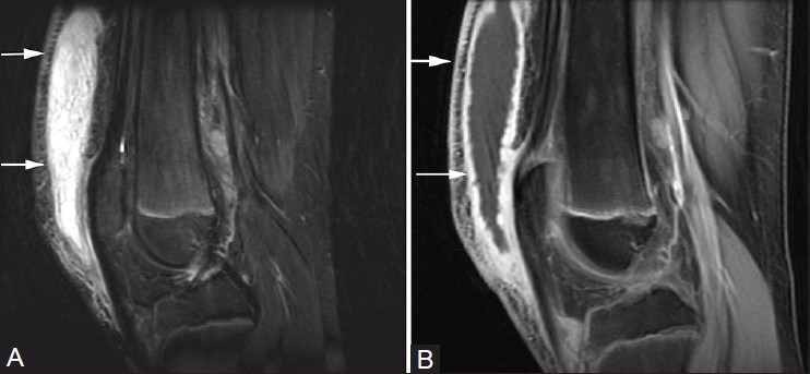
MRISagittal knee MRI: fluid collection distending the prepatellar bursa, arrows on the bursa.
Source : Telischak et al., The Indian journal of radiology & imaging, 2014, figure 5 · CC BY-NC-SA
Which questions should you ask to understand the patient and their history?
The clinical history is the first decisive step. It must be systematic and focused:- Triggering mechanism: direct trauma (a fall, a blow)? Occupational or leisure activity involving prolonged or repetitive kneeling (tiling, plumbing, gardening, mechanics)? This distinction points towards an acute origin rather than a chronic one from microtrauma.¹٬²
- History of the symptoms: first episode or recurrence? Time of onset (acute over hours or days suggests a traumatic or septic cause; insidious over weeks suggests chronic mechanical overload)? Course under any empirical treatment already taken.²
- Characteristics of the pain: strictly anterior, prepatellar location, with typical pain on direct pressure (kneeling) or at end-range knee flexion (stretching of the skin over the inflamed bursa). Conversely, walking on level ground is generally well tolerated.¹٬³
- Systemic signs (CRITICAL): fever > 38 °C, shivering, general malaise? ⚠️ Important: fever is not always present in septic bursitis (found in only 40-60% of patients across published series).⁴٬⁵ Its absence does not rule out infection.
- Skin portal of entry: systematic search for a wound, an abrasion, an insect bite or a chronic skin lesion (eczema, psoriasis), whether overlying the bursa or remote from it.⁴
- Comorbidities carrying an infectious risk: diabetes, chronic alcohol misuse, corticosteroid therapy, immunosuppressants, kidney failure, active malignancy.⁵
- Inflammatory history: gout (a previous attack, a known tophus, hyperuricaemia), rheumatoid arthritis, other inflammatory rheumatic diseases.⁶
Which clinical tests should you perform and which other conditions must be ruled out?
The physical examination must be comparative (against the other side), focusing on inspection, palpation and assessment of mobility, while keeping the differential diagnoses in mind. Inspection: swelling that is usually well defined, round or oval, directly in front of the patella.¹ Look for erythema (redness), local warmth and a skin lesion acting as a portal of entry. Redness spreading well beyond the bursa, or lymphangitis, points strongly to a septic bursitis with associated cellulitis.⁵ Palpation: confirms the fluctuant nature of the swelling (a fluid collection) and exquisite pain on direct pressure.¹ Compare local warmth with the other side. Assess the tenderness of adjacent structures (patellar tendon, inferior pole of the patella, tibial tuberosity, Hoffa's fat pad). Joint mobility (KEY TEST FOR DIFFERENTIAL DIAGNOSIS):- In prepatellar bursitis that is uncomplicated, active knee range is completely preserved or only painfully limited at end-range flexion (skin tension over the bursa).¹
- Conversely, a marked painful limitation across all ranges, with inability to bear weight or major functional loss, suggests intra-articular pathology : septic arthritis of the knee (a surgical emergency), an acute flare of chondrocalcinosis, haemarthrosis.⁷
🩺 Comparative examination: prepatellar bursitis versus septic arthritis of the knee
Telling these two entities apart is crucial: septic arthritis is a surgical emergency
| Criterion | Prepatellar bursitis | Septic arthritis of the knee |
|---|---|---|
| Location of the swelling | Strictly anterior, prepatellar, well defined | Diffuse around the knee, patellar tap positive |
| Joint range | Preserved, or limited only at end-range flexion | Major limitation across all ranges |
| Weight-bearing / walking | Tolerated (normal walking on level ground) | Unable to bear weight, functional loss |
| Intra-articular effusion | Absent | Present (patellar tap positive, ballottement sign) |
| Aspiration | Prepatellar bursa | Joint (knee at 20° of flexion) |
| Emergency | Medical (if septic) | Surgical (urgent joint lavage) |
Sources: Rishor-Olney 2024, Mathews 2007 (Ann Rheum Dis; systematic review on septic arthritis). If doubt persists, ultrasound-guided aspiration of both compartments (bursa and joint) is recommended.
- Septic bursitis (see the dedicated section below): the absolute priority.⁴٬⁵
- Septic arthritis of the knee: diffuse pain, major limitation, intra-articular effusion.⁷
- Gout / pseudogout (chondrocalcinosis): an inflammatory flare that can mimic septic bursitis. Looking for crystals under polarised light microscopy in the aspirated fluid is the reference diagnostic test.⁶
- Patellar or quadriceps tendinopathy: pain over the tendon, reproduced by resisted contraction, with no bursal collection.
- Prepatellar Morel-Lavallée haematoma: post-traumatic shearing of the skin from the subcutaneous plane, which can mimic a chronic haemorrhagic bursitis.⁸
- Osgood-Schlatter disease (in adolescents): pain over the anterior tibial tuberosity, not over the patella.
- Gouty tophus and bursal tuberculosis (rare): to be considered in resistant chronic forms (see the Kosmoliaptsis 2007 and MacLean 2013 cases).⁹٬¹⁰
🚩 Red flags in a prepatellar swelling
- Spreading cellulitis extending well beyond the bursa + ascending lymphangitis
- Fever > 38.5 °C, shivering, sepsis (tachycardia, hypotension)
- Major limitation of knee mobility with functional loss → suspected adjacent septic arthritis
- Immunosuppression : poorly controlled diabetes, alcohol misuse, corticosteroid therapy, immunosuppressants, malignancy
- Failure of empirical antibiotic therapy at 48-72h → reassess (atypical organism, MRSA, surgical indication)
- Chronic non-healing wound or overlying ulceration → suspected deep infection, osteomyelitis
- Chronic form resistant to conservative care for > 6 months → consider gout, tuberculosis, tumour (rare)
- Non-traumatic bilateral involvement + systemic signs → rheumatological workup (rheumatoid arthritis, tophaceous gout)
⚠️ Any red flag → prompt medical referral (GP, emergency department, orthopaedic surgeon) BEFORE any active physiotherapy management.
Should patients with prepatellar bursitis be classified, and what is gained by doing so?
Yes: classification has a direct impact on treatment strategy. It is built around two axes : aetiology and time course.² 1. Aetiological classification:- Aseptic, mechanical or traumatic (the dominant case, ≈ 65%): repeated microtrauma or a direct blow. Conservative treatment (PRICE, NSAIDs, knee pads, activity modification).¹٬²
- Septic, infectious (≈ 33%): a medical emergency, empirical anti-staphylococcal antibiotic therapy plus aspiration ± surgical drainage.⁴٬⁵
- Inflammatory or crystal-induced (rare, ≈ 2%): gout, rheumatoid arthritis, chondrocalcinosis. Treatment of the underlying disease plus local management.⁶٬⁹
- Acute (< 6 weeks): rapid onset, marked inflammatory signs, often post-traumatic or septic.
- Chronic (> 6 weeks): repeated loading, a thickened, fibrous bursal wall, persistent but less painful swelling. Poorer response to conservative treatment → potential indication for a selective corticosteroid injection (under strict conditions) or bursectomy.²٬¹¹
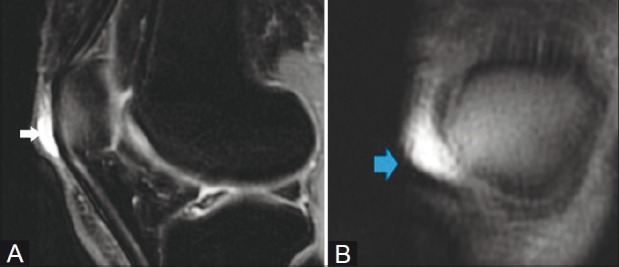
MRISagittal (A) and axial (B) knee MRI, fat-saturated T2 sequences: prepatellar bursa distended by an effusion, arrows on the collection.
Source : Chatra, The Indian journal of radiology & imaging, 2012, figure 1 · CC BY-NC-SA
Key points
- The diagnosis is essentially clinical : a focused history plus a comparative examination of inspection / palpation / mobility.
- Absolute priority: telling aseptic from septic. Fever is inconsistent (40-60%); spreading erythema and cellulitis discriminate better.
- Key test: knee mobility is preserved in bursitis (except at end-range flexion), and severely limited in adjacent septic arthritis.
- The aspiration of the bursal fluid with analysis (Gram stain, culture, crystals, cytology) remains the gold standard whenever infection or gout is in doubt.
- The ultrasound scan (sensitivity ≈ 87%, specificity 100% for knee bursitis, Draghi 2015) is the first-line imaging modality and guides aspirations.
- Classifying (aetiology × time course) structures the treatment strategy and avoids over-medicalisation.
References
- Rishor-Olney CR, Taqi M, Pozun A. Prepatellar Bursitis. In: StatPearls. Treasure Island (FL): StatPearls Publishing; 2024. NBK557508.
- Baumbach SF, Lobo CM, Badyine I, Mutschler W, Kanz KG. Prepatellar and olecranon bursitis: literature review and development of a treatment algorithm. Arch Orthop Trauma Surg. 2014;134(3):359-370. PMID 24305696.
- Khodaee M. Common Superficial Bursitis. Am Fam Physician. 2017;95(4):224-231. PMID 28290630.
- Lormeau C, Cormier G, Sigaux J, Arvieux C, Semerano L. Management of septic bursitis. Joint Bone Spine. 2019;86(5):583-588. PMID 31615686.
- García-Porrúa C, González-Gay MA, Ibañez D, García-País MJ. The clinical spectrum of severe septic bursitis in northwestern Spain: a 10 year study. J Rheumatol. 1999;26(3):663-667. PMID 10090179.
- Truong J, Mabrouk A, Ashurst JV. Septic Bursitis. In: StatPearls. 2023. NBK470331.
- Mathews CJ, Kingsley G, Field M, et al. Management of septic arthritis: a systematic review. Ann Rheum Dis. 2007;66(4):440-445. PMID 17223664.
- Retal H, Cavez N, De Smet E, Abid W, Kadi R. Prepatellar Morel-Lavallée Effusion Mimicking a Bursitis. Cureus. 2024;16(4):e59129. PMC 11129518. PMID 38803733.
- Kosmoliaptsis V, Soni R. Tophaceous gout mass distending the prepatellar bursa. J Clin Rheumatol. 2007;13(6):359. PMID 18176150.
- MacLean S, Kulkarni S. Tuberculosis of the patella masquerading as prepatellar bursitis. Ann R Coll Surg Engl. 2013;95(1):e17-e19. PMC 3964674. PMID 23317718.
- Draghi F, Corti R, Urciuoli L, Alessandrino F, Rotondo A. Knee bursitis: a sonographic evaluation. J Ultrasound. 2015;18(3):251-257. PMC 4529416. PMID 26261475.
- Lueders DR, Smith J, Sellon JL. Ultrasound-Guided Knee Procedures. Phys Med Rehabil Clin N Am. 2016;27(3):631-648. PMID 27468670.
- Chatra PS. Bursae around the knee joints. Indian J Radiol Imaging. 2012;22(1):27-30. PMC 3354353.
Septic bursitis: how do you avoid missing it? (Specialised section) 🚨
Criteria for telling septic from aseptic
The major clinical challenge is that no single sign is sensitive or specific enough to distinguish formally between a septic bursitis and a highly inflammatory aseptic one.³ It is the accumulation of features that guides the clinician:🔬 Discriminating criteria, septic versus aseptic: sensitivity varies
Lormeau 2019 (systematic review), García-Porrúa 1999 (n=75), Truong StatPearls 2023
| Criterion | Septic (sensitivity) | Aseptic | Discriminating value |
|---|---|---|---|
| Skin erythema extending beyond the bursa / cellulitis | Very common (> 80%) | Rare | High |
| Local warmth > 2 °C vs the other side | Present | Often present too | Moderate |
| Fever > 38 °C | 40-60% only | Absent | Moderate (inconsistent) |
| Visible skin portal of entry | ≈ 50% of cases | Possible (traumatic) | Moderate |
| At-risk comorbidities (diabetes, immunosuppression) | Over-represented | Possible | Moderate |
| CRP > 50 mg/L, leucocytosis | Common but not specific | Possible (highly inflammatory) | Moderate |
| Bursal fluid white cells > 2000-5000/mm³ | Suggestive | Rare | High |
| Gram stain with bacteria on microscopy | Diagnostic | Sterile | Diagnostic |
| Positive culture | GOLD STANDARD | Sterile | Diagnostic |
Source: Lormeau C et al. Joint Bone Spine 2019;86(5):583-588 (PMID 31615686). No single clinical criterion has a sensitivity > 80%, hence the importance of aspiration at the slightest doubt.
- Staphylococcus aureus > 80 % (most often methicillin-sensitive, MRSA depending on local prevalence).¹٬³٬⁴
- Streptococcus sp. (β-haemolytic groups A, B, G): 5-10%.³
- Gram-negative bacteria (rare, in immunosuppressed patients and seawater divers).³
- Mycobacteria (tuberculosis, atypical): exceptional, but to be considered in resistant chronic forms (MacLean 2013).⁵
Aspiration, empirical antibiotic therapy and drainage
🔀 Decision tree: suspected septic prepatellar bursitis
Adapted from Lormeau 2019, Brown 2022 and Truong StatPearls 2023
Simplified tree. Adapt locally according to microbial ecology (MRSA prevalence) and institutional guidance. The choice of IV versus oral antibiotics depends on severity and comorbidities (Lormeau 2019, Brown 2022).
- Strict surgical asepsis, skin disinfection, sterile gloves.
- An 18G or 20G needle, lateral approach, avoiding erythematous skin.
- Ultrasound guidance is recommended for a small collection or when the anatomy is unclear (Lueders 2016).⁶
- Always send for: Gram stain, aerobic and anaerobic culture, sensitivities, cytology (white cell count), search for crystals.³
- If the aspiration is dry, do not rule out infection: repeat it if the course is unfavourable.
- Mild outpatient case, MSSA suspected : an oral anti-staphylococcal penicillin (cloxacillin, dicloxacillin) or a first-generation cephalosporin (cefalexin) for 7-14 days.³
- Penicillin allergy : clindamycin, doxycycline, TMP-SMX.
- MRSA risk (depending on local prevalence): IV vancomycin or linezolid, TMP-SMX, clindamycin according to sensitivities.³
- Severe illness or sepsis : hospital admission, IV antibiotics (vancomycin + cefazolin), surgical assessment.³٬⁷
- Failure of well-conducted antibiotic therapy (> 7 days).
- Recurrent septic bursitis.
- A purulent collection that cannot be drained by aspiration.
- Fistula formation through the skin.
- Refractory chronic aseptic bursitis (> 6 months of conservative care).¹٬²
Key points
- Septic bursitis = 1/3 of prepatellar bursitis cases, a medical emergency. S. aureus > 80 %.
- No single clinical sign is discriminating enough. Spreading cellulitis, fever and immunosuppression are the most suggestive features, but their absence does not rule out infection.
- The bursal aspiration (Gram stain, culture, crystals, cytology) is the diagnostic gold standard.
- Empirical antibiotic therapy: an anti-staphylococcal penicillin or a first-generation cephalosporin as first-line therapy for MSSA; vancomycin if there is MRSA risk or sepsis.
- Duration of antibiotic therapy: shorten it where possible (< 8 days suggested for uncomplicated cases, Brown 2022).
- Endoscopic bursectomy in case of failure, recurrence or refractory chronic bursitis: superior to the open route in terms of skin complications.
References
- Baumbach SF, Lobo CM, Badyine I, Mutschler W, Kanz KG. Prepatellar and olecranon bursitis: literature review and development of a treatment algorithm. Arch Orthop Trauma Surg. 2014;134(3):359-370. PMID 24305696.
- Brown OS, Smith TO, Parsons T, Benjamin M, Hing CB. Management of septic and aseptic prepatellar bursitis: a systematic review. Arch Orthop Trauma Surg. 2022;142(10):2445-2457. PMID 33721054.
- Lormeau C, Cormier G, Sigaux J, Arvieux C, Semerano L. Management of septic bursitis. Joint Bone Spine. 2019;86(5):583-588. PMID 31615686.
- García-Porrúa C, González-Gay MA, Ibañez D, García-País MJ. The clinical spectrum of severe septic bursitis in northwestern Spain: a 10 year study. J Rheumatol. 1999;26(3):663-667. PMID 10090179.
- MacLean S, Kulkarni S. Tuberculosis of the patella masquerading as prepatellar bursitis. Ann R Coll Surg Engl. 2013;95(1):e17-e19. PMC 3964674. PMID 23317718.
- Lueders DR, Smith J, Sellon JL. Ultrasound-Guided Knee Procedures. Phys Med Rehabil Clin N Am. 2016;27(3):631-648. PMID 27468670.
- Mathews CJ, Kingsley G, Field M, et al. Management of septic arthritis: a systematic review. Ann Rheum Dis. 2007;66(4):440-445. PMID 17223664.
- Huang YC, Yeh WL. Endoscopic treatment of prepatellar bursitis. Int Orthop. 2011;35(3):355-358. PMID 20521045. doi:10.1007/s00264-010-1033-5.
- Luk WCB, Lui TH. Endoscopic Resection of Prepatellar Bursa. Arthrosc Tech. 2020;9(7):e1057-e1060. PMID 32714819.
- Reilly D, Kamineni S. Olecranon bursitis. J Shoulder Elbow Surg. 2016;25(1):158-167. PMID 26577126.
- Nchinda NN, Wolf JM. Clinical Management of Olecranon Bursitis: A Review. J Hand Surg Am. 2021;46(6):501-506. PMID 33840568.
- Qadeer H, Suwal A, Oke I. The Journey From Knee to Heart: A Case of MSSA Infective Endocarditis Secondary to Septic Prepatellar Bursitis. Cureus. 2023;15(3):e36806. PMC 10146384.
Which treatment strategies are the most effective for prepatellar bursitis?
Where do you start? What is the hierarchy of interventions?
For aseptic bursitis, the hierarchy follows a progressive path:- First-line conservative treatment: the PRICE protocol (Protection, Rest, Ice, Compression, Elevation), strict avoidance of kneeling activities, oral or topical NSAIDs for the painful phase (on medical prescription). Systematic wearing of protective knee pads as soon as activity resumes.¹⁻³
- Bursal aspiration: can quickly relieve pressure and pain. The recurrence rate is high, however, if the underlying mechanical cause is not corrected. Best reserved for marked functional discomfort or diagnostic doubt (cytobacteriological analysis).⁴
- Corticosteroid injection (controversial): an option for resistant chronic aseptic bursitis. Documented risks: skin atrophy, depigmentation, iatrogenic infection (in a bursa that was aseptic to begin with) and a rare tendon risk through diffusion.⁵٬⁶ Several recent reviews recommend caution and selectivity in this indication, because of the skin complications specific to the superficial prepatellar bursa.⁴
- Surgical bursectomy: a last resort for chronic, recurrent or refractory septic forms. The endoscopic route is recommended (Huang 2011, Sasaki 2020): non-inferior to the open route with fewer skin complications (Brown 2022).⁴٬⁷٬⁸
📊 Treatment modalities for prepatellar bursitis: level of evidence (adapted GRADE)
Synthesis of Brown 2022 (systematic review, n=702), Lormeau 2019 (systematic review) and Khodaee 2017: most recommendations rest on observational series and expert opinion
| Intervention | Aseptic form | Septic form | Evidence level (GRADE) |
|---|---|---|---|
| PRICE + avoidance + knee pads | First line | Adjunct | Moderate (expert opinion, plausibility) |
| Oral or topical NSAIDs | First line (short term) | Symptomatic | Moderate (extrapolated from other bursitides) |
| Diagnostic + therapeutic aspiration | Marked discomfort or doubt | Essential (Gram stain/culture) | High (diagnostic gold standard) |
| Empirical anti-staphylococcal antibiotics | — | Essential | High (consensus, Lormeau 2019) |
| Intrabursal corticosteroid injection | Selected chronic forms | CONTRAINDICATED | Low (controversial, skin risks) |
| Endoscopic bursectomy | Conservative failure > 6 months | Recurrent / refractory | Moderate (Brown 2022, comparative series) |
| Open bursectomy | If endoscopy is unavailable | Complex cases / fistula | Moderate (more skin complications) |
| Local manual therapy | Adjunct in the post-acute phase | — | Very low (extrapolation) |
| Ultrasound / laser / TENS | Not recommended routinely | — | Very low (no specific benefit demonstrated) |
| Shockwave therapy (ESWT) | No specific evidence | — | Very low (evidence in other tendinopathies, Mani-Babu 2015) |
| Patient education + ergonomics + recurrence prevention | First line (always) | Essential after the episode | Moderate (plausibility, mechanism) |
Grading: High = international consensus, solid data; Moderate = systematic review or converging cohorts; Low = controversial or limited data; Very low = extrapolation, expert opinion.
What is the place of exercise in rehabilitation?
💡 There are no randomised controlled trials defining a superior exercise programme for prepatellar bursitis. The approach relies on general physiological principles and on extrapolation from protocols validated in other knee conditions (knee osteoarthritis, patellar tendinopathy). Acute phase (days 0-10):- NO direct strengthening and no aggressive stretching of the prepatellar region.
- Aim: settle things down, reduce inflammation, avoid direct pressure.
- Gentle pain-free active knee mobilisations to prevent stiffness.
- Recovery of full joint range (passive then active flexion and extension).
- Isometric quadriceps strengthening (quad sets), then progression to closed-chain work (mini-squats, leg press) without skin pressure over the patella.
- Gentle pain-free stretching of the quadriceps, hamstrings and triceps surae.
- Progressive open- and closed-chain strengthening.
- Gradual reintroduction of at-risk positions (kneeling) with a knee pad, in short sessions at first.
- Proprioceptive and task-specific work according to occupational or sporting demands.
Manual therapies and technologies: how effective are they really?
The level of evidence for manual therapies and physical modalities (laser, ultrasound, TENS, shockwave) applied specifically to prepatellar bursitis is low to very low. Their use must remain selective and critical:- Manual therapy (gentle peripatellar soft tissue mobilisation): no robust evidence, but it may be offered in the post-acute phase to improve local mobility. Avoid any manoeuvre that compresses the bursa directly.
- Cryotherapy (ice) : the simplest and best validated modality in the acute phase, for its analgesic effect.⁹
- Extracorporeal shockwave therapy (ESWT) : favourable evidence in some lower limb tendinopathies (Mani-Babu 2015) but no specific data on non-calcifying aseptic prepatellar bursitis. Not recommended routinely.¹⁰
- Therapeutic ultrasound and low-level laser : no evidence of a specific significant benefit. Their use is not scientifically demonstrated for this indication.
- TENS : possibly as an adjunctive painkiller, without changing the course of the condition.
Beyond the physical: how do you educate the patient about prevention?
🧘♂️ The education of the patient (therapeutic patient education) is probably the most important component of long-term management. It is essential to prevent the recurrences that are common in this occupational condition.¹ The pillars of that education:- Understanding the causal mechanism: the patient must know WHY the bursa became inflamed (repeated direct pressure) in order to stick to preventive measures.
- Ergonomic modification of activities: alternating positions, using a low stool, taking regular breaks to stand up and restore blood flow.²
- Systematic wearing of suitable protective knee pads (high-density foam, gel) during at-risk activities: the preventive measure with the best empirical support.¹⁻³
- Progressive load management : recognising the early signs of an inflammatory flare (swelling, warmth, pain) and adapting activities immediately.
- Addressing kinesiophobia in chronic forms: reassuring language, explaining that progressive pain-free movement is beneficial, is crucial to avoid deconditioning.¹¹
Critique and controversies: the grey areas of management
The literature on prepatellar bursitis suffers from a lack of high-quality randomised controlled trials, particularly on physiotherapy interventions. The Brown 2022 systematic review, the most rigorous to date, included only 10 studies and 702 patients for such a common topic.⁴ Many current recommendations rest on expert opinion and physiological extrapolation. That calls for considerable humility from the clinician. Three specific controversies deserve attention: 1. Corticosteroid injections. Widely used in routine practice for their rapid anti-inflammatory effect, their benefit-to-risk balance is now being questioned. The iatrogenic risk of introducing infection into an aseptic bursa, together with the skin atrophy and depigmentation peculiar to the thin prepatellar skin, has led several authors to reserve this option for highly selected chronic aseptic cases, after a well-conducted conservative approach has failed and infection has been formally ruled out.⁵٬⁶ 2. The endoscopic versus open route. Brown 2022 concludes that endoscopic bursectomy is non-inferior, with fewer skin complications. The technique is not equally available everywhere, however, there is a learning curve, and cost-effectiveness has not been formally demonstrated in every health system.⁴ 3. The duration of antibiotic therapy in septic forms. Brown 2022 suggests that a course of < 8 days may be enough in uncomplicated cases, against the classic 14-21 days. This aligns with the general trend towards shorter courses (antibiotic stewardship) but needs prospective validation in dedicated trials.⁴Key points
- Absolute priority: telling apart aseptic from septic. The latter is a medical emergency.
- First-line treatment of the aseptic form: conservative (PRICE + avoidance of kneeling + knee pads + NSAIDs).
- The exercise programme is introduced after the acute phase, focusing on restoring range and on progressive strengthening without direct pressure on the bursa.
- The education of the patient about ergonomics and the systematic wearing of knee pads is the cornerstone of preventing recurrence.
- ⚠️ Bear in mind that corticosteroid injections are controversial: skin and infection risks peculiar to prepatellar skin, so use them selectively after conservative treatment has failed.
- ❌ The evidence supporting the effectiveness of manual therapies or of most technologies (ultrasound, laser, ESWT) for prepatellar bursitis is very weak.
- 🔪 Where surgery is needed, endoscopic bursectomy is the recommended surgical route when conservative treatment fails or when a septic bursitis is refractory (Brown 2022).
References
- Rishor-Olney CR, Taqi M, Pozun A. Prepatellar Bursitis. StatPearls. 2024. NBK557508.
- Khodaee M. Common Superficial Bursitis. Am Fam Physician. 2017;95(4):224-231. PMID 28290630.
- Aaron DL, Patel A, Kayiaros S, Calfee R. Four common types of bursitis: diagnosis and management. J Am Acad Orthop Surg. 2011;19(6):359-367. PMID 21628647.
- Brown OS, Smith TO, Parsons T, Benjamin M, Hing CB. Management of septic and aseptic prepatellar bursitis: a systematic review. Arch Orthop Trauma Surg. 2022;142(10):2445-2457. PMID 33721054.
- Brinks A, Koes BW, Volkers AC, Verhaar JA, Bierma-Zeinstra SM. Adverse effects of extra-articular corticosteroid injections: a systematic review. BMC Musculoskelet Disord. 2010;11:206. PMC 2945953.
- Chatra PS. Bursae around the knee joints. Indian J Radiol Imaging. 2012;22(1):27-30. PMC 3354353.
- Huang YC, Yeh WL. Endoscopic treatment of prepatellar bursitis. Int Orthop. 2011;35(3):355-358. PMID 20521045.
- Luk WCB, Lui TH. Endoscopic Resection of Prepatellar Bursa. Arthrosc Tech. 2020;9(7):e1057-e1060. PMID 32714819.
- Malanga GA, Yan N, Stark J. Mechanisms and efficacy of heat and cold therapies for musculoskeletal injury. Postgrad Med. 2015;127(1):57-65. PMID 25526231.
- Mani-Babu S, Morrissey D, Waugh C, Screen H, Barton C. The effectiveness of extracorporeal shock wave therapy in lower limb tendinopathy: a systematic review. Am J Sports Med. 2015;43(3):752-761. PMID 24817008.
- Wertli MM, Rasmussen-Barr E, Weiser S, Bachmann LM, Brunner F. The role of fear avoidance beliefs as a prognostic factor for outcome in nonspecific low back pain: a systematic review. Spine J. 2014;14(5):816-836. PMID 24412032.
How do you secure lasting recovery and prevent flares?
How do you make the patient an active participant in their recovery?
🧑🏫 Empowering the patient is the most durable strategy for preventing recurrence. It rests on four pillars: 1. Understanding the causal mechanism: the patient must make the explicit link between a specific action (unprotected kneeling, a direct blow) and the pain that follows. That realisation is what drives behaviour change.³ 2. Modifying activities:- Favour alternative positions: a low stool, brief squatting.
- Break kneeling tasks into blocks (5-10 min, then a standing break).
- Adapt the workstation where possible (a raised tiling board, a plumbing bench).
- The systematic wearing of suitable knee pads of good quality (high-density foam, gel, a rigid shell depending on the trade).
- Regular checks on the state of the knee pad (wear, flattened foam).
- Choose comfortable models to encourage adherence: uncomfortable equipment will not be worn.
- Recognise the early signs of irritation: morning swelling, local warmth, residual pain after activity.
- Adapt immediately (reduce activity for 24-48h, ice, topical NSAIDs).
- Reintroduce gradually, never abruptly.
When and how do you plan a safe return to activity?
The return to work or sport must be a structured, criteria-based process rather than an arbitrary delay. 📅 Returning too early is a major risk factor for recurrence or for becoming chronic. Essential criteria before a full return:- Complete resolution of symptoms: no pain on direct palpation of the bursa, none during knee movement or basic functional activities. Any residual effusion has gone.
- Range of movement restored: full flexion and extension, symmetrical with the other side, pain-free.
- Muscle strength normalised: quadriceps and hamstring strength ≥ 90-95 % of the uninvolved limb (isokinetic testing or a functional proxy such as a single-leg hop).
- Functional tests specific to the job or sport: the ability to kneel (on a knee pad), stand up, pivot, jump and climb stairs without pain or apprehension.
- Psychological confidence: assessment of kinesiophobia (fear of movement, fear of recurrence). A high score on the Tampa Scale for Kinesiophobia should prompt the inclusion of cognitive behavioural strategies.⁴
- Tiler : short days at first (3-4h), high-quality knee pads, frequent breaks, full-time return in 2-4 weeks depending on progress.
- Wrestler : first a return to non-contact technical work (drills), then controlled bouts on the mat with a knee pad, then competition.
- Gardener : alternate standing, squatting and protected kneeling positions; avoid long sessions on hard paving.
Critique and controversies: the difficulty of evaluating prevention
The literature is sorely lacking in studies with a high level of evidence (randomised controlled trials, systematic reviews) devoted specifically to preventing recurrence of prepatellar bursitis. Most current recommendations are extrapolated from the management of other overuse conditions, or based on pathophysiological reasoning and consensus.¹ One persistent controversy: compliance with wearing knee pads in real working life. Productivity pressures, the perceived discomfort of some models and the lack of a preventive culture in certain sectors (construction in particular) hold back systematic uptake. Structured workplace ergonomic programmes are needed but are poorly documented scientifically.²Key points
- ✅ Preventing recurrence rests on patient education and on lasting modification of at-risk activities.
- 🛡️ Using suitable protective knee pads systematically is the most important preventive measure for occupations that involve kneeling.
- 📈 Return to activity must be progressive and guided by functional criteria (no pain, full range, strength ≥ 90%, functional tests) rather than by an arbitrary timetable.
- 🧠 The empowerment of the patient through structured self-management strategies is the key to long-term success.
- ⚠️ Compliance with wearing protection remains a challenge in the workplace: a company ergonomics programme strengthens individual adherence.
References
- Khodaee M. Common Superficial Bursitis. Am Fam Physician. 2017;95(4):224-231. PMID 28290630.
- Aaron DL, Patel A, Kayiaros S, Calfee R. Four common types of bursitis: diagnosis and management. J Am Acad Orthop Surg. 2011;19(6):359-367. PMID 21628647.
- Bandura A. Self-efficacy: toward a unifying theory of behavioral change. Psychol Rev. 1977;84(2):191-215. PMID 847061.
- Wertli MM, Rasmussen-Barr E, Weiser S, Bachmann LM, Brunner F. The role of fear avoidance beliefs as a prognostic factor for outcome in nonspecific low back pain: a systematic review. Spine J. 2014;14(5):816-836. PMID 24412032.
- Brown OS, Smith TO, Parsons T, Benjamin M, Hing CB. Management of septic and aseptic prepatellar bursitis: a systematic review. Arch Orthop Trauma Surg. 2022;142(10):2445-2457. PMID 33721054.
- Hoffmann TC, Montori VM, Del Mar C. The connection between evidence-based medicine and shared decision making. JAMA. 2014;312(13):1295-1296. PMID 25268434.
- Cook JL, Rio E, Purdam CR, Docking SI. Revisiting the continuum model of tendon pathology: what is its merit in clinical practice and research? Br J Sports Med. 2016;50(19):1187-1191. PMID 27127294.
What do published clinical cases teach us about prepatellar bursitis? 📚
Analysis of a typical case: from diagnosis to resolution
The typical case is a middle-aged man in a kneeling occupation, presenting with a well-defined fluctuant swelling in front of the knee and no systemic signs. The clinical diagnosis is made, ultrasound confirms the fluid collection and rules out chronic fibrous thickening. Management combines avoidance of kneeling, ice, a short course of NSAIDs, the wearing of knee pads and therapeutic education. Complete resolution of symptoms is seen within 4 to 6 weeks in most uncomplicated cases.¹ ⚠️ Important, a methodological note: many reviews quote "typical profiles" of patient (a precise age, an occupation, a duration of symptoms) without always referencing a single publication. These teaching syntheses, which are not meant to be genuine clinical cases, must be told apart from the published and indexed case reports, set out below.The diagnostic challenge: when bursitis mimics another condition
Case 1: massive bilateral chronic bursitis (Maguire 2024, Cureus, PMC 11668264) A 60-year-old man, a painter and decorator, presented with bilateral anterior swelling of both knees that had been developing for 6 years after an initial fall onto both knees.² The swellings were large enough to interfere with putting on trousers and with leaning against a ladder. Surgical management consisted of sequential excision of the bursae: on the right, organised solid content (a chronic haematoma with a "chocolate paste" appearance); on the left, thick brown fluid drained in addition to fibrous tissue. At more than 2 years of follow-up there was no recurrence. Lesson: massive bilateral chronic bursitis is rare but can arise from repeated occupational microtrauma. The "solid" form (an organised fibrous haematoma) rules out simple drainage and requires bursectomy. The open route was chosen here because of the size of the lesion; in everyday practice endoscopy remains preferred for bursae of moderate size. Case 2: gouty tophus mimicking a chronic bursitis (Kosmoliaptsis 2007, PMID 18176150) A patient presented with a firm, chronic prepatellar mass initially labelled fibrous bursitis. Imaging showed a heterogeneous signal. Histological analysis after excision revealed a gouty tophus (needle-shaped birefringent sodium urate crystals): an unexpected diagnosis that justified long-term urate-lowering treatment.³ Lesson: in patients with a history of gout (hyperuricaemia, attacks of podagra), a prepatellar mass should raise tophus among the differential diagnoses. Cytological analysis of the fluid or the tissue (looking for birefringent crystals under polarised light microscopy) is diagnostic. Rheumatoid arthritis can also cause chronic inflammation of the prepatellar bursa. Case 3: patellar tuberculosis mimicking a prepatellar bursitis (MacLean 2013, PMC 3964674) A 25-year-old woman of Bangladeshi origin had insidious swelling of the right knee for 3 months following a fall. Initial treatment with oral flucloxacillin brought no improvement. Radiograph: a central necrotic nidus in the patella. MRI: osteomyelitis confirmed. Microbiology of the bone sequestrum: Mycobacterium tuberculosis (acid-fast bacilli on culture). Prolonged antituberculous treatment.⁴ Lesson: in an at-risk population (origin in an endemic country, immunosuppression, previous pulmonary TB), a chronic bursitis that does not respond to standard antibiotics should raise the possibility of tuberculosis. The index of suspicion must be high, because the disease is paucibacillary and the early signs are non-specific.Study of a complex case: septicaemia and endocarditis arising from a bursitis
Case 4: MSSA endocarditis secondary to a septic prepatellar bursitis (Qadeer 2023, Cureus, PMC 10146384) A patient with a history of recurrent prepatellar bursitis requiring several catheter drainages presented to the emergency department with sepsis and infective endocarditis. Blood cultures: Staphylococcus aureus methicillin-sensitive (MSSA). Echocardiography: valvular vegetations. The source identified was the poorly controlled prepatellar bursitis. Treatment: prolonged IV antibiotics plus cardiological management.⁵ 💔 Lesson: although rare, endocarditis secondary to a poorly treated septic bursitis illustrates the potential severity of a neglected septic bursitis. The clinical message is clear: a recurrent septic bursitis must be managed aggressively (surgical drainage if it recurs after aspiration, bursectomy if that fails), and systemic bacteraemia must always be looked for in a septic patient with an obvious skin or bursal focus. Case 5: bursitis treated by endoscopic bursectomy (Huang 2011, PMID 20521045) A prospective series of 60 patients (mean age 33.5 years) with traumatic prepatellar bursitis refractory to conservative treatment. Bursectomy performed endoscopically as day surgery under local anaesthesia, 2-3 arthroscopic mini-portals, mean duration 18 minutes. Mean follow-up 36.3 months: all patients were asymptomatic, knee function was normal, with no infection, no loss of range and no skin revision.⁶ Lesson: endoscopic bursectomy gives excellent functional results and a very favourable complication profile compared with the open route. Confirmed by the Brown 2022 systematic review (n=10 studies).⁷Critique and controversies in the case literature
The clinical case literature on prepatellar bursitis remains fragmented. Several areas of debate persist: 1. The threshold for performing a diagnostic aspiration. Unavoidable when infection is suspected. Performing it routinely in apparently aseptic bursitis is debated, however, because of the iatrogenic risk of inoculation.⁸ The literature offers no clear consensus, leaving the decision to clinical judgement. 2. The place of corticosteroid injections. Effective in the short term but carrying skin and infection risks peculiar to the thin prepatellar skin. Several recent reviews recommend caution and reserving them for selected chronic aseptic cases, once infection has been formally ruled out.⁷٬⁹ 3. The management of septic bursitis. Routine admission versus outpatient care, duration of antibiotics, early surgery versus surgery after failure: practice varies on every one of these. Complex cases (such as the Qadeer 2023 endocarditis) argue for an aggressive stance when there is no response to initial treatment.⁵٬¹⁰ ⚠️ Level of evidence: a case report = level 5 (the lowest in the CEBM hierarchy). It illustrates, it never demonstrates efficacy or superiority. Where findings diverge, meta-analyses and systematic reviews (level 1a) must take precedence.Key points
- ✅ The typical case of prepatellar bursitis (a manual worker, a fluctuant swelling, conservative treatment, resolution in 4-6 weeks) remains the most common.
- 🤔 Conditions such as gouty tophus (Kosmoliaptsis 2007) or patellar tuberculosis (MacLean 2013) can mimic a chronic bursitis: consider them if the presentation is atypical or resistant.
- ⚠️ The most dreaded complication is MSSA endocarditis secondary to a neglected septic bursitis (Qadeer 2023, Cureus): hence the imperative of aggressive management when there is no response.
- 🔪 The endoscopic bursectomy performed as day surgery (Huang 2011, series of n=60) gives excellent functional results with a very favourable complication profile.
- 📊 A case report = level 5: it illustrates but does not demonstrate. Meta-analyses (level 1a) take precedence in case of conflict.
References
- Rishor-Olney CR, Taqi M, Pozun A. Prepatellar Bursitis. StatPearls. 2024. NBK557508.
- Maguire A, Lawrence C, Nicolai P, Rosenbloom C. Massive Bilateral Haemorrhagic Prepatellar Bursitis: A Case Report. Cureus. 2024;16(11):e74351. PMC 11668264. PMID 39720372.
- Kosmoliaptsis V, Soni R. Tophaceous gout mass distending the prepatellar bursa. J Clin Rheumatol. 2007;13(6):359. PMID 18176150.
- MacLean S, Kulkarni S. Tuberculosis of the patella masquerading as prepatellar bursitis. Ann R Coll Surg Engl. 2013;95(1):e17-e19. PMC 3964674. PMID 23317718.
- Qadeer H, Suwal A, Oke I. The Journey From Knee to Heart: A Case of Methicillin-Sensitive Staphylococcus aureus Infective Endocarditis Secondary to Septic Prepatellar Bursitis. Cureus. 2023;15(3):e36806. PMC 10146384.
- Huang YC, Yeh WL. Endoscopic treatment of prepatellar bursitis. Int Orthop. 2011;35(3):355-358. PMID 20521045.
- Brown OS, Smith TO, Parsons T, Benjamin M, Hing CB. Management of septic and aseptic prepatellar bursitis: a systematic review. Arch Orthop Trauma Surg. 2022;142(10):2445-2457. PMID 33721054.
- Lormeau C, Cormier G, Sigaux J, Arvieux C, Semerano L. Management of septic bursitis. Joint Bone Spine. 2019;86(5):583-588. PMID 31615686.
- Brinks A, Koes BW, Volkers AC, Verhaar JA, Bierma-Zeinstra SM. Adverse effects of extra-articular corticosteroid injections: a systematic review. BMC Musculoskelet Disord. 2010;11:206. PMC 2945953.
- García-Porrúa C, González-Gay MA, Ibañez D, García-País MJ. The clinical spectrum of severe septic bursitis in northwestern Spain: a 10 year study. J Rheumatol. 1999;26(3):663-667. PMID 10090179.
How do you apply these recommendations concretely in your practice?
When should you refer, and to which professionals?
The fundamental skill of the physiotherapist is to recognise the limits of their scope and to identify the situations that call for interprofessional collaboration. The identification of red flags (see the red flag box in the Diagnosis chapter) is the first, non-negotiable step. An international framework was set out by Finucane 2020 for spinal conditions, and its methodological principles apply here: red flags must be assessed cumulatively and within the whole clinical picture, not as isolated items with poor predictive value.¹ Referrals to consider in prepatellar bursitis:- Emergency department / urgent GP appointment : suspected septic bursitis (spreading cellulitis, fever, sepsis, immunosuppression), suspected adjacent septic arthritis, an uncontrolled infected skin wound.
- Orthopaedic surgeon : chronic bursitis > 6 months resistant to conservative treatment, septic bursitis refractory to antibiotics, suspected associated bone or joint lesion, potential indication for bursectomy.
- Infectious disease specialist : atypical organisms (MRSA, tuberculosis, atypical mycobacteria), severe immunosuppression, failure of empirical antibiotics at 48-72h.
- Rheumatologist : suspected systemic inflammatory disease (rheumatoid arthritis, tophaceous gout, chondrocalcinosis, spondyloarthritis).
- Occupational physician : recognition as an occupational disease (schedule 79 of the French general scheme, for chronic knee hygromas), workstation adaptation, collective prevention.
- Psychologist : marked yellow flags (kinesiophobia, catastrophising, anxiety about returning to work), psychosocial factors holding back recovery.²
How do you measure outcomes and overcome the barriers to implementation?
To evaluate the effectiveness of interventions, you have to go beyond simply measuring joint range. The patient-reported outcome measures (PROMs) capture the patient's perception of pain, function and quality of life.⁵ PROMs relevant to prepatellar bursitis and the knee:- KOOS (Knee injury and Osteoarthritis Outcome Score) : 5 dimensions (pain, symptoms, activities of daily living, sport, quality of life): broad functional coverage.
- IKDC (International Knee Documentation Committee) : subjective knee function, validated in general and sporting populations.
- NRS (Numeric Rating Scale) or pain VAS : simple, usable in a short consultation.
- Tampa Scale for Kinesiophobia (TSK-11) : for chronic patients with a fear-of-movement component.
- EQ-5D : generic quality of life for health economic evaluation.
- Lack of time : use short PROMs and synthesised summaries of systematic reviews (Cochrane summaries, BMJ Best Practice).
- Lack of access to articles : make use of open resources (PubMed Central, institutional access).
- Lack of critical appraisal skills : targeted continuing education, mentoring, multidisciplinary journal clubs.
- Organisational culture : support from managers, dedicated time allocated to continuing education.
- Patient compliance : co-build the goals, simplify the advice, check understanding with open questions.
Critique and controversies: reflections on the challenges of application
🧠 Three tensions deserve critical thought: 1. The red flag paradox. Essential for safety, they each have a low positive predictive value on their own.⁶٬⁷ Focusing on them excessively can lead to over-ordering investigations and to iatrogenic anxiety. Clinical skill lies in the ability to weigh the cumulative weight of the signs rather than reacting to a single item: that kind of reasoning is hard to standardise. 2. The gap between research and practice. The literature on prepatellar bursitis illustrates the problem: 10 studies in the best systematic review available (Brown 2022) for such a common condition. Science needs to produce more pragmatic evidence (real-world trials, care registries, real-life data) that transfers directly to daily clinical work.⁸ 3. Standardisation versus personalisation. Protocols and core outcome sets are needed for quality and for research, but the heart of physiotherapy is personalisation. Expert practice navigates between the two: using standardised tools for follow-up while designing treatment plans that respect each patient's own goals (a 45-year-old tiler, an elite athlete, an older person at home).Key points
- Referral is crucial for safety: red flags (cellulitis, fever, immunosuppression, antibiotic failure) → prompt medical assessment; yellow flags → collaboration with a psychologist.
- Close interprofessional collaboration (GP, emergency physician, orthopaedic surgeon, infectious disease specialist, occupational physician) is essential for complex cases and for recognition as an occupational disease.
- Measure outcomes with PROMs suited to the knee (KOOS, IKDC, NRS, TSK-11) and consider a Core Outcome Set to standardise follow-up.
- The barriers to evidence-based implementation (time, training, access) are real: multi-level strategies are needed (individual, organisational, systemic).
- Genuine shared decision-making (Hoffmann 2014) is a powerful lever for treatment adherence and for preventing recurrence.
References
- Finucane LM, Downie A, Mercer C, et al. International Framework for Red Flags for Potential Serious Spinal Pathologies. J Orthop Sports Phys Ther. 2020;50(7):350-372. PMID 32438853. doi:10.2519/jospt.2020.9971.
- Wertli MM, Rasmussen-Barr E, Weiser S, Bachmann LM, Brunner F. The role of fear avoidance beliefs as a prognostic factor for outcome in nonspecific low back pain: a systematic review. Spine J. 2014;14(5):816-836. PMID 24412032.
- Kamper SJ, Apeldoorn AT, Chiarotto A, et al. Multidisciplinary biopsychosocial rehabilitation for chronic low back pain: Cochrane systematic review and meta-analysis. BMJ. 2015;350:h444. PMID 25694111.
- Hoffmann TC, Montori VM, Del Mar C. The connection between evidence-based medicine and shared decision making. JAMA. 2014;312(13):1295-1296. PMID 25268434.
- Roos EM, Lohmander LS. The Knee injury and Osteoarthritis Outcome Score (KOOS): from joint injury to osteoarthritis. Health Qual Life Outcomes. 2003;1:64. PMID 14613558.
- Verhagen AP, Downie A, Maher CG, Koes B. Most red flags for malignancy in low back pain guidelines lack empirical support: a systematic review. Pain. 2017;158(10):1860-1868. PMID 28708761.
- Henschke N, Maher CG, Ostelo RWJG, de Vet HCW, Macaskill P, Irwig L. Red flags to screen for malignancy in patients with low-back pain. Cochrane Database Syst Rev. 2013;(2):CD008686. PMID 23450586.
- Glasgow RE, Lichtenstein E, Marcus AC. Why don't we see more translation of health promotion research to practice? Rethinking the efficacy-to-effectiveness transition. Am J Public Health. 2003;93(8):1261-1267. PMID 12893608.
- Brown OS, Smith TO, Parsons T, Benjamin M, Hing CB. Management of septic and aseptic prepatellar bursitis: a systematic review. Arch Orthop Trauma Surg. 2022;142(10):2445-2457. PMID 33721054.
What next after this read?
This article is part of a collection of evidence-based clinical syntheses. A question, some feedback, a correction to suggest? Contact us directly through the WhatsApp button at the bottom right of the screen.

