
















Patients with ataxia rarely arrive with their diagnosis. They arrive with a widened gait, falls they play down, and a complaint that three different systems can produce. This article deals with what is decided in the clinic: classifying the ataxia, rating it with the scale that the learned societies actually recommend, knowing what rehabilitation shifts and at what price in intensity, and recognising the rapid onset that is not a matter for physiotherapy.
- Updated August 2026
- Level clinical summary
- Sources 48 verified references (PMID / DOI)
- Reading time about 45 minutes
Three figures that frame management
Frequency, risk, and the size of the effect obtained by rehabilitation
Sources: Ruano 2014, Neuroepidemiology (PMID 24603320); Fonteyn 2013, Eur Neurol (PMID 23146840); Matsugi 2024, Front Neurol (PMID 39866519). The prevalence of 1/10,000 is an aggregate estimate that brings together hereditary ataxias and hereditary spastic paraplegias; the effect of −1.41 points comes with a serious risk of bias and a low certainty of evidence.
In brief
- Three ataxias, one single word. Cerebellar, proprioceptive and vestibular ataxia produce the same complaint of unsteadiness and call for different rehabilitation. The sorting is done at the bedside, not on MRI: how much the patient leans on vision, associated cerebellar motor signs, distal deep sensation.
- The SARA is the working scale. The Movement Disorder Society scales committee rates the ICARS and the SARA as "recommended" for the spinocerebellar ataxias; the BARS appears in none of its recommendation lists6. Eighteen of the SARA's 40 points bear on posture and gait9 : it is a physiotherapist's scale.
- Rehabilitation is the mainstay treatment. The international consensus places it, together with occupational therapy and speech and language therapy, as a pillar of the treatment of degenerative cerebellar ataxias, for want of an effective drug1.
- The effect is real, modest, and does not last without maintenance. −1.41 SARA points in meta-analysis3, −1.51 points at 30 weeks in the best multicentre trial4. To be set against a natural progression of 1.5 to 2.1 SARA points a year depending on genotype5.
- Intensity is not negotiable. The protocols that produce an effect run to 4 to 6 dense weeks, followed by a home programme which alone makes the gain hold at one year12.
- An ataxia that sets in over hours is a stroke until proven otherwise and a normal early diffusion MRI does not rule it out: it is falsely reassuring in 12 % of cases before 48 hours19.
What does the cerebellum do, and what happens when it gives way?
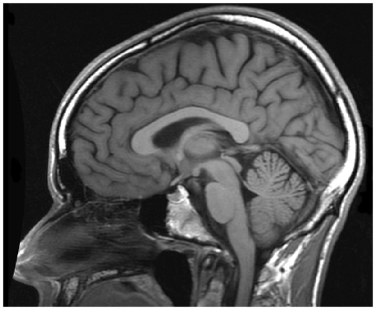
MRIMidsagittal brain MRI, T1-weighted: cerebellar atrophy, with widened sulci between the cerebellar folia.
Source : Kasperavičiūtė et al., PLoS One, 2011, figure 3 · CC BY
The cerebellum does not command movement, it calibrates it. Understanding precisely what it calibrates explains why the ataxic patient trembles on arrival and not on departure, why the base of support widens, and why some rehabilitation strategies work while others fail by construction.
A predictive corrector, not an executor
The cerebellum receives a copy of the motor command and sensory information in return, and it constantly compares the predicted movement with the movement actually performed. That comparison serves two purposes: correcting the movement under way, and updating the internal model that will serve the next one. When it is damaged, what is missing is neither strength nor the programme, it is the adjustment. The patient knows what to do and does it badly.
The heaviest clinical consequence is not motor, it is educational. Amy Bastian's work has established that cerebellar damage consistently disrupts motor adaptation, the learning that works by error correction, whereas lesions of other brain regions often spare it17. In other words: the learning route on which a large part of our neurological protocols implicitly rests is precisely the one that is broken in this patient.
"Cerebellar damage consistently disrupts adaptation; lesions of other brain regions, often, do not." Bastian, Current Opinion in Neurology, 2008
This does not condemn rehabilitation: the trials show the opposite. But it steers its content. A patient who no longer adjusts finely through error makes progress through the repetition of whole tasks, through the building of substitution strategies, and through the upkeep of the capacities that do not depend on the cerebellum: strength, endurance, flexibility, a safe environment. That is exactly the profile of the programmes that worked in the trials, and it is also why very technical, very segmental protocols disappoint.
The cerebellar syndrome is not one, it is three
The reference clinical description, restated in 2026 by Manto, Mitoma, Burt and Schmahmann, divides the cerebellar syndrome into three categories7 :
- The cerebellar motor syndrome (CMS) : dysmetria, kinetic tremor, asynergia, dysdiadochokinesia, dyschronometria. This is the side that the SARA rates.
- The vestibulo-cerebellar syndrome (VCS) : saccadic dysmetria, saccadic smooth pursuit, inferior vertical nystagmus (downbeat), gaze-evoked nystagmus. This is what the ICARS rates in part and what the SODA scale rates specifically.
- The cerebellar cognitive affective syndrome, or Schmahmann syndrome (CCAS/SS) : impairment of executive function, spatial cognition, language processing and emotional regulation.
This third category deserves a word for the physiotherapist, because it is the one most often missed and the heaviest in practical consequences. A patient whose executive functions are impaired no longer organises their home programme, no longer plans their sequences, no longer transfers an instruction from one context to another. They are labelled "poorly adherent" when they are cerebellar right down to their capacity to adhere. A dedicated scale exists, the CCAS/Schmahmann Scale8 ; its use is the neuropsychologist's business, but simply knowing of it changes the way an exercise instruction is written.
Key point
The cerebellum calibrates; it does not execute. Learning through error is the route most affected, which explains why effective programmes rest on repeating whole tasks and not on fine correction. And the cerebellar syndrome carries a cognitive and affective side that conditions adherence to the home programme.
How many patients, and which ones?
Degenerative ataxias are rare diseases, but taken together they are not so rare. The systematic review by Ruano and colleagues, which brought together 22 studies and 14,539 patients in 16 countries, puts the mean prevalence of autosomal dominant hereditary ataxias at 2.7 per 100,000 (range 1.5 to 4.0) and that of the recessive forms at 3.3 per 100,000 (1.8 to 4.9). SCA3, or Machado-Joseph disease, is the commonest dominant form, followed by SCA2 and SCA6; Friedreich's ataxia dominates the recessive forms2.
Two figures from that same review speak even more directly to practice. The first: bringing together hereditary ataxias and hereditary spastic paraplegias, the authors estimate that about one person in 10,000 is affected. The second, often left unsaid: in population series, 33 to 92 % of families with dominant transmission and 40 to 46 % of recessive families remain without an identified mutation after systematic genetic testing2. The patient who tells you "they didn't find anything" is not recounting a diagnostic odyssey: they are describing the statistical norm.
Four epidemiological markers
What the orders of magnitude are worth before reasoning about a patient
Sources: Ruano 2014, Neuroepidemiology (PMID 24603320); de Silva 2019, Orphanet J Rare Dis (PMID 30786918). The prevalences are means of ranges that are highly heterogeneous across populations and survey methods.
What has changed since 2023: SCA27B
A physiotherapy article has no business settling questions of genetics, but this one has to flag a recent shift, because it concerns exactly the patient seen in private practice: the 60-year-old adult who has been walking badly for a few years, and in whom "nothing was found".
In January 2023, a deep intronic GAA expansion of the FGF14 gene was identified as a cause of late-onset cerebellar ataxia. It was present in 61 % of French-Canadian index cases, 18 % of German, 15 % of Australian and 10 % of Indian cases of late-onset ataxia24. The clinical characterisation published the same year in 50 patients describes a late-onset pancerebellar syndrome, median age at onset 60.0 years, associated with an afferent sensory deficit in 55 % of patients and with dysautonomia in 28 %25.
Two findings from that series bear directly on rehabilitation. First the slowness: the measured progression is 0.29 SARA points a year, with a maximum score of 18 points even at advanced stages, and only half the patients use a unilateral walking aid after eight years of disease25. Then the treatment: 86 % of patients treated with 4-aminopyridine report a response that matters for their daily life, confirmed by three individual prospective experiments in on/off25. On this ground, "idiopathic" late-onset ataxia is no longer necessarily idiopathic, nor necessarily untreatable.
What this changes in the session
Faced with a late-onset cerebellar ataxia labelled "sporadic, no cause found" in a patient whose genetic work-up predates 2023, it is legitimate to suggest that the patient raise it again with their neurologist. The physiotherapist prescribes nothing and diagnoses nothing: they point out that a frequent and potentially treatable diagnostic lead did not exist at the time of the work-up.
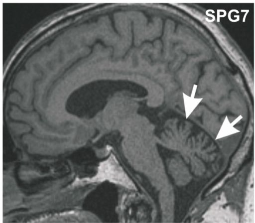
MRIMidsagittal brain MRI: cerebellar vermis atrophy with widened sulci (arrows), in a 70-year-old patient.
Source : Thal et al., International Journal of Molecular Sciences, 2015, figure 1 · CC BY
How do you tell cerebellar ataxia from proprioceptive or vestibular ataxia?
This is the question that decides everything else. The three ataxias produce an unsteady gait and an identical complaint, but they are not rehabilitated in the same way, and one of them responds far less well to coordinative training than the other two. The sorting is done with your hands, a chair, and five minutes.
The principle: which channel is missing, and what compensates
Balance rests on three inputs, visual, vestibular and somatosensory, and on one integrator, the cerebellum. Each ataxia corresponds to the failure of one link, and is signalled by the compensation the patient sets up spontaneously.
- Proprioceptive (or sensory, or afferent) ataxia. The somatosensory input is missing. Vision compensates, effectively. The patient watches their feet, walks better in full light, and comes apart in the dark or in the shower. This is the mechanism of sensory neuropathies, posterior column lesions and certain deficiency states.
- Vestibular ataxia. The vestibular input is missing or delivers a false signal. The patient has spinning symptoms, nystagmus, a lateralised deviation, and compensates by fixing on the horizon.
- Cerebellar ataxia. It is the integrator that is damaged: all three inputs arrive, but they are no longer fused or converted into a calibrated command. No sensory compensation is possible, since nothing is missing upstream. That is why the patient is already unstable with the eyes open.
The proprioceptive patient deteriorates when you close their eyes. The cerebellar patient was already unstable before you closed them.
The Romberg, and what it is wrongly made to say
The Romberg test, standing with the feet together and the eyes closed, is taught as a test of proprioception, and so it is. But a review by Halmágyi and Curthoys published in 2021 recalls a decisive nuance: standing with the eyes closed on a soft surface rather than on a firm floor tests vestibular function more than it tests proprioception18. The foam, added to "toughen" the test, changes the question being asked. A pathological Romberg on foam in a patient who is normal on a firm floor points to the vestibule, not to the posterior columns.
Three practical points follow from this:
- Do the Romberg on a firm floor first, and record performance with the eyes open and with the eyes closed separately. It is the gap between the two that informs, not the absolute value.
- Do not conclude "positive Romberg" from mere unsteadiness. In the cerebellar patient there is sway with the eyes open and a slight worsening on closing them. In the proprioceptive patient the worsening is marked, often with a fall if you are not there.
- Add the foam afterwards, as a second question and not as a hardening of the first.
The sorting table
| Item | Cerebellar | Proprioceptive | Vestibular |
|---|---|---|---|
| Spontaneous complaint | "I walk like a drunk", clumsy hands | "I can't feel my feet any more", worse in the dark | "The room spins", "I veer off to one side" |
| Effect of closing the eyes | Slight worsening; the instability was already there | Major worsening, possible fall | Clear worsening, often lateralised |
| Romberg (firm floor) | Unstable with the eyes open, little difference | Normal or near-normal with the eyes open, frankly pathological with the eyes closed | Moderate difference, deviation to one side |
| Distal deep sensation | Normal (except in mixed forms) | Impaired: position sense, vibration sense | Normal |
| Dysmetria, dysdiadochokinesia, intention tremor | Present, including with the eyes open | Absent; the error is corrected by looking | Absent |
| Oculomotor function | Saccadic pursuit, gaze-evoked nystagmus, possible downbeat nystagmus | Normal | Unidirectional nystagmus, abnormal head impulse on the affected side |
| Speech | Cerebellar dysarthria (scanning, irregular) | Normal | Normal |
| Gait | Widened base, unsystematised lurches, increased intra-limb temporal variability | Stamping, controlled by vision, widened base | Reproducible deviation to one side, star-shaped walking |
| Expected response to coordinative training | Good (the population of the positive trials) | Lower: patients with afferent involvement make less progress | Primarily a matter for vestibular rehabilitation |
The last row of this table is not an opinion. In the founding trial by Ilg and colleagues, the patients whose afferent pathways were also affected made less progress than patients with pure cerebellar ataxia, in gait speed, lateral sway and intra-limb coordination; and balance regulation in static and dynamic tasks improved significantly only in the patients without afferent involvement11. The differential diagnosis is therefore not an academic exercise: it predicts part of the response to treatment.
A gait signature specific to the cerebellum
There is a kinematic marker that separates cerebellar ataxia from the other causes of instability, and it lights up clinical reasoning even without a gait laboratory. Ilg and colleagues compared the gait of 13 patients with cerebellar degeneration, 6 patients with peripheral vestibular loss and 8 patients with Parkinson's disease. All three groups shared balance-related gait abnormalities and increased general variability. But the increase in the temporal variability of intra-limb coordination was specific to cerebellar damage, markedly smaller in the two other groups16.
Translated into clinical observation: in a vestibular or parkinsonian patient, the instability produces an irregular step but a rhythm that is relatively preserved. In the cerebellar patient it is the tempo itself that breaks down: the stance and swing phases do not last the same from one step to the next. That is what you hear, literally, when you listen to an ataxic patient walking down a corridor.
Sorting tree at the first examination
Four questions asked in order, before any further investigation
Sorting tree built from the description of the cerebellar syndrome in Manto 2026 (PMID 41663552), the Ataxia UK guidelines of de Silva 2019 (PMID 30786918) and the criteria for urgent referral of Kattah 2009 (PMID 19762709). It steers the rehabilitation professional's reasoning; it does not replace the medical diagnostic process.
The trap of mixed forms
The tree above files patients into one box; clinical practice puts many of them into two. Friedreich's ataxia combines cerebellar and afferent pathway involvement from the outset. SCA27B carries an afferent sensory deficit in 55 % of patients25. A diabetic neuropathy can be superimposed on any degenerative ataxia. The right question is therefore not "which of the three?" but "which one dominates, and is there a second?", because an associated afferent component signals a poorer response to coordinative training11 and calls for a substitution strategy to be added.
Key point
The differential turns on three observations: how much the patient leans on vision (the gap between eyes open and eyes closed on a firm floor), the presence of cerebellar motor signs in the limbs and in speech, and the state of distal deep sensation. The Romberg on foam asks a different question from the Romberg on a firm floor: it interrogates the vestibule. And an associated afferent component is no detail: it changes the rehabilitation prognosis.
Which scale should be used to rate ataxia: SARA, ICARS or BARS?
Three scales are in circulation, all valid, all published, and one of them is faster than the others. That does not make it the one the learned societies recommend. Here is what each of them measures, what their measurement properties are worth, and which one really serves physiotherapy follow-up.
What the Movement Disorder Society says
The Movement Disorder Society scales review committee sifted through 14 instruments, 9 rating scales and 5 functional tests, and classified them as "recommended", "suggested" or "listed" according to pre-set criteria. The result, published in Movement Disorders in 2021, is clear-cut6 :
- Spinocerebellar ataxias : ICARS and SARA "recommended".
- Friedreich's ataxia : FARS, ICARS and SARA "recommended".
- Ataxia-telangiectasia : ICARS and SARA.
- Brain tumour ataxia : SARA.
- Cerebellar signs in multiple sclerosis : ICARS.
- Multiple system atrophy, cerebellar type : UMSARS and ICARS.
- FXTAS syndrome : ICARS. CDG-PMM2 : ICARS.
The BARS appears in none of these recommendation lists. This is not a judgement on its intrinsic quality: it was validated by its authors, and its psychometric properties are good. It is an observation about status: in 2026, the reference scale for documenting an ataxia in a record, a prior-approval request or a publication remains the SARA, or possibly the ICARS.
The SARA in detail
The SARA (Scale for the Assessment and Rating of Ataxia) was developed and tested on two series of 167 and 119 patients with spinocerebellar ataxia. Its published characteristics10 :
- Administration time: 14.2 ± 7.5 minutes (range 5 to 40 minutes).
- Inter-rater reliability: ICC = 0.98. Test-retest reliability: ICC = 0.90.
- Internal consistency: Cronbach's alpha = 0.94, and factor analysis finds a single factor: the SARA measures one thing only.
- Linear relationship with global assessment on a visual analogue scale (r² = 0.98), strong correlation with the Barthel index (r = −0.80) and with part IV of the UHDRS (r = −0.89), but weak correlation with disease duration (r = 0.34).
This last point deserves a pause. A scale that correlates strongly with independence and weakly with how long the disease has lasted is exactly what follow-up needs: it describes a functional state, not a calendar. Two patients at the same chronological stage can have very different SARA scores, and it is the SARA that is right.
For the physiotherapist, one further argument, noted by Marquer, Barbieri and Pérennou in their systematic review: 18 of the SARA's 40 points bear on postural disorders (gait, standing and sitting)9. Nearly half the score is therefore played out on exactly the ground of rehabilitation. The remaining 22 points are split between speech and the four limb coordination tasks.
| Item | Points | What it captures for the rehabilitation professional |
|---|---|---|
| Gait | 0–8 | The heaviest item on the scale: base of support, tandem, independence in getting about |
| Standing | 0–6 | Feet apart, feet together, tandem: the very progression of our exercises |
| Sitting | 0–4 | Trunk ataxia, the one that responds best to intensive rehabilitation |
| Speech | 0–6 | Dysarthria; a matter for the speech and language therapist but rated here |
| Finger chase | 0–4 | Upper limb dysmetria, rated on the right and on the left then averaged |
| Finger-to-nose test | 0–4 | Intention tremor |
| Fast alternating hand movements | 0–4 | Dysdiadochokinesia |
| Heel-shin slide | 0–4 | Lower limb coordination, predictive of gait |
| Total | 0–40 | Of which 18 points postural (gait, standing, sitting) |
Where the SARA score is played out
Distribution of the 40 points between posture, limb coordination and speech
Scoring after Schmitz-Hübsch 2006, Neurology (PMID 16769946); the count of 18 postural points is that of Marquer 2014, Ann Phys Rehabil Med (PMID 24582474). The four limb tasks are rated on the right and on the left then averaged, hence 16 points and not 32.
ICARS and BARS: what they are for
The ICARS (International Cooperative Ataxia Rating Scale) is the oldest of the three. Proposed in 1997 by an ad hoc committee of the World Federation of Neurology, it is semi-quantitative, scored out of one hundred points, and compartmentalised: posture and stance disorders, limb ataxia, dysarthria and oculomotor disorders are quantified separately, each compartment carrying a deliberately calibrated weight31. It therefore covers both the cerebellar motor syndrome and the vestibulo-cerebellar syndrome7, which explains why it remains the only one "recommended" in the indications where oculomotor function weighs heavily: multiple sclerosis, FXTAS, multiple system atrophy. Its cost in time, however, makes it barely realistic in private practice.
The BARS (Brief Ataxia Rating Scale) was built in 2009 by Schmahmann and his team precisely to lift that obstacle. Their method is interesting: the authors first extended the ICARS by adding seven tasks (the MICARS), then searched computationally for the subset of five tasks that correlated best with the total, under the clinical constraint of keeping one per domain: gait, upper limb, lower limb, speech, oculomotor function. The result comes down to five tasks scored out of 30 points in all (gait 0 to 8, heel-shin and finger-to-nose rated on each side, dysarthria 0 to 4, oculomotor function 0 to 2), and to three figures13 : a correlation of 0.952 between the BARS and the rest of the MICARS; a Cronbach's alpha of 0.90 for the BARS against 0.92 for the SARA ; inter-rater reliability of 0.91 against 0.93. The authors conclude that the BARS is "valid, reliable, and sufficiently rapid and accurate for clinical use".
Two honest readings of this result sit side by side. The first: the BARS does almost as well as the SARA with fewer tasks, and it has the merit of including oculomotor function, which the SARA ignores. The second: "almost as well" on properties that are already excellent does not make up for the absence of a formal recommendation and for the thinner body of longitudinal data. In private practice the reasoning settles simply: the SARA remains the follow-up scale, the BARS can serve as a rapid screen when time is short, provided that measurements made with different scales are never compared with one another.
| Criterion | SARA | ICARS | BARS |
|---|---|---|---|
| Year | 2006 | 1997 | 2009 |
| Total score | 0 to 40, 8 items | 0 to 100, 19 items | 0 to 30, 5 tasks |
| Duration | 14.2 ± 7.5 min (5 to 40) | Clearly longer | Not quantified by the authors |
| Oculomotor function rated | No | Yes | Yes (1 task) |
| Inter-rater reliability | ICC 0.98 (0.93 in direct comparison) | Good, long established | ICC 0.91 |
| Internal consistency | α 0.94 (0.92 in direct comparison) | Good | α 0.90 |
| MDS status 2021 | Recommended (SCA, Friedreich, A-T, tumours) | Recommended (SCA, Friedreich, A-T, MS, MSA-C, FXTAS, CDG) | Absent from the recommendation lists |
| Relevant physiotherapy use | Reference follow-up | Research, oculomotor function | Rapid screening |
The trap of the clinical significance threshold
It is tempting to tell the patient "your score has dropped by 2 points, that is clinically significant". No threshold for a minimal clinically important difference commands consensus for the SARA in degenerative cerebellar ataxia. What does exist, by contrast, are benchmarks of natural progression by genotype5, together with a judgement of clinical responsibility. The honest wording is: "your score has improved by 2 points, whereas an untreated SCA3 gains 1.6 a year on average". That is not a threshold, it is a comparison, and it is far more informative.
What the SARA does not rate, and what has to be rated elsewhere
Three blind spots, all of them clinically weighty:
- Oculomotor function. The vestibulo-cerebellar syndrome escapes the SARA entirely. A dedicated scale has existed since 2022, the SODA (Scale for Ocular motor Disorders in Ataxia)14.
- Cognition and affect. The CCAS/Schmahmann Scale explores them8 ; a review by the Task Force of the Society for Research on the Cerebellum has set out its conceptual framework15.
- The patient's lived experience. No clinician-rated scale says what the disease does to a life. Patient-reported measures, among them the PROM-Ataxia cited in the 2026 review of the scales7 fill this gap.
In everyday practice, though, the most profitable addition is none of these scales: it is a functional goal written with the patient. The Ilg 2009 trial and the Ilg 2010 trial both used a goal attainment score for each individual, and that is where the most telling gains were observed: the ability to achieve personally meaningful goals of everyday life11.
Key point
SARA for follow-up, systematically, because it is recommended, quick, reproducible (ICC 0.98) and 45 % postural. ICARS when oculomotor function counts. BARS as a screen, never mixed with SARA scores on the same curve. And a personal goal attainment scale alongside it, because that is the one that speaks to the patient.
What goes into a physiotherapy assessment of ataxia that is of any use?
An assessment serves to decide and to compare. Anything that does neither costs session time. Here are the five measurements that carry the treatment decision, and the one history question that must never be asked open-endedly.
The question of falls, asked properly
"Do you fall?" gets "no, not really" from patients whose prospective diary shows the opposite. In the prospective study run within the EuroSCA cohort, 113 patients with SCA1, SCA2, SCA3 or SCA6 kept a falls diary between two annual visits: 84.1 % reported at least one fall over twelve months, injuries were frequent and their frequency rose along with the frequency of falls26. The authors had in fact run the same study retrospectively beforehand, precisely because recall bias made the reported figures unreliable.
One result from that same study steers the assessment: the presence of non-ataxic symptoms was associated with a higher frequency of falls26. In other words, the ataxia score is not enough to predict fall risk: it is the associated neurological comorbidities (spasticity, neuropathy, oculomotor disorders, cognitive impairment) that tip the balance. Earlier work by the same team had already established that falls are very frequent in degenerative cerebellar ataxias, often serious, and a source of fear of falling27.
In practice: ask how many falls there have been over the last three months, in what circumstances, with what consequences, and offer a diary. The diary is not a research gadget, it is the only instrument that makes the real frequency visible to the patient themselves.
The tandem, the earliest test
Tandem walking, one foot in front of the other, heel against toes, deserves a place of its own. The international consensus on ataxic gait establishes that it is impaired at the early stages of cerebellar disorders and can be particularly useful for assessing the pre-ataxic stages of progressive ataxias30. It is the test that gives way first, before free walking deteriorates. Its simplest measure: the number of consecutive steps taken without a deviation, out of the ten asked for.
That same consensus recalls a fact that structures the whole of management: ataxia of stance and gait is one of the very first deficits to appear in degenerative cerebellar disorders, it is disabling and it carries a high risk of falling30. Gait is not one goal among others, it is the goal.
The five measurements that are enough
| Measurement | Time | What it decides |
|---|---|---|
| SARA | ≈ 15 min | Overall severity and longitudinal follow-up. The only measurement that is comparable from one practitioner to another and from one year to the next. |
| Tandem walking, 10 steps | 1 min | Detects early involvement; serves as a training target that transfers immediately. |
| Berg Balance Scale | ≈ 15 min | Responsiveness to change demonstrated in meta-analysis (+2.6 points under therapeutic exercise); guides the progression of balance exercises. |
| Number of falls over 3 months, by diary | Continuous | Decides the walking aid, home safety measures, and the intensity of balance work. |
| Two to three written functional goals | 10 min | They carry adherence to the home programme; it is on this type of measure that the trials show the gains that matter most to the patient. |
A word about the functional independence measure (FIM). The Milne 2025 multicentre trial had made it its primary outcome, in its motor domain, the mFIM, and the result is worth knowing: rehabilitation improves it significantly at 7 weeks (mean difference 2.26 points, 95 % CI 0.26 to 4.26; p = 0.028) but no longer significantly at 30 weeks (1.74; 95 % CI −0.32 to 3.81; p = 0.098), even though the SARA score does remain improved at 30 weeks4. The two measures do not say the same thing and do not decay at the same rate: documenting both avoids concluding too quickly one way or the other.
Key point
Five measurements are enough: SARA, tandem, Berg, falls diary, written goals. The question of falls is asked in numbers and not as "yes/no". The tandem gives way before free walking does. And non-ataxic comorbidities predict fall risk better than the ataxia score itself.
Which red flags in an ataxia of rapid onset?
A degenerative ataxia sets in over years. Any other speed of onset changes the nature of the problem, and sometimes the time scale available for acting. This is the chapter to reread before taking on a new patient.
The speed of onset is the first sort
| Time frame | What to consider | What the physiotherapist should do |
|---|---|---|
| Minutes to hours | Cerebellar stroke (ischaemic or haemorrhagic), trauma, acute poisoning | Urgent referral. Do not delay, do not "see how it is at the next session" |
| Hours to days | Wernicke encephalopathy (thiamine deficiency), infectious cause, drug iatrogenesis (antiepileptics, lithium), withdrawal | Same-day referral; thiamine deficiency is treatable and delay costs dearly |
| Weeks to months | Paraneoplastic cerebellar degeneration, autoimmune ataxia, posterior fossa space-occupying lesion, deficiency state | Written report to the general practitioner, cautious continuation of treatment |
| Months to years | Hereditary and sporadic degenerative ataxias, multiple system atrophy, chronic toxic cause | The usual field of rehabilitation |
| Episodic, in attacks | Episodic ataxias; episodic ataxia type 2 responds to acetazolamide and to the aminopyridines | Neurological referral: this is one of the few pictures with an established drug treatment |
Red flags: stop the session and refer
- Ataxia that has set in over a few hours, all the more so in a patient with vascular risk factors.
- Associated severe headache, vomiting, altered alertness: the oedema of a cerebellar infarction can compress the brainstem and threaten life.
- Recent-onset downbeat nystagmus, new diplopia, dysphagia or dysphonia.
- Rapid worsening over a few weeks in a patient stable until then, especially with weight loss: paraneoplastic degeneration often precedes the discovery of the cancer.
- Confusion, oculomotor disorders and ataxia in a malnourished or alcohol-dependent patient, after bariatric surgery or prolonged vomiting : consider Wernicke encephalopathy, a therapeutic emergency.
- Fever, meningeal signs, or any febrile ataxia.
A normal MRI is no reassurance: the case of acute vestibular syndrome
The physiotherapist regularly meets the patient who was "seen in the emergency department, scan normal, they told me vestibular neuritis". Two publications call for caution.
The first is the original study of the HINTS examination. Kattah and colleagues prospectively examined 101 high-risk patients presenting with an acute vestibular syndrome (vertigo, nystagmus, nausea, intolerance of head movement, unsteadiness), of whom 25 turned out to have peripheral involvement and 76 central involvement, including 69 ischaemic strokes. The combination of three signs, a horizontal head impulse test that was normal, direction-changing nystagmus on eccentric gaze, or skew deviation was 100 % sensitive and 96 % specific for stroke. And above all: the initial diffusion MRI was falsely negative in 12 % of cases, all of them performed less than 48 hours after the onset of symptoms19.
A three-minute clinical examination proved more sensitive than early diffusion MRI for detecting stroke in acute vestibular syndrome.
The second publication tempers the first, and it would be dishonest to cite only one of them. A 2020 systematic review with meta-analysis, covering 5 studies and 617 participants, compared the performance of the HINTS examination according to who carried it out. Performed by neurologists: sensitivity 96.7 % (95 % CI 93.1 to 98.5) and specificity 94.8 % (91 to 97.1). Performed by a cohort that included emergency physicians: sensitivity 83 % (63 to 95) and specificity 44 % (36 to 51)20. The authors conclude that the HINTS examination, used on its own by emergency physicians, has not demonstrated sufficient accuracy to rule out a stroke.
The HINTS examination is worth only as much as the examiner
Sensitivity and specificity for stroke in acute vestibular syndrome, according to who performs the examination
Source: Ohle 2020, Acad Emerg Med 27(9):887-896, a meta-analysis of 5 studies and 617 participants (PMID 32167642). The cohort "including emergency physicians" combined board-certified emergency physicians with neurologists subspecialised in neuro-otology or vascular neurology; the fall in specificity from 94.8 % to 44 % means that a badly conducted examination classes more than one peripheral patient in two as "central".
The lesson for the physiotherapist is twofold and unambiguous. This is not a test you make your own by reading an acronym : its performance collapses outside trained hands, and a "reassuring" result badly obtained is more dangerous than no test at all. But knowing its components remains useful: knowing that a head impulse test that is normal in an acutely vertiginous patient is a worrying sign and not a reassuring one is exactly the kind of counter-intuition that makes you ask for a further opinion.
The counter-intuition to remember
In acute vestibular syndrome, a head impulse test that is abnormal points to a peripheral cause, and is therefore rather reassuring. A test that is normal in a patient with frank vertigo points to a central cause. This is the opposite of the spontaneous reflex, and it is the reason why the HINTS examination is not performed from memory.
The reversible causes, which on their own justify vigilance
The British guidelines on the progressive ataxias, drawn up by more than 30 health professionals and published in 2019, insist on a point that concerns everyone: more than a hundred different diseases can cause an ataxia, there is no disease-modifying treatment for most of them, but many aspects are treatable, and the early diagnosis of the few reversible causes is of paramount importance28.
The physiotherapist does not have to make these diagnoses. They do, however, hold a privileged observation post: they see the patient repeatedly, over time, and are often the first to notice a change of trajectory. A patient whose SARA gains three points in three months does not have a degenerative ataxia running its course: they have something else, and the only right answer is a letter to the general practitioner.
Key point
The speed of onset sorts better than any sign. Hours: vascular emergency, early MRI falsely negative one time in eight. Days: think thiamine and iatrogenesis. Weeks: think paraneoplastic. And an unexpected acceleration in a patient already under care is grounds for a letter, not for intensifying the programme.
What does rehabilitation really change, and by how much?
The question is not whether rehabilitation "does good". It is by how many scale points, for how long, at what price in intensity, and how that compares with the speed at which the disease advances. The figures exist, they are modest, and they are more interesting than unmeasured enthusiasm.
The starting point: it is the only treatment
The 2014 international consensus on the management of degenerative cerebellar disorders, signed by fifteen teams, reaches a conclusion that has not been contradicted since: no drug has proved its efficacy, with the exception of the aminopyridines and acetazolamide in episodic ataxia type 2 and of the aminopyridines in certain downbeat nystagmus: "all authors agreed that the mainstays of the treatment of degenerative cerebellar ataxia are currently physiotherapy, occupational therapy and speech and language therapy"1.
The same text adds a sentence that this chapter sets out to honour: there is consensus that evidence-based guidelines for the physiotherapy of degenerative cerebellar ataxia remain to be developed1. Twelve years later, they still do not exist. What we have are trials and two meta-analyses.
The three founding trials
Ilg 2009: intensive coordinative training
Sixteen patients with progressive ataxia from cerebellar degeneration, four weeks of intensive coordinative training, an intra-individually controlled protocol in which each patient was their own control, with four assessments: eight weeks before, just before, just after, and eight weeks after the training. Result: significant improvement in motor performance and a reduction of ataxic symptoms on the clinical scales, maintained at the eight-week check11.
The authors conclude in terms worth quoting precisely: coordinative training improves motor performance and reduces symptoms, "enabling them to achieve personally meaningful goals of everyday life", and "for both groups, continuous training seems crucial to stabilise the improvements and should become a standard of care"11. Level of evidence stated by the review: class III.
Ilg 2010: durability at one year
The same team reassessed these patients one year after the intensive four-week block, which had been followed by a home exercise programme. The result is nuanced, and that is what makes it valuable: despite a progressive decline in motor performance and a progressive increase in ataxic symptoms due to disease progression, the improvements in performance and the gains in activities of daily living persisted12.
The disease went on advancing; the patient stayed above the trajectory they would have followed without training. That is the right mental model for talking to the patient: not "we are going to recover", but "we are going to shift the curve".
Miyai 2012: the randomised trial of intensive rehabilitation
Forty-two patients with pure cerebellar degeneration, randomised between an immediate group and a delayed-entry control group. The intervention: two hours of inpatient physiotherapy and occupational therapy a day on weekdays and one hour at the weekend, for four weeks, centred on coordination, balance and activities of daily living. Results21 :
- Significantly greater functional gains in the immediate group, on ataxia, gait speed and activities of daily living.
- The improvement in trunk ataxia was more marked than the improvement in limb ataxia.
- The gains on ataxia held at 12 weeks, those on gait at 24 weeks.
- At 24 weeks, 22 patients out of 42 still had at least one measure better than at inclusion; the authors note that functional status tended overall to return to baseline within 24 weeks.
It is this trial that the American Academy of Neurology retained, in its 2018 systematic review, to state that in patients with degenerative ataxias, four weeks of inpatient rehabilitation probably improves ataxia and function (a single class I study)29. That review now carries the label [RETIRED] in PubMed: the AAN has withdrawn it from its documents in force. It remains a valid historical source, it is no longer an active recommendation, and it would be misleading to cite it as one.
What the meta-analyses say
Three quantitative syntheses frame the question today, and they do not give the same figure, which is in itself a piece of information.
Milne 2017 reviewed 17 studies and 292 participants with genetic degenerative ataxias (148 dominant, 85 recessive), including 5 randomised trials, most of the work being of level III-3 or IV. Fifteen of the seventeen studies showed a statistically significant improvement on at least one measure of ataxia, function, gait or balance. The authors' conclusion: "there is consistent evidence that rehabilitation improves function, mobility, ataxia and balance". Major reservation, stated by them: fewer than half the studies assessed long-term outcomes23.
Winser 2023 meta-analysed 8 of the 26 studies included, with GRADE assessment. "Low" to "moderate" quality evidence in favour of therapeutic exercise for reducing disease severity as measured by the SARA: weighted mean difference −3.3 points (95 % CI −3.7 to −2.8; p < 0.01), and for improving balance as measured by the Berg scale: +2.6 points (95 % CI 1.1 to 4.2; p < 0.01). By contrast, the effect on functional independence is not significant : +1.6 points (95 % CI −1.5 to 4.6; p = 0.31)22.
Matsugi 2024 is the most recent and the most cautious. Eighteen randomised trials, 398 participants, of whom 315 were assessed with the SARA and included in the meta-analysis, with prior registration on the PROSPERO register. Overall effect of physiotherapy: −1.41 SARA points (95 % CI −2.16 to −0.66). The authors describe the risk of bias as "serious" and the certainty of the evidence as "low" to "very low", with high heterogeneity3.
The gap between Winser's −3.3 and Matsugi's −1.41 is not a contradiction: the populations, the inclusion criteria and the periods are not the same, and Matsugi restricted himself to randomised trials in cerebellar ataxia that is degenerative. The cautious reading is to keep the lowest and the most recent estimate (a gain of the order of 1.4 SARA points, with a low certainty of evidence).
The trial that was missing: Milne 2025
Published in Annals of Neurology, this multicentre, randomised, single-blind superiority trial is the most robust work available. Five Australian sites, participants with dominant or recessive hereditary ataxia, aged 15 years and over, randomised 1:1 between 6 weeks of outpatient physiotherapy followed by 24 weeks of a home exercise programme (n = 39) and continuation of usual activity (n = 37). Intention-to-treat analysis on 71 participants, with blinded assessors4.
- At 7 weeks : mFIM improved by 2.26 points (95 % CI 0.26 to 4.26; p = 0.028) and SARA by −1.21 points (95 % CI −2.32 to −0.11; p = 0.032).
- At 30 weeks : SARA held at −1.51 points (95 % CI −2.76 to −0.27; p = 0.017), but mFIM not significant (1.74; 95 % CI −0.32 to 3.81; p = 0.098).
- Frequent adverse effects in both groups : fatigue, pain and falls.
This last point is to be read carefully. Falls and fatigue are among the frequent adverse events on both sides (in the patients who were rehabilitated as much as in those who carried on with their usual activity). Rehabilitating an ataxic patient is not a risk-free activity, and saying so to the patient is part of the contract.
The result one would rather keep quiet
Seven years earlier, the same team had run a delayed-start randomised trial in Friedreich's ataxia: six weeks of outpatient rehabilitation followed by six weeks of a home programme. It included only 19 participants: of 159 people assessed for eligibility, 92 were excluded and 48 declined to take part. Result: no significant difference on the functional independence measure between the groups at six weeks (2.00 ± 3.16 against 0.56 ± 4.06). One quality-of-life subscale, the "body movement" domain of the Friedreich Ataxia Impact Scale, did by contrast improve significantly (p = 0.003). And above all: the within-group improvements obtained after the supervised phase were not maintained after the home programme phase39.
Two lessons follow, and neither is comfortable. The first concerns recruitment: when 140 people out of 159 do not take part, the trial population is not the practice population. The second concerns the home programme: prescribing it is not enough for it to preserve the gains. In the Ilg 2010 trial it preserved them over a year; in this one it did not preserve them over six weeks. The difference probably lies in the support given to the programme rather than in its content, and that is exactly where the physiotherapist's work is played out.
The effect of rehabilitation, on the scale of natural progression
SARA points gained each year by the disease, compared with the points taken back by rehabilitation
Annual progressions: Jacobi 2015, Lancet Neurol, EUROSCA cohort, 462 patients followed for a median of 49 months (PMID 26377379); SCA27B: Wilke 2023, Brain, 50 patients (PMID 37165652). Rehabilitation gains: Matsugi 2024 (PMID 39866519) and Milne 2025 (PMID 39520242). A comparison of orders of magnitude: rehabilitation gains do not add up year after year, and natural progression is not suspended during treatment.
The comparison that gives it meaning
A gain of 1.4 SARA points, on its own, says nothing to anyone. Set against the speed at which the disease advances, it takes on immediate meaning. The European EUROSCA cohort followed 462 patients with at least one follow-up visit, over a median observation period of 49 months, and established an annual progression of the SARA score5 of 2.11 points for SCA1, 1.56 for SCA3, 1.49 for SCA2 and 0.80 for SCA6.
The right thing to say to the patient then becomes: "what we get through rehabilitation is roughly the equivalent of one year of progression of your disease. It does not stop it, and you will have to keep going to hold on to that gain." It is true, it is quantified, and it is more motivating than a vague promise.
Do not turn a comparison into an equivalence
Saying that rehabilitation "takes back a year of disease" is an image of orders of magnitude, not a trial result. No trial has shown that the gain accumulates from year to year, nor that the trajectory of progression is slowed. What has been shown is narrower and more solid: a one-off gain of the order of 1.2 to 1.5 SARA points, maintained up to 30 weeks when the home programme is continued.
Key point
Rehabilitation is the mainstay treatment, for want of a drug. The measured effect is of the order of −1.4 SARA points in meta-analysis, with a serious risk of bias and low certainty, and of −1.5 points at 30 weeks in the best trial. It compares with a natural progression of 0.8 to 2.1 points a year depending on genotype. Trunk ataxia responds better than limb ataxia. And without a maintenance programme, the benefit fades within a few months.
Which modalities for which level of evidence?
Session time is a scarce and non-expandable resource. This chapter allocates it according to what has been measured, distinguishing what has a demonstrated effect, what has none, and what is still entirely unknown: three categories that are readily confused.
The effects by modality, as measured
The subgroup analysis of the Matsugi 2024 meta-analysis is the most directly usable document, because it compares the modalities with one another on the same outcome measure, the SARA3.
Effect of rehabilitation modalities on the SARA score
Mean differences and 95 % confidence intervals. To the left of zero: improvement
Single source: Matsugi 2024, Front Neurol 15:1491142, subgroup analysis (PMID 39866519). PROSPERO registration CRD42024493883. The authors describe the risk of bias as serious, the certainty of the evidence as low to very low, and the heterogeneity as high: these estimates are liable to change. The dual-task interval (−6.4 to +6.88) runs off the frame on both sides, which the arrows mark.
Three readings force themselves on you in front of this graph.
First, the three effective modalities are effective within a hair's breadth of one another : −1.65 for aerobic training, −1.59 for multi-component training, −1.58 for balance. None dominates the others. There is no superior "method" to be discovered, there is training time to be spent.
Second, the confidence interval for multi-component training is very wide (−5.15 to −0.03): the upper bound skims zero. The estimate is significant by a whisker, on five heterogeneous trials. It has to be treated as such.
Third, dual tasking is the only modality whose point estimate goes the wrong way (+0.24), on a single trial and with an interval that covers practically the whole imaginable spectrum (−6.4 to +6.88). That is not proof of ineffectiveness, it is a total absence of information. Faced with a patient who falls, devoting session time to an unevaluated modality when three evaluated ones exist is a choice you must be able to justify.
The modality by level of evidence table
| Modality | Measured effect | Certainty | What can be done with it |
|---|---|---|---|
| Multi-component training (strengthening, coordination, gait, activities of daily living) | −1.59 SARA pts (−5.15 to −0.03) | Low | The backbone of the programme. It is the format of the trials that produced the best functional results |
| Balance training | −1.58 SARA pts (−2.55 to −0.62); Berg +2.6 (1.1 to 4.2) | Low to moderate | Systematic, progressive, with the working area genuinely made safe |
| Aerobic training | −1.65 SARA pts (−2.53 to −0.77); 1.9 pts vs 0.6 in a direct trial | Low | The modality most under-prescribed relative to its level of evidence |
| Intensive coordinative training (4 weeks) | Significant improvement, maintained at 8 weeks and at 1 year with maintenance work | Low (class III) | The starting format of choice when the organisation allows it |
| Intensive inpatient rehabilitation (2 h/day, 4 weeks) | Gains on ataxia, gait speed and daily life; trunk > limbs | Moderate (1 class I trial) | A stay in a rehabilitation centre to be discussed at stages of worsening |
| Exergames at home | Improved postural control in patients at an advanced stage | Very low (intra-individually controlled trial, n = 10) | A maintenance option for motivated and equipped patients |
| Dynamic trunk orthoses | Reduced trunk sway and step variability | Very low (uncontrolled longitudinal study, n = 11) | A lead, not a standard |
| Vibration | −0.56 SARA pts (−2.05 to 0.93) | Not significant | Do not devote session time to it |
| Dual-task training | +0.24 SARA pts (−6.4 to 6.88) | Not significant, 1 trial | No data; do not present it as evidence-based |
| Body-weight-supported treadmill | No significant effect after cerebellar stroke in the only controlled series | Very low | Do not make it the main axis |
| Cerebello-spinal transcranial stimulation (tDCS) | Improvement in SARA, ICARS, 8 m walk time; long-term motor and cognitive effects | Moderate (class II then a trial of 61 patients) | Outside the physiotherapy scope of practice in France; worth knowing, because the patient will hear about it |
The grading, in plain terms
MODERATE
Four weeks of intensive multidisciplinary rehabilitation. A class I randomised trial (42 patients) shows gains on ataxia, gait speed and independence, with the trunk responding better than the limbs21. A recent multicentre trial confirms the effect in a prolonged outpatient format4.
Cerebello-spinal direct current stimulation. A class II crossover trial35 then a trial of 61 patients with one-year follow-up36. Outside the physiotherapist's scope of practice in France.
LOW
Therapeutic exercise in general (multi-component, balance, aerobic). A significant but modest and heterogeneous effect, in trials at serious risk of bias322. It is the foundation of the programme, and it is also the foundation whose evidence is the most fragile, both at once.
Four weeks of coordinative training followed by a home programme, with the gains persisting at one year despite disease progression12.
VERY LOW
OR NONE
Whole-body vibration : effect not significant in meta-analysis (2 trials)3. The AAN review already concluded that the data were insufficient to support or refute stochastic whole-body vibration29.
Dual-task training : one trial, null effect, an uninterpretable confidence interval3.
Dynamic trunk orthoses : an objectively measured reduction in trunk sway and in swing phase variability, but in an uncontrolled longitudinal study of 11 patients37.
Pressure splints in multiple sclerosis ataxia : possibly without additional benefit compared with neuromuscular rehabilitation alone (1 class II study)29.
The particular case of aerobic training
This is the surprise of the recent literature, and it deserves some space, because it is counter-intuitive: why would pedalling improve an ataxia?
A randomised trial, single-blind for the assessor, compared six months of home aerobic training (19 patients) with balance training (17 patients) in ambulant patients with cerebellar ataxia. Thirty-one participants completed the trial, adherence exceeded 70 %, and there were no serious adverse events related to the training. Mean improvement: 1.9 SARA points (standard deviation 1.62) in the aerobic group against 0.6 points (standard deviation 1.34) in the balance group. The authors conclude that a phase III trial is warranted32.
One must be careful not to over-interpret a feasibility trial of 36 randomised patients, and its authors are explicitly careful themselves. But two practical lessons stand up. The first: home aerobic training is feasible and safe in the ambulant ataxic patient, with high adherence. The second: it is the modality that costs the least in supervision and is the easiest to keep up over time, and keeping it up over time is what decides the result.
The modality that produces the best result is not the most sophisticated one: it is the one the patient will still be doing in six months.
And in children, and at advanced stages?
Two populations are regularly left aside, and two studies document them.
In children, a trial showed that coordinative training based on body-controlled video games improves ataxia in children with degenerative ataxia34. The 2014 consensus already relied on this result to state that coordinative training improves motor function in the adult as well as in the juvenile patient1.
At advanced stages, where people give up soonest, a rater-blinded, intra-individually controlled trial studied ten young subjects with advanced degenerative ataxia, unable to stand or barely able to do so, with twelve weeks of home training using body-controlled video games, structured in two six-week phases so as to fit the training to individual progression. Postural control improved33. Ten patients do not make a proof, but they are enough to forbid the sentence "at this stage there is nothing more to be done".
Key point
Three modalities have a measured and equivalent effect: multi-component, balance, aerobic. Two have none: vibration and dual tasking. The rest is a lead. Aerobic training is the most under-used relative to its evidence, and it is also the most sustainable at home.
How do you build a programme, session by session?
The trials that worked share one architecture: a dense block that creates the gain, a home relay that preserves it. This chapter turns that architecture into real sessions, with the published doses and not invented orders of magnitude.
The two-stage architecture, as it was tested
Durability of the gains, trial by trial
What each protocol measured, and how long the benefit held
Sources: Ilg 2009, Neurology (PMID 19864636); Ilg 2010, Mov Disord (PMID 20737551); Miyai 2012, Neurorehabil Neural Repair (PMID 22140200); Milne 2025, Ann Neurol (PMID 39520242). The durations are those of the published measurements; the colours convey the authors' verdict, not a continuous measurement.
This graph says one thing and one thing only, but it is decisive: the only protocol whose benefit can still be read at one year is the one that included a home programme12. The most intensive inpatient protocol, with no relay, saw functional status return towards its starting level within 24 weeks21. Time spent building the home programme is not time stolen from the exercises: it is what determines whether the exercises will have been of any use.
The published doses
| Trial | Dose | Population | Main result |
|---|---|---|---|
| Miyai 2012 | 2 h/day on weekdays + 1 h at the weekend, 4 weeks, inpatient | 42 patients, pure cerebellar degeneration | Ataxia, gait speed and daily life; trunk > limbs |
| Ilg 2009 | 4 weeks of intensive coordinative training | 16 patients | Improvement maintained at 8 weeks; smaller if afferent involvement |
| Ilg 2010 | The same block, then a home programme for 1 year | Same patients | Gains persisting at 1 year despite progression |
| Milne 2025 | 6 weeks of outpatient physiotherapy + 24 weeks at home | 71 patients, hereditary ataxias | SARA −1.51 at 30 weeks; motor FIM not significant at that point |
| Barbuto 2023 | Home aerobic training, 6 months | 36 randomised, ambulant cerebellar ataxia | SARA −1.9 (vs −0.6 for balance); adherence > 70 % |
| Schatton 201733 | 12 weeks of home exergames, in two 6-week phases | 10 young subjects, advanced stage, standing impossible or difficult | Improved postural control in severely affected patients |
The content of a typical session
No publication prescribes a session plan. What follows is a transposition of the content of the positive protocols: coordination, balance, gait training, strengthening, activities of daily living3 and it is flagged as such: this is a proposed way of organising a session, not a trial result.
1. Balance, in a real progression (15 to 20 minutes)
The progression follows the SARA's own scoring, which has the advantage of making progress measurable with the follow-up tool: feet naturally apart, then feet together, then tandem, then single leg. At each level, vary four variables and only one at a time: surface (firm then foam), vision (open, reduced, closed), task (static then dynamic), hand support (two hands, one hand, light touch, none).
One safety point that is not negotiable: in a patient 84 % of whose peers fall within the year26, balance exercise is done in a corner of the room, between parallel bars, or with a gait belt. Falls are among the frequent adverse events of the Milne 2025 trial4 ; they happen in the session too.
2. Gait and transfers (15 minutes)
The tandem is both the earliest test30 and an exercise that transfers directly. Add to it: walking with changes of direction, counted turns, stepping over low obstacles, stops on command, going up and down stairs. Since trunk ataxia is the component that responds best to intensive training21, sitting work without a backrest, on an unstable surface and then with upper limb movements, deserves a fixed place.
3. Limb coordination (10 minutes)
Tasks of the finger-to-nose type, finger chase, fast alternating movements, heel-shin slide, that is to say the very items of the SARA, worked as exercises. Progressive amplitude, progressive speed, then a precision constraint (targets, templates). Recall here the fundamental constraint: in the cerebellar patient, learning by fine error correction is the route that is damaged17. The instruction is better aimed at the whole task and its result than at segmental correction of the movement.
4. Aerobic endurance (15 to 20 minutes)
This is the part most often sacrificed, even though it carries one of the three significant effects of the meta-analysis3 and a direct trial gives it the advantage over balance training alone32. The equipment matters less than safety: stationary bike, arm ergometer, treadmill with support, supervised walking. In the patient who cannot stand, a seated ergometer remains within reach.
5. Building the home programme (5 to 10 minutes, every session)
Three to five exercises, written down, illustrated, doable without equipment and without any risk of an unsupervised fall. Since fatigue is a frequent adverse event in both arms of the Milne 2025 trial4, the home dose is negotiated with the patient rather than prescribed.
The home programme and the cognitive syndrome
A patient whose executive functions are affected by the cerebellar cognitive affective syndrome7 will not build the organisation of their programme alone. In concrete terms: anchor the exercises to existing landmarks in the day (after breakfast, before the evening news) rather than to clock times; limit them to three; involve a family member. This is not social support, it is the treatment of a symptom of the disease.
What belongs to other professionals, and must be referred on
The international consensus puts physiotherapy, occupational therapy and speech and language therapy on the same level1. Two referrals deserve to be offered systematically.
Speech and language therapy, for the dysarthria. The state of the evidence has to be presented honestly, though: the Cochrane review devoted to the treatment of speech disorders in Friedreich's ataxia and the other hereditary ataxias identified 14 trials and 721 participants, of which thirteen evaluated drug treatments ; no meta-analysis was possible, for want of two studies sharing the same assessment procedure44. Ataxic dysarthria remains a field without established evidence, which does not excuse you from referring, but does forbid you from promising.
Occupational therapy, for home adaptation and assistive equipment. It is the component that the Miyai 2012 trial never separates itself from21 : the protocol combined physiotherapy and occupational therapy, and the gains also bore on activities of daily living.
Key point
A dense block of 4 to 6 weeks, then a home relay: that is the architecture of the positive trials, and the relay is not optional, it is what makes the difference at one year. Five blocks per session: progressive balance, gait and trunk, limb coordination, aerobic work, building the home programme. And a genuinely safe working area, because falls happen during our sessions too.
What do real clinical cases teach us?
Four published cases, all verifiable, chosen as much for what they contradict as for what they show. One ends better than predicted, another less well than hoped, and the third does not work at all, and that is the most useful one.
Case 1: a grim prognosis announced, and fourteen months later
A 51-year-old woman, sporty and with no medical history, suffered acute bilateral cerebellar infarcts. Craniotomy to remove the infarcted areas, complicated by postoperative haemorrhage and hydrocephalus. The prognosis was judged poor and a palliative care consultation was requested.
The management described by the authors begins in intensive care with multimodal sensory stimulation and early mobilisation, until the patient could tolerate conventional neuromuscular rehabilitation. The initial deficits included proximal weakness, global ataxia, vertical diplopia, dysphagia, communication difficulties and emotional lability. Fourteen months after the infarcts, the patient was living at home with her husband, walking, standing with help, and carrying out most activities of daily living with someone simply present or with set-up assistance.
What the case teaches. The authors stress two factors: the steady slowness of the progress, "slow, steady, consistent gains" over complex and intensive management, and the involvement of the family. What the case does not say: it is a single observation, without a comparator, and an unfavourable prognosis given in the acute phase remains a prognosis, not a mistake.
Wilson CM, Mitchell CL, Hebert KM. Cerebellar Stroke Occupational Therapy and Physical Therapy Management from Intensive Care Unit to Outpatient: A Case Report. Cureus. 2017;9(12):e1949. PMID 29468104.
Case 2: intensive rehabilitation at a severe stage, small and real gains
A 26-year-old woman with systemic lupus erythematosus (cerebellar involvement occurs in fewer than 2 % of lupus cases, and cerebellar atrophy on imaging is rarer still). MRI shows cerebellar atrophy, PET-CT marked hypometabolism in the bilateral temporal and parietal regions and in the cerebellum. Examination finds bilateral nystagmus, intention tremor, and bilateral dysmetria and dysdiadochokinesia.
Before the programme: Berg Balance Scale 4/56, modified Barthel index 37/100. Two inpatient rehabilitation admissions, combining physiotherapy, occupational therapy, hydrotherapy and robot-assisted gait training. At discharge: Berg 9/56, modified Barthel 46/100, and walking more than 20 metres with an anterior walking frame. The last two weeks were devoted to learning to use a powered wheelchair for getting about outdoors.
What the case teaches. Five Berg points and nine Barthel points are nothing spectacular, and that is precisely the interest of the case: at this level of severity, the gain is counted in steps up the staircase of independence, not in recovery. The sequence chosen by the team, intensive rehabilitation then learning to use the powered wheelchair, is an honest model of what "not giving up without overestimating" means.
Cha JM, Kim HS. Functional Improvement after Taking Rehabilitation Program in Cerebellar Ataxia in a Patient with Systemic Lupus Erythematosus: a Case Report. Brain Neurorehabil. 2020;13(2):e11. PMID 36744189.
Case 3: thirty sessions, and postural stability that gets worse
A 56-year-old man with clinically diagnosed spinocerebellar ataxia, the onset of symptoms preceded by a COVID-19 infection. Thirty physiotherapy sessions centred on muscle strengthening, balance and coordination. Assessment by timed sit-to-stand test, Romberg test, Dizziness Handicap Inventory, baropodometry and stabilometry.
Published results: the patient reports a subjective improvement in his dizziness and his score on the Dizziness Handicap Inventory improves by nearly 50 %, mainly on the physical domain. Baropodometry shows a normalisation of bilateral contact area with the eyes closed. But no significant change in balance or mobility, and stabilometry that gets worse: centre-of-pressure area and mean sway velocity increased in both visual conditions.
What the case teaches. Three things, all useful. First, a clearly improving subjective experience can coexist with an objective measure that gets worse: if you measure only one of the two, you go wrong in one direction or the other. Second, thirty non-intensive sessions on a progressive disease are not necessarily enough to counter the progression, and the authors themselves conclude that complementary strategies and more intensive functional training are needed. Lastly, this is a single case: it does not refute the trials, it illustrates what can happen in one particular practice.
Monteiro LHF, Rêgo INS, da Conceição ABS, et al. Physiotherapy in Spinocerebellar Ataxia Following COVID-19: A Biomechanical and Biopsychosocial Case Report. Physiother Res Int. 2026;31(1):e70122. PMID 41267344.
Case 4: trunk and locomotion, with a protocol that withdraws and returns
A 23-year-old man with severe ataxia following a head injury thirteen months earlier. A single-subject withdrawal protocol of the A-B-A type: the measurement is repeated before, during and after the intervention, which allows the change to be attributed to the intervention rather than to the passage of time. The intervention combined trunk stabilisation work with locomotor training on a treadmill with body-weight support, then over ground.
After ten weeks: improvement on the Berg Balance Test, on standing time without support, on the functional ambulation category, on the ten-metre walk test and on functional self-assessment. Analytically, the symmetry of transversus abdominis at rest improved, as did endurance time in the right side plank.
What the case teaches. Trunk work is not a decorative preliminary to gait: it is the component that responds best to intensive rehabilitation in the reference randomised trial21, and this case shows how to combine it with locomotor training. Note, however, that the only controlled work available on the treadmill after cerebellar stroke found no significant effect of treadmill training38. One case, however well built, does not make a modality.
Freund JE, Stetts DM. Use of trunk stabilization and locomotor training in an adult with cerebellar ataxia: a single system design. Physiother Theory Pract. 2010;26(7):447-458. PMID 20649489.
Key point
Four cases, four lessons. An unfavourable acute prognosis does not rule out a return home at fourteen months. At a severe stage, the gain is measured in points of independence, not in recovery. The patient's experience and the objective measure can diverge, so measure both. And an isolated case never establishes a modality, however well written up it is.
How do you apply this in the clinic?
This chapter is the operational translation of everything above: what you do at the first session, what you write in the record, what you say to the patient, and when you hand over.
The first session, in 45 minutes
- Focused history (10 min). Date and mode of onset. Number of falls over three months, circumstances, consequences. Current medication, in particular antiepileptics, lithium, benzodiazepines. Alcohol intake. Family history of gait disorders. Whether genetic testing was done, and when: the answer "before 2023" has a value of its own.
- Differential sorting (10 min). Cerebellar motor signs in the limbs and in speech; Romberg on a firm floor, eyes open then closed; distal deep sensation; a search for nystagmus. The sorting tree of chapter 2 unfolds in this order.
- SARA (15 min). Rated in full, recorded item by item and not as an overall score: it is the split between posture and limbs that steers the programme.
- Two functional measures (5 min). Tandem over ten steps, and one gait measure.
- Written goals (5 min). Two or three, framed by the patient, in their own words, with an observable success criterion.
What to write in the record
The overall SARA score on its own cannot usefully be reread. What can be reread in six months: the date, the postural subtotal (gait + standing + sitting, out of 18) and the limb subtotal, the number of tandem steps, the number of falls in the quarter, the walking aid used, and progress on the goals. Six lines.
Three true things to say to the patient
- On the expected effect. "Rehabilitation does not cure the disease and does not stop it. What it does, as measured in the trials, is take back roughly the equivalent of one year of progression. That is little if you hope for a cure, and a great deal if you count in years of independence."
- On duration. "The benefit fades within a few months if you stop. The only published protocol whose effect can still be seen a year later included a programme to do at home."
- On risk. "We are going to work on balance, so at the limit of your stability. In the trials, falls and fatigue are among the frequent adverse effects. That is why we work in a space that has been made safe, and why I will ask you not to repeat the hardest exercises on your own."
When to hand over, and to whom
| Situation | To whom | Time frame |
|---|---|---|
| Onset over hours, headache, vomiting, altered alertness | Emergency department | Immediate |
| Confusion, oculomotor disorders, a background of malnutrition or alcohol dependence | Emergency department or general practitioner the same day | The same day |
| Unusual worsening over a few weeks in a patient under care | General practitioner, written letter | Within 8 days |
| Late-onset ataxia "with no cause found", genetic testing predating 2023 | Neurologist, through the general practitioner | Not urgent, but not to be forgotten |
| Dysarthria affecting communication | Speech and language therapist | From the initial assessment |
| Difficulties organising the home, assistive equipment | Occupational therapist | From the initial assessment |
| Repeated falls despite the programme | General practitioner, review of the walking aid and of the medication | No particular deadline, but documented |
The five mistakes that keep coming back
- Concluding "positive Romberg" without having compared eyes open and eyes closed on a firm floor. It is the gap that informs, and foam asks a different question18.
- Treating a mixed ataxia as a pure cerebellar ataxia. The afferent component predicts a poorer response to coordinative training11 and calls for a substitution strategy on top.
- Devoting the session to limb coordination. It is the trunk that responds best to intensive training21, and it is gait that decides independence.
- Neglecting aerobic work. It is one of the three significant modalities in the meta-analysis3, and the easiest to carry on with at home.
- Stopping at the end of the prescribed sessions without having set up the home programme. Without a relay, the gain erodes within a few months21 ; with a relay, it holds at one year12.
Key point
First session: differential sorting, a full SARA recorded item by item, tandem, falls, goals. Record: six lines that can be reread. What you say: a quantified effect, a duration conditional on maintenance, a risk stated up front. And a referral list written in advance, so that you do not have to build it on the day you need it.
Frequently asked questions
How do you tell cerebellar ataxia from proprioceptive ataxia?
By how much the patient leans on vision, first of all. Proprioceptive ataxia deteriorates massively with the eyes closed on a firm floor, because vision was making up for the missing joint information; cerebellar ataxia is already unstable with the eyes open and worsens little on closing them. Then by the associated signs: dysmetria, dysdiadochokinesia, intention tremor and dysarthria mark the cerebellar motor side7 ; a deficit of position sense and of vibration sense marks the afferent side. Take care not to make the Romberg harder with foam in the belief that you are testing the same thing: on a soft surface, you are mainly testing the vestibule18.
Which scale should be used to rate cerebellar ataxia in 2026?
The SARA, in the great majority of situations. The Movement Disorder Society scales committee rates the ICARS and the SARA as "recommended" for the spinocerebellar ataxias, and the SARA for Friedreich's ataxia, ataxia-telangiectasia and brain tumour ataxias; the BARS appears in none of these lists6. The SARA rates 8 items over 40 points in 14.2 ± 7.5 minutes, with an inter-rater reliability of 0.9810, and 18 of its 40 points bear on posture and gait9.
Does rehabilitation really change anything in a degenerative ataxia?
Yes, with a modest, reproducible effect and a still low certainty of evidence. The most recent meta-analysis, 18 randomised trials and 398 participants, measures a mean reduction of 1.41 SARA points (95 % CI −2.16 to −0.66), with a serious risk of bias3. The most robust multicentre trial finds −1.21 points at 7 weeks and −1.51 points at 30 weeks against usual care4. To be set against a natural progression of 0.80 to 2.11 SARA points a year depending on genotype5.
What intensity is needed to obtain an effect?
The effective protocols are dense and short, then prolonged at home. Miyai 2012: 2 hours a day on weekdays and 1 hour at the weekend for 4 weeks as an inpatient21. Ilg 2009: 4 weeks of intensive coordinative training11, in which it was the home programme that made the gains hold at one year12. Milne 2025: 6 outpatient weeks then 24 weeks of a home programme4.
What are the red flags in an ataxia of rapid onset?
An onset over minutes or hours is a cerebellar stroke until proven otherwise, and a normal early diffusion MRI does not rule it out: it is falsely negative in 12 % of cases before 48 hours19. Over a few days: Wernicke encephalopathy, iatrogenesis, an infectious cause. Over a few weeks to months: paraneoplastic degeneration, autoimmune ataxia, a posterior fossa space-occupying lesion. Any severe headache, vomiting or altered alertness associated with it calls for immediate referral.
Should dual-task work be used in an ataxic patient?
Nothing supports it to date. In the Matsugi 2024 meta-analysis, dual tasking is the only modality whose point estimate goes the wrong way (+0.24 SARA points), on a single trial, with a confidence interval from −6.4 to +6.883. That is not proof of ineffectiveness, it is an absence of proof, and three evaluated modalities exist alongside it.
Can the ataxic patient cycle or do endurance sport?
Aerobic training is one of the three modalities with a significant effect in the meta-analysis3, and a six-month randomised trial comparing home aerobic training with balance training in ambulant patients reported adherence above 70 % and no serious adverse events related to the training32. The choice of equipment is made on safety grounds: a stationary bike or an ergometer rather than a road bike, a treadmill with support rather than without.
Is there an effective drug for cerebellar ataxia?
Not in the general case. The 2014 international consensus concludes that no drug has proved its efficacy, with two exceptions: the aminopyridines and acetazolamide in episodic ataxia type 2, and the aminopyridines in certain downbeat nystagmus1. One recent special case is worth knowing: in GAA-FGF14 ataxia (SCA27B), 86 % of patients treated with 4-aminopyridine report a response that matters for their daily life25. The decision is a neurological one; the physiotherapist need only know that the question exists.
How long should an ataxic patient be kept in rehabilitation?
The question of duration is badly framed: it is the question of the relay that counts. Without a maintenance programme, functional status tended to return towards its starting level within 24 weeks in the Miyai 2012 trial, even though more than half the patients still had at least one improved measure21. With a maintenance programme, the gains could still be read at one year12. A reasonable model is that of the trials: a dense block, then independent maintenance with periodic reassessments.
Can a cerebellar ataxia regress?
That depends entirely on the cause. Hereditary or sporadic degenerative ataxias progress. Acquired ataxias, on the other hand, can recover: after an isolated cerebellar infarction, a series of 23 patients showed that the postural impairment had recovered completely at 3 months, leaving only a moderate ataxia of the lower limbs and of gait, particularly in speed; the differences between vascular territories were no longer significant at that point38. Some toxic, deficiency-related or immune-mediated causes also regress under treatment of the cause.
Elsewhere in neurology
This article treats cerebellar ataxia as a clinical entity and deals with its differential diagnosis. The neurological conditions within which an ataxia may sit, and the forms of vertigo it is confused with, are dealt with separately:
- Rehabilitation after a stroke (for ataxias of vascular origin, including cerebellar infarction).
- Multiple sclerosis: the physiotherapist's role (cerebellar involvement is one of the components of the picture there).
- Parkinson's and physiotherapy (instability of another mechanism, often confused with this one from a distance).
- Vestibular neuritis and benign paroxysmal positional vertigo (the vestibular side of the differential diagnosis).
- Peripheral neuropathy, particularly diabetic (the leading cause of proprioceptive ataxia in private practice).
- The Boubée scale (for rating sitting balance in other neurological contexts).
Bibliography: 48 verified references
- Ilg W, Bastian AJ, Boesch S, et al. Consensus paper: management of degenerative cerebellar disorders. Cerebellum. 2014;13(2):248-268. PMID 24222635
- Ruano L, Melo C, Silva MC, Coutinho P. The global epidemiology of hereditary ataxia and spastic paraplegia: a systematic review of prevalence studies. Neuroepidemiology. 2014;42(3):174-183. PMID 24603320
- Matsugi A, Bando K, Kondo Y, et al. Effects of physiotherapy on degenerative cerebellar ataxia: a systematic review and meta-analysis. Front Neurol. 2024;15:1491142. PMID 39866519
- Milne SC, Roberts M, Williams S, et al. Goal-Directed Rehabilitation Versus Standard Care for Individuals with Hereditary Cerebellar Ataxia: A Multicenter, Single-Blind, Randomized Controlled Superiority Trial. Ann Neurol. 2025;97(3):409-424. PMID 39520242
- Jacobi H, du Montcel ST, Bauer P, et al. Long-term disease progression in spinocerebellar ataxia types 1, 2, 3, and 6: a longitudinal cohort study. Lancet Neurol. 2015;14(11):1101-1108. PMID 26377379
- Perez-Lloret S, van de Warrenburg B, Rossi M, et al. Assessment of Ataxia Rating Scales and Cerebellar Functional Tests: Critique and Recommendations. Mov Disord. 2021;36(2):283-297. PMID 33022077
- Manto M, Mitoma H, Burt AL, Schmahmann JD. Cerebellar clinical syndromes: the triad and rating scales. J Neurol. 2026;273(2):129. PMID 41663552
- Hoche F, Guell X, Vangel MG, Sherman JC, Schmahmann JD. The cerebellar cognitive affective/Schmahmann syndrome scale. Brain. 2018;141(1):248-270. PMID 29206893
- Marquer A, Barbieri G, Pérennou D. The assessment and treatment of postural disorders in cerebellar ataxia: a systematic review. Ann Phys Rehabil Med. 2014;57(2):67-78. PMID 24582474
- Schmitz-Hübsch T, du Montcel ST, Baliko L, et al. Scale for the assessment and rating of ataxia: development of a new clinical scale. Neurology. 2006;66(11):1717-1720. PMID 16769946
- Ilg W, Synofzik M, Brötz D, Burkard S, Giese MA, Schöls L. Intensive coordinative training improves motor performance in degenerative cerebellar disease. Neurology. 2009;73(22):1823-1830. PMID 19864636
- Ilg W, Brötz D, Burkard S, Giese MA, Schöls L, Synofzik M. Long-term effects of coordinative training in degenerative cerebellar disease. Mov Disord. 2010;25(13):2239-2246. PMID 20737551
- Schmahmann JD, Gardner R, MacMore J, Vangel MG. Development of a brief ataxia rating scale (BARS) based on a modified form of the ICARS. Mov Disord. 2009;24(12):1820-1828. PMID 19562773
- Shaikh AG, Kim JS, Froment C, et al. Scale for Ocular motor Disorders in Ataxia (SODA). J Neurol Sci. 2022;443:120472. PMID 36403298
- Argyropoulos GPD, van Dun K, Adamaszek M, et al. The Cerebellar Cognitive Affective/Schmahmann Syndrome: a Task Force Paper. Cerebellum. 2020;19(1):102-125. PMID 31522332
- Ilg W, Golla H, Thier P, Giese MA. Specific influences of cerebellar dysfunctions on gait. Brain. 2007;130(Pt 3):786-798. PMID 17287287
- Bastian AJ. Understanding sensorimotor adaptation and learning for rehabilitation. Curr Opin Neurol. 2008;21(6):628-633. PMID 18989103
- Halmágyi GM, Curthoys IS. Vestibular contributions to the Romberg test: Testing semicircular canal and otolith function. Eur J Neurol. 2021;28(9):3211-3219. PMID 34160115
- Kattah JC, Talkad AV, Wang DZ, Hsieh YH, Newman-Toker DE. HINTS to diagnose stroke in the acute vestibular syndrome: three-step bedside oculomotor examination more sensitive than early MRI diffusion-weighted imaging. Stroke. 2009;40(11):3504-3510. PMID 19762709
- Ohle R, Montpellier RA, Marchadier V, et al. Can Emergency Physicians Accurately Rule Out a Central Cause of Vertigo Using the HINTS Examination? A Systematic Review and Meta-analysis. Acad Emerg Med. 2020;27(9):887-896. PMID 32167642
- Miyai I, Ito M, Hattori N, et al. Cerebellar ataxia rehabilitation trial in degenerative cerebellar diseases. Neurorehabil Neural Repair. 2012;26(5):515-522. PMID 22140200
- Winser S, Chan HK, Chen WK, et al. Effects of therapeutic exercise on disease severity, balance, and functional independence among individuals with cerebellar ataxia: A systematic review with meta-analysis. Physiother Theory Pract. 2023;39(7):1355-1375. PMID 35212247
- Milne SC, Corben LA, Georgiou-Karistianis N, Delatycki MB, Yiu EM. Rehabilitation for Individuals With Genetic Degenerative Ataxia: A Systematic Review. Neurorehabil Neural Repair. 2017;31(7):609-622. PMID 28595509
- Pellerin D, Danzi MC, Wilke C, et al. Deep Intronic FGF14 GAA Repeat Expansion in Late-Onset Cerebellar Ataxia. N Engl J Med. 2023;388(2):128-141. PMID 36516086
- Wilke C, Pellerin D, Mengel D, et al. GAA-FGF14 ataxia (SCA27B): phenotypic profile, natural history progression and 4-aminopyridine treatment response. Brain. 2023;146(10):4144-4157. PMID 37165652
- Fonteyn EM, Schmitz-Hübsch T, Verstappen CC, et al. Prospective analysis of falls in dominant ataxias. Eur Neurol. 2013;69(1):53-57. PMID 23146840
- van de Warrenburg BPC, Steijns JAG, Munneke M, Kremer BPH, Bloem BR. Falls in degenerative cerebellar ataxias. Mov Disord. 2005;20(4):497-500. PMID 15645525
- de Silva R, Greenfield J, Cook A, Bonney H, Vallortigara J, Hunt B, Giunti P. Guidelines on the diagnosis and management of the progressive ataxias. Orphanet J Rare Dis. 2019;14(1):51. PMID 30786918
- Zesiewicz TA, Wilmot G, Kuo SH, et al. Comprehensive systematic review summary: Treatment of cerebellar motor dysfunction and ataxia [RETIRED]. Report of the Guideline Development, Dissemination, and Implementation Subcommittee of the American Academy of Neurology. Neurology. 2018;90(10):464-471. PMID 29440566 (document withdrawn by the AAN, cited here as a historical source).
- Cabaraux P, Agrawal SK, Cai H, et al. Consensus Paper: Ataxic Gait. Cerebellum. 2023;22(3):394-430. PMID 35414041
- Trouillas P, Takayanagi T, Hallett M, et al. International Cooperative Ataxia Rating Scale for pharmacological assessment of the cerebellar syndrome. J Neurol Sci. 1997;145(2):205-211. PMID 9094050
- Barbuto S, Kuo SH, Winterbottom L, et al. Home Aerobic Training for Cerebellar Degenerative Diseases: a Randomized Controlled Trial. Cerebellum. 2023;22(2):272-281. PMID 35303255
- Schatton C, Synofzik M, Fleszar Z, Giese MA, Schöls L, Ilg W. Individualized exergame training improves postural control in advanced degenerative spinocerebellar ataxia: A rater-blinded, intra-individually controlled trial. Parkinsonism Relat Disord. 2017;39:80-84. PMID 28365204
- Ilg W, Schatton C, Schicks J, Giese MA, Schöls L, Synofzik M. Video game-based coordinative training improves ataxia in children with degenerative ataxia. Neurology. 2012;79(20):2056-2060. PMID 23115212
- Benussi A, Dell'Era V, Cantoni V, et al. Cerebello-spinal tDCS in ataxia: A randomized, double-blind, sham-controlled, crossover trial. Neurology. 2018;91(12):e1090-e1101. PMID 30135258
- Benussi A, Cantoni V, Manes M, et al. Motor and cognitive outcomes of cerebello-spinal stimulation in neurodegenerative ataxia. Brain. 2021;144(8):2310-2321. PMID 33950222
- Serrao M, Casali C, Ranavolo A, et al. Use of dynamic movement orthoses to improve gait stability and trunk control in ataxic patients. Eur J Phys Rehabil Med. 2017;53(5):735-743. PMID 28627859
- Bultmann U, Pierscianek D, Gizewski ER, et al. Functional recovery and rehabilitation of postural impairment and gait ataxia in patients with acute cerebellar stroke. Gait Posture. 2014;39(1):563-569. PMID 24119775
- Milne SC, Corben LA, Roberts M, et al. Can rehabilitation improve the health and well-being in Friedreich's ataxia: a randomized controlled trial? Clin Rehabil. 2018;32(5):630-643. PMID 29072092
- Cha JM, Kim HS. Functional Improvement after Taking Rehabilitation Program in Cerebellar Ataxia in a Patient with Systemic Lupus Erythematosus: a Case Report. Brain Neurorehabil. 2020;13(2):e11. PMID 36744189
- Wilson CM, Mitchell CL, Hebert KM. Cerebellar Stroke Occupational Therapy and Physical Therapy Management from Intensive Care Unit to Outpatient: A Case Report. Cureus. 2017;9(12):e1949. PMID 29468104
- Monteiro LHF, Rêgo INS, da Conceição ABS, et al. Physiotherapy in Spinocerebellar Ataxia Following COVID-19: A Biomechanical and Biopsychosocial Case Report. Physiother Res Int. 2026;31(1):e70122. PMID 41267344
- Freund JE, Stetts DM. Use of trunk stabilization and locomotor training in an adult with cerebellar ataxia: a single system design. Physiother Theory Pract. 2010;26(7):447-458. PMID 20649489
- Vogel AP, Folker J, Poole ML. Treatment for speech disorder in Friedreich ataxia and other hereditary ataxia syndromes. Cochrane Database Syst Rev. 2014;2014(10):CD008953. PMID 25348587
- Sinha S, Kataria A, Kolla BP, Thusius N, Loukianova LL. Wernicke Encephalopathy — Clinical Pearls. Mayo Clin Proc. 2019;94(6):1065-1072. PMID 31171116
- Edlow JA, Newman-Toker DE, Savitz SI. Diagnosis and initial management of cerebellar infarction. Lancet Neurol. 2008;7(10):951-964. PMID 18848314
- McKeon A, Tracy J. Paraneoplastic movement disorders. Handb Clin Neurol. 2024;200:211-227. PMID 38494279
- Mitoma H, Adhikari K, Aeschlimann D, et al. Consensus Paper: Neuroimmune Mechanisms of Cerebellar Ataxias. Cerebellum. 2016;15(2):213-232. PMID 25823827
Every reference in this article was checked one by one against the PubMed database on 14 August 2026: existence of the identifier, accuracy of the journal, the year and the pagination, and agreement between the claim cited and the actual content of the publication. The quantified estimates reproduce the values published by the authors, with their confidence intervals and the reservations they themselves state about the risk of bias and the certainty of the evidence.
Behind this article
An author who explains, a reviewer who checks.
How we write and check our content

Physiotherapist · co-founder of Physio Learning
Marked for life by his first four-hour lecture without a single image, he took a master’s in instructional design so that it would never happen to anyone again. He hunts down publication bias and unreadable slides with the same intransigence.

Head of scientific content
Physiotherapist specialising in neuro-musculoskeletal practice and holder of a master’s in public health. He checks the methodological rigour of every article: primary sources, levels of evidence, no exceptions.