















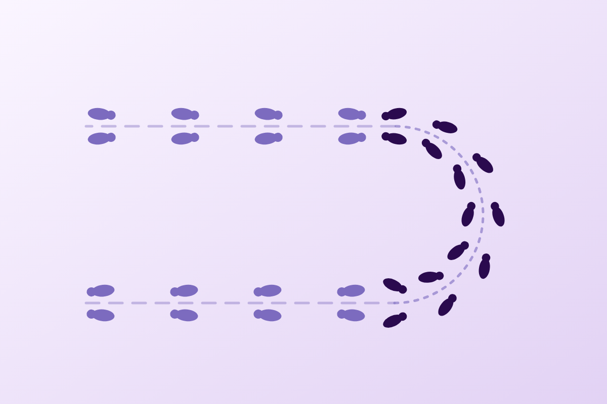

A patient of 78 walks with short steps, feet as though magnetised to the floor, widens his base of support, breaks his turns into five or six steps. He has come because of falls. Faced with this picture, most records conclude ageing, fear of falling, sometimes early parkinsonism. In some of these patients the cause is normal pressure hydrocephalus, and it is one of the very few gait disorders of the older adult that surgery can reverse. The physiotherapist who follows this patient for his falls sees him walk every week, measures that walking in any case, and is often better placed than anyone to think of it.
- Updated August 2026
- Level clinical summary
- Reading time about 55 minutes
- Sources 49 verified references (PMID / DOI)
Three figures that frame the problem
Frequency in the oldest, share actually treated, size of the surgical effect on gait
Sources: Jaraj D, et al. Neurology 2014;82(16):1449-54 (PMID 24682964); Luciano MG, et al. N Engl J Med 2025;393(22):2198-209 (PMID 40960253). The 0.21 m/s gain is the between-group difference, with a 95% confidence interval of 0.12 to 0.31.
In brief
- Gait is the earliest sign, the most consistent, and the only one whose surgical benefit has been demonstrated against placebo. Among 429 patients, 90% have a gait disorder6. In the only double-blind randomised trial against a placebo valve, gait speed and the Tinetti score improve, cognition and incontinence do not24.
- The full triad is in the minority. Only 41% of patients have symptoms in all four domains assessed; waiting for the complete picture means waiting too long6.
- The gait signature is measurable and distinct from that of Parkinson's disease. Broad base of support, outward-turned feet, reduced step height, and above all, the visual and auditory cues that unlock a parkinsonian gait have only a mild effect in NPH8.
- The cerebrospinal fluid tap test is read in one direction only. Positive predictive value above 90%, negative predictive value below 20%: a positive test points towards surgery, a negative test excludes nobody18.
- The physiotherapist's role is a measuring role before it is a treating role. The test lasts a few hours, and the response window closes within 90 to 100 hours10 : without a measurement before and after, the surgical decision is taken on an impression.
- After surgery, only one programme has done better than nothing in a randomised trial: dynamic balance training during walking, which improves the Functional Gait Assessment and reduces the incidence of falls at six months. Standard exercise, for its part, did no better than the natural course33.
- Delay costs outcome. Operated within three months of the decision, 57% of patients improve; beyond six months, 46%29.
Contents
- Why does normal pressure hydrocephalus go unnoticed?
- What exactly does the gait of normal pressure hydrocephalus look like?
- How is it told apart from a parkinsonian gait and from the other gaits of the older adult?
- Is the triad really a triad?
- Which diagnostic criteria are in force, and what are they worth?
- What does the cerebrospinal fluid tap test contribute?
- Which gait scales should be quantified in practice, and with which thresholds?
- What does ventriculoperitoneal shunting really change?
- What is the physiotherapist's role before and after shunting?
- What do real clinical cases teach us?
- How do you apply this in the clinic?
- Frequently asked questions
Why does normal pressure hydrocephalus go unnoticed?
Because its three signs resemble ordinary ageing, because its pressure is normal (that is its name), and because the measurement of its frequency depends so heavily on the criteria used that it long gave the impression of being rare. It is not.
A hydrocephalus that does not behave like a hydrocephalus
The name says what matters and misleads at the same time. There is indeed a hydrocephalus: the cerebral ventricles are dilated, and this dilatation is visible on a CT scan or on magnetic resonance imaging. But cerebrospinal fluid pressure, when measured by lumbar puncture, comes back normal. None of the classic signs of raised intracranial pressure is present: no headache, no vomiting, no papilloedema. In their place, a slow, insidious picture that settles over months or years in a person over 60.
The Cochrane review devoted to the subject gives the working definition: « Normal pressure hydrocephalus occurs when the brain ventricles expand, causing a triad of gait, cognitive, and urinary impairment », a ventricular dilatation responsible for a triad combining gait, cognition and continence25. When no cause is found (no meningitis, no severe head injury, no subarachnoid haemorrhage in the history), the normal pressure hydrocephalus is called idiopathic. This is the form dealt with here, and by far the most common in the older adult. The 2021 Japanese guidelines have in fact formalised this separation, clearly distinguishing the idiopathic form from congenital, developmental and acquired hydrocephalus16.
What makes this entity clinically interesting comes down to one word: it is treatable. The excess fluid can be shunted surgically, and part of the symptoms regress. In a field, that of gait disorders and cognitive disorders of the older adult, where almost every diagnosis opens onto supportive care, here is one that opens onto an intervention. That is what justifies thinking of it, and it is also what makes forgetting it costly.
Four landmarks for placing the disease
Frequency of the signs, gap between disease and surgery, place of gait
Sources: Agerskov S, et al. J Neurol Sci 2018;391:54-60 (PMID 30103972); Martín-Láez R, et al. World Neurosurg 2015;84(6):2002-9 (PMID 26183137). Bringing together the two figures on the fourth card is a reading of this article: the authors report them separately and themselves conclude that the disease is « extremely underdiagnosed ».
A prevalence that depends on the criteria more than on the country
The published frequency figures range from 0.2% to nearly 9%. Such a spread usually passes for methodological noise calling for an apology. Here it is the other way round: the spread is the information, because it measures exactly what the definition adopted changes.
The clearest demonstration comes from a Swedish prospective study. In a randomly drawn sample of residents over 65 in Jämtland County, the authors applied two sets of criteria in turn to the same people. With the American-European criteria, the prevalence of probable NPH is 3.7 %. With the Japanese criteria, in the same sample, it falls to 1.5 %2. A factor of two and a half, without a single patient having changed.
The same phenomenon plays out again with the imaging thresholds. In a population survey of 1,491 Shanghai residents over 60, the crude prevalence is 3.09% using a DESH score of 6 or more, and 2.62% using a Radscale score of 7 or more5. The authors say so explicitly: prevalence « fluctuates according to the radiological scoring systems and the thresholds used ».
What does not vary from one study to another, by contrast, is the effect of age. It is massive and constant.
Measured prevalence of normal pressure hydrocephalus, by study and by criteria
Each bar carries its population, its imaging method and the definition used
Sources: Jaraj D, et al. Neurology 2014;82(16):1449-54 (PMID 24682964); Constantinescu C, et al. Neurology 2024;102(2):e208037 (PMID 38165321); Andersson J, et al. PLoS One 2019;14(5):e0217705 (PMID 31141553); Martín-Láez R, et al. World Neurosurg 2015;84(6):2002-9 (PMID 26183137); Fang X, et al. Alzheimers Dement 2025;21(2):e14525 (PMID 39950414). The two boxed bars concern the same population : only the diagnostic criteria differ. The 11.6% refers to residents meeting the NPH criteria described as suspected, a broader definition than the other bars.
Two readings stand out. The first: age multiplies the frequency. In the Gothenburg cohort, the figure goes from 0.2% in those aged 70-79 to 5.9% in those aged 80 and over1. In Jämtland, from 2.1% in those aged 65-79 to 8.9% in those aged 80 and over, with a p below 0.0012. In Shanghai, the estimated prevalence goes from 2.59% after 60 to 7.99% after 905. In other words: the population the physiotherapist sees most often for falls is also the one in which this disease is most common.
The second reading is more unsettling. A recent population study using magnetic resonance imaging, covering 791 people born in 1944, finds a prevalence of 1.5% as early as age 70, which its authors call « considerably higher than what had been reported so far in this age band ». And 5.1% of the sample already shows the characteristic radiological signs, without necessarily meeting the full clinical criteria3. The authors suggest that isolated ventriculomegaly might be, in some people, an early sign of the disease to come.
Underdiagnosis, measured rather than assumed
It would be easy to claim that this disease is underdiagnosed without ever putting a number on it. Two measurements do so.
The first is that of Jaraj and his team, and it is probably the most telling figure in the whole file. Among 1,238 people examined, 26 met the criteria for probable NPH. The authors then write, in a single sentence: « Only 2 of these persons had been treated for iNPH »1. Two out of twenty-six. Twenty-four people living with a disease for which a treatment exists, and not receiving it.
The second is that of a systematic review of 21 epidemiological studies. It establishes that the pooled prevalence in those over 65, in studies specifically designed to detect NPH, is 1.3%, that is « almost 50-fold higher », almost fifty times more, than what the door-to-door surveys devoted to dementia or parkinsonism suggested4. The same review puts the incidence at 1.20 cases per 1,000 people per year in the only prospective population survey available, that is fifteen times the estimates drawn from hospital caseloads. And the number of shunts actually placed for this indication: fewer than 2 per 100,000 people per year.
Setting these last two numbers side by side is not a trivial move, and it has to be done honestly: they are not measured in the same populations, and the authors do not themselves relate them arithmetically. But the order of magnitude they sketch, many patients and very few operated on, is precisely the conclusion the review states: « iNPH appears to be extremely underdiagnosed ».
Key points
- Dilated ventricles, normal cerebrospinal fluid pressure, slow onset after 60: that is the frame.
- The published prevalence runs from 0.2% to 8.9% depending on age, and from 1.5% to 3.7% depending on the criteria alone within one and the same population.
- After 80, the estimates converge around 6 to 9%.
- In the Gothenburg cohort, 2 of the 26 people concerned had been treated.
- No French population study was identified: the figures quoted are Swedish, Japanese, Chinese and Spanish.
The trap of isolated ventriculomegaly
An Evans index above 0.3, the ratio between the width of the frontal horns and the maximum skull diameter, is found in 20.7 % of people over 701, and in 11% of 70-year-olds in a magnetic resonance imaging cohort3. In other words: one person in five over 70 has « dilated » ventricles in the sense of this criterion, and the overwhelming majority does not have normal pressure hydrocephalus. An imaging report mentioning ventriculomegaly therefore does not make the diagnosis, and the absence of such a mention does not rule it out either, since the radiologist looks only for what he is asked to look for.
What exactly does the gait of normal pressure hydrocephalus look like?
This is the central chapter, for a simple reason: gait is the first symptom to appear, the most frequent, the one that responds best to treatment, and the only one the physiotherapist measures in any case. Everything else in this article follows from it.
The description one can give from memory, and the one the sensors confirm
The classic picture comes down to a few features. The steps are short. The base of support is broad, sometimes markedly so: the feet move apart, and often turn outwards. The feet barely leave the ground any more, hence the image of the « magnetised » patient, which gives this type of gait its English name, magnetic gait. The turn falls apart: instead of pivoting in one piece, the patient strings together four, five, six small steps. And starting off is laborious, as if the first steps cost more than the ones that follow.
This description is not only clinical: it has been measured. An instrumented analysis comparing ten patients with twelve age-matched controls, on a treadmill and overground, isolates a triad of parameters. The patients walk with a reduced step length, a reduced step height above the ground, or, in the authors' words, foot-to-floor clearance, and a broadened base of support. Dorsiflexion of the forefoot before heel strike is insufficient. Step width and foot rotation angles are significantly increased9.
One detail of this study deserves attention, because it runs against intuition: these balance parameters are increased with lower variability than in the controls. The widening of the base of support is therefore not a disordered oscillation, it is a stable, sustained strategy. The patient has found a position and no longer leaves it. This clearly separates the picture from a cerebellar ataxia, where the widening comes with marked irregularity from one step to the next, and it explains why these patients often look « careful » rather than « unsteady » to the naked eye.
The numerical thresholds that separate the disease from ageing
One question remains, the one every clinician asks in front of an older person who walks slowly: from what point is it abnormal? A German study answered it in the only way that counts, comparing 55 patients with 55 age-matched controls on a pressure-sensor walkway, in eight different walking conditions11.
The two most discriminating conditions are walking at self-selected speed and the semantic dual task (walking while listing animal names, for example). And at self-selected speed, three thresholds stand out, with diagnostic performance that few clinical tests reach.
Three thresholds that separate NPH gait from that of a healthy older adult
Walking at self-selected speed, 55 patients versus 55 age-matched controls, pressure-sensor walkway
Sources: Möhwald K, et al. Sci Rep 2022;12(1):18295 (PMID 36316420) for the three thresholds; Kihlstedt CJ, et al. Fluids Barriers CNS 2024;21(1):22 (PMID 38454478) for the speeds of 0.59 and 0.89 m/s, measured respectively in patients with and without freezing. These thresholds separate NPH from normal ageing; they are not criteria for referral to the neurologist, which do not exist.
A step length of 1.02 metres or less reaches a sensitivity of 0.93 and a specificity of 0.91, for an area under the curve of 0.96. A speed of 0.83 m/s or less gives 0.80 and 0.91, for an area under the curve of 0.9311. These values must be read for what they are: they separate affected patients from a matched healthy control group, not affected patients from an unselected geriatric population where other conditions compete for the same slowing. They still say something useful: a patient walking at 0.6 m/s with 60-centimetre steps is not within the normal spread for his age.
The patient does not come to complain about his step length. He comes because he has fallen. Between the two there is a measurement, and that measurement takes four minutes.
The turn, the place where everything shows
If a single movement had to be watched, it would be the turn. Two sets of data converge.
The first concerns freezing, that abrupt block where the feet seem glued to the floor while the trunk keeps moving forward. Long regarded as a parkinsonian signature, it was systematically looked for in 139 patients with NPH, on standardised video recordings reviewed by two observers with excellent agreement (Cohen's kappa of 0.9). The result: 22 patients, or 16%, had freezing before surgery. And above all: « The symptom was most frequently exhibited during turning (n = 16, 73%) », in 73% of cases it occurred during the turn12.
These patients are not just any patients: they are older (77.5 against 74.6 years), markedly slower (0.59 against 0.89 m/s), with a collapsed Tinetti score (6.8 against 10.8), a lower MMSE (21.3 against 24.0), and a disease of longer standing (4.2 against 2.3 years)12. Freezing therefore marks a stage, not a subtype. Notably, neither the white matter lesion load on imaging nor the cerebrospinal fluid biomarkers set them apart from the others: this is not a disguised comorbidity.
An encouraging detail: after shunting, the number of patients with freezing falls from 22 to 7, that is from 16% to 8% (p = 0.039)12. The symptom responds.
The second set of data comes from a direct comparison between 21 patients with NPH and parkinsonism and 21 parkinsonian patients not yet treated, all assessed with an instrumented Timed Up and Go at the time of diagnosis. The turn there is longer and slower in NPH. And in multivariable analysis adjusted for age and cognitive status, it is neither gait speed nor step length that best separates the two groups: it is the mean angular velocity of the turn performed before sitting down13.
That parameter calls for an inertial measurement unit. But a version for the practice exists, and it is better validated than one might think: counting the steps of the turn. In a Thai study of 27 patients assessed before and after a subtractive lumbar puncture, three measures improve significantly, the sit-to-stand time (p = 0.046) and the 3-metre walking time (p = 0.048), and a fourth goes far beyond them: the number of steps in the 180-degree turn, with a p of 0.001. The time of the turn, for its part, does not reach the significance threshold (p = 0.064)14.
In other words: in this study, counting steps did better than the stopwatch. This is information that can be used directly in practice, where counting steps costs nothing and calls for no equipment.
Step variability, and why strength is not the problem
One last property of this gait deserves to be known, because it reorients the content of sessions. In 63 patients fitted with a triaxial accelerometer placed at the level of L3 during a 10-metre walk test, having fallen in the previous six months is correlated with the temporal variability of the step (the coefficient of variation of step time), and with the Functional Gait Assessment and Berg scores. It is not correlated with isometric quadriceps strength15.
The three variables independently associated with fall risk are, in this work: the coefficient of variation of step time, the FGA score, and age. A study by the same group, in 68 patients, reaches the same conclusion by another route and states it bluntly: « Patients with iNPH who fall experience falls due to dynamic balance dysfunction during gait rather than lower limb muscle strength »32.
This does not remove the need to strengthen a deconditioned patient, which he often is in any case. But it shows where his fall risk is decided: in dynamic balance control during walking, not in quadriceps strength. A programme centred on analytical strengthening misses the target.
Red flags in a gait disorder of the older adult
- An onset over a few days or a few weeks does not fit the profile of NPH, which takes months to years to build up: think vascular, tumoural, infectious, iatrogenic.
- A focal sensory or motor deficit, a unilateral Babinski sign, cranial nerve involvement point elsewhere and call for a neurological opinion.
- Spinal pain, claudication on walking relieved by trunk flexion suggest lumbar spinal stenosis, which can moreover coexist with NPH and blur the picture entirely.
- Acute urinary retention, an anal sphincter deficit, saddle anaesthesia : cauda equina syndrome, an emergency.
- Repeated falls with head injury in an anticoagulated patient : chronic subdural haematoma gives a similar picture, and it too is surgical.
Key points
- The measured signature is short step, foot that does not clear the ground, broad base of support with external rotation, and a variability of these balance parameters that is lower than in the healthy subject.
- Discriminating thresholds at self-selected speed: step length ≤ 1.02 m (AUC 0.96), speed ≤ 0.83 m/s (AUC 0.93), double support ≥ 27% of the cycle.
- Freezing exists in NPH in 16% of patients, and it occurs in 73% of cases during the turn.
- The number of steps in the 180° turn is more sensitive than the time of the turn for detecting change.
- Fall risk is linked to step variability and to dynamic balance, not to quadriceps strength.
How is it told apart from a parkinsonian gait and from the other gaits of the older adult?
Confusion with Parkinson's disease is the most frequent and the most costly, because the two pictures share their most visible feature: short steps in an older person who is slowing down. Yet there is a test of differentiation that the physiotherapist can run in a session, with no equipment, and whose result has been published since 2001.
What the two gaits have in common, and what explains the error
A comparative analysis set 11 patients with NPH, 10 parkinsonian patients and 12 age-matched controls face to face, on a treadmill and overground. The common ground is clear: in both diseases speed is reduced, and it is reduced by the same mechanics: a reduced and highly variable step length, with cadence relatively preserved8.
Better still: after treatment, 30 mL of cerebrospinal fluid removed on one side, 150 mg of levodopa given on the other, both groups improve in the same way, through a lengthening of the step, which becomes less variable, with no change in the other parameters. The two diseases therefore resemble each other right down to their response to treatment, which explains why a therapeutic trial of levodopa can mislead as much as it enlightens.
What separates them, and what can be tested in a session
The differences do exist, and they are of two kinds.
The first kind is morphological : « Specific features of the gait disturbance in normal pressure hydrocephalus were a broad based gait pattern with outward rotated feet and a diminished height of the steps »8. Broad base of support, outward-turned feet, steps that do not clear the ground. The parkinsonian patient, for his part, keeps a narrow base of support, walking « on a line », and his disorder comes with a stooped trunk, a loss of arm swing and frequently a clear asymmetry.
The second kind is the more useful in practice, because it can be tested. The authors gave both groups external cues: strips stuck to the floor as a visual cue, a metronome as an auditory cue. The result, in their own words: « External cues only mildly improved gait in normal pressure hydrocephalus, whereas they were highly effective in raising the stride length and cadence in Parkinson's disease »8.
The visual or auditory cue unlocks a parkinsonian gait. It does not unlock a normal pressure hydrocephalus gait. This is a test the physiotherapist already performs, often without knowing that it carries diagnostic information.
The practical reach is direct. External cueing is a routine tool in the rehabilitation of Parkinson's disease, where it is a recommended modality; the subject is developed in our summary on Parkinson's disease and physiotherapy. A patient labelled parkinsonian in whom neither the floor strips nor the metronome change anything should raise questions, just as a broad and irregular gait should bring to mind a cerebellar ataxia, or as an abrupt onset should prompt a search for a stroke. This is not a validated diagnostic criterion, and that must be said plainly: the study covers 21 patients in all and was not designed to establish diagnostic performance. But it is a signal, available free of charge, in a patient seen twice a week.
Postural instability does not have the same geometry
A second difference was measured on a force platform, in 27 patients with NPH, 20 parkinsonian patients and 20 controls, during tasks of voluntary leaning of the centre of pressure in four directions. In Parkinson's disease, control is impaired forwards and backwards. In NPH, it is impaired in every direction, and significantly more than in Parkinson's disease laterally, to the right as to the left17.
The authors explicitly link this lateral instability to the widening of the base of support and to fall risk: a patient who cannot control his lateral weight transfers must widen his feet to stay upright. The widening of the base of support is not the disease, it is the response to the disease. A notable point for anyone tempted to rely on the stopwatch alone: in that same work, the Timed Up and Go did not separate the two groups, nor did postural sway in quiet standing.
A Turkish study, on smaller numbers (13 NPH, 20 Parkinson, 13 controls) fitted with inertial sensors, points the same way and adds two things: patients with NPH have a step length and a speed lower than those of parkinsonian patients, and they struggle particularly on the most demanding balance tasks: feet apart with eyes closed on foam, and tandem stance. On the Timed Up and Go, their time is longer and their turning speed lower compared with parkinsonian patients19.
The differential table of gaits in the older adult
This table gathers the entities that present with a slowly progressive gait disorder after 65. It is not meant to produce a diagnosis, which is the neurologist's work, but to organise what the physiotherapist observes, and to show when to ask for an opinion.
| Entity | Base of support | Foot on the ground | Turn | Effect of visual or auditory cues | What should suggest something else |
|---|---|---|---|---|---|
| Normal pressure hydrocephalus | Broad, outward-turned feet, with variability that is low | Barely clears the ground, « magnetised » | Broken into several steps; freezing in 16%, of which 73% during the turn | Weak (« only mildly improved ») | No cognitive or urinary trouble, even subtle, after several years |
| Parkinson's disease | Narrow, walking « on a line » | Drags, festination possible | In one piece, freezing frequent | Marked on step length and cadence | No rest tremor, no rigidity and no asymmetry; no response to levodopa |
| Cerebellar ataxia | Broad, with variability that is high from one step to the next | Clears the ground normally, imprecise landing | Clear loss of balance, lurching | Little or no effect | Dysarthria, nystagmus, upper limb dysmetria |
| Cautious gait (fear of falling) | Slightly broad | Clears the ground normally | Careful but fluid | Little effect; verbal reassurance and a light support, by contrast, transform the picture | A normal neurological examination and a disorder that recedes when confidence returns |
| Lumbar spinal stenosis | Variable | Normal at first | Normal | No effect | Walking distance limited by pain or numbness, relieved by trunk flexion and by stopping; cycling remains possible |
| Chronic subdural haematoma | Variable | Variable | Variable | No effect | Onset over weeks, recent fall or head injury, anticoagulant, lateralised signs, headache |
The two are not mutually exclusive
Some patients with NPH have a genuine associated parkinsonism, described in the literature under the term iNPH-P : that is exactly the population of the turning study cited above13. The reasoning is therefore not « either one or the other » but « what if it were also the other ». A known parkinsonian patient, on well-conducted treatment, whose gait deteriorates faster than his disease would warrant, and in whom urinary trouble and a widening of the base of support appear, deserves to have the question put back to the neurologist.
Key points
- NPH and Parkinson's share the reduced speed through a short and variable step, and both improve their step under treatment: the resemblance goes a long way.
- What separates them: broad base of support and externally rotated feet in NPH, narrow base of support in Parkinson's.
- External cues markedly improve parkinsonian gait and very little that of NPH. A test available in a session, to be treated as a signal and not as a criterion.
- Postural instability in NPH is multidirectional, and worse laterally than in Parkinson's; the TUG alone does not separate them.
- The two diagnoses can coexist.
Is the triad really a triad?
No, or rather: it is rarely complete at the moment when the diagnosis could still be made in time. Waiting for the three signs means waiting for the stage where surgery yields least.
What counting across 429 patients gives
The so-called Hakim and Adams triad, gait disorder, cognitive disorder and sphincter disorder, is taught as a set. A single-centre series of 429 patients, mean age 71, makes it possible to know what the reality is, by distinguishing four domains rather than three: gait, balance, neuropsychology and continence6.
72% of patients have symptoms in three or four domains. Only 41% have them in all four. In other words, nearly three patients in five reach diagnosis with an incomplete picture, and nearly one in three with two domains or fewer.
Frequency of the signs in 429 patients with normal pressure hydrocephalus
Single-centre series, mean age 71, detailed examinations before and after surgery
Source: Agerskov S, et al. J Neurol Sci 2018;391:54-60 (PMID 30103972). The four domains assessed are gait, balance, neuropsychology and continence. The 30% freezing rate in this series is higher than the 16% measured on standardised video by Kihlstedt 2024 (PMID 38454478): the collection methods differ, and the gap illustrates how hard this sign is to rate.
The order of appearance, and why it favours the physiotherapist
Gait comes first, at 90%, and it is usually the first sign to appear. Continence follows closely in frequency, at 86%, but it is rarely reported spontaneously: few older patients mention nocturia or urge leakage of their own accord, and few professionals ask about it. The cognitive side is present in a little over half, with an MMSE below 25 in 53%6.
This hierarchy has a practical consequence. The professional who sees the first sign, several times a week, in a setting where he assesses it formally, is the physiotherapist. The neurologist will later see a more complete picture, therefore a later one, therefore a less reversible one.
Looking for the full triad means giving oneself a comfortable and late criterion. The 59% of patients who do not have all four domains do exist, they walk badly, and they can be operated on.
The cognitive side, and its border with the degenerative dementias
The cognitive profile of NPH is classically described as subcortical-frontal: slowed information processing, impaired attention and executive function, apathy, with recall memory relatively better preserved than in typical Alzheimer's disease. It is a useful distinction, but it is not enough to settle the question, and the overlap is real.
Two things are worth knowing. The first is an imaging result: the callosal angle, measured on a coronal slice through the posterior commissure, averages 66 ± 14 degrees in NPH, against 104 ± 15 degrees in Alzheimer's disease and 112 ± 11 degrees in the healthy subject. With a threshold of 90 degrees, the discrimination between NPH and Alzheimer's disease reaches an accuracy of 93%, a sensitivity of 97% and a specificity of 88%21. This measurement is not the physiotherapist's business, but it appears in imaging reports when the question is asked, and the question still has to be asked.
The second is more uncertain, and it must be presented as such. A Taiwanese national cohort of 2,053 patients, with propensity score weighting and 16 years of follow-up, finds that a shunt is associated with a reduced risk of later developing dementia (subdistribution hazard ratio 0.74; 95% CI 0.55-0.99) and Alzheimer's disease (0.15; 95% CI 0.04-0.61), but not vascular dementia30. This is an observational association, on administrative health data, with everything that implies in terms of possible residual bias; it does not show that surgery protects against cognitive decline. It does indicate, however, that the question is open, and that it is being asked seriously.
The differential diagnosis with major neurocognitive disorders proper, Alzheimer's disease, Lewy body disease and frontotemporal lobar degeneration, goes beyond the scope of this article and belongs to neurological and neuropsychological assessment; these three entities and what they change in the session are covered in our summary Major neurocognitive disorders: FTD, Alzheimer's and Lewy body disease. What has to be retained here comes down to one sentence: NPH is one of the causes of cognitive decline not to be missed, because it is the only one of the set from which improvement can be hoped for.
The urinary side, better documented than people think
The only prospective assessment of urinary symptoms in newly diagnosed NPH, in 55 consecutive patients with standardised questionnaires and urodynamic testing, gives a precise picture. The incontinence is mild to moderate, of the urgency type. Nocturia is the most frequent symptom ; urge incontinence is the most troublesome. And on urodynamic testing, 100% of the patients tested had detrusor overactivity, with a mean bladder capacity of 200 mL31.
Two consequences. First, the question to ask is not « do you leak? », to which many will answer no, but « how many times do you get up at night? » and « when the urge comes, do you have time to get there? ». Second, this mechanism is not that of stress incontinence: the usual pelvic floor strengthening protocols are not the answer to this situation, and a patient referred on that ground without the central cause having been identified receives a treatment that cannot work.
Key points
- Only 41% of patients have symptoms in all four domains; 72% have them in three or four.
- Measured frequencies: gait 90%, continence 86%, balance 53%, MMSE < 25 in 53%.
- Gait appears first: the physiotherapist is in a position to be the first witness.
- The urinary trouble is detrusor overactivity (nocturia and urgency), not stress incontinence.
- The callosal angle separates NPH and Alzheimer's disease well (threshold 90°, Se 97%, Sp 88%). Careful, though: keep apart diagnosing and predicting the response to surgery, two different questions dealt with further on.
Which diagnostic criteria are in force, and what are they worth?
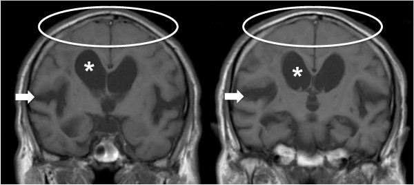
MRICoronal brain MRI showing the two characteristic signs of normal pressure hydrocephalus: tight sulci at the convexity (circle) and a dilated Sylvian fissure (arrow), with a dilated lateral ventricle (asterisk).
Source : Hashimoto et al., Cerebrospinal Fluid Research, 2010, figure 1 · CC BY
Two bodies of guidelines coexist, one American-European and the other Japanese. They do not give the same prevalence on the same population, as we have seen. Knowing them matters less for making the diagnosis, which is not our role, than for understanding what the neurologist will look for, and what the physiotherapist can bring him.
The American-European criteria: probable, possible, unlikely
Published in 2005, they are the first attempt at evidence-based criteria for this disease. Their method is explicit: built from the literature of 1966 to 2003, supplemented by expert opinion where the evidence was missing. Their main contribution is a three-level classification: probable, possible and unlikely NPH, intended to harmonise the selection of patients in studies20.
The elements that make up the « probable » category are, in short: an age of at least 40, an insidious onset over at least three to six months, the absence of an identifiable cause, progression over time, a gait disorder combined with at least one other domain (cognition or continence), ventriculomegaly with an Evans index above 0.3, and a cerebrospinal fluid opening pressure within normal limits20.
These criteria are the ones applied by most of the prevalence studies cited above, which explains, in passing, why some of them state that they had to depart from them: measuring an opening pressure requires a lumbar puncture, a procedure that a population survey cannot impose on asymptomatic volunteers2.
The Japanese guidelines and the place of DESH
The third edition of the Japanese guidelines, published in 2021 by a multidisciplinary working group, introduces two notable changes. The first is a new classification that clearly separates idiopathic NPH from NPH of congenital, developmental or acquired origin. The second concerns imaging: the authors discuss the « essential role of disproportionately enlarged subarachnoid-space hydrocephalus (DESH) in the imaging diagnosis and decision for further management », and propose an algorithm for diagnosis and surgical decision-making16.
DESH refers to a particular configuration: dilated ventricles and widened sulci in the sylvian region, whereas the subarachnoid spaces of the high convexity and of the midline are, for their part, compressed. It is this disproportion, hence the name, that is characteristic, and not ventricular dilatation alone. In the Gothenburg cohort, effacement of the high convexity sulci was found in only 5.4% of the people examined, against 20.7% for an Evans index above 0.31 : it is a far more specific sign.
The most important point of this chapter: diagnosing is not predicting
Here is a distinction that the literature draws clearly and that practice often blurs.
Imaging serves to identify the patients who probably have NPH. It does not serve, or serves very little, to predict which of them will benefit from surgery. A systematic review with meta-analysis sifted 301 articles, kept 28 studies and identified 26 different radiological markers22. Five met the conditions for a meta-analysis: DESH, the callosal angle, periventricular white matter abnormalities, cerebral blood flow and CT cisternography.
The result is harsh: only the callosal angle and the periventricular abnormalities significantly separate responders from non-responders, and they do so with diagnostic odds ratios of 1.88 and 1.01. The authors conclude that they are « insufficient as sole predictors » and should be used only in combination with other tests. DESH, for its part, does not separate responders from non-responders22.
A diagnostic odds ratio of 1.01 means that a marker says nothing. Imaging identifies the disease; it does not say who will walk better after surgery. That question is settled by measuring walking.
This finding is what gives weight to the next chapter, and to the physiotherapist's role as a whole. If imaging predicted the response, a good radiologist would be enough. It does not predict it: what predicts it is how gait behaves when fluid is removed.
Key points
- Two bodies coexist: the American-European criteria of 2005 (probable / possible / unlikely) and the Japanese guidelines, 3rd edition 2021.
- Evans index > 0.3: necessary, very unspecific: 20.7% of those over 70 have it.
- The DESH (disproportion between widened sylvian sulci and a compressed high convexity) is far more specific than ventricular dilatation alone.
- No imaging marker usefully predicts the response to surgery. Callosal angle: diagnostic odds ratio 1.88; periventricular abnormalities: 1.01; DESH: not discriminating.
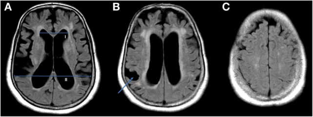
MRIAxial brain MRI showing ventriculomegaly with measurement of the Evans index (A), marked ventricular dilation (B, arrow) and tight sulci at the vertex (C).
Source : Afonso et al., Frontiers in Neurology, 2017, figure 1 · CC BY
What does the cerebrospinal fluid tap test contribute?
This is the examination that decides, in practice, whether a patient goes to surgery. Its principle is simple: remove fluid and see whether the patient is better. Carrying it out is less simple, and its interpretation obeys an asymmetry that has to be borne in mind before reading the report.
The principle and how it is carried out
The fluid removal test, known as the tap test in the literature, consists of taking off by lumbar puncture a volume of cerebrospinal fluid, usually 30 to 50 mL, then reassessing the patient. The studies cited here use 30 mL9,26 or 50 mL27.
A variant exists, heavier and better performing: the external lumbar drain, where a catheter left in place removes fluid continuously for one to three days. In a prospective series of 68 patients, a positive response to external drainage correctly predicted improvement after shunting in 87.9 % of patients, the best performance of all the tests assessed, including the measures of cerebrospinal fluid dynamics. The price to pay: a complication rate for the invasive tests of 5.4 % in this series23.
The measurement window, and why it decides everything
One technical point governs the whole practical organisation of the test, and it is established by meta-regression across 17 studies and 527 patients: the effect of the test on gait speed reaches its maximum between 24 and 48 hours, then returns to baseline within 90 to 100 hours10.
Kinetics of the response to the cerebrospinal fluid tap test
Effect on gait speed, from a meta-regression covering 17 studies and 527 patients
Source: Passaretti M, et al. Mov Disord Clin Pract 2023;10(11):1574-84 (PMID 38026510). The two coloured windows and the three measurement points are carried over from the meta-regression values; the curve itself is a schematic illustration, it does not reproduce a trajectory measured point by point. The 2-4 hour point corresponds to the Gallagher 2018 protocol (PMID 29702069), the 24-hour point to that of Bovonsunthonchai 2018 (PMID 29581614).
This kinetics has three immediate consequences.
A patient assessed too early may look like a non-responder. The Australian Gallagher protocol measures 2 to 4 hours after the puncture and detects clear changes26 ; but the effect is not then at its maximum.
A patient assessed too late will not look like a responder either. A reassessment the following week measures nothing at all, or measures noise.
And above all: the family is not a measuring instrument. A response that lasts two days and vanishes without a trace is almost impossible to establish retrospectively by questioning the patient and his family a week later. If nobody measured before and after, the information is lost, definitively, since repeating the test means another lumbar puncture.
The asymmetry of interpretation: a positive test counts, a negative test does not
This is the most important property of this examination, and the least intuitive.
The European multicentre study, carried out on 115 patients whose test results were kept blinded from the care team, with reassessment at 12 months, reaches a striking conclusion: neither the resistance to cerebrospinal fluid outflow nor the tap test correlates with the outcome at 12 months. Only one element correlates significantly, and it is worth noting: the improvement on the walking task (ten metres at free speed), with a coefficient of 0.22 (p = 0.02)18.
The predictive values, for their part, are unambiguous: positive predictive value above 90%, negative predictive value below 20%. The authors put it in a sentence that should appear in every report: « Rout and CSF TT can be used for selecting patients for shunt surgery but not for excluding patients from treatment ».
A meta-analysis covering 17 trials and 812 subjects estimated the performance of the simple tests carried out after fluid removal: 18-metre walk test, sensitivity 0.83 (95% CI 0.57-0.99) and specificity 0.67 (0.33-0.95); Timed Up and Go, sensitivity 0.89 (0.79-0.95) and specificity 0.63 (0.24-0.90)42. The same asymmetry is found: these tests detect responders well, they rule out non-responders badly.
A Swedish study of 116 operated patients, using the Hellström iNPH scale, refines the picture further and delivers a useful result: 63.8% of patients respond on the gait domain, 44.3% on the balance domain ; and against the overall postoperative outcome, sensitivity and specificity are 68.1% and 52.0% for gait, against 47.8% and 68.0% for balance. The authors conclude that « the gait domain may be used to predict outcomes for gait, but the balance domain is too insensitive »44. It is gait that must be measured, not static balance.
An independent systematic review confirms the order of magnitude across eight studies: positive predictive value of 92 % (range 73 to 100%), negative predictive value of 37 % (18 to 50%), specificity 75%, sensitivity 58%28.
What a negative test does not mean
With a negative predictive value between 18% and 50%, a negative tap test leaves in place a substantial probability that the patient will respond to surgery anyway. A patient whose test came back negative and whose condition keeps deteriorating is therefore not a closed file: the external lumbar drain, which is more sensitive, exists. It is not for the physiotherapist to reopen the file, but it is for him to report a documented worsening rather than treat the question as settled.
What the test changes in concrete terms, measured test by test
A Swedish study from 2026 supplies what was missing: a direct comparison of the responsiveness to change of a broad range of physiotherapy tests, in 95 patients of mean age 77, before and after removal of 50 mL27.
Which tests move most after removal of 50 mL of cerebrospinal fluid
95 selected patients, mean age 77, all tests significant at p < 0.001
Source: Akar K, Kollén L, Tullberg M, Persson HC. Fluids Barriers CNS 2026;23(1):35 (PMID 41703573). A selected population of patients due for surgery: these percentages describe the size of the observable change, not the diagnostic sensitivity of the test. The two blocks have different scales and are not comparable with each other.
Three lessons. First, the 3-metre backward walk is the test most responsive to change in the whole battery, in both of its scoring modes. Next, the hardest balance tests do not move : tandem with eyes closed and single-leg stance stay on the floor, which means they are of no use in this indication, since a patient who could not hold three seconds before will hold no longer after. Finally, the authors explicitly recommend demanding tests that probe gait in its direction and in its capacity, rather than the ten-metre there-and-back alone.
One point about the differences between men and women, often badly reported: the men showed significant gains on two balance tasks that the women did not show, but after adjustment for baseline performance, no sex-related difference in response remains27. The apparent gap came from the starting level, not from sex.
Key points
- Removal of 30 to 50 mL by lumbar puncture, or an external lumbar drain over one to three days (correct prediction in 87.9%, but 5.4% complications from the invasive tests).
- Response window: maximum at 24-48 h, return to baseline within 90-100 h. Measuring too early underestimates, measuring too late measures nothing.
- PPV > 90%, NPV < 20% : the test selects, it does not exclude.
- In the European study, the only element correlated with the outcome at 12 months is the improvement on the 10-metre walk test.
- Tests most responsive to change: 3 m backward walk (−32%), TUG (−27%), 6-minute test (+25%), 10 m walk (−21%).
- Tandem with eyes closed and single-leg stance: useless here, they do not move.
Which gait scales should be quantified in practice, and with which thresholds?
None of these tests calls for equipment beyond a stopwatch, a tape measure and a chair. That is precisely what makes their absence costly: the measurement that will be missing from the surgical file is a measurement the practice could have made.
The reference deltas: what removing fluid actually moves
An Australian prospective study of 74 patients, all put through a battery of tests before and 2 to 4 hours after removal of 30 mL, provides the comparison most directly usable in practice. The patients were then classified as responders, those to whom a shunt was offered by the neurologist or the neurosurgeon, and non-responders: 40 on one side, 34 on the other26.
In the responders, the mean changes measured are as follows: Tinetti +3.88 points, Timed Up and Go −3.98 seconds, 10-metre walk test +0.08 m/s, Berg scale +5.29 points. In the non-responders, only the Tinetti (+0.91 point) and the Berg move slightly26. The differences between the two groups are significant for every test.
One detail of this study deserves to be picked out, because it shifts the cursor: the patients also rated their own perceived change on a global scale, and their self-rating separated the two groups (+2 for walking in the responders against 0 in the non-responders, +2.5 against 0 for balance). The authors conclude that « Patients appear to be able to accurately identify if change has occurred ». Asking the patient what he felt is therefore not a nicety: it is a piece of data that discriminates.
The benchmark that gives the figures their meaning
Is a gain of 0.08 m/s a lot? The question is settled by the reference work on meaningful change in physical performance measures in older adults, established by anchor-based and distribution-based methods in nearly 700 subjects: the small but meaningful change sits around 0.05 m/s of gait speed, and the substantial change around 0.10 m/s. For the 6-minute test, the benchmarks are 20 metres and 50 metres respectively34.
The +0.08 m/s observed after fluid removal in the responders therefore sits between the two: real, perceptible, without reaching the threshold of substantial change. Which fits with the fact that a lumbar puncture is only a temporary glimpse of what surgery will do. We shall see in the next chapter that shunting does far better.
The Functional Gait Assessment, and why it is worth learning
If a single test had to be added to routine practice for this population, it would be this one. Two studies by the same Japanese group show it.
The first compares the ability of four tests to separate patients who fall from those who do not, in 68 patients. The areas under the curve are: FGA 0.869 (95% CI 0.761-0.933), Berg 0.796, 10-metre walk test 0.692, Timed Up and Go 0.651, and isometric quadriceps strength is not predictive at all32.
The second point is decisive for practice: in the most independent patients, those whose Timed Up and Go is below 15 seconds, that is half the sample, the FGA is the only test that remains significant, with an area under the curve of 0.84232. The TUG, the 10-metre test and strength no longer discriminate anything.
In the patient who still walks well, the stopwatch no longer sees the fall risk. The Functional Gait Assessment does, because it puts him in difficulty instead of watching him walk in a straight line.
The reason is mechanical: the FGA has ten items that deliberately disturb walking, changes of speed, horizontal and vertical head turns, a rapid pivot turn, stepping over an obstacle, tandem walking, walking with eyes closed, walking backwards, going up and down stairs. That is exactly the register in which NPH fails, since its instability is multidirectional and its turn disorganised. A patient can cover ten metres in a straight line at a respectable speed and come apart on the first head-turn item.
The 3-metre backward walk, the best value test for picking up change
This test, timing a 3-metre course walked backwards and counting the steps, is the clear winner of the two recent Swedish studies. After fluid removal, it changes by −32% in time and −23% in the number of steps, ahead of the Timed Up and Go (−27%) and the 10-metre test (−21%)27. After shunting, in 291 operated patients reassessed at a median of 5.4 months, it changes by −43 %, against −28% for the Timed Up and Go and −19 to −23% for the 10-metre test35.
That same study brings two prognostic pieces of information worth knowing before setting goals with a patient. First, women have poorer performance before and after surgery, but their improvement does not differ from that of men once adjusted for the starting level. Second, each additional five years of age reduces the gain by 3 to 8%. Age does not rule out surgery; it reduces its yield, which is one more argument for not waiting.
On the complementarity of the measures, the correlations reported are instructive: the 6-minute test is strongly correlated with the 10-metre test (ρ = −0.81) and the backward walk with the Timed Up and Go (ρ = 0.75), whereas the correlation between the 6-minute test and the backward walk is only moderate (ρ = −0.58). In practical terms: a speed test and a capacity test say roughly the same thing; a speed test and a directional test do not. Taking the 10-metre test and the 6-minute test together adds little; taking the 10-metre test and the backward walk adds more.
The table of scales usable in practice
| Test | What it captures | Equipment and duration | Change after CSF removal | Change after shunting | What you need to know |
|---|---|---|---|---|---|
| 10 m walk, self-selected speed | Speed, step length | 14 m corridor, stopwatch. 3 min | +0.08 m/s in responders, ≈ 0 in non-responders; −21% in time | −19 – −23 % in time | The only element correlated with the outcome at 12 months in the European multicentre study. Change benchmarks in older adults: 0.05 m/s (small), 0.10 m/s (substantial) |
| Backward walk 3 m | Directional control, dynamic balance | 3 m of clear space, stopwatch, step counter. 2 min | −32% in time, −23% in steps, the most responsive in the battery | −43 % in time | Little used in France, very good value here. Make the space behind the patient safe |
| Timed Up and Go | Transfer, walking, turn | Chair, 3 m, stopwatch. 2 min | −3.98 s in responders; −27% | −28 % | Does not separate NPH from Parkinson's disease. Loses its discriminating power on fall risk when it is below 15 s |
| Functional Gait Assessment | Dynamic balance during walking, 10 disturbing items | Corridor, obstacle, stairs. 10-12 min | Not reported in the tap test studies cited | The only outcome improved by rehabilitation in the 2023 randomised trial | AUC 0.869 for fall risk, and 0.842 in patients whose TUG is below 15 s, where all the other tests fail |
| Berg Balance Scale | Static balance and transfers, 14 items | Chair, step stool, ruler. 15 min | +5.29 points in responders, slight movement in non-responders | Not reported in the studies cited | AUC 0.796 for fall risk. Useful but less discriminating than the FGA in independent patients |
| Tinetti (POMA) | Balance and gait, observational rating | Chair, corridor. 10 min | +3.88 points in responders, +0.91 in non-responders | +2.9 points against +0.5 with a placebo valve (PENS randomised trial) | The only secondary outcome of the placebo-controlled trial to reach significance alongside gait speed |
| 6-minute test | Walking endurance capacity | Marked 30 m corridor, stopwatch. 8 min | +25 % in distance | Responsive to postoperative change | Strongly correlated with the 10-metre test (ρ = −0.81): the two together add little extra information |
| Number of steps in the 180° turn | Organisation of the turn | None. 1 min | p = 0.001 and more sensitive than the time of the turn (p = 0.064) | Not reported | Free, fast, and it is there that 73% of freezing episodes happen |
| Romberg and balance on foam | Static postural control | Foam cushion, stopwatch. 5 min | Romberg +4.9 s ; foam with eyes open +3.8 to +3.9 s; tandem with eyes open +3.1 s | Not reported | Tandem with eyes closed and single-leg stance do not move : do not include them in this indication |
Two method errors that make a measurement unusable
- Changing the instruction between the two measurements. A comparison between 10 metres at self-selected speed before and 10 metres at maximum speed after measures nothing: they are two different tests. A Japanese study compared them during a tap test and concludes that self-selected speed is easier to measure and more sensitive, whereas maximum speed has better specificity and better overall diagnostic performance, at the cost of a possible ceiling effect36. Their analysis covers 29 patients, among whom only one non-responder, which rules out drawing any firm rule from it. The usable message lies elsewhere: choose one instruction once and for all, and repeat it identically.
- Measuring one day, comparing with an impression. If there is no numerical starting value, no comparison will be possible after the puncture or after surgery. The baseline is taken at the first session, not when the puncture is scheduled.
Key points
- Reference deltas after fluid removal, in responders: Tinetti +3.88; TUG −3.98 s; 10 m +0.08 m/s; Berg +5.29.
- Change benchmarks in older adults: 0.05 m/s (small) and 0.10 m/s (substantial).
- The FGA is the best test of fall risk in this population (AUC 0.869), and the only one that holds up in independent patients.
- The 3-metre backward walk is the test most responsive to change, before as after surgery.
- The patient's self-rating separates responders from non-responders: asking for it is part of the measurement.
- Do not include tandem with eyes closed or single-leg stance: they do not move.
What does ventriculoperitoneal shunting really change?
Until 2025, the answer rested on surgical series reporting 70 to 80% improvement, figures that were impressive and fragile, since nobody knew what a sham operation would have given. A placebo-controlled trial has since answered, and its answer is more interesting than a plain success would have been.
The principle and the variants
The procedure consists of placing a catheter in a cerebral ventricle, connected by a subcutaneous tube to a cavity where the fluid will be reabsorbed, the peritoneum in the vast majority of cases, hence the name ventriculoperitoneal shunt. A valve, adjustable or not, controls the flow. Two variants exist: the lumboperitoneal shunt, which draws from the lumbar level and avoids opening the skull7, and endoscopic third ventriculostomy, which creates an internal communication with no hardware.
A meta-analysis covering 33 studies and 2,461 patients concludes that outcomes do not differ significantly between these techniques, with more than 75% of patients improved. It does, however, bring a clear argument about the hardware: adjustable valves reduce the revision surgery rate from 32% to 12% and reduce subdural collections38.
The PENS trial: what surgery does, and what it does not do
Published at the end of 2025, this trial is the first to compare an active shunt with a credible placebo. The design is elegant: every participant is operated on and receives a genuinely implanted valve, but one that can be set remotely without a further procedure. Half receive an open setting, an opening pressure of 110 mm of water, the other half a placebo setting above 400 mm of water, which leaves the system in place without draining. Neither the patients nor the assessors know who has what. Ninety-nine participants were randomised, all selected on one precise criterion: having improved their gait speed during a temporary drainage of cerebrospinal fluid24.
PENS trial: active shunt against placebo valve, at 3 months
99 patients randomised double-blind, all carrying an implanted valve, all selected on their response to temporary drainage
Source: Luciano MG, et al. N Engl J Med 2025;393(22):2198-209 (PMID 40960253), PENS trial, NCT05081128. Gait speed assessed in 49 participants per group at 3 months. The authors themselves describe the adverse event balance as « mixed ».
The main result is clear: at three months, gait speed increases by 0.23 ± 0.23 m/s in the open valve group against 0.03 ± 0.23 m/s on placebo, that is a difference of 0.21 m/s (95% CI 0.12 to 0.31; p < 0.001). Set against the benchmarks for older adults, this gain is worth more than twice the threshold of substantial change34. This is not a statistical improvement without clinical translation: it is a difference a patient feels, and that those around him see.
The Tinetti score follows: +2.9 points against +0.5 (p = 0.003)24.
And then there is the other half of the result, the half that did not make the headlines. The MoCA cognitive score does not reach significance, 1.3 point against 0.3. Nor does the overactive bladder questionnaire, −3.3 against −1.5. Two of the three components of the triad, in patients selected to be the best possible candidates, do not differ from placebo at three months.24
The first placebo-controlled trial in the history of this disease confirms that the operation works, on walking. The physiotherapist assesses precisely the domain where the benefit is demonstrated.
The safety profile also cuts both ways, and the authors describe it as « mixed ». On one side, patients on the placebo valve fall more, 46% against 24%, which is hardly surprising since they walk less well. On the other, the open valve comes with 12% of subdural haematomas against 2%, and with 59% of positional headaches against 28%24. Cerebral haemorrhage, for its part, occurs in 2% of patients in both groups. These figures are worth knowing: an operated patient who complains of headache coming on when standing and easing when lying down is not making it up, it is the most frequent effect of the device, and a new neurological deterioration after a phase of improvement should raise the possibility of a subdural haematoma and lead to a prompt opinion.
What the Cochrane synthesis says, and what it cannot say
The 2024 Cochrane review gathered four randomised trials, three of them combinable, that is 140 participants of mean age 75. Its conclusions are graded using the GRADE method, which makes it possible to know exactly how far each of them can be leaned on25.
That last line is important and rarely said: to date, no randomised trial documents outcomes beyond six months. Everything known about the long term comes from uncontrolled series.
Response rates, and why they vary so much
The published figures run from 65% to 82%. As with prevalence, the spread is not noise: it measures selection. The largest synthesis available, 64 studies and 3,063 patients, reports improvement in 71% of patients on average, and 82% in the studies published in its last five years alone, with mortality falling from 1% to 0.2% and a common complication rate of 8.2%43. This progression does not say that the operation has got better: it says first of all that the indications have narrowed.
| Study | Sample | Patient selection | Improvement rate | On which criterion |
|---|---|---|---|---|
| SINPHONI-2, 2015 (PMID 25934242) | 93 randomised | Ventriculomegaly + high convexity compression on MRI; open randomised trial, immediate surgery against surgery delayed by 3 months | 65% against 5% at 3 months; 67% against 58% at 12 months after surgery | Gain of at least 1 point on the modified Rankin scale |
| Agerskov, 2018 (PMID 30103972) | 429 | Caseload of a specialist centre | 68 % | Composite measure built by the authors |
| Toma, 2013 (PMID 23975646) | 3,063, across 64 studies | All series combined, 1966 to 2013 | 71 % on average; 82 % for the studies of the last 5 years alone | « Positive improvement » reported by the authors of each series |
| Giordan, 2019 (PMID 30497150) | 2,461, across 33 studies | Opening pressure < 25 mm Hg, age > 60, idiopathic hydrocephalus | > 75 % overall | Breakdown: gait 75%, cognition > 60%, continence 55% |
| Chidiac, 2022 (PMID 34970701) | 3,007, Swedish national registry | All patients operated on in Sweden from 2004 to 2019 | 57 % if operated on within 3 months; 46 % beyond 6 months | Gain of at least 5 points on the modified iNPH scale, at 3 months |
| PENS, 2025 (PMID 40960253) | 99 randomised | Strict selection: gait speed response to temporary drainage | No binary rate; +0.21 m/s attributable to the shunt | Comparison against a placebo valve, the only trial that allows it |
Reading this table comes down to two sentences. The stricter the selection, the higher the rate, which is mechanical, and it is the reason why Toma finds 82% in the recent series against 71% overall: the indications have narrowed. And the more demanding the criterion, the lower the rate, which is why continence, measured separately, improves in only 55% of patients when the overall rate exceeds 75%.
The Giordan gradient is in fact worth keeping as it stands, because it matches exactly the result of the placebo-controlled trial, ten years earlier and by another method: gait 75%, cognition more than 60%, continence 55%38. The three symptoms of the triad do not respond equally.
Delay, a modifiable variable
The Swedish national registry, on 3,007 patients operated on between 2004 and 2019, measured what waiting costs. The delay is defined between the decision to operate and the operation itself. At three months after surgery, significant improvement concerns 57% of patients operated on within 3 months, 52% of those operated on between 3.1 and 5.9 months, and 46% of those operated on after 6 months. The authors conclude that surgery should be carried out within three months of the decision29.
Eleven percentage points between the two extremes. That delay starts upstream of the decision: in the time it takes for someone to think of the diagnosis. That is where the physiotherapist's contribution lies, and it can be put in numbers.
The long term, maintenance, and the absence of any drug alternative
An Italian series of 50 patients with programmable valves, followed for at least ten years, gives the best available glimpse of the late outcome, on the understanding that these are not randomised data. Two lessons. First, gait is the symptom that improves best and most durably ; cognition and incontinence improve at first and then decline over the years. Second, 10 patients out of 50, that is 20%, needed revision surgery for shunt malfunction, and 93.3% of those revisions improved the clinical picture39.
That last figure has a direct practical bearing on follow-up in the practice. A shunt can stop working, and the warning sign will most often be the gradual return of the gait disorder in a patient who had been doing better. A physiotherapist who documents a numerical deterioration, gait speed falling back, the Timed Up and Go rising again, supplies exactly the element that will trigger a check of the valve.
Finally, on the question every patient hesitating over surgery asks: is there a drug? The answer, now, is documented. A randomised double-blind placebo-controlled phase 2 trial tested acetazolamide in 50 patients awaiting a shunt, on a composite gait score combining the 10-metre test, the Timed Up and Go and the 3-metre backward walk. Acetazolamide did not improve gait compared with placebo, and it was poorly tolerated: 36% of withdrawals for adverse effects against 8% on placebo. The authors conclude that these results do not support its use as a routine treatment40.
Key points
- Placebo-controlled trial: +0.21 m/s of gait speed and +2.9 Tinetti points against +0.5 on placebo ; neither cognition nor continence differs from placebo at 3 months.
- Cochrane: the certainty is moderate for gait speed and the reduction of disability; very low for cognition; no randomised data beyond 6 months.
- Improvement rates from 65 to 82% depending on selection. By domain: gait 75%, cognition > 60%, continence 55%.
- Operating within 3 months: 57% improved. Beyond 6 months: 46%.
- Complications to know about: subdural haematoma 12%, positional headache 59%, revision surgery in 20% at 10 years, and adjustable valves take revisions from 32% to 12%.
- There is no drug treatment : acetazolamide failed against placebo and was poorly tolerated.
What is the physiotherapist's role before and after shunting?
This has to be said in the right order, because the reverse order is the one adopted spontaneously and it is wrong. Before the shunt, the physiotherapist brings a measurement, and that contribution is solidly grounded. After the shunt, he brings a treatment, and there the evidence base is far thinner than one would hope.
Before: the best-grounded contribution, and it is not therapeutic
Let us take up what was established in the previous chapters, and put it end to end.
No imaging marker usefully predicts the response to surgery: diagnostic odds ratios of 1.88 for the callosal angle, 1.01 for the periventricular abnormalities, and DESH not discriminating22. In the European multicentre study, neither the outflow resistance nor the tap test overall correlates with the outcome at twelve months: only one element does, the improvement on the ten-metre walk test18. And the trial that demonstrated the efficacy of surgery against placebo selected its patients on that precise criterion: their gait speed improvement under temporary drainage24.
In other words: what decides referral for surgery, in the present state of knowledge, is a gait measurement repeated before and after fluid removal. Nobody is better placed to make that measurement than a physiotherapist, and when it does not exist, the decision is taken on a clinical impression collected after the event from a patient and a family, about a change that we now know had vanished within four days.
What the physiotherapist can supply to the file, in concrete terms
- A numerical baseline, dated, taken before the question arises. Gait speed over 10 metres, Timed Up and Go, 3-metre backward walk, number of steps in the turn. Four minutes of a session.
- A trajectory. Three measurements two months apart are worth more than one isolated measurement: they show progression, which is a diagnostic criterion in its own right in the guidelines.
- A framing of the tap test : one measurement the day before or the same morning, one measurement in the 24 to 48 hour window, with the same tests, the same instruction, the same operator if possible.
- The question put to the patient : « since the puncture, do you feel different when you walk? » This self-rating separates responders from non-responders26.
- After surgery, the same battery to document the result objectively, and to spot a later deterioration that would signal a valve malfunction.
What is not demonstrated, and what should stop being implied
It would be tempting to move straight on to a preoperative rehabilitation programme. There is no data to support it. A scoping review published in 2025, which searched five databases, kept only six articles corresponding to five distinct studies, the whole of the available literature on exercise in this condition37. None of them deals specifically with the period before surgery.
This does not mean that nothing should be done with a waiting patient: fall prevention, maintaining mobility and adapting the home belong to ordinary practice and need no evidence specific to this disease. It means that one must not promise a preoperative benefit, and that above all a rehabilitation programme must not be allowed to delay referral. Delay costs eleven percentage points of improvement between three and six months of waiting29.
After: the only trial that showed something, and exactly what it showed
A Japanese three-arm randomised trial included 70 patients who had undergone shunting, divided between six weeks of dynamic balance training during walking (23 patients), six weeks of standard exercise (23 patients) and the natural course with no additional treatment. Sixty-five completed the study. The assessments covered the Functional Gait Assessment, the 10-metre walk test, the Timed Up and Go, life-space mobility and the incidence of falls, measured at the end of the intervention and at six months after surgery33.
Three results, to be taken exactly for what they are.
First result. The dynamic balance training group recovers significantly better, but only on the Functional Gait Assessment, at the end of the intervention as at six months. Neither the 10-metre test nor the Timed Up and Go sets it apart from the other groups. This is not a disappointment: it fits everything that comes before, since the FGA is precisely the test that captures dynamic balance, that is to say what the intervention was training, and the only one that separates fallers among independent patients.
Second result, the most important in practice. The trained group has an incidence of falls significantly lower at six months. That is the result that counts for a patient of 75.
Third result, and it is awkward. The « standard exercise » group did no better than the natural course. Six weeks of non-specific exercise after a shunt brought nothing measurable compared with doing nothing at all.
In the only randomised trial available, standard exercise did no better than nothing. It is the content of the session that makes the difference, not the fact of prescribing one.
The trial that did not work, and why it counts too
A Swedish two-centre trial with a blinded assessor randomised 127 operated patients between a high-intensity functional exercise programme, supervised twice a week for twelve weeks, and simple written information. It did not reach its primary outcome (the total score of the iNPH scale), and its authors point to a high dropout rate in the exercise group41.
Two secondary results survive: the exercise group improves more on the balance domain at six months, and in per-protocol analysis it attains more of the personal goals that the patients had set before surgery.
What has to be retained is not only the failure of the primary outcome. It is the mechanism of that failure: a supervised high-intensity programme, twice a week for three months, in patients over 70 who have just undergone neurosurgery, has insufficient adherence. Feasibility is a therapeutic variable, not a logistical detail.
The table of modalities and their level of evidence
The levels shown in the left-hand column are an editorial judgement based on the study design, the sample size and the replication of the results. These are not formal GRADE ratings: none of this work, apart from the Cochrane review, has been the subject of a published GRADE assessment. The 2025 scoping review itself in fact concludes on « the current paucity of well-powered randomized controlled trials » and on an urgent need for research37. It also notes that these interventions improved gait, sit-to-stand, mobility, balance and disability, but not quality of life.
What that means for a session
Putting together what is supported, a rehabilitation session after shunting should devote most of its time to what has been measured as effective, that is to dynamic balance during walking and not to static standing balance.
- Walking with head perturbation: horizontal and vertical turns while moving forward. This is an FGA item, and one of the most revealing.
- Commanded changes of speed during walking, accelerations and decelerations.
- Turns worked on specifically : this is where 73% of freezing episodes happen12, and the angular velocity of the turn is the most discriminating parameter against Parkinson's disease13.
- Backward walking: the modality most responsive to change, and therefore probably one of the most demanding for these patients.
- Lateral weight transfers, since it is laterally that postural control is most deficient, more so than in Parkinson's disease17.
- Stepping over obstacles and dual tasking, the semantic dual task being one of the two most discriminating conditions of pathological gait11.
And one thing not to expect: that cueing by visual or auditory means transforms walking as it does in Parkinson's disease. It has been measured as weakly effective here8. It can serve as an adjunct, it cannot serve as a foundation.
After shunting: what should lead to a prompt opinion
- Headache coming on when standing and easing when lying down : very frequent (59% in the PENS trial), most often linked to overdrainage, to be reported so that the valve can be adjusted.
- Neurological deterioration after a phase of improvement, drowsiness, new confusion, focal deficit: consider a subdural haematoma, 12% with an open valve in the PENS trial.
- Gradual return of the gait disorder in a stabilised patient: consider a shunt malfunction. 20% of patients need a revision at ten years, and 93.3% of those revisions improve the clinical picture.
- Fever, pain or redness along the catheter track, abdominal pain : consider an infection or a complication at the peritoneal site.
Key points
- Before: measure. This is the best-grounded contribution, because gait improvement under fluid removal is the only element correlated with the outcome at 12 months and the selection criterion of the trial that demonstrated the efficacy of surgery.
- No data supports a specific preoperative rehabilitation, and nothing must delay referral.
- After: a single positive randomised trial, dynamic balance training during walking, 6 weeks: gain on the FGA and a fall in the incidence of falls.
- Standard exercise did no better than the natural course. Content matters more than the prescription.
- A supervised high-intensity programme ran into insufficient adherence: feasibility is part of the treatment.
What do real clinical cases teach us?
Three published cases, chosen for what they contradict as much as for what they show. The first recounts a diagnostic delay of more than two years and analyses its causes. The second is a rehabilitation case, with partial results and a rebound. The third falls outside the expected frame by its age.
Case 1: Two years to recognise a triad, and why
A man of 70. The diagnosis of normal pressure hydrocephalus will be made after more than two years of moving through the medical system48.
The sequence of the symptoms explains a good part of the delay. He first presents with a depressive mood and a change of behaviour. The gait disorder appears next. Urinary incontinence last. At no point in the early consultations are the three elements therefore present together, and each of them, taken on its own, allows a commonplace explanation in a man of that age.
The authors describe multiple consultations and investigations that provided no overall explanation. The diagnosis comes when a new doctor, at the hospital, takes the picture up again from the beginning and recognises the triad that has formed in the meantime; the radiologist confirms. Their conclusion bears less on the disease than on the pathway: the diagnosis is difficult because the symptoms appear over time, and a broader interdisciplinary look could have brought it forward.
This case illustrates exactly the point of the chapter on the triad. Waiting for the three signs means waiting two years. And it places the physiotherapist at a precise point in the story: he is probably the professional who saw the second symptom appear, the gait one, with a first already in place.
Van Brabander L, Huyghebaert L, Vermoere MS. Case report of idiopathic normal pressure hydrocephalus: a challenging diagnosis. J Rehabil Med Clin Commun. 2023;6:11631. PMID 37927824.
Case 2: A programme borrowed from Parkinson's disease, and its partial results
A man of 62, with normal pressure hydrocephalus that has been evolving for sixteen years47. He has hypokinesia, impaired balance and cognitive disorder, with frequent falls and limited outdoor walking as consequences.
Starting from the observation that there was no literature on physiotherapy interventions in this condition, the authors apply the standardised protocol LSVT BIG over four weeks (a movement amplification programme developed for Parkinson's disease), then five booster sessions seven months later.
The results are mixed, and that is what makes them instructive. Immediately after the intervention, the gains exceed the minimal detectable change on the Berg Balance Scale and the balance confidence scale. Floor transfers are faster. But there is no change at all on the Timed Up and Go, nor on the TUG with a cognitive or manual dual task, nor on the five-times sit-to-stand. And at four months the scores decline, before rising again on the Berg after the booster sessions.
The authors draw a cautious conclusion from it: this kind of programme can be considered, but a longer programme or regular maintenance sessions are probably needed. The case is also consistent with the previous chapter: what moved was balance; what did not move was the stopwatch.
Fillmore S, Cavalier G, Franke H, Hajec M, Thomas A, Moriello G. Outcomes Following LSVT BIG in a Person With Idiopathic Normal Pressure Hydrocephalus: A Case Report. J Neurol Phys Ther. 2020;44(3):220-227. PMID 32516302.
Case 3: Fifty-five years old, which is not the expected age
A woman aged 55 years presents to the emergency department with progressive ataxia, cognitive slowing and urinary incontinence49. The authors point out the anomaly straight away: « Reports of NPH in patients younger than 60 are rare », advanced age being closely associated with the disease.
Brain imaging shows a compatible ventriculomegaly. She undergoes continuous lumbar drainage, with a marked clinical improvement, then an inpatient rehabilitation programme that brings further gains in mobility and cognition.
This case is kept here for two reasons. The first is demographic: the 60-year threshold is a statistical rule, not a biological boundary, and a younger patient presenting the full picture must not be set aside for that reason alone. The second is chronological: the drainage came before the rehabilitation, and the rehabilitation brought something extra. That is the order that makes sense given the data of the previous chapter.
Ogbu CE, Oo SL, Gupta A, Doad J, Ezechukwu M, Medina Y, Onyeaso E. Early Presentation and Management of Normal Pressure Hydrocephalus in a Middle-Aged Patient: A Case Report. Cureus. 2025;17(8):e89838. PMID 40937258.
What these three cases have in common
- None of them presented the full triad from the outset.
- In two cases out of three, the gait disorder was not the reason for the first consultation.
- In the two cases where rehabilitation is described, it comes after the removal of fluid, and its gains bear on balance more than on speed.
How do you apply this in the clinic?
This chapter does not offer a protocol: there is no validated one. It offers a course of action, built on what was established in the previous chapters, and nothing more.
When to think of it
The trigger is not an isolated sign, it is a combination. In a patient over 65 followed for falls or a progressive gait disorder, four elements should raise the hypothesis:
- A broad base of support with outward-turned feet and steps that do not clear the ground. This is the most specific morphology. A narrow base of support points elsewhere.
- A turn broken into several steps, with or without a clear block.
- A slow, progressive onset over months, with no jolt and no acute episode.
- At least one associated sign, to be looked for actively because it is almost never reported spontaneously: repeated nocturia, urgency, slowing, apathy, a complaint from those around the patient about organisation or initiative.
Three of these four elements in a patient whose gait is deteriorating justify putting the question in writing to the general practitioner. It is not a matter of offering a diagnosis: it is a matter of passing on a dated and numerical observation, and naming the hypothesis.
The minimum assessment, in four minutes
This battery covers the parameters for which data exist in this condition, with no equipment other than a stopwatch, a tape measure and a chair.
| Measure | How | To record |
|---|---|---|
| 10 m walk | Flying start and finish over a 14 m corridor; instruction « walk at your usual speed » or « as fast as possible without running », choose one and stick to it | Time in seconds and speed in m/s |
| Timed Up and Go | Chair with armrests, 3 m, turn, return, sit down | Time in seconds |
| Backward walk 3 m | Clear path, someone standing safely behind the patient | Time and number of steps |
| 180° turn | On the spot, with no speed instruction | Number of steps, presence of a block |
| Gait morphology | Observation over 10 m, from the front and from behind | Width of the base of support, foot rotation, step height, arm swing |
| External cue test | Repeat 10 m with a metronome or floor markers | Clear, weak or absent improvement: a signal, not a criterion |
If time allows, add the Functional Gait Assessment : it is the most informative test on fall risk in this population, and the only one that stays discriminating in patients whose Timed Up and Go is below 15 seconds32.
If a tap test is scheduled
This is the moment when the practice's contribution is at its highest, and when the opportunity is most easily lost.
- Measure the day before or the same morning, with the full battery.
- Measure again in the 24 to 48 hour window, where the effect is at its maximum, and not a week later, when there is nothing left to measure10.
- Change nothing : same corridor, same shoes, same instruction, same time of day if possible.
- Ask the patient what he perceives, and record it, since this self-rating discriminates responders26.
- Pass on both sets of figures, not a conclusion. The neurologist will interpret; the physiotherapist supplies the data.
And if the test comes back negative while the picture remains suggestive, remember the negative predictive value: between 18% and 50%28. A file is not closed because a test has been. Documenting a later deterioration, if it occurs, is useful.
After surgery
Repeat the same battery, under the same conditions, at one and three months. Then steer the content of the sessions towards what has been measured as effective: dynamic balance during walking, turns, backward walking, lateral weight transfers, head perturbations while moving forward, dual tasking.
And watch three things: positional headache, a neurological deterioration after a phase of improvement, and a gradual return of the gait disorder in a patient who had been doing better. All three are to be reported, and the last is the one the physiotherapist is best placed to document, because he has the figures from before.
Three things not to do
- Do not announce the diagnosis to the patient. The physiotherapist describes what he observes and passes it on. Raising with a patient of 78 the idea of a « disease that can be operated on », on the strength of a short-stepped gait, creates an expectation that what follows may not meet.
- Do not let a rehabilitation programme delay referral. Eleven percentage points of improvement separate an operation done within three months from one done after six months.
- Do not centre the programme on analytical strengthening. Quadriceps strength is not associated with fall risk in this condition, and standard exercise did no better than the natural course in the only randomised trial available.
Frequently asked questions
Can a physiotherapist raise this diagnosis with the doctor?
Yes, and that is the whole point of this article. It is not a matter of making a diagnosis (that is the doctor's remit, and confirmation goes through imaging and a neurological assessment) but of passing on a structured observation: a gait morphology, dated measurements, a trajectory over several weeks, and the hypothesis named. The fact that 90% of patients have a gait disorder6 and that this disorder usually appears first places the physiotherapist in the position of early witness.
Should the full triad be awaited before raising the hypothesis?
No. Among 429 patients, only 41% had symptoms in all four domains assessed, and 72% in three or four6. Waiting for the complete picture means waiting for a more advanced stage, at the very time when delay reduces the benefit of surgery29.
How can this gait be told apart from a parkinsonian gait during a session?
Three things. The base of support : broad with outward-turned feet in normal pressure hydrocephalus, narrow in Parkinson's disease. The response to external cues : marked in Parkinson's disease, weak in normal pressure hydrocephalus8. And lateral postural control, more impaired in hydrocephalus17. One caveat all the same: the two diagnoses can coexist, and the Timed Up and Go alone does not separate them.
Does a negative tap test rule out the diagnosis?
No. The negative predictive value lies between 18% and 50% depending on the series28, and the European multicentre study places it below 20%18. Its authors write that the test « can be used for selecting patients for shunt surgery but not for excluding patients from treatment ». An external lumbar drain, which performs better, exists for doubtful cases.
Does surgery improve memory and incontinence?
Surgical series report improved cognition in more than 60% of patients and improved continence in 55%38. But in the only randomised trial against a placebo valve, neither the MoCA cognitive score nor the overactive bladder questionnaire differed from placebo at three months24, and the Cochrane review grades the evidence on cognition as « very low » certainty25. Gait is the domain where the benefit is best established.
Is it too late to operate on a patient of 85?
Age is not in itself an exclusion criterion, but it reduces the yield of surgery: every additional five years lowers the postoperative gait gain by 3 to 8%35. That is an argument for not waiting, not for giving up. The decision rests with the neurosurgical team, which weighs the expected benefit against the operative risks.
Is there a drug treatment?
No. Acetazolamide was tested in a randomised phase 2 placebo-controlled trial in 50 patients awaiting a shunt: it did not improve gait and was poorly tolerated, with 36% of withdrawals for adverse effects against 8% on placebo40.
What rehabilitation should be prescribed after shunting?
The only positive randomised trial concerns dynamic balance training during walking over six weeks: it improves the Functional Gait Assessment and reduces the incidence of falls at six months, whereas standard exercise did no better than the natural course33. The overall evidence base remains very thin: five studies in all, every modality combined37.
Can a shunt stop working?
Yes. In a series of 50 patients followed for at least ten years, 20% needed revision surgery, and 93.3% of those revisions improved the clinical picture39. The most common warning sign is the gradual return of the gait disorder in a patient who had been doing better, which is why earlier measurements are worth having.
How long does a patient stay improved after surgery?
The honest answer is that this is not known with certainty: no randomised trial documents outcomes beyond six months25. Uncontrolled series followed for ten years suggest that gait remains the symptom that improves best and most durably, while cognition and incontinence improve at first and then decline39.
References: 49 verified sources
- Jaraj D, Rabiei K, Marlow T, Jensen C, Skoog I, Wikkelsø C. Prevalence of idiopathic normal-pressure hydrocephalus. Neurology. 2014;82(16):1449-1454. PMID 24682964
- Andersson J, Rosell M, Kockum K, Lilja-Lund O, Söderström L, Laurell K. Prevalence of idiopathic normal pressure hydrocephalus: A prospective, population-based study. PLoS One. 2019;14(5):e0217705. PMID 31141553
- Constantinescu C, Wikkelsø C, Westman E, Ziegelitz D, Jaraj D, Rydén L, Skoog I, Tullberg M. Prevalence of Possible Idiopathic Normal Pressure Hydrocephalus in Sweden: A Population-Based MRI Study in 791 70-Year-Old Participants. Neurology. 2024;102(2):e208037. PMID 38165321
- Martín-Láez R, Caballero-Arzapalo H, López-Menéndez LÁ, Arango-Lasprilla JC, Vázquez-Barquero A. Epidemiology of Idiopathic Normal Pressure Hydrocephalus: A Systematic Review of the Literature. World Neurosurg. 2015;84(6):2002-2009. PMID 26183137
- Fang X, Xu X, Liu C, Li S, Deng Y, Tang F, Zhang L, Xing Y, Mao R, Hu J. Prevalence of idiopathic normal pressure hydrocephalus in older adult population in Shanghai, China: A population-based observational study. Alzheimers Dement. 2025;21(2):e14525. PMID 39950414
- Agerskov S, Hellström P, Andrén K, Kollén L, Wikkelsö C, Tullberg M. The phenotype of idiopathic normal pressure hydrocephalus - a single center study of 429 patients. J Neurol Sci. 2018;391:54-60. PMID 30103972
- Kazui H, Miyajima M, Mori E, Ishikawa M ; SINPHONI-2 Investigators. Lumboperitoneal shunt surgery for idiopathic normal pressure hydrocephalus (SINPHONI-2): an open-label randomised trial. Lancet Neurol. 2015;14(6):585-594. PMID 25934242
- Stolze H, Kuhtz-Buschbeck JP, Drücke H, Jöhnk K, Illert M, Deuschl G. Comparative analysis of the gait disorder of normal pressure hydrocephalus and Parkinson's disease. J Neurol Neurosurg Psychiatry. 2001;70(3):289-297. PMID 11181848
- Stolze H, Kuhtz-Buschbeck JP, Drücke H, Jöhnk K, Diercks C, Palmié S, Mehdorn HM, Illert M, Deuschl G. Gait analysis in idiopathic normal pressure hydrocephalus - which parameters respond to the CSF tap test? Clin Neurophysiol. 2000;111(9):1678-1686. PMID 10964082
- Passaretti M, Maranzano A, Bluett B, Rajalingam R, Fasano A. Gait Analysis in Idiopathic Normal Pressure Hydrocephalus: A Meta-Analysis. Mov Disord Clin Pract. 2023;10(11):1574-1584. PMID 38026510
- Möhwald K, Wuehr M, Decker J, Asch EM, Schenkel F, Illigens B, Schniepp R. Quantification of pathological gait parameter thresholds of idiopathic normal pressure hydrocephalus patients in clinical gait analysis. Sci Rep. 2022;12(1):18295. PMID 36316420
- Kihlstedt CJ, Malm J, Fasano A, Bäckström D. Freezing of gait in idiopathic normal pressure hydrocephalus. Fluids Barriers CNS. 2024;21(1):22. PMID 38454478
- Mostile G, Contrafatto F, Terranova R, Terravecchia C, Luca A, Sinitò M, Donzuso G, Cicero CE, Sciacca G, Nicoletti A, Zappia M. Turning and Sitting in Early Parkinsonism: Differences Between Idiopathic Normal Pressure Hydrocephalus Associated with Parkinsonism and Parkinson's Disease. Mov Disord Clin Pract. 2023;10(3):466-471. PMID 36949785
- Bovonsunthonchai S, Witthiwej T, Ngamsombat C, Sathornsumetee S, Vachalathiti R, Muangpaisan W, Hengsomboon P, Thong-On S, Jankhum S, Yangyoo P. Effect of spinal tap test on the performance of sit-to-stand, walking, and turning in patients with idiopathic normal pressure hydrocephalus. Nagoya J Med Sci. 2018;80(1):53-60. PMID 29581614
- Nikaido Y, Urakami H, Akisue T, Okada Y, Katsuta N, Kawami Y, Ikeji T, Kuroda K, Hinoshita T, Ohno H, Kajimoto Y, Saura R. Associations among falls, gait variability, and balance function in idiopathic normal pressure hydrocephalus. Clin Neurol Neurosurg. 2019;183:105385. PMID 31207457
- Nakajima M, Yamada S, Miyajima M, et al. ; research committee of idiopathic normal pressure hydrocephalus. Guidelines for Management of Idiopathic Normal Pressure Hydrocephalus (Third Edition): Endorsed by the Japanese Society of Normal Pressure Hydrocephalus. Neurol Med Chir (Tokyo). 2021;61(2):63-97. PMID 33455998
- Nikaido Y, Akisue T, Kajimoto Y, Tucker A, Kawami Y, Urakami H, Iwai Y, Sato H, Nishiguchi T, Hinoshita T, Kuroda K, Ohno H, Saura R. Postural instability differences between idiopathic normal pressure hydrocephalus and Parkinson's disease. Clin Neurol Neurosurg. 2018;165:103-107. PMID 29331870
- Wikkelsø C, Hellström P, Klinge PM, Tans JT ; European iNPH Multicentre Study Group. The European iNPH Multicentre Study on the predictive values of resistance to CSF outflow and the CSF Tap Test in patients with idiopathic normal pressure hydrocephalus. J Neurol Neurosurg Psychiatry. 2013;84(5):562-568. PMID 23250963
- Çakmak ÖÖ, Akar K, Youssef H, Samancı MY, Ertan S, Vural A. Comparative Assessment of Gait and Balance in Patients with Parkinson's Disease and Normal Pressure Hydrocephalus. Sisli Etfal Hastan Tip Bul. 2023;57(2):232-237. PMID 37899810
- Relkin N, Marmarou A, Klinge P, Bergsneider M, Black PM. Diagnosing idiopathic normal-pressure hydrocephalus. Neurosurgery. 2005;57(3 Suppl):S4-16. PMID 16160425
- Ishii K, Kanda T, Harada A, Miyamoto N, Kawaguchi T, Shimada K, Ohkawa S, Uemura T, Yoshikawa T, Mori E. Clinical impact of the callosal angle in the diagnosis of idiopathic normal pressure hydrocephalus. Eur Radiol. 2008;18(11):2678-2683. PMID 18500524
- Thavarajasingam SG, El-Khatib M, Vemulapalli K, Iradukunda HAS, K SV, Borchert R, Russo S, Eide PK. Radiological predictors of shunt response in the diagnosis and treatment of idiopathic normal pressure hydrocephalus: a systematic review and meta-analysis. Acta Neurochir (Wien). 2023;165(2):369-419. PMID 36435931
- Mahr CV, Dengl M, Nestler U, Reiss-Zimmermann M, Eichner G, Preuß M, Meixensberger J. Idiopathic normal pressure hydrocephalus: diagnostic and predictive value of clinical testing, lumbar drainage, and CSF dynamics. J Neurosurg. 2016;125(3):591-597. PMID 26824377
- Luciano MG, Williams MA, Hamilton MG, Katzen HL, Dasher NA, Moghekar A, Hua J, Malm J, Eklund A, Alpert Abel N, Raslan AM, Elder BD, Savage JJ, Barrow DL, Shahlaie K, Jensen H, Zwimpfer TJ, Wollett J, Hanley DF, Holubkov R ; PENS Trial Investigators and the Adult Hydrocephalus Clinical Research Network. A Randomized Trial of Shunting for Idiopathic Normal-Pressure Hydrocephalus. N Engl J Med. 2025;393(22):2198-2209. PMID 40960253
- Pearce RKB, Gontsarova A, Richardson D, Methley AM, Watt HC, Tsang K, Carswell C. Shunting for idiopathic normal pressure hydrocephalus. Cochrane Database Syst Rev. 2024;8(8):CD014923. PMID 39105473
- Gallagher R, Marquez J, Osmotherly P. Gait and Balance Measures Can Identify Change From a Cerebrospinal Fluid Tap Test in Idiopathic Normal Pressure Hydrocephalus. Arch Phys Med Rehabil. 2018;99(11):2244-2250. PMID 29702069
- Akar K, Kollén L, Tullberg M, Persson HC. Sensitivity of physiotherapy-based clinical tests in detecting change in gait and balance performance following a 50 mL CSF tap test in idiopathic normal pressure hydrocephalus. Fluids Barriers CNS. 2026;23(1):35. PMID 41703573
- Mihalj M, Dolić K, Kolić K, Ledenko V. CSF tap test - Obsolete or appropriate test for predicting shunt responsiveness? A systemic review. J Neurol Sci. 2016;362:78-84. PMID 26944123
- Chidiac C, Sundström N, Tullberg M, Arvidsson L, Olivecrona M. Waiting time for surgery influences the outcome in idiopathic normal pressure hydrocephalus - a population-based study. Acta Neurochir (Wien). 2022;164(2):469-478. PMID 34970701
- Tseng PH, Huang WT, Wang JH, Huang BR, Huang HY, Tsai ST. Cerebrospinal fluid shunt surgery reduces the risk of developing dementia and Alzheimer's disease in patients with idiopathic normal pressure hydrocephalus: a nationwide population-based propensity-weighted cohort study. Fluids Barriers CNS. 2024;21(1):16. PMID 38355601
- Krzastek SC, Bruch WM, Robinson SP, Young HF, Klausner AP. Characterization of lower urinary tract symptoms in patients with idiopathic normal pressure hydrocephalus. Neurourol Urodyn. 2017;36(4):1167-1173. PMID 27490149
- Nikaido Y, Kajimoto Y, Akisue T, Urakami H, Kawami Y, Kuroda K, Ohno H, Saura R. Dynamic Balance Measurements Can Differentiate Patients Who Fall From Patients Who Do Not Fall in Patients With Idiopathic Normal Pressure Hydrocephalus. Arch Phys Med Rehabil. 2019;100(8):1458-1466. PMID 30731067
- Nikaido Y, Urakami H, Okada Y, Akisue T, Kawami Y, Ishida N, Kajimoto Y, Saura R. Rehabilitation effects in idiopathic normal pressure hydrocephalus: a randomized controlled trial. J Neurol. 2023;270(1):357-368. PMID 36071284
- Perera S, Mody SH, Woodman RC, Studenski SA. Meaningful change and responsiveness in common physical performance measures in older adults. J Am Geriatr Soc. 2006;54(5):743-749. PMID 16696738
- Andersson J, Kollén L, Persson HC, Tullberg M. 6-minute walk test and the 3-metre backward walk test are sensitive to postoperative gait improvement in idiopathic normal pressure hydrocephalus. BMJ Neurol Open. 2026;8(1):e001615. PMID 42206097
- Matsuoka T, Fujimoto K, Kawahara M. Comparison of comfortable and maximum walking speed in the 10-meter walk test during the cerebrospinal fluid tap test in iNPH patients: A retrospective study. Clin Neurol Neurosurg. 2022;212:107049. PMID 34871990
- Bovonsunthonchai S, Witthiwej T, Hengsomboon N, Tongkongharn D, Siriwannaphar N, Sanguankwamdee N, Rattanatreyanupab J, Chokchaijaroensin P, Richards J. Effects of Exercise on Gait and Functional Performance in Individuals With Idiopathic Normal Pressure Hydrocephalus: A Scoping Review. J Geriatr Phys Ther. 2025;48(4):E175-E187. PMID 40729120
- Giordan E, Palandri G, Lanzino G, Murad MH, Elder BD. Outcomes and complications of different surgical treatments for idiopathic normal pressure hydrocephalus: a systematic review and meta-analysis. J Neurosurg. 2019;131(4):1024-1036. PMID 30497150
- Grasso G, Torregrossa F, Leone L, Frisella A, Landi A. Long-Term Efficacy of Shunt Therapy in Idiopathic Normal Pressure Hydrocephalus. World Neurosurg. 2019;129:e458-e463. PMID 31154105
- Virhammar J, Fasth O, Ekblom M, Ekman O, Holmstrand N, Fällmar D, Nyholm D. Safety, tolerability, and efficacy of acetazolamide in idiopathic normal pressure hydrocephalus (DRAIN) in Sweden: a randomised, double-blind, placebo-controlled, phase 2 trial. Lancet Neurol. 2026;25(6):550-559. PMID 42127932
- Rydja J, Kollén L, Hellström P, Owen K, Lundgren Nilsson Å, Wikkelsø C, Tullberg M, Lundin F. Physical exercise and goal attainment after shunt surgery in idiopathic normal pressure hydrocephalus: a randomised clinical trial. Fluids Barriers CNS. 2021;18(1):51. PMID 34809666
- Scully AE, Lim ECW, Teow PP, Tan DML. A systematic review of the diagnostic utility of simple tests of change after trial removal of cerebrospinal fluid in adults with normal pressure hydrocephalus. Clin Rehabil. 2018;32(7):942-953. PMID 29514517
- Toma AK, Papadopoulos MC, Stapleton S, Kitchen ND, Watkins LD. Systematic review of the outcome of shunt surgery in idiopathic normal-pressure hydrocephalus. Acta Neurochir (Wien). 2013;155(10):1977-1980. PMID 23975646
- Rydja J, Eleftheriou A, Lundin F. Evaluating the cerebrospinal fluid tap test with the Hellström iNPH scale for patients with idiopathic normal pressure hydrocephalus. Fluids Barriers CNS. 2021;18(1):18. PMID 33827613
- Modesto PC, Pinto FCG. Home physical exercise program: analysis of the impact on the clinical evolution of patients with normal pressure hydrocephalus. Arq Neuropsiquiatr. 2019;77(12):860-870. PMID 31939583
- Hnin HH, Bovonsunthonchai S, Witthiwej T, Vachalathiti R, Ariyaudomkit R. Feasibility of action observation effect on gait and mobility in idiopathic normal pressure hydrocephalus patients. Dement Neuropsychol. 2021;15(1):79-87. PMID 33907600
- Fillmore S, Cavalier G, Franke H, Hajec M, Thomas A, Moriello G. Outcomes Following LSVT BIG in a Person With Idiopathic Normal Pressure Hydrocephalus: A Case Report. J Neurol Phys Ther. 2020;44(3):220-227. PMID 32516302
- Van Brabander L, Huyghebaert L, Vermoere MS. Case report of idiopathic normal pressure hydrocephalus: a challenging diagnosis. J Rehabil Med Clin Commun. 2023;6:11631. PMID 37927824
- Ogbu CE, Oo SL, Gupta A, Doad J, Ezechukwu M, Medina Y, Onyeaso E. Early Presentation and Management of Normal Pressure Hydrocephalus in a Middle-Aged Patient: A Case Report. Cureus. 2025;17(8):e89838. PMID 40937258
Method and limitations
The 49 references in this article were checked one by one by querying the PubMed E-utilities API directly on 15 August 2026: existence of the identifier, accuracy of the journal, the year and the pagination, and agreement between the claim cited and the actual content of the publication. The numerical estimates take up the values published by the authors, with their confidence intervals and the reservations they themselves state about the risk of bias and the certainty of the evidence.
Four limitations deserve to be stated. No French data : no population study conducted in France was identified, and the prevalence figures quoted are Swedish, Japanese, Chinese and Spanish. No validated referral threshold : the gait values presented separate affected patients from matched healthy controls, they do not constitute criteria for referral to the neurologist, which do not exist. A very thin evidence base in rehabilitation : the most recent scoping review identifies only five unique studies, every modality combined. No randomised follow-up beyond six months on surgery, as the 2024 Cochrane review explicitly establishes.
This article is a training document intended for health professionals. It replaces neither the clinical examination nor a doctor's opinion, and both the diagnosis of normal pressure hydrocephalus and the surgical indication belong to specialist neurological and neurosurgical assessment.
Behind this article
An author who explains, a reviewer who checks.
How we write and check our content

Physiotherapist · co-founder of Physio Learning
Marked for life by his first four-hour lecture without a single image, he took a master’s in instructional design so that it would never happen to anyone again. He hunts down publication bias and unreadable slides with the same intransigence.

Head of scientific content
Physiotherapist specialising in neuro-musculoskeletal practice and holder of a master’s in public health. He checks the methodological rigour of every article: primary sources, levels of evidence, no exceptions.